Article

Ropporin-1 and 1B Are Widely Expressed in Human Melanoma and Evoke Strong Humoral Immune Responses

1
Olivia Newton-John Cancer Research Institute, Heidelberg, VIC 3084, Australia
2
School of Cancer Medicine, La Trobe University, Bundoora, VIC 3086, Australia
3
Department of Clinical Pathology, Melbourne Medical School, University of Melbourne, Parkville, VIC 3010, Australia
4
Department of Integrative Biomedical Sciences, Faculty of Health Sciences, University of Cape Town, Cape Town 7925, South Africa
5
Institute for Infectious Disease and Molecular Medicine, University of Cape Town, Cape Town 7925, South Africa
6
Medical Oncology Unit, Austin Health, Heidelberg, VIC 3084, Australia
7
Department of Medicine—Austin, Melbourne Medical School, University of Melbourne, Parkville, VIC 3010, Australia
*
Author to whom correspondence should be addressed.
Cancers 2021, 13(8), 1805; https://doi.org/10.3390/cancers13081805
Submission received: 22 March 2021 / Revised: 6 April 2021 / Accepted: 7 April 2021 / Published: 9 April 2021
(This article belongs to the Special Issue Challenges and Opportunities for Effective Cancer Immunotherapies)

Abstract

:

Simple Summary

Despite the unprecedented clinical benefit of immunotherapy in melanoma, some patients still do not respond to treatment, arguing the need for novel therapeutic targets. The aim of this study was to investigate the therapeutic potential of two understudied proteins, Ropporin-1 (ROPN1) and 1B (ROPN1B). We confirmed that these proteins are widely expressed in melanoma patients using gene data derived from public datasets, and protein data derived from 61 patient tumours. Moreover, these proteins were able to evoke strong immune responses in 104 melanoma patients. These findings therefore suggest that ROPN1 and ROPN1B may be valuable targets for immunotherapy, alone or in combination with existing treatments.

Abstract

Antibodies that block immune regulatory checkpoints (programmed cell death 1, PD-1 and cytotoxic T-lymphocyte-associated antigen 4, CTLA-4) to mobilise immunity have shown unprecedented clinical efficacy against cancer, demonstrating the importance of antigen-specific tumour recognition. Despite this, many patients still fail to benefit from these treatments and additional approaches are being sought. These include mechanisms that boost antigen-specific immunity either by vaccination or adoptive transfer of effector cells. Other than neoantigens, epigenetically regulated and shared antigens such as NY-ESO-1 are attractive targets; however, tissue expression is often heterogeneous and weak. Therefore, peptide-specific therapies combining multiple antigens rationally selected to give additive anti-cancer benefits are necessary to achieve optimal outcomes. Here, we show that Ropporin-1 (ROPN1) and 1B (ROPN1B), cancer restricted antigens, are highly expressed and immunogenic, inducing humoral immunity in patients with advanced metastatic melanoma. By multispectral immunohistochemistry, 88.5% of melanoma patients tested (n = 54/61) showed ROPN1B expression in at least 1 of 2/3 tumour cores in tissue microarrays. Antibody responses against ROPN1A and ROPN1B were detected in 71.2% of melanoma patients tested (n = 74/104), with increased reactivity seen with more advanced disease stages. Thus, ROPN1A and ROPN1B may indeed be viable targets for cancer immunotherapy, alone or in combination with other cancer antigens, and could be combined with additional therapies such as immune checkpoint blockade.

1. Introduction

Immune checkpoint blockade (ICB) targeting programmed cell death 1 (PD-1) and cytotoxic T-lymphocyte-associated antigen 4 (CTLA-4) has revolutionised the treatment of melanoma, with clinical benefit seen in up to 70% of patients when compared to either PD-1 (54%) or CTLA-4 blockade (41%) alone [1]. This is mediated by the induction or reactivation of antigen-specific effector T lymphocytes. There is considerable evidence that the peptide products of mutated genes [2] or aberrant post-translational changes are important immune targets in cancer [3]. These neoantigens have been correlated with patient responses to ICB. Additionally, a study in melanoma demonstrated immunogenicity of personalised neoantigen vaccines, designed to selectively target the mutated antigens of each patient [4]. However, next-generation sequencing and algorithms for human leukocyte antigen (HLA) binding indicate that as many as two thirds of human cancers do not generate mutational neoantigens at sufficiently high frequencies to ensure immune recognition [5,6]. Clearly, approaches that extend options for immunotherapy need to also account for tumours with low numbers or absent mutated antigens. Indeed, there is abundant evidence in pre-clinical models and in human clinical trials, indicating that ICB can be highly effective if combined with vaccines or adoptive cell transfer (ACT) of effector T lymphocytes, even in tumour models with few mutations [7,8,9]. Thus, combinations with antigen-specific therapeutic approaches are amendable for increasing the scope of ICB as long as appropriate antigens with optimal characteristics can be identified.
For practical purposes, vaccines that utilise shared antigens are attractive since this avoids the complexity and expense of generating customised products for individual patients. For such vaccines, the epigenetically-regulated cancer-testis antigens (CTAgs) can be considered as immune-neoantigens, since their expression is newly acquired as part of the malignant phenotype. A variety of CTAgs have been widely studied for their use as vaccine antigens or for ACT [10,11,12,13]. Arguably, the prototype CTAg is NY-ESO-1 (CTAG1A or identical gene copy CTAG1B), which induces spontaneous cellular and humoral immune responses in melanoma and other cancers [14,15,16]. However, while CTAgs have the distinct advantage of being tumour-specific, expression is often heterogeneous, patchy or weak [17]. To overcome this, cancer vaccines have been designed using several immunogenic epitopes from different CTAgs in combination [18,19,20]. To date, success with these treatment types has been limited. However, peptide vaccines are currently being reconsidered as combination treatments with ICB and ACT to boost immunity and broaden the cohort of responders [21,22,23]. Thus, identification of novel tumour-specific antigens continues to be of importance in the development of effective anti-cancer therapies. The ideal antigen would be one that is immunogenic and expressed in a large percentage of cancer patients, while limited in any normal tissue expression. Here, we describe and characterise ropporin-1 (ROPN1) and 1B (ROPN1B, 96% sequence homology with ROPN1) as antigens with the aforementioned properties. Based on studies of tissue distribution, CTAgs have been broadly classified as testis-restricted, testis/brain-restricted, and testis-selective, with ROPN1 being classified as the latter [24]. Despite this, the testis is an immune-privileged site that is protected from systemic immune attack, and hence ROPN1 or ROPN1B-specific therapeutic approaches should not result in testicular toxicity [25]. Hence, although poorly studied, we propose they represent promising targets for further clinical development.
Ropporin was firstly described in 1999 as a testis-restricted, rhophilin-binding protein by Fujita et al., using a yeast two hybrid screen on a mouse testis cDNA library [26]. Human ropporin has been shown to directly interact with and bind to A-kinase anchoring proteins (AKAPs) [27], predominantly AKAP110. While the exact function of ropporin remains elusive, its localization in sperm cells and expression data from patients with asthenozoospermia suggested a potential role in sperm motility [28]. The expression pattern of human ropporin is tissue-restricted, with the majority of expression detected in testis and foetal liver, as well as a variety of hematologic malignancies [29] and breast cancers [30].
A recent study using triple negative breast cancer cells suggests that ROPN1 activates RhoA signalling via rhophilin-1 (RHPN1), promoting cell migration, invasion and metastatic potential [30]. It was shown to be overexpressed in triple negative breast cancer cell lines and tissue, with high levels predictive of a poor prognosis [30]. Ropporin was identified as a potential target for immunotherapy in multiple myeloma, where its expression was detected in 44% of cases and its immunogenic potential was confirmed by the presence of antibodies and cytotoxic lymphocytes [31]. We therefore hypothesised that this may also be the case in melanoma and aimed to investigate the expression and immunogenicity of ROPN1 and ROPN1B. Here, we found that both ROPN1A and ROPN1B have favourable features as tumour antigens based on distribution and immunogenicity.

2. Results

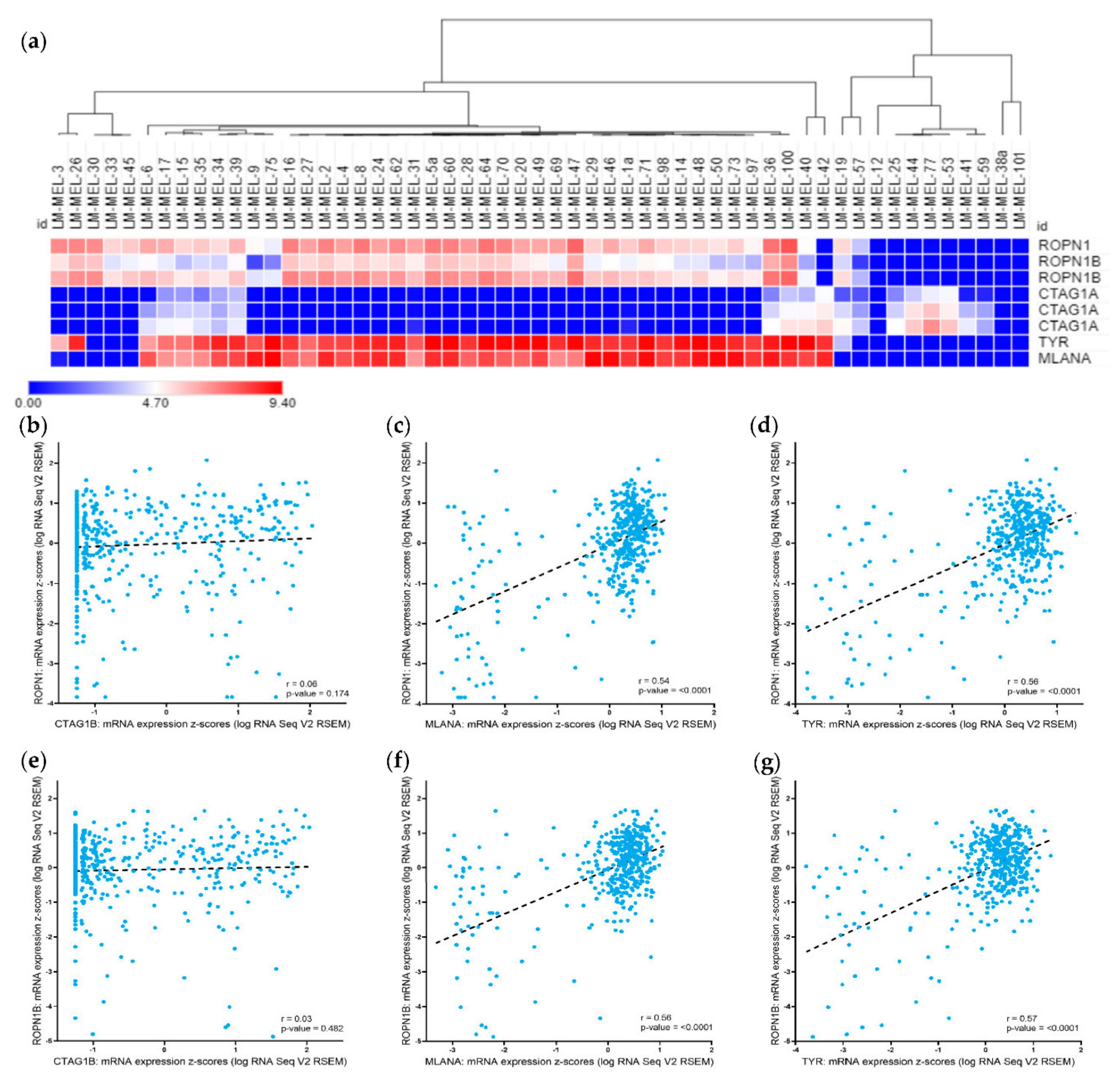
2.1. Ropporin-1 (ROPN1) and Ropporin-1B (ROPN1B) Genes Are Expressed in Melanoma Samples and Correlated with Melanoma Differentiation Antigens

We tested the gene expression levels of ROPN1 and ROPN1B (96% sequence homology with ROPN1) in a panel of melanoma cell lines generated in-house [32] (Table 1). In 45 out of 55 (81.8%) cell lines, we detected ROPN1 gene expression compared to 46 out of 55 (83.6%) with ROPN1B expression (Figure 1A). For the purposes of comparison with a well-validated CTAg, we also determined the gene expression of CTAG1A (identical gene copy to CTAG1B) in the same cohort of cell lines. CTAG1A expression was detected in 20 out of 55 (36.4%) cell lines (Figure 1A), with 14 cell lines showing expression of both CTAgs. Expression of either ROPN1/ROPN1B or CTAG1A was observed in 52 out of 55 cell lines (94.5%).
The expression of differentiation antigens that are commonly present and have been previously used as immunological targets in melanoma, namely, TYR (tyrosinase) and MLANA (Melan-A/MART1) [20,33], were compared with CTAG1A and ROPN1 or ROPN1B in our panel of melanoma cell lines. We found a strong correlation in the expression of both TYR and MLANA with ROPN1 and ROPN1B among our cell line panel (Figure 1A, TYR vs. ROPN1: r = 0.73, p-value < 0.0001; TYR vs. ROPN1B: r = 0.73, p-value < 0.0001; MLANA vs. ROPN1: r = 0.61, p-value < 0.0001; MLANA vs. ROPN1B: r = 0.61, p-value < 0.0001). In contrast, CTAG1A expression was found in more differentiated cell lines with high TYR and/or MLANA expression, and in less differentiated ones without TYR and/or MLANA expression (Figure 1A).
We further evaluated the gene expression levels of ROPN1, ROPN1B, CTAG1B (identical gene copy to CTAG1A for which no gene expression data is available), TYR and MLANA in 472 additional melanoma samples using data accessible via The Cancer Genome Atlas (TCGA) (Table 1). As expected, expression of ROPN1 and its paralog ROPN1B was highly correlated (r = 0.86, p-value = 8.71 × 10−141). ROPN1 and ROPN1B were expressed at transcript level in nearly all melanomas (ROPN1: n = 467/472, 98.9%, ROPN1B: n = 466/472, 98.7%), more frequently than CTAG1B (n = 322/472, 68.2%), and sometimes exclusively (Figure 1B, ROPN1: r = 0.06, p-value=0.174; Figure 1E, ROPN1B: r = 0.03, p-value = 0.482). As expected, TYR (n = 469/472, 99.4%) and MLANA (n = 471/472, 99.8%) were commonly expressed. We further demonstrated co-expression of ROPN1/ROPN1B and TYR (ROPN1: r = 0.56, p-value < 0.0001; ROPN1B: r = 0.57, p-value < 0.0001) or MLANA (ROPN1: r = 0.54, p-value < 0.0001; ROPN1B: r = 0.56, p-value < 0.0001) in the majority of patient samples (Figure 1C, 1F and 1D, 1G, respectively), and found no correlation between differentiation antigens and CTAG1B expression (Figure S1, TYR: r = −0.06, p-value=0.165, MLANA: r = −0.05, p-value = 0.268), reflecting the results of our cell line analysis.
We further investigated ROPN1, ROPN1B and CTAG1B gene expression across different stages of disease and genders in melanoma. ROPN1, ROPN1B and CTAG1B were expressed across all AJCC disease stages, without apparent differences (Figure S2, ROPN1: p-value = 0.179; ROPN1B: p-value = 0.140; CTAG1B: p-value = 0.338). Similarly, no difference was observed between males and females (Figure S3, ROPN1: p-value = 0.944; ROPN1B: p-value = 0.362; CTAG1B: p-value = 0.829).

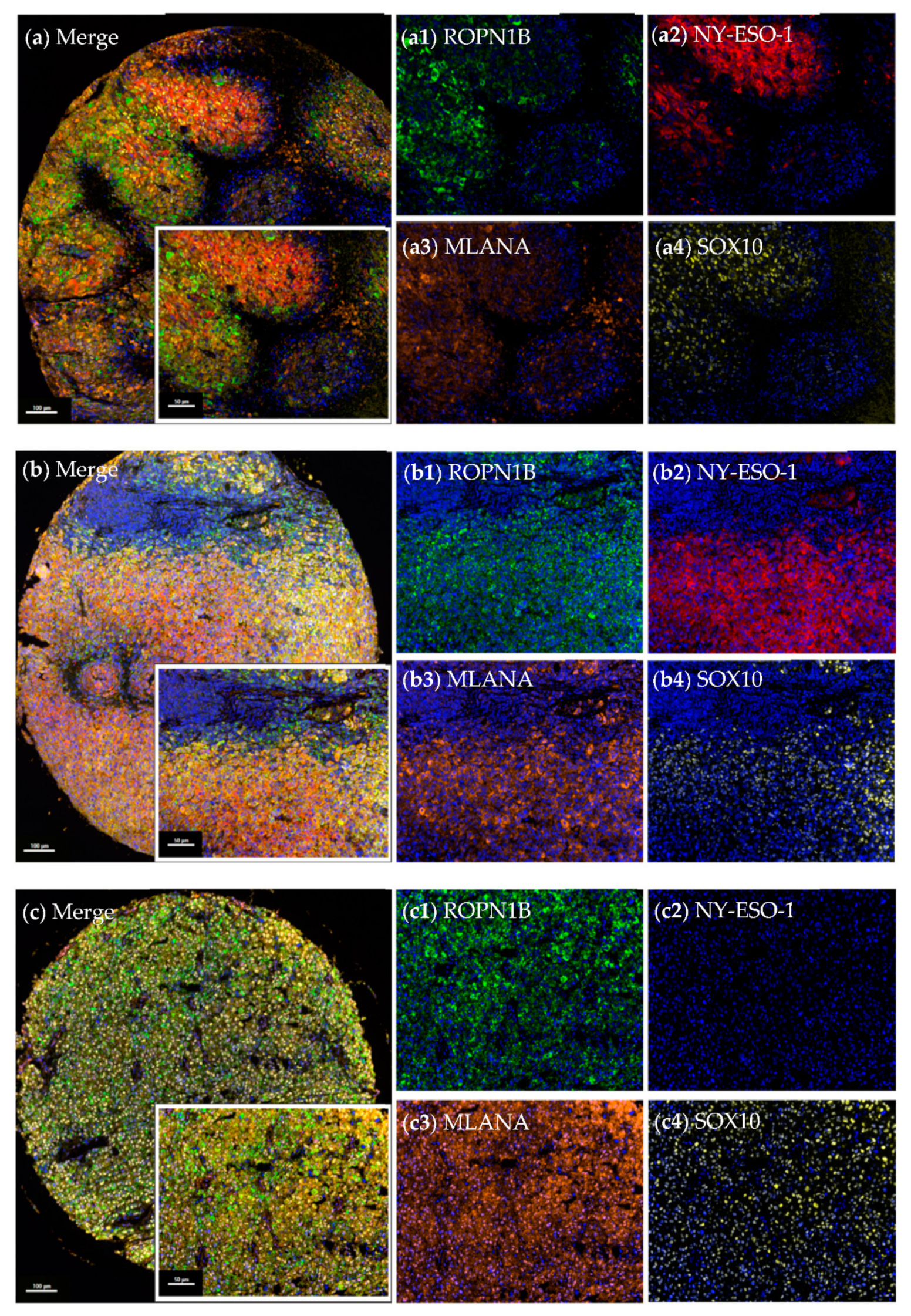
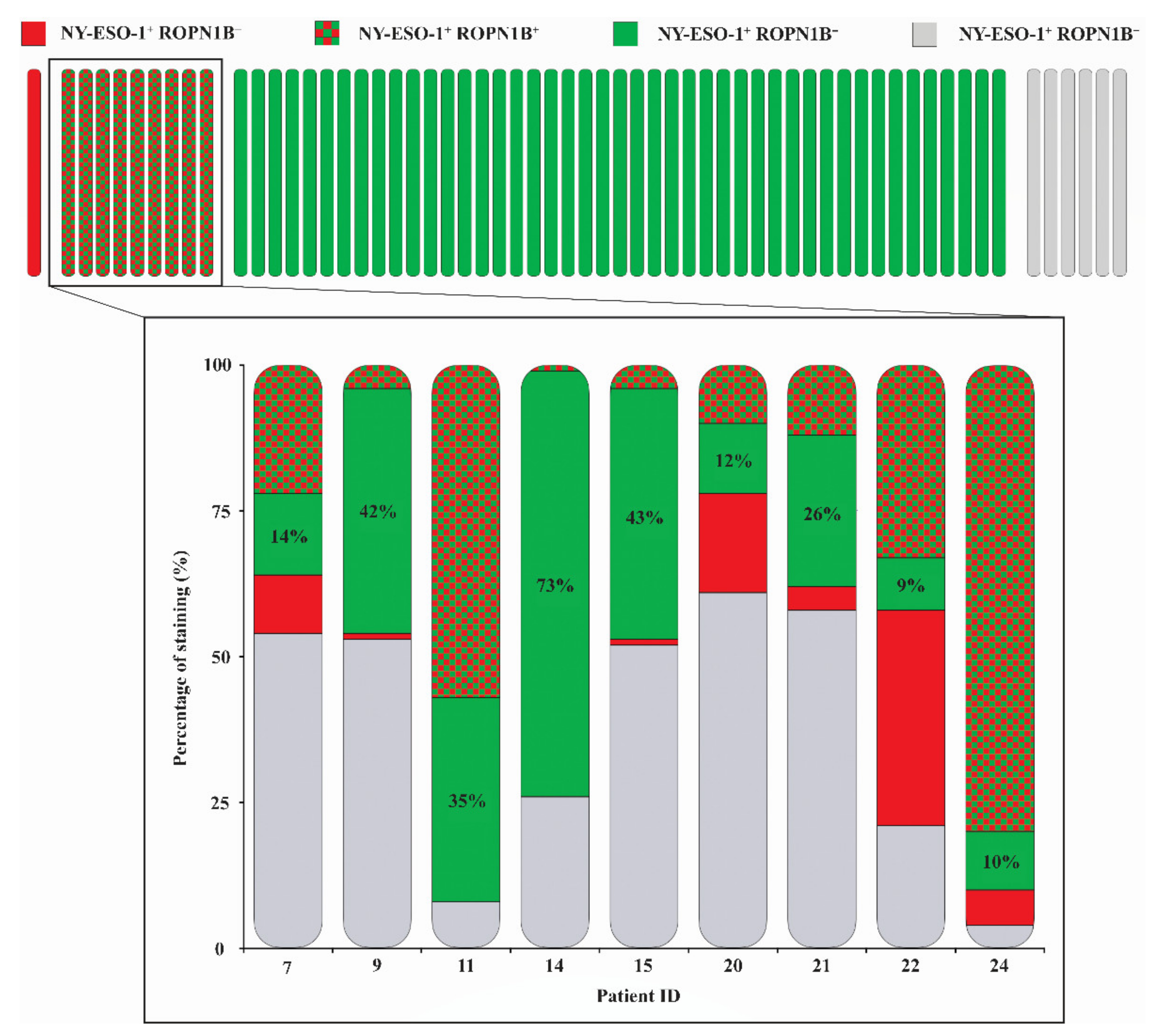
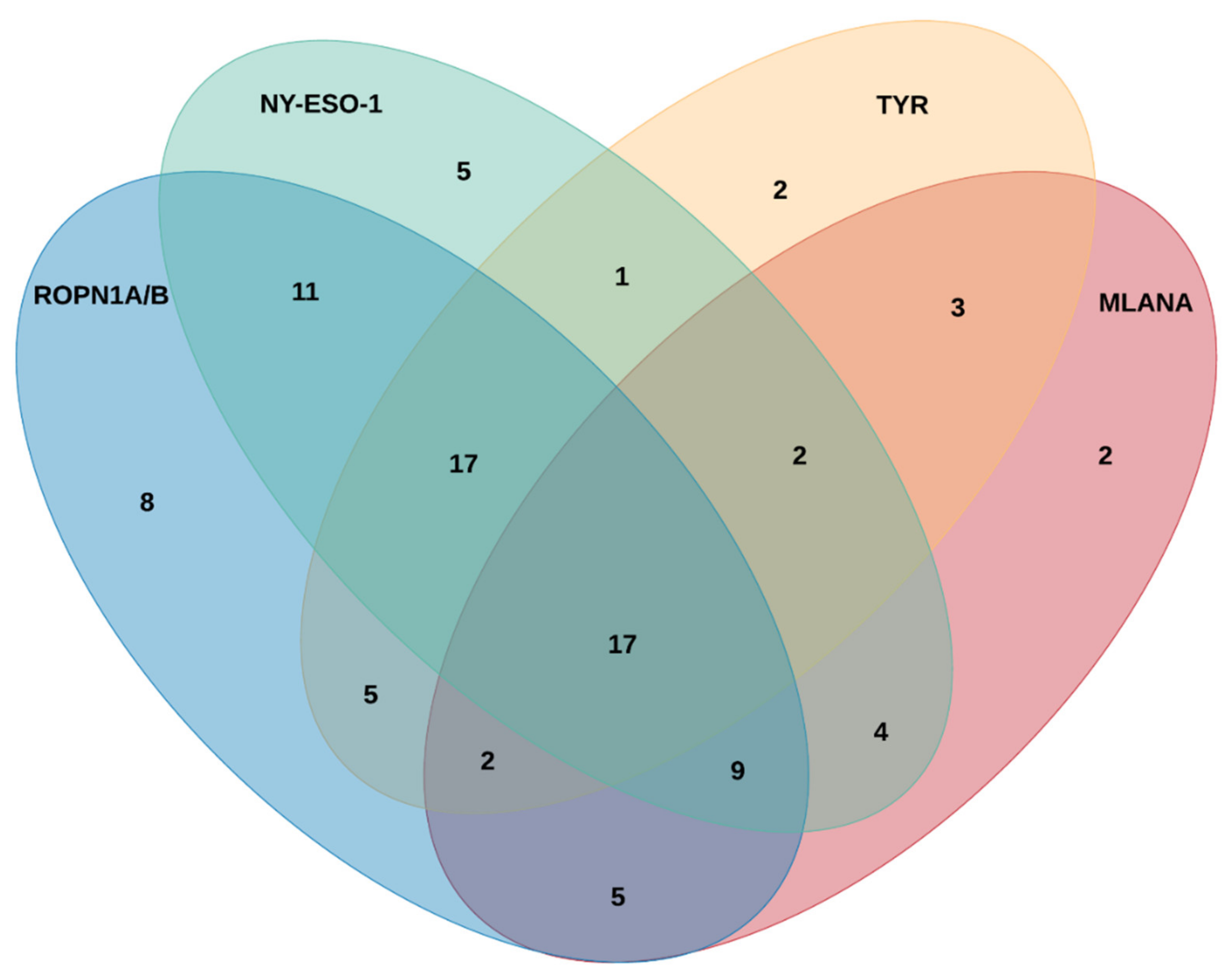
2.2. Ropporin-1B (ROPN1B) Protein Is Expressed in Melanoma Tumours

We assessed expression of ROPN1B, NY-ESO-1 (single protein from CTAG1A or CTAG1B genes), MLANA and SOX10 (used here as a melanoma tumour marker) at the protein level by multispectral immunohistochemistry on melanoma tissue microarrays (TMAs) comprising two or three cores from 61 patient tumours (Table 1). Protein expression levels were at times heterogeneous amongst patient cores, and lack of expression was only reported in cases where all cores per patient were assessable. ROPN1B cytoplasmic expression was observed in 88.5% (n = 54/61) of patient samples, while cytoplasmic NY-ESO-1 was observed in 16.4% (n = 10/61) of samples (Figure S4). Expression of both ROPN1B and NY-ESO-1 was seen in 9 cases (n = 9/61, 14.8%), where tumour cells within cores showed instances of co-expression or exclusive expression of ROPN1B and/or NY-ESO-1 (Figure 2 and Figure 3). Moreover, ROPN1B expression was often detected ubiquitously, whereas NY-ESO-1 appeared more scattered and diffuse throughout tumour cores (Figure S5). In patient samples with expression of both ROPN1B and NY-ESO-1, cells with exclusive ROPN1B positivity increased tumour cell coverage by 29.3% on average (range from 9.0% to 73.0%, Figure 3). Nuclear SOX10 (n = 60/61) or cytoplasmic MLANA (n = 60/61) expression was detected in nearly all tumour cores, as expected (Figure 2 and Figure S5).
ROPN1B expression was seen in both early (I and II, n = 19/26, 73.1%) and late stages (III and IV, n = 29/29, 100.0%) of disease, more so than NY-ESO-1 (n = 1/26, 3.8% vs. n = 9/29, 31.0%, respectively). Nonetheless, expression was more predominant in advanced disease (ROPN1B: chi-square p-value = 0.003; NY-ESO-1: chi-square p-value = 0.009), with evidence of more abundant melanoma cell expression in tumour cores, in contrast to sparse, dispersed cells commonly identified in early-stage disease. ROPN1B (females: 88.5%, n = 23/26 vs. males: 88.6%, n = 31/35, chi-square p-value = 0.989) and NY-ESO-1 (females: 11.5%, n = 3/26 vs. males: 20.0%, n = 7/35, chi-square p-value = 0.377) protein expression did not differ between genders.

2.3. Ropporin-1A (ROPN1A) and Ropporin-1B (ROPN1B) Are Immunogenic Antigens in Melanoma

To explore the immunogenic potential of ROPN1A and ROPN1B, we screened sera or plasma from 104 melanoma patients (Table 1). These were tested for the presence and titre of antibodies against ROPN1A/B, NY-ESO-1, MLANA and TYR (Table S2). Antibodies were detected above noise threshold against ROPN1A/B in 71.2% (n = 74/104) of patients, and against NY-ESO-1 in 63.5% (n = 66/104) of patients. Although there were instances where reactivity was exclusive to either ROPN1A/B (19.2%, n = 20/104) or NY-ESO-1 (11.6%, n = 12/104), co-reactivity was seen in many cases (51.9%, n = 54/104) (Figure 4). Alternatively, a small subset of patients had reactivity to neither (17.3%, n = 18/104). In addition, co-reactivity against both ROPN1A/B and TYR (39.4%, n = 41/104) or ROPN1A/B and MLANA (31.7%, n = 33/104) was also seen, although to a lesser degree. Only 10.6% (n = 11/104) of the patient cohort had no antibody reactivity to any of these antigens.
This cohort consisted predominately of stage III and IV melanoma patients, with 74.4% (n = 64/86) of these seropositive for ROPN1A/B, and 62.8% (n = 54/86) seropositive for NY-ESO-1. Furthermore, it included 60.6% males and 38.8% females, with no gender-related differences seen for ROPN1A/B (females: 75.0%, n = 30/40 vs. males: 69.8%, n = 44/63, chi-square p-value = 0.571) or NY-ESO-1 antibody reactivity (females: 62.5%, n = 25/40 vs. males: 63.5%, n = 40/63 chi-square p-value = 0.919) (Figure S6).

3. Discussion

Neoantigens that arise from somatic mutations can play an important role in immune tumour rejection [34] but generally differ from tumour to tumour. In order to create personalised vaccines against these targets, individual therapies need to be tailored on a patient-by-patient basis, requiring gene sequencing and manufacturing of antigens. This approach has limitations for routine clinical use across hospitals both in terms of the associated costs and extensive timeframes. Nevertheless, a small study in melanoma designed personalised neoantigen-based peptide vaccines for six patients, which led to the generation of tumour-specific CD4+ and CD8+ T lymphocyte responses and clinical benefit (four out of six patients remaining cancer free at a 25-month follow up) [4]. One reason cited for the failure of prior therapies in melanoma, including cancer vaccine- based treatments, has been the considerable heterogeneity of melanoma cells [35,36]. Phenotypic plasticity and differentiation/de-differentiation is a likely contributor to this heterogeneity as it has been known to result in antigen down-regulation. Indeed, epithelial to mesenchymal transition-like phenotype switching has been implicated as an escape mechanism following vaccination or ACT [37,38]. CTAgs comprise a large group of antigens, many of which are expressed following de-repression of epigenetic silencing in cancer [39]. In contrast to the unique products of mutations, their expression is shared between tumours and restricted normal tissues, mostly at immune-privileged sites. Therefore, they are able to stimulate potent immune responses [40] and many have been widely trialled as vaccine antigens [39]. Namely, an NY-ESO-1 vaccine was able to induce antigen-specific cellular and humoral responses but did not affect overall survival, possibly due to its limited tumour expression [41].
In this study, gene-expression profiling of a large panel of early-passage melanoma cell lines [32] identified ROPN1 and ROPN1B as being highly expressed in melanoma tumour samples (ROPN1: 81.8%, ROPN1B: 83.6%), more so than CTAG1 (36.4%). Similarly, gene expression of ROPN1 and ROPN1B was also commonly detected in a melanoma patient cohort accessed via the TCGA (ROPN1: 98.9%, ROPN1B: 98.7% vs. CTAG1B: 68.2%). When investigating melanoma TMAs, 88.5% of patient tumours expressed ROPN1B, a much larger proportion compared to NY-ESO-1 (16.4%). Even in instances of tumours expressing both NY-ESO-1 and ROPN1B (14.8%), the addition of ROPN1B increased the tumour cell coverage substantially.
The role of antibodies against tumour antigens in melanoma is unclear [42,43,44], and while an antibody response is not necessarily accompanied by cellular immunity, these often go hand-in-hand [45,46]. Antibody profiling using a custom protein array [47] detected high-titre antibodies against ROPN1A/B in the serum or plasma of a large cohort of melanoma patients (71.2%), more so when compared to NY-ESO-1 (63.5%). NY-ESO-1 antibodies were more commonly detected than expected, based on the above gene and protein expression studies. In previous studies that assessed the frequency of NY-ESO-1 antibody responses, reports have described NY-ESO-1-specific antibodies in ~10% [15] to 45% [48] of patients. However, the protein array used here is more sensitive than ELISA [47], which would contribute to a higher percentage of patients with detectable anti-NY-ESO-1 and/or anti-ROPN1A/B antibody responses when compared to historical data. Alternatively, NY-ESO-1 is known to be highly immunogenic, and hence it is possible that the detected circulating antibodies may be a result of earlier NY-ESO-1+ cell eradication. Similarly, the prevalence of detectable antibodies against MLANA and TYR were below expected levels, particularly when considering the above gene expression data, arguing for inferior humoral immunogenicity when compared to ROPN1A/B and NY-ESO-1. Furthermore, a trend towards increasing ROPN1A/B and NY-ESO-1 antibody reactivity was seen with progression of disease stage. For example, 54.5% of Stage I/II had antibody responses to ROPN1A/B, compared with 74.4% of stage III/IV patients. The number of stage I and II patients screened here (n = 11) was too small to allow us to draw definitive conclusions, however, prior studies have also demonstrated a correlative increase in the proportion of NY-ESO-1 seropositivity with disease stage [17,49]. In addition, we observed that ROPN1A/B reactivity was slightly increased in females, albeit not significantly. This observation was also noted in a recent study where antibody responses to ROPN1 were significantly higher in female multiple myeloma patients [50]. An increased humoral response in females may explain the reduced melanoma incidence and the increased survival benefit independent of disease stage reported in women [51,52]. Together, these data show that (i) NY-ESO-1, ROPN1 and ROPN1B are highly immunogenic; and (ii) patients who have no evidence of NY-ESO-1 immunity can have antibody titres against other CTAgs, including ROPN1A and ROPN1B, thereby validating that the addition of ROPN1A and ROPN1B as target antigens can potentially enlarge the population of eligible patients who might benefit from a combined vaccine approach.
Ongoing immune editing occurring over the course of disease allows cancer escape from immune targeting of individual antigens [53]. For these reasons, the potential to achieve increased tumour coverage by targeting NY-ESO-1, ROPN1A and ROPN1B combined makes these immunogenic CTAgs highly compatible for combination in a cancer vaccine with the potential to generate synergistic immune responses to the tumour. It is tempting to speculate that further immunogenic tumour antigens could also be incorporated, leading to the design of a cancer vaccine that “covers all bases” in terms of heterogeneity and plasticity. As strategies are being pursued to widen the applicability of cancer immunotherapy, vaccination is now being proposed and trialled (NCT03092453) in combination with other immunotherapies, such as ICB [22,54,55]. Our study indicates that ROPN1A and ROPN1B may serve as promising candidates for such antigen-specific approaches, and therefore further studies are warranted and should be pursued to explore this.

4. Materials and Methods

4.1. Human Ethics Approval

Blood samples used in this study were derived from patients who provided written informed consent to participate in a clinical research protocol approved by the Austin Health Human Research Ethics Committee (HREC/14/Austin/425, approved on 6 November 2014) (Table 1).

4.2. Melanoma Cell Lines and Cell Culture

Establishment and characterisation of the melanoma cell lines used has been previously described [32]. Cells were cultured in RPMI 1640, 2 mM Glutamax, 100 U/mL Penicillin, 100 µg/mL Streptomycin and 10% foetal calf serum (RF10) (all Invitrogen, Carlsbad, CA, USA).

4.3. Cell Pellet DNA Extraction

DNA was extracted from pelleted cells using a DNeasy® Blood and Tissue kit (Qiagen, Hilden, Germany) as per the manufacturer’s instructions. Briefly, pellets were suspended in 200 µL of PBS with 36 µL of Proteinase K (600 mAU/mL, Scimar, Templestowe, VIC, Australia) and 200µL AL buffer (Qiagen, Hilden, Germany), then incubated at 56 °C overnight. Clean-up was as per the manufacturer’s instructions and samples were eluted in 50 µL of AE buffer (Qiagen, Hilden, Germany).

4.4. Gene Expression—Cell Lines

The gene expression array method and analysis have been previously described (Gene Expression Omnibus, GEO dataset ID GSE89438) [32,56]. Briefly, genomic DNA was purified from 55 melanoma cell lines originating from 52 patients (Qiagen AllPrep kit, Hilden, Germany) and assayed using Illumina standard protocols. Samples were subjected to whole-genome expression arrays (Illumina HT12, San Diego, CA, USA), and hierarchical clustering by Pearson correlation of ROPN1 (probe ID: 5420739), ROPN1B (probe IDs: 730521 and 460291; 96% sequence homology with ROPN1), CTAG1A (probe IDs: 6770332, 1430215 and 1070577; identical gene copy to CTAG1B), TYR (probe ID: 5260253) and MLANA (probe ID: 7330367) was performed using the Morpheus software (https://software.broadinstitute.org/morpheus/index.html, Broad Institute, Cambridge, MA, USA). Absolute values were used to determine level of expression across cell lines, with any level above 0.3 considered “positive” for expression.

4.5. Gene Expression—The Cancer Genome Atlas (TCGA)

The gene expression data of ROPN1, ROPN1B, CTAG1B (identical gene copy to CTAG1A for which no gene expression data is available), TYR and MLANA in melanoma tumour samples was accessible via TCGA research network (http://cancergenome.nih.gov/) and analysed using the cBioPortal [57,58]. The TCGA dataset used was the Skin Cutaneous Melanoma, TCGA, Firehose Legacy, consisting of 472 samples (469 patients). Absolute mRNA transcript values were used to determine level of expression across TCGA patient samples, with any transcript level above zero considered “positive” for expression. mRNA expression of the respective genes is shown as z-scores (RNA Seq V2 RSEM) and represented in a log2 scale. Co-expression of mRNA is analysed using the Pearson correlation.

4.6. Multispectral Immunohistochemistry

Two (ME1002b, US Biomax, Derwood, MD, USA and 07 TMA Mel 1.8, in-house) melanoma TMAs containing two or three cores for 61 melanoma patient tumours (59 patients) were baked at 65 °C for 2 h, dewaxed in xylene three times for 10 min, rehydrated in ethanol twice for 10 min and manually stained. The staining included initial blocking of endogenous peroxidases using 3% hydrogen peroxide for 30 min, followed by sequential 15 min rounds of heat-induced epitope retrieval (microwave at 20% power); 10 min blocking of non-specific binding sites; 30 min primary (anti-NY-ESO-1 (single protein from CTAG1A or CTAG1B genes); ROPN1B, MLANA or SOX10); 10 min secondary (anti-mouse and anti-rabbit Opal™ horseradish peroxidase) antibody incubation; and 10 min fluorophore-tyramide signal amplification using OpalTM 520, 570, 620 and 690 fluorophores labelling target proteins, respectively (Akoya Biosciences®, Marlborough, MA, USA) (Table S1). Slides were counterstained with spectral DAPI, and tissue cores were scanned using the Vectra 3 Automated Quantitative Pathology Imaging System (Akoya Biosciences®, Marlborough, MA, USA) where target proteins were detected and imaged using the FITC, Cy3, Texas red and Cy5 filter cubes. Images were spectrally unmixed and analysed using inForm® Cell Analysis software version 3.0.5 or HALO™ Image Analysis Software version 3.2 (Akoya Biosciences®, Marlborough, MA, USA) with the assistance of a pathologist. Whole tumour cores with more than 2% of the tissue cells staining for the target proteins in at least one out of two or three cores per patient tumour were considered positive. Staining pattern of localization was defined as nuclear (if co-localised with DAPI) or cytoplasmic. Cytoplasmic staining was distinguished from membranous staining by performing immunohistochemistry using anti-MHC class I antibody (membranous expression on melanoma cells that have not experienced MHC class I loss, as well as stromal cells) with either anti-ROPN1B, anti-NY-ESO-1 or anti-MLANA antibodies, along with DAPI counterstain in melanoma tumours.

4.7. Antibody Profiling

Serum or plasma from melanoma patients was used to measure antibody titres towards ROPN1A/B, NY-ESO-1, MLANA and TYR using a custom tumour antigen protein microarray platform [47]. Blood samples originated from melanoma patients without available matched tumour tissue. Following the printing of previously expressed biotinylated antigens to a streptavidin-coated microarray slide using a QArray2 robotic arrayer (Genetix, Berkshire, UK), slides were immersed in blocking buffer (50 µM biotin in PBST) and incubated on ice in a plastic chamber protected from light for 1 h. Slides were then washed three times for 5 min in PBST and dried. Individual arrays were incubated with a unique serum or plasma sample (100 µL at 1:800 or 1:400 dilution, respectively) for 1 h at RT, washed in PBST and dH2O, and incubated with 100 µL of 20 µg/mL Alexa Fluor 647 Goat anti-Human IgG (H + L) (Invitrogen, Carlsbad, CA, USA, 1:100 dilution in PBST) for 30 min at RT. The individual arrays were then washed, dried and scanned using a Tecan LS Reloaded fluorescence microarray scanner (Tecan Group Ltd., Männedorf, Switzerland) in automatic gain control (AGC) mode. All liquid handling steps were performed using QuadChambers on a Tecan HS4800 Pro automated hybridization station (Tecan Group Ltd., Männedorf, Switzerland). The resulting arrays were viewed using the ArrayPro Analyzer software Version 6.3 (Media Cybernetics, Rockville, MD, USA), and raw data was extracted. Finally, these data were processed using a custom bioinformatic tool for protein microarray data processing and normalisation, and the resulting data were analysed accordingly [59].

5. Conclusions

In this study, we have identified ROPN1 and ROPN1B as compelling novel therapeutic antigen targets for further clinical evaluation. Features include frequent humoral immunogenicity and abundance, with virtually universal tumour-specific expression across all melanomas tested.

Supplementary Materials

The following are available online at https://www.mdpi.com/article/10.3390/cancers13081805/s1, Figure S1: Gene expression of CTAG1B and differentiation antigens TYR and MLANA in melanoma tumours, Figure S2: Gene expression of ROPN1, ROPN1B and CTAG1B by AJCC disease stage codes in melanoma tumours, Figure S3: Gene expression of ROPN1, ROPN1B and CTAG1B by gender in melanoma tumours, Figure S4: Cytoplasmic expression of ROPN1B, NY-ESO-1 and MLANA in melanoma tumours, Figure S5: Variability seen between concentrated ROPN1B and diffuse NY-ESO-1 staining patterns in melanoma tumours, Figure S6: Venn diagram displaying predominance of ROPN1A/B and NY-ESO-1-specific antibodies in melanoma patients by gender, Table S1: Specifications of antibodies and conditions used for multispectral immunohistochemistry, Table S2: ROPN1A, ROPN1B, NY-ESO-1, MLANA and TYR-specific antibodies in melanoma patients.

Author Contributions

Conceptualization, J.D.G.D., K.W., A.D., J.M.B., J.C. and A.B.; methodology, J.D.G.D., K.W., L.T.Q., C.D., C.T., T.W., S.O., C.H., S.C.-H.T., A.P. and A.B.; formal analysis, J.D.G.D., K.W., L.T.Q., C.D., C.T., T.W., S.O., C.H., S.C.-H.T. and A.P.; investigation, J.D.G.D., K.W., L.T.Q., C.D., C.T., T.W., S.O., C.H., S.C.-H.T., A.P.; resources, A.D., J.M.B., J.C. and A.B.; data curation, J.D.G.D., K.W., L.T.Q., C.D., C.T., T.W., S.O., C.H., S.C.-H.T. and A.P.; writing—original draft preparation, J.D.G.D., K.W., and A.B.; writing—review and editing, J.D.G.D., K.W., L.T.Q., and A.B.; supervision, J.D.G.D., K.W., A.D., J.M.B., J.C. and A.B.; funding acquisition, A.D., J.M.B., J.C. and A.B. All authors have read and agreed to the published version of the manuscript.

Funding

This project was partially funded by a Clinical and Laboratory Integration Program (CLIP) grant from the Cancer Research Institute (CRI). This project was also funded in part by the Ludwig Institute for Cancer Research, Melanoma Research Alliance (MRA) and the Melbourne Research Victoria (MRV). The Olivia Newton-John Cancer Research Institute acknowledges the support of the Victorian Government Operational Infrastructure Support Program and the Ian Potter Foundation for providing funds to purchase the Vectra System. The contents of the published material are solely the responsibility of La Trobe University and do not reflect the views of Cancer Australia. J.D.G.D. is supported by Cure Cancer Australia through the Cancer Australia Priority-driven Cancer Research Scheme (#1187815). J.M.B. is supported by a research chair from the National Research Foundation (South Africa). A.B. is supported by a fellowship from the Department of Health and Human Services acting through the Victorian Cancer Agency (VCA).

Institutional Review Board Statement

Clinical research protocol approved on 6 November 2014 by the Austin Health Human Research Ethics Committee (HREC/14/Austin/425).

Informed Consent Statement

Informed consent was obtained from all subjects involved in the study.

Data Availability Statement

Publicly available datasets were analysed in this study. This data can be found here: https://www.ncbi.nlm.nih.gov/geo/query/acc.cgi?acc=GSE89438; and here: http://www.cbioportal.org/study/summary?id=skcm_tcga. The remaining data presented in this study are available within the article or in the supplementary material.

Acknowledgments

The authors would like to thank Ryan Farid and Nektaria Dimopoulous for their technical support.

Conflicts of Interest

The authors declare no conflict of interest. The funders had no role in the design of the study; in the collection, analyses, or interpretation of data; in the writing of the manuscript; or in the decision to publish the results.

References

  1. Larkin, J.; Chiarion-Sileni, V.; Gonzalez, R.; Grob, J.J.; Rutkowski, P.; Lao, C.D.; Cowey, C.L.; Schadendorf, D.; Wagstaff, J.; Dummer, R.; et al. Five-year survival with combined nivolumab and ipilimumab in advanced melanoma. N. Engl. J. Med. 2019, 381, 1535–1546. [Google Scholar] [CrossRef] [PubMed] [Green Version]
  2. Rizvi, N.A.; Hellmann, M.D.; Snyder, A.; Kvistborg, P.; Makarov, V.; Havel, J.J.; Lee, W.; Yuan, J.; Wong, P.; Ho, T.S.; et al. Cancer immunology. Mutational landscape determines sensitivity to PD-1 blockade in non-small cell lung cancer. Science 2015, 348, 124–128. [Google Scholar] [CrossRef] [PubMed] [Green Version]
  3. Malaker, S.A.; Penny, S.A.; Steadman, L.G.; Myers, P.T.; Loke, J.C.; Raghavan, M.; Bai, D.L.; Shabanowitz, J.; Hunt, D.F.; Cobbold, M. Identification of Glycopeptides as Posttranslationally Modified Neoantigens in Leukemia. Cancer Immunol. Res. 2017, 5, 376–384. [Google Scholar] [CrossRef] [PubMed] [Green Version]
  4. Ott, P.A.; Hu, Z.; Keskin, D.B.; Shukla, S.A.; Sun, J.; Bozym, D.J.; Zhang, W.; Luoma, A.; Giobbie-Hurder, A.; Peter, L.; et al. An immunogenic personal neoantigen vaccine for patients with melanoma. Nature 2017, 547, 217–221. [Google Scholar] [CrossRef] [PubMed]
  5. Alexandrov, L.B.; Nik-Zainal, S.; Wedge, D.C.; Aparicio, S.A.J.R.; Behjati, S.; Biankin, A.V.; Bignell, G.R.; Bolli, N.; Borg, A.; Børresen-Dale, A.L.; et al. Signatures of mutational processes in human cancer. Nature 2013, 500, 415–421. [Google Scholar] [CrossRef] [Green Version]
  6. Chalmers, Z.R.; Connelly, C.F.; Fabrizio, D.; Gay, L.; Ali, S.M.; Ennis, R.; Schrock, A.; Campbell, B.; Shlien, A.; Chmielecki, J.; et al. Analysis of 100,000 human cancer genomes reveals the landscape of tumor mutational burden. Genome Med. 2017, 9, 1–14. [Google Scholar] [CrossRef] [PubMed]
  7. Fourcade, J.; Sun, Z.; Pagliano, O.; Chauvin, J.-M.; Sander, C.; Janjic, B.; Tarhini, A.A.; Tawbi, H.A.; Kirkwood, J.M.; Moschos, S.; et al. PD-1 and Tim-3 regulate the expansion of tumor antigen-specific CD8+ T cells induced by melanoma vaccines. Cancer Res. 2014, 74, 1045–1055. [Google Scholar] [CrossRef] [PubMed] [Green Version]
  8. Mahvi, D.A.; Meyers, J.V.; Tatar, A.J.; Contreras, A.; Suresh, M.; Leverson, G.E.; Sen, S.; Cho, C.S. Ctla-4 blockade plus adoptive T-cell transfer promotes optimal melanoma immunity in mice. J. Immunother. 2015, 38, 54–61. [Google Scholar] [CrossRef] [Green Version]
  9. Kuramitsu, S.; Yamamichi, A.; Ohka, F.; Motomura, K.; Hara, M.; Natsume, A. Adoptive immunotherapy for the treatment of glioblastoma: Progress and possibilities. Immunotherapy 2016, 8, 1393–1404. [Google Scholar] [CrossRef]
  10. Davis, I.D.; Chen, W.; Jackson, H.; Parente, P.; Shackleton, M.; Hopkins, W.; Chen, Q.; Dimopoulos, N.; Luke, T.; Murphy, R.; et al. Recombinant NY-ESO-1 protein with ISCOMATRIX adjuvant induces broad integrated antibody and CD4(+) and CD8(+) T cell responses in humans. Proc. Natl. Acad. Sci. USA 2004, 101, 10697–10702. [Google Scholar] [CrossRef] [Green Version]
  11. Maraskovsky, E. NY-ESO-1 Protein Formulated in ISCOMATRIX Adjuvant Is a Potent Anticancer Vaccine Inducing Both Humoral and CD8+ T-Cell-Mediated Immunity and Protection against NY-ESO-1+ Tumors. Clin. Cancer Res. 2004, 10, 2879–2890. [Google Scholar] [CrossRef] [Green Version]
  12. Phan, G.Q.; Rosenberg, S.A. Adoptive cell transfer for patients with metastatic melanoma: The potential and promise of cancer immunotherapy. Cancer Control 2013, 20, 289–297. [Google Scholar] [CrossRef]
  13. Geldmacher, A.; Freier, A.; Losch, F.O.; Walden, P. Therapeutic vaccination for cancer immunotherapy: Antigen selection and clinical responses. Hum. Vaccin. 2011, 7, 115–119. [Google Scholar] [CrossRef]
  14. Jackson, H.; Dimopoulos, N.; Mifsud, N.A.; Tai, T.Y.; Chen, Q.; Svobodova, S.; Browning, J.; Luescher, I.; Stockert, L.; Old, L.J.; et al. Striking immunodominance hierarchy of naturally occurring CD8+ and CD4+ T cell responses to tumor antigen NY-ESO-1. J. Immunol. 2006, 176, 5908–5917. [Google Scholar] [CrossRef] [Green Version]
  15. Stockert, E.; Jäger, E.; Chen, Y.T.; Scanlan, M.J.; Gout, I.; Karbach, J.; Arand, M.; Knuth, A.; Old, L.J. A survey of the humoral immune response of cancer patients to a panel of human tumor antigens. J. Exp. Med. 1998, 187, 1349–1354. [Google Scholar] [CrossRef] [Green Version]
  16. Knights, A.J.; Nuber, N.; Thomson, C.W.; De La Rosa, O.; Jäger, E.; Tiercy, J.M.; Van Den Broek, M.; Pascolo, S.; Knuth, A.; Zippelius, A. Modified tumour antigen-encoding mRNA facilitates the analysis of naturally occurring and vaccine-induced CD4 and CD8 T cells in cancer patients. Cancer Immunol. Immunother. 2009, 58, 325–338. [Google Scholar] [CrossRef]
  17. Barrow, C.; Browning, J.; MacGregor, D.; Davis, I.D.; Sturrock, S.; Jungbluth, A.A.; Cebon, J. Tumor antigen expression in melanoma varies according to antigen and stage. Clin. Cancer Res. 2006, 12, 764–771. [Google Scholar] [CrossRef] [Green Version]
  18. Filipazzi, P.; Pilla, L.; Mariani, L.; Patuzzo, R.; Castelli, C.; Camisaschi, C.; Maurichi, A.; Cova, A.; Rigamonti, G.; Giardino, F.; et al. Limited induction of tumor cross-reactive T cells without a measurable clinical benefit in early melanoma patients vaccinated with human leukocyte antigen class I-modified peptides. Clin. Cancer Res. 2012, 18, 6485–6496. [Google Scholar] [CrossRef] [Green Version]
  19. Slingluff, C.L. The present and future of peptide vaccines for cancer: Single or multiple, long or short, alone or in combination? Cancer J. 2011, 17, 343–350. [Google Scholar] [CrossRef] [Green Version]
  20. Slingluff, C.L.; Petroni, G.R.; Yamshchikov, G.V.; Hibbitts, S.; Grosh, W.W.; Chianese-Bullock, K.A.; Bissonette, E.A.; Barnd, D.L.; Deacon, D.H.; Patterson, J.W.; et al. Immunologic and clinical outcomes of vaccination with a multiepitope melanoma peptide vaccine plus low-dose interleukin-2 administered either concurrently or on a delayed schedule. J. Clin. Oncol. 2004, 22, 4474–4485. [Google Scholar] [CrossRef]
  21. Kumai, T.; Fan, A.; Harabuchi, Y.; Celis, E. Cancer immunotherapy: Moving forward with peptide T cell vaccines. Curr. Opin. Immunol. 2017, 47, 57–63. [Google Scholar] [CrossRef] [PubMed]
  22. Hersey, P.; Gallagher, S.J.; Kirkwood, J.M.; Cebon, J. Melanoma Vaccines. In Cutaneous Melanoma; Balch, C.M., Atkins, M.B., Garbe, C., Gershenwald, J.E., Halpern, A.C., Kirkwood, J.M., McArthur, G.A., Thompson, J.F., Sober, A.J., Eds.; Springer International Publishing: Cham, Switzerland, 2020; pp. 1243–1265. ISBN 978-3-030-05070-2. [Google Scholar]
  23. Grenier, J.M.; Yeung, S.T.; Qiu, Z.; Jellison, E.R. Combining Adoptive Cell Therapy with Cytomegalovirus-Based Vaccine Is Protective against Solid Skin Tumors. Front. Immunol. 2017, 8, 1–10. [Google Scholar] [CrossRef] [PubMed] [Green Version]
  24. Hofmann, O.; Caballero, O.L.; Stevenson, B.J.; Chen, Y.-T.Y.; Cohen, T.; Chua, R.; Maher, C.A.; Panji, S.; Schaefer, U.; Kruger, A.; et al. Genome-wide analysis of cancer/testis gene expression. Proc. Natl. Acad. Sci. USA 2008, 105, 20422–20427. [Google Scholar] [CrossRef] [PubMed] [Green Version]
  25. Zhao, S.; Zhu, W.; Xue, S.; Han, D. Testicular defense systems: Immune privilege and innate immunity. Cell. Mol. Immunol. 2014, 11, 428–437. [Google Scholar] [CrossRef] [Green Version]
  26. Fujita, A.; Nakamura, K.I.; Kato, T.; Watanabe, N.; Ishizaki, T.; Kimura, K.; Mizoguchi, A.; Narumiya, S. Ropporin, a sperm-specific binding protein of rhophilin, that is localized in the fibrous sheath of sperm flagella. J. Cell Sci. 2000, 113, 103–112. [Google Scholar]
  27. Carr, D.W.; Fujita, A.; Stentz, C.L.; Liberty, G.A.; Olson, G.E.; Narumiya, S. Identification of Sperm-specific Proteins that Interact with A-kinase Anchoring Proteins in a Manner Similar to the Type II Regulatory Subunit of PKA. J. Biol. Chem. 2001, 276, 17332–17338. [Google Scholar] [CrossRef] [Green Version]
  28. Chen, J.; Wang, Y.; Wei, B.; Lai, Y.; Yan, Q.; Gui, Y.; Cai, Z. Functional expression of ropporin in human testis and ejaculated spermatozoa. J. Androl. 2011, 32, 26–32. [Google Scholar] [CrossRef]
  29. Li, Z.; Li, W.; Meklat, F.; Wang, Z.; Zhang, J.; Zhang, Y.; Lim, S.H. A yeast two-hybrid system using Sp17 identified Ropporin as a novel cancer-testis antigen in hematologic malignancies. Int. J. Cancer 2007, 121, 1507–1511. [Google Scholar] [CrossRef]
  30. Liu, Q.; Huang, X.; Li, Q.; He, L.; Li, S.; Chen, X.; Ouyang, Y.; Wang, X.; Lin, C. Rhophilin-associated tail protein 1 promotes migration and metastasis in triple negative breast cancer via activation of RhoA. FASEB J. 2020, 34, 9959–9971. [Google Scholar] [CrossRef]
  31. Chiriva-Internati, M.; Mirandola, L.; Yu, Y.; Jenkins, M.R.; Gornati, R.; Bernardini, G.; Gioia, M.; Chiaramonte, R.; Cannon, M.J.; Kast, W.M.; et al. Cancer testis antigen, ropporin, is a potential target for multiple myeloma immunotherapy. J. Immunother. 2011, 34, 490–499. [Google Scholar] [CrossRef]
  32. Behren, A.; Anaka, M.; Lo, P.-H.; Vella, L.J.; Davis, I.D.; Catimel, J.; Cardwell, T.; Gedye, C.; Hudson, C.; Stan, R.; et al. The Ludwig institute for cancer research Melbourne melanoma cell line panel. Pigment. Cell Melanoma Res. 2013, 26, 597–600. [Google Scholar] [CrossRef] [Green Version]
  33. Tarhini, A.A.; Leng, S.; Moschos, S.J.; Yin, Y.; Sander, C.; Lin, Y.; Gooding, W.E.; Kirkwood, J.M. Safety and immunogenicity of vaccination with MART-1 (26-35, 27L), gp100 (209-217, 210M), and tyrosinase (368-376, 370D) in adjuvant with PF-3512676 and GM-CSF in metastatic melanoma. J. Immunother. 2012, 35, 359–366. [Google Scholar] [CrossRef] [Green Version]
  34. Schumacher, T.N.; Schreiber, R.D. Realising the Promise: Neoantigens in cancer immunotherapy. Sci. Mag. 2015, 348. [Google Scholar]
  35. Roesch, A. Tumor heterogeneity and plasticity as elusive drivers for resistance to MAPK pathway inhibition in melanoma. Oncogene 2015, 34, 2951–2957. [Google Scholar] [CrossRef]
  36. Monjazeb, A.M.; Zamora, A.E.; Grossenbacher, S.K.; Mirsoian, A.; Sckisel, G.D.; Murphy, W.J. Immunoediting and antigen loss: Overcoming the Achilles heel of immunotherapy with antigen non-specific therapies. Front. Oncol. 2013, 3, 1–10. [Google Scholar] [CrossRef] [Green Version]
  37. Landsberg, J.; Kohlmeyer, J.; Renn, M.; Bald, T.; Rogava, M.; Cron, M.; Fatho, M.; Lennerz, V.; Wölfel, T.; Hölzel, M.; et al. Melanomas resist T-cell therapy through inflammation-induced reversible dedifferentiation. Nature 2012, 490, 412–416. [Google Scholar] [CrossRef]
  38. Woods, K.; Pasam, A.; Jayachandran, A.; Andrews, M.C.; Cebon, J. Effects of epithelial to mesenchymal transition on t cell targeting of melanoma cells. Front. Oncol. 2014, 4, 1–7. [Google Scholar] [CrossRef] [Green Version]
  39. Cebon, J.; Caballero, O.; John, T.; Klein, O. Cancer Testis Antigens. In Tumor-Associated Antigens; Gires, O., Seliger, B., Eds.; Wiley-VCH Verlag GmbH & Co. KGaA: Weinheim, Germany, 2009; pp. 161–177. ISBN 9783527320844. [Google Scholar]
  40. Scanlan, M.J.; Gure, A.O.; Jungbluth, A.A.; Old, L.J.; Chen, Y. Cancer/testis antigens: An expanding family of targets for cancer immunotherapy. Immunol. Rev. 2002, 188, 22–32. [Google Scholar] [CrossRef]
  41. Cebon, J.S.; Gore, M.; Thompson, J.F.; Davis, I.D.; McArthur, G.A.; Walpole, E.; Smithers, M.; Cerundolo, V.; Dunbar, P.R.; MacGregor, D.; et al. Results of a randomized, double-blind phase II clinical trial of NY-ESO-1 vaccine with ISCOMATRIX adjuvant versus ISCOMATRIX alone in participants with high-risk resected melanoma. J. Immunother. Cancer 2020, 8, 1–11. [Google Scholar] [CrossRef] [Green Version]
  42. Maire, C.; Vercambre-Darras, S.; Devos, P.; D’Herbomez, M.; Dubucquoi, S.; Mortier, L. Metastatic melanoma: Spontaneous occurrence of auto antibodies is a good prognosis factor in a prospective cohort. J. Eur. Acad. Dermatol. Venereol. 2013, 27, 92–96. [Google Scholar] [CrossRef]
  43. Kupsch, J.M.; Tidman, N.H.; Kang, N.V.; Truman, H.; Hamilton, S.; Patel, N.; Bishop, J.A.N.; Leigh, I.M.; Crowe, J.S. Isolation of human tumor-specific antibodies by selection of an antibody phage library on melanoma cells. Clin. Cancer Res. 1999, 5, 925–931. [Google Scholar]
  44. Ohue, Y.; Wada, H.; Oka, M.; Nakayama, E. Antibody response to cancer/testis (CT) antigens: A prognostic marker in cancer patients. Oncoimmunology 2014, 3, e970032-1–e970032-3. [Google Scholar] [CrossRef]
  45. Casadevall, A.; Pirofski, L. Antibody-mediated regulation of cellular immunity and the inflammatory response. Trends Immunol. 2003, 24, 474–478. [Google Scholar] [CrossRef]
  46. Jäger, E.; Chen, Y.T.; Drijfhout, J.W.; Karbach, J.; Ringhoffer, M.; Jäger, D.; Arand, M.; Wada, H.; Noguchi, Y.; Stockert, E.; et al. Simultaneous humoral and cellular immune response against cancer-testis antigen NY-ESO-1: Definition of human histocompatibility leukocyte antigen (HLA)-A2-binding peptide epitopes. J. Exp. Med. 1998, 187, 265–270. [Google Scholar] [CrossRef]
  47. Beeton-Kempen, N.; Duarte, J.; Shoko, A.; Serufuri, J.-M.; John, T.; Cebon, J.; Blackburn, J. Development of a novel, quantitative protein microarray platform for the multiplexed serological analysis of autoantibodies to cancer-testis antigens. Int. J. Cancer 2014, 135, 1842–1851. [Google Scholar] [CrossRef]
  48. Gnjatic, S.; Atanackovic, D.; Jäger, E.; Matsuo, M.; Selvakumar, A.; Altorki, N.K.; Maki, R.G.; Dupont, B.; Ritter, G.; Chen, Y.-T.; et al. Survey of naturally occurring CD4+ T cell responses against NY-ESO-1 in cancer patients: Correlation with antibody responses. Proc. Natl. Acad. Sci. USA 2003, 100, 8862–8867. [Google Scholar] [CrossRef] [Green Version]
  49. Svobodová, S.; Browning, J.; MacGregor, D.; Pollara, G.; Scolyer, R.A.; Murali, R.; Thompson, J.F.; Deb, S.; Azad, A.; Davis, I.D.; et al. Cancer-testis antigen expression in primary cutaneous melanoma has independent prognostic value comparable to that of Breslow thickness, ulceration and mitotic rate. Eur. J. Cancer 2011, 47, 460–469. [Google Scholar] [CrossRef]
  50. Mirandola, L.; Wade, R.; Verma, R.; Pena, C.; Hosiriluck, N.; Figueroa, J.A.; Cobos, E.; Jenkins, M.R.; Chiriva-Internati, M. Sex-Driven Differences in Immunological Responses: Challenges and Opportunities for the Immunotherapies of the Third Millennium. Int. Rev. Immunol. 2015, 34, 134–142. [Google Scholar] [CrossRef]
  51. Joosse, A.; Collette, S.; Suciu, S.; Nijsten, T.; Lejeune, F.; Kleeberg, U.R.; Coebergh, J.W.W.; Eggermont, A.M.M.; de Vries, E. Superior outcome of women with stage I/II cutaneous melanoma: Pooled analysis of four European Organisation for Research and Treatment of Cancer phase III trials. J. Clin. Oncol. 2012, 30, 2240–2247. [Google Scholar] [CrossRef]
  52. Joosse, A.; Collette, S.; Suciu, S.; Nijsten, T.; Patel, P.M.; Keilholz, U.; Eggermont, A.M.M.; Coebergh, J.W.W.; de Vries, E. Sex is an independent prognostic indicator for survival and relapse/progression-free survival in metastasized stage III to IV melanoma: A pooled analysis of five European organisation for research and treatment of cancer randomized controlled trials. J. Clin. Oncol. 2013, 31, 2337–2346. [Google Scholar] [CrossRef]
  53. Mittal, D.; Gubin, M.M.; Schreiber, R.D.; Smyth, M.J. New insights into cancer immunoediting and its three component phases--elimination, equilibrium and escape. Curr. Opin. Immunol. 2014, 27, 16–25. [Google Scholar] [CrossRef] [PubMed] [Green Version]
  54. Ott, P.A.; Hodi, F.S.; Kaufman, H.L.; Wigginton, J.M.; Wolchok, J.D. Combination immunotherapy: A road map. J. Immunother. Cancer 2017, 5, 1–15. [Google Scholar] [CrossRef] [PubMed] [Green Version]
  55. Gjerstorff, M.F.; Andersen, M.H.; Ditzel, H.J. Oncogenic cancer/testis antigens: Prime candidates for immunotherapy. Oncotarget 2015, 6, 15772–15787. [Google Scholar] [CrossRef] [PubMed] [Green Version]
  56. Anaka, M.; Freyer, C.; Gedye, C.; Caballero, O.; Davis, I.D.; Behren, A.; Cebon, J. Stem cell media culture of melanoma results in the induction of a nonrepresentative neural expression profile. Stem Cells 2012, 30, 336–343. [Google Scholar] [CrossRef]
  57. Cerami, E.; Gao, J.; Dogrusoz, U.; Gross, B.E.; Sumer, S.O.; Aksoy, B.A.; Jacobsen, A.; Byrne, C.J.; Heuer, M.L.; Larsson, E.; et al. The cBio Cancer Genomics Portal: An open platform for exploring multidimensional cancer genomics data. Cancer Discov. 2012, 2, 401–404. [Google Scholar] [CrossRef] [Green Version]
  58. Gao, J.; Aksoy, B.A.; Dogrusoz, U.; Dresdner, G.; Gross, B.; Sumer, S.O.; Sun, Y.; Jacobsen, A.; Sinha, R.; Larsson, E.; et al. Integrative analysis of complex cancer genomics and clinical profiles using the cBioPortal. Sci. Signal. 2013, 6, pl1. [Google Scholar] [CrossRef] [Green Version]
  59. Da Gama Duarte, J.; Goosen, R.W.; Lawry, P.J.; Blackburn, J.M. PMA: Protein Microarray Analyser, a user-friendly tool for data processing and normalization. BMC Res. Notes 2018, 11, 156. [Google Scholar] [CrossRef] [Green Version]
Figure 1. Gene expression of ROPN1, ROPN1B, CTAG1A/B, TYR and MLANA in melanoma cell lines and tumours. (a) Heat map representing the hierarchical clustering by Pearson correlation of ROPN1, ROPN1B, CTAG1A, TYR and MLANA gene expression in 55 in-house generated melanoma cell lines derived from 52 patients [32]. Each row shows absolute level of expression results for one specific probe for the respective gene. The full gene expression dataset can be found under GEO dataset ID GSE89438. (b) mRNA co-expression plots for ROPN1 and CTAG1B (r = −0.06, p-value = 0.174), or (c) TYR (r = 0.56, p-value < 0.0001) or (d) MLANA (r = 0.54, p-value < 0.0001); and (e) mRNA co-expression plots for ROPN1B and CTAG1B (r = 0.03, p-value = 0.482), or (f) TYR (r = 0.57, p-value < 0.0001) or (g) MLANA (r = 0.56, p-value < 0.0001) in 472 TCGA melanoma patient samples (469 patients). mRNA expression is shown as z-scores relative to all samples (log RNA Seq V2 RSEM), and correlation is analysed using the Pearson correlation. Gene Expression Omnibus, GEO; TCGA, The Cancer Genome Atlas.
Cancers 13 01805 g001
Figure 2. Patterns of ROPN1B and NY-ESO-1 expression in melanoma tumours. Multispectral immunohistochemistry of melanoma tumour cores showing staining for ROPN1B, NY-ESO-1, MLANA and SOX10. Representative cores showing ROPN1B and NY-ESO-1 co-expression in discreet (a) or shared (b) tumour regions, as well as exclusive (c) ROPN1B expression. Tissue microarrays were stained with anti-ROPN1B (green), anti-NY-ESO-1 (red), anti-MLANA (orange) and anti-SOX10 (yellow) antibodies with DAPI (blue) counterstain, and are displayed as merge (a,b,c) and single colour (a1a4,b1b4, c1c4) cores. Whole tumour cores with more than 2% of the tissue cells staining for the target proteins were considered positive. All images were taken using a 20× objective, and scale bars indicate 100 µm (core) or 50 µm (region of interest).
Cancers 13 01805 g002
Figure 3. Tumour cell coverage of ROPN1B and NY-ESO-1 expression in investigated melanoma tumours. Upper panel indicates number of tumours with any expression of NY-ESO-1 alone (red), NY-ESO-1 and ROPN1B (chequered red and green), ROPN1B alone (green) or neither (grey) across the cohort. Magnified panel depicts quantitative distribution of NY-ESO-1 and ROPN1B staining relative to SOX10+ melanoma cells across patients displaying both NY-ESO-1 and ROPN1B expression. Percentages on bars show ROPN1B expression alone.
Cancers 13 01805 g003
Figure 4. Venn diagram displaying predominance of ROPN1A/B, NY-ESO-1, TYR and MLANA-specific antibodies in melanoma patients. Antibody titres were measured in 104 melanoma patients using a custom protein microarray platform, and all resulting intensities above 500 RFU (defined noise threshold) were considered positive signals and plotted using a 4-way Venn diagram. RFU, relative fluorescent units.
Cancers 13 01805 g004
Table 1. Summary of patient characteristics across all cohorts. This includes sample numbers, age, gender and disease stage. Cohort 1 was used to generate gene expression data (GEO dataset ID GSE89438) from 55 in-house generated melanoma cell lines derived from 52 patients [32]. Cohort 2 was used to generate gene expression data from 472 melanoma patient tumours derived from the TCGA Skin Cutaneous Melanoma Firehose Legacy dataset with 469 patients. Cohort 3 was used to generate protein expression data from 61 melanoma patient tumours across 2 TMAs (ME1002b, US Biomax, Derwood, MD, USA and 07 TMA Mel 1.8, in-house) derived from 61 patients. Cohort 4 was used to generate circulating antibody data from 104 melanoma patient serum or plasma. Gene Expression Omnibus, GEO; TCGA, The Cancer Genome Atlas; TMAs, tissue microarrays; yr, years.
Cohort 1 (n = 52)Cohort 2 (n = 469)Cohort 3 (n = 61)Cohort 4 (n = 104)
Sample TypeCell linesTumoursTumoursSerum or Plasma
Used to DetermineGene ExpressionGene ExpressionProtein ExpressionCirculating Antibodies
Age–yr
Median56585354
Range25–8315–9021–8821–87
Gender–no. (%)
Unknown0 (0)0 (0)0 (0)1 (0.9)
Male33 (63.5)289 (61.6)35 (57.4)63 (60.6)
Female19 (36.5)180 (38.4)26 (42.6)40 (38.5)
Stage–no. (%)
Unknown9 (17.3)59 (12.6)6 (9.8)7 (6.7)
I0 (0)77 (16.4)5 (8.2)2 (1.9)
II1 (1.9)140 (29.9)21 (34.4)9 (8.7)
III22 (42.3)170 (36.2)4 (6.6)35 (33.7)
IV20 (38.5)23 (4.9)25 (41.0)51 (49.0)
Publisher’s Note: MDPI stays neutral with regard to jurisdictional claims in published maps and institutional affiliations.

