Article

Cost-Effectiveness of Digital Breast Tomosynthesis vs. Abbreviated Breast MRI for Screening Women with Intermediate Risk of Breast Cancer—How Low-Cost Must MRI Be?

by
Fabian Tollens
1,
Pascal A.T. Baltzer
2,
Matthias Dietzel
3,
Johannes Rübenthaler
4,
Matthias F. Froelich
1 and
Clemens G. Kaiser
1,*
1
Department of Radiology and Nuclear Medicine, University Medical Centre Mannheim, Medical Faculty Mannheim, University of Heidelberg, Theodor-Kutzer-Ufer 1-3, 68167 Mannheim, Germany
2
Department of Biomedical Imaging and Image-Guided Therapy, Vienna General Hospital, Medical University of Vienna, 1090 Wien, Austria
3
Department of Radiology, Friedrich-Alexander-University Hospital Erlangen, 91054 Erlangen, Germany
4
Department of Radiology, Ludwig-Maximilians-University Munich, 80331 München, Germany
*
Author to whom correspondence should be addressed.
Cancers 2021, 13(6), 1241; https://doi.org/10.3390/cancers13061241
Submission received: 22 December 2020 / Revised: 9 March 2021 / Accepted: 10 March 2021 / Published: 12 March 2021
(This article belongs to the Special Issue Novel Insights into Biology and Cancers)

Abstract

:

Simple Summary

Women with dense breasts have an increased risk of breast cancer and a smaller likelihood of detection in conventional mammographic screening. It is an ongoing challenge of breast imaging techniques to increase sensitivity in dense breasts. Tomosynthesis, as well as abbreviated breast MR, intends to close the gap between optimal cancer detection rate and cost-effectiveness. The aim of this economic evaluation was to analyse the cost-effectiveness of these two imaging techniques in screening women of intermediate risk of breast cancer due to dense breasts. The model-based analysis revealed that abbreviated breast MRI can be considered cost-effective, being below the willingness to pay-threshold of $100,000 per quality-adjusted life-year gained.

Abstract

Background: Digital breast tomosynthesis (DBT) and abbreviated breast MRI (AB-MRI) offer superior diagnostic performance compared to conventional mammography in screening women with intermediate risk of breast cancer due to dense breast tissue. The aim of this model-based economic evaluation was to analyze whether AB-MRI is cost-effective in this cohort compared to DBT. Methods: Decision analysis and Markov simulations were used to model the cumulative costs and quality-adjusted life-years (QALYs) over a time horizon of 30 years. Model input parameters were adopted from recent literature. Deterministic and probabilistic sensitivity analyses were applied to test the stability of the model. Results: In the base-case scenario, the costs of an AB-MRI examination were defined to equal the costs of a full protocol acquisition. Two-yearly screening of women with dense breasts resulted in cumulative discounted costs of $8798 and $9505 for DBT and AB-MRI, and cumulative discounted effects of 19.23 and 19.27 QALYs, respectively, with an incremental cost-effectiveness ratio of $20,807 per QALY gained in the base-case scenario. By reducing the cost of an AB-MRI examination below a threshold of $241 in sensitivity analyses, AB-MRI would become cost-saving compared to DBT. Conclusion: In comparison to DBT, AB-MRI can be considered cost-effective up to a price per examination of $593 in screening patients at intermediate risk of breast cancer.

1. Introduction

Up to this day, mammography is the main diagnostic pillar of breast cancer screening in women with average risk of breast cancer worldwide. However, considering the age groups eligible for screening, almost half of the women have heterogeneously or extremely dense breast tissue [1].
Dense breasts are associated with an increased risk of breast cancer and a reduced likelihood of breast cancer detection on conventional mammography [2,3,4]. In order to improve the diagnostic performance of x-ray-based methods, digital breast tomosynthesis (DBT) is increasingly being applied either in addition to or as a replacement for breast cancer screening. The 3-dimensional mammographic imaging in DBT offers increased cancer detection rates and reduced false-positive findings [5,6].
MR-mammography (MRM) is the most sensitive technique in breast cancer detection. Diagnostic performances with sensitivities above 95% and specificities of about 95% have consistently been reported [7,8]. Recent prospective multicenter studies could not show a substantial diagnostic benefit of conventional imaging, even in combination for high-risk patients [9,10,11], when MRM had additionally been performed.
However, concerns about the cost of MRM, as well as the discussion about its specificity in non-expert hands, have confined it to the screening of high-risk patients or as problem solver in individual cases [12].
The benefits of extending the use of MRM to screening women with intermediate and average risk of breast cancer have been indicated by an increasing number of studies [4,13,14]. At the same time, the cost-effectiveness of MRM in screening women at intermediate risk of breast cancer has recently been shown with an incremental cost-effectiveness ratio (ICER) far below commonly accepted willingness to pay (WTP)-thresholds [15,16].
Nevertheless, the abbreviation of MRM protocols (AB-MRI) with the aim of reducing both acquisition and image reading times has been in the scientific focus in the previous years in order to find the optimal balance between cost and diagnostic performance [17]. Abbreviated protocols are supposed to improve the cost-effectiveness of breast MRI, particularly in populations that are not at high risk of breast cancer and in health care systems with conservative WTP-thresholds.
Recently, the first prospective head-to-head comparison of DBT and AB-MRI for screening patients at intermediate risk of breast cancer has shown the superior diagnostic value of AB-MRI [14]. In light of evolving data in new patient collectives, we intend to perform an economic evaluation of the cost-effectiveness of DBT vs. AB-MRI from a U.S. healthcare system perspective in order to address the following questions:
  • Compared to DBT, is AB-MRI a cost-effective modality in screening women with dense breasts for breast cancer?
  • Considering the cost benefits by abbreviating breast MRI: How cheap do we have to abbreviate MRI in order to compensate for the diagnostic difference?

2. Results

2.1. Cost-Effectiveness Analysis

Over a time horizon of 30 years, the simulation of long-term costs and effects resulted in average cumulative costs of $8798 and an average cumulative effect of 19.23 quality-adjusted life-years (QALYs) for DBT (Table 1). In contrast, using AB-MRI resulted in average costs of $9505 and a cumulative quality of life of 19.27 QALYs. Even though the average results per woman might naturally appear small on a population level, the individual benefits, i.e., gains in QALYs for affected women, may be substantial. The consecutive ICER of AB-MRI was $20,807 per QALY (Table 1), below the accepted willingness-to-pay-threshold of $100,000 per QALY. Therefore, the use of AB-MRI in screening women with dense breasts for breast cancer may be regarded cost-effective at the selected WTP-threshold in the base-case scenario.

2.2. Sensitivity Analysis

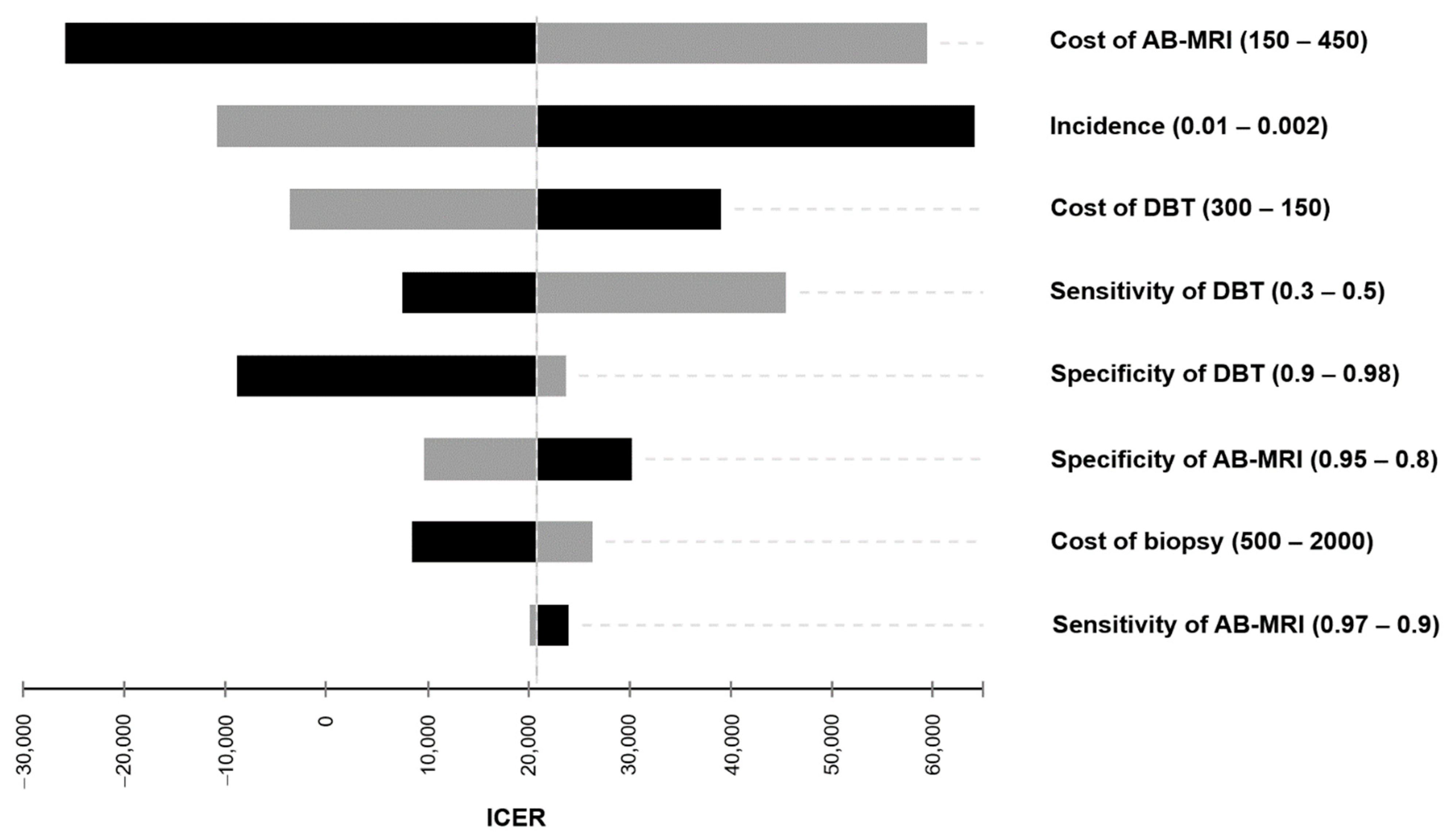
2.2.1. Deterministic Sensitivity Analysis

The results of the one-way sensitivity analysis are illustrated in Figure 1 and Table S1. Cost of AB-MRI and incidence were identified as the most influential drivers of cost-effectiveness. Considering diagnostic factors with the most impact on the ICER, the sensitivity of DBT was the biggest driver of cost-effectiveness. A variation of all input parameters within reasonable ranges resulted in AB-MRI consistently remaining cost-effective with ICER values below a WTP-threshold of $100,000.

2.2.2. Costs of AB-MRI

In the base case scenario, the costs of AB-MRI were defined to equal the costs of a full protocol breast MRI examination. The average cumulative costs of an AB-MRI screening program over a time horizon of 30 years exceeded the costs of DBT by $706 (Table 1). The cost of an AB-MRI examination was set to $314 compared to $214 for DBT (Table 2). Variations of the costs of MRI were associated with significant changes of the resulting overall cost-effectiveness. Decreasing costs of AB-MRI were associated with a smaller corresponding ICER (Figure 2). Up to examination cost of $593 for AB-MRI, the resulting ICER fell below a hypothetical WTP-threshold of $100,000. Below AB-MRI examination costs of $240.79, screening with AB-MRI may be assumed to not only be diagnostically superior, but also cheaper. The resulting cumulative costs and ICERs for varying costs of AB-MRI and DBT are reported in Table 3.

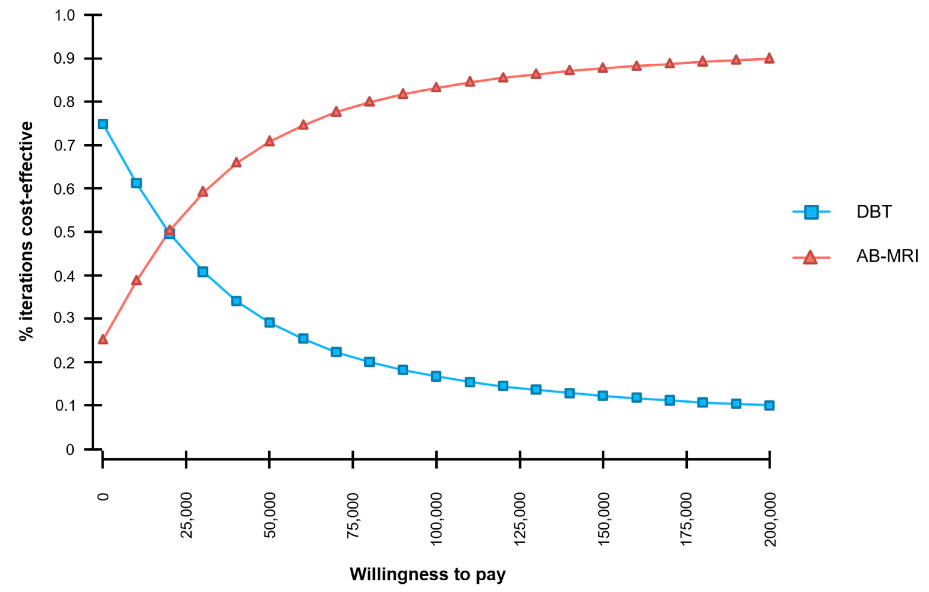
2.2.3. Probabilistic Sensitivity Analysis

The distribution of the resulting ICER-values based on the probabilistic sensitivity analysis and Monte Carlo simulation is shown in Figure 3. The majority of iterations resulted in higher average costs of AB-MRM and higher average outcomes.
The stability of the model is illustrated by a cost-effectiveness acceptability curve (Figure 4 and Table S2). At a WTP-threshold of $100,000, 83.2% of the simulations were cost-effective.

3. Discussion

In 2014, Kuhl et al. first introduced the concept of abbreviated breast MRI as a means to significantly reduce costs of MRM while maintaining an acceptable specificity [17]. In the following years, various protocols have been proposed: Non-contrast T1- and T2-weighted images followed by contrast-enhanced images, ultrafast contrast-enhanced sequences, or diffusion-weighted images as a contrast-free alternative [18,19,20,21,22,23].
At the same time, DBT was introduced in order to enhance the diagnostic performance of conventional mammography [5,24,25] and is now widely implemented in place of two-dimensional mammography.
While literature reveals a better diagnostic performance of AB-MRI [17], DBT may be significantly less expensive per examination, raising the question as to their incremental cost-effectiveness.
Recently, Comstock et al. have published the first head-to-head comparison of DBT and AB-MRI in a screening setting for women of intermediate risk of breast cancer due to their elevated levels of breast density [14]. The study’s multicenter approach addressed abbreviated protocols of various composition and length, yet all including T2-weighted and pre- and post-contrast T1-weighted images and a time limit of 10 min per examination. This data on the comparative diagnostic performance of the two screening methods now unprecedentedly allows for the economic evaluation of cost-effectiveness.
In our model-based evaluation, we are able to demonstrate that AB-MRI is not only the diagnostically superior strategy in terms of long-term outcomes, but also the more cost-effective alternative in screening women in this specific screening cohort—below the WTP-threshold of most westernized countries as described above. Our results, therefore, deem AB-MRI to be the superior screening technique in dense breasts, especially considering MRM to be a radiation-free technique.
However, the drawbacks of abbreviating MRM need to be discussed in detail: Even though Kuhl et al. initially defined the use of abbreviated protocols, current data suggest variations to be accepted forms of abbreviation (i.e., additional T2 weighted images, etc.). Depending on the composition of the abbreviated protocols, variations of the diagnostic performances in terms of specificity and various effects on cost-effectiveness must be assumed.
Further studies are required in order to examine the optimal cut-off between accuracy and costs, also with medical and economic consequences of future diagnostic or histologic clarification of unclear findings in mind (recall rates). Standardization of abbreviated protocols will ensure optimal quality of breast MRI as a screening tool.
Currently, there is no consented cost estimation or Medicare CPT code for AB-MRI. Therefore, in the base-case scenario of our analysis, the costs of AB-MRI were defined to equal the costs of a full protocol.
In the next step, the impact of varying prices per examination of AB-MRI on the overall cost-effectiveness was examined in sensitivity analyses in order to consider a range of possible cost constellations (Figure 2 and Table 3). The cost of AB-MRI was identified as one of the key determinants of cost-effectiveness in the general sensitivity analysis (Figure 1).
By reducing the cost per examination of AB-MRI below $241, the technique becomes cost-saving compared to DBT—due to the superior diagnostic performance of AB-MRI compared to DBT as well as its lower price in this case. Assuming a reasonable range of costs per examination up to $314 (current Medicare average cost of the full protocol MRI-examination), the ICER increases to $20,807 per QALY within this range.
Prior studies have demonstrated that full MRM-protocols can be considered cost-effective compared to conventional mammography in screening women at intermediate risk of breast cancer with an incremental cost-effectiveness ratio of $ 8797.60 per QALY gained [15,16]. The authors believe it may be important to emphasize that “full protocols” are equally undefined in their composition up to this point as AB-MRI protocols, also leaving a certain spectrum for economical interpretation.
Further studies should compare the diagnostic performance of varying AB-MRI- as well as varying full-MRM protocols with their consecutive economic effects, also in further patient cohorts, such as women of average risk and women with less dense breast parenchyma (density categories A & B) as opposed to extremely dense breast tissue (category D) in order to identify the diagnostic benefit for different risk-stratified subgroups.
The model-based economic evaluation is afflicted with certain limitations:
The follow-up interval reported by Comstock et al. was below two years, and long-term data on the diagnostic performance of the imaging modalities in subsequent screening rounds are not available so far.
The data used as input parameters for this study was adopted from published literature. In line with common practice in conducting economic evaluations, the corresponding authors of underlying literature were not contacted individually. Even though common practice, this may be considered a possible weakness of our study.
The United States healthcare system perspective has been selected for this economic evaluation for the purpose of standardization and comparability to other available literature. It would obviously be important to interpret our results in the light of varying input parameters of different healthcare systems, such as Europe or Asia. Screening methods, costs, and QOL might differ in other countries. However, the authors do not believe that the impact of effects to be significantly altered by international differences.
Our underlying Markov model included various possible stages of local and regional breast cancer to reflect the diversity of different stages of the disease upon detection in a screening program. Throughout the time frame of 30 years, false-positive and false-negative findings and their corresponding costs and impairments of QOL were included in the simulations as well as true positives and true negatives. Variations of the incidence of breast cancer resulted in significant effects on cost-effectiveness, which confirms the validity of the Markov model design and the assumptions in our model-based analysis. However, a Markov model can only represent simplified pathways of cancer detection and subsequent treatment. It will never accurately reflect any possible manifestation of clinical reality.

4. Materials and Methods

4.1. Screening Collective

The economic evaluation was conducted based on published data of a study investigating 1516 women at intermediate risk of breast cancer (median age of 54 years, range 40–75 years) [14], resembling an unprecedented study population. The multicenter program was dedicated to women with dense breasts (ACR BI-RADS categories C or D) and evaluated the diagnostic performance of DBT and AB-MRI. The cross-sectional study with longitudinal follow-up at 11–13 months after the study baseline was conducted at 47 institutions in the United states and 1 institution in Germany.

4.2. Cost-Effectiveness Modelling

4.2.1. Decision Model

To compare the diagnostic strategies, a decision analytic model including DBT and AB-MRI was constructed, and the respective outcomes true-positive, false-positive, true negative, and false-negative were defined for each diagnostic strategy (Figure 5a). The probability of each diagnostic result depended on the performance of the diagnostic modality as determined in the screening program [14].

4.2.2. Markov Model

To model long-term costs and outcomes of screening women for breast cancer, the Markov model designed by the publishing research group [16] was used and refined to reflect the characteristics of the screening collective and a two-yearly screening program (Figure 5b). Markov states were constructed to represent the absence of cancer, undetected breast cancer, detected malignancy, post-treatment states, and death. The cycle length was set to one year so that each Markov state was characterized by its annual costs and the associated quality of life (QoL). The screening interval was set to 2 years. A true-positive finding resulted in a timely treatment, whereas a false-negative finding consecutively resulted in delayed treatment and a higher probability of progressive disease with more extensive and costly therapy. In case of false-positive findings, unnecessary follow-ups with associated costs and impairment of quality of life (QoL) were assumed. Positive findings of DBT resulted in biopsy, whereas positive findings of AB-MRI resulted in a full protocol breast MRI examination followed by a biopsy only in case of a confirmed finding. Timely detection of breast malignancies depended on the sensitivity of the applied screening modality.

4.3. Input Parameters

For this economic evaluation comparing DBT and AB-MRI in women with dense breasts, relevant input parameters were extracted from the literature (Table 2), closely following international recommendations on the conduct of cost-effectiveness analyses [26].

4.3.1. Diagnostic Efficacy Parameters

The diagnostic performance of DBT and AB-MRI in a screening setting for women with dense breasts was recently compared [14]. Sensitivities of 95.7% and 39.1% and specificities of 86.7% and 97.4% were reported for AB-MRI and DBT, respectively.

4.3.2. Utilities

Quality of life was estimated for each Markov state based on available literature. To account for treatment-related differences in the QOL depending on the size of the tumor at the time of detection, a utility of 0.87 was applied for tumors smaller than 1 cm, a utility of 0.74 for tumors larger than 1 cm and a utility of 0.62 for regional breast cancer in an advanced stage [27,28,29]. Extensive and systemic treatment of large tumors was assumed to result in reduced QOL in the long term (0.95) compared to the QOL after treatment of localized disease (0.99). Women without detected breast cancer were assumed to have unimpaired QOL.

4.3.3. Cost Estimates

Costs of the diagnostic procedures were collected based on the Medicare-specific Current Procedural Terminology (CPT) codes of each modality and on reported average Medicare costs [24,30]. For AB-MRI examinations, Medicare costs of an MRI examination of both breasts were applied. Potential savings in MRI examination costs were evaluated in the sensitivity analysis (Figure 2). Treatment costs from a Medicare setting were also extracted from the literature depending on the stage of disease [31].

4.3.4. Transition Probabilities

Incidence rates were extracted from cancer statistics based on the Centers for Disease Control and Prevention [32]. Tumor-unrelated, age-specific death rates of women of all ethnicities in the United States were collected from U.S. Life Tables 2017 [33]. Tumor-related death rates were estimated based on the PREDICT prognostication model [34]. The probability of nodal disease and R0 resection rates were extracted from the literature [35,36].

4.4. Economic Analysis

4.4.1. Cost-Effectiveness Analysis

The model-based analyses were conducted using a dedicated decision analysis software (TreeAge Pro 2020, TreeAge Software, Williamstown, MA, USA). A United States healthcare system perspective was selected for this economic evaluation with all costs calculated in US-$. Outcomes were expressed by QALYs. Costs and outcomes were discounted at an annual discount rate of 3.0% in line with current recommendations [26]. A WTP-threshold of $100,000 per QALY was selected [37,38,39]. Using a cycle length of one-year, long-term costs and outcomes were simulated across a time frame of 30 years. All analyses complied with recommendations on the conduct and reporting of cost-effectiveness analyses (Table S3).

4.4.2. Sensitivity Analysis

A deterministic sensitivity analysis was performed to study the impact of changes to single input parameters on the incremental cost-effectiveness ratio (ICER). Costs and diagnostic performance were varied within a plausible range to emphasize their impact; the resulting ICERs were computed and visualized in a tornado diagram (Figure 1).
A probabilistic sensitivity analysis was performed to simulate the overall uncertainty of the input parameters and their combined impact on cost-effectiveness, based on the probability distributions reported in Table 2. A Monte Carlo simulation with 30,000 iterations was applied to investigate the stability of the model outputs.

5. Conclusions

Within the limits of the willingness to pay usually applied to a U.S. healthcare system perspective, i.e., $100,000 per QALY gained, AB-MRI can be considered cost-effective in comparison to DBT in screening women at intermediate risk of breast cancer due to high breast density, although various willingness to pay-thresholds have been proposed for the United States and different Western countries.

Supplementary Materials

The following are available online at https://www.mdpi.com/2072-6694/13/6/1241/s1, Table S1: Deterministic sensitivity analysis of the incremental cost-effectiveness ratio (ICER) of AB-MRI compared to DBT in the base-case scenario, Table S2: Cost-effectiveness acceptability analysis, Table S3: Consolidated Health Economic Evaluation Reporting Standards (CHEERS)-checklist.

Author Contributions

Conceptualization, P.A.T.B., M.D. and C.G.K.; Data curation, F.T., J.R., M.F.F. and C.G.K.; Formal analysis, F.T. and M.F.F.; Investigation, F.T., P.A.T.B., M.D., M.F.F. and C.G.K.; Methodology, F.T., J.R. and M.F.F.; Project administration, F.T., M.F.F. and C.G.K.; Resources, M.F.F. and C.G.K.; Software, F.T., M.F.F. and C.G.K.; Supervision, P.A.T.B., M.D. and J.R.; Visualization, F.T.; Writing–original draft, F.T. and C.G.K.; Writing–review & editing, F.T., P.A.T.B., M.D., J.R., M.F.F. and C.G.K. All authors have read and agreed to the published version of the manuscript.

Funding

This research received no external funding.

Institutional Review Board Statement

Not applicable.

Informed Consent Statement

Not applicable.

Data Availability Statement

The datasets used and/or analyzed during the current study are available from the corresponding author on reasonable request.

Conflicts of Interest

The research reported was funded by Bayer Crop Science and the researchers involved in this work were employees of Bayer Crop Science and its predecessors in business.

Abbreviations

AB-MRIAbbreviated breast MRI
DBTDigital breast tomosynthesis
ICERIncremental cost-effectiveness ratio
MRMMR-mammography
MRIMagnetic resonance imaging
PSAProbabilistic sensitivity analysis
QALYQuality-adjusted life-year
QOLQuality of life
WTPWillingness to pay

References

  1. Sprague, B.L.; Gangnon, R.E.; Burt, V.; Trentham-Dietz, A.; Hampton, J.M.; Wellman, R.D.; Kerlikowske, K.; Miglioretti, D.L. Prevalence of Mammographically Dense Breasts in the United States. J. Natl. Cancer Inst. 2014, 106. [Google Scholar] [CrossRef]
  2. Pisano, E.D.; Hendrick, R.E.; Yaffe, M.J.; Baum, J.K.; Acharyya, S.; Cormack, J.B.; Hanna, L.A.; Conant, E.F.; Fajardo, L.L.; Bassett, L.W.; et al. Diagnostic Accuracy of Digital versus Film Mammography: Exploratory Analysis of Selected Population Subgroups in DMIST. Radiology 2008, 246, 376–383. [Google Scholar] [CrossRef] [Green Version]
  3. Boyd, N.F.; Guo, H.; Martin, L.J.; Sun, L.; Stone, J.; Fishell, E.; Jong, R.A.; Hislop, G.; Chiarelli, A.; Minkin, S.; et al. Mammographic Density and the Risk and Detection of Breast Cancer. N. Engl. J. Med. 2007, 356, 227–236. [Google Scholar] [CrossRef] [PubMed] [Green Version]
  4. Bakker, M.F.; De Lange, S.V.; Pijnappel, R.M.; Mann, R.M.; Peeters, P.H.; Monninkhof, E.M.; Emaus, M.J.; Loo, C.E.; Bisschops, R.H.; Lobbes, M.B.; et al. Supplemental MRI Screening for Women with Extremely Dense Breast Tissue. N. Engl. J. Med. 2019, 381, 2091–2102. [Google Scholar] [CrossRef] [PubMed]
  5. Friedewald, S.M.; Rafferty, E.A.; Rose, S.L.; Durand, M.A.; Plecha, D.M.; Greenberg, J.S.; Hayes, M.K.; Copit, D.S.; Carlson, K.L.; Cink, T.M.; et al. Breast Cancer Screening Using Tomosynthesis in Combination with Digital Mammography. JAMA 2014, 311, 2499–2507. [Google Scholar] [CrossRef] [PubMed] [Green Version]
  6. Park, J.M.; Franken, E.A.; Garg, M.; Fajardo, L.L.; Niklason, L.T. Breast Tomosynthesis: Present Considerations and Future Applications. Radiogr. Rev. Publ. Radiol. Soc. N. Am. Inc. 2007, 27, 231–240. [Google Scholar] [CrossRef]
  7. Kaiser, C.G.; Reich, C.; Dietzel, M.; Baltzer, P.A.T.; Krammer, J.; Wasser, K.; Schoenberg, S.O.; Kaiser, W.A. DCE-MRI of the breast in a stand-alone setting outside a complementary strategy—Results of the TK-study. Eur. Radiol. 2015, 25, 1793–1800. [Google Scholar] [CrossRef]
  8. Bennani-Baiti, B.; Bennani-Baiti, N.; Baltzer, P.A. Diagnostic Performance of Breast Magnetic Resonance Imaging in Non-Calcified Equivocal Breast Findings: Results from a Systematic Review and Meta-Analysis. PLoS ONE 2016, 11, e0160346. [Google Scholar] [CrossRef]
  9. Kuhl, C.; Weigel, S.; Schrading, S.; Arand, B.; Bieling, H.B.; König, R.; Tombach, B.; Leutner, C.; Rieber-Brambs, A.; Nordhoff, D.; et al. Prospective Multicenter Cohort Study to Refine Management Recommendations for Women at Elevated Familial Risk of Breast Cancer: The EVA Trial. J. Clin. Oncol. 2010, 28, 1450–1457. [Google Scholar] [CrossRef] [Green Version]
  10. Sardanelli, F.; Podo, F.; Santoro, F.; Manoukian, S.; Bergonzi, S.; Trecate, G.; Vergnaghi, D.; Federico, M.; Cortesi, L.; Corcione, S.; et al. Multicenter surveillance of women at high genetic breast cancer risk using mammography, ultrasonography, and contrast-enhanced magnetic resonance imaging (the high breast cancer risk italian 1 study): Final results. Investig. Radiol. 2011, 46, 94–105. [Google Scholar] [CrossRef]
  11. Riedl, C.C.; Luft, N.; Bernhart, C.; Weber, M.; Bernathova, M.; Tea, M.-K.M.; Rudas, M.; Singer, C.F.; Helbich, T.H. Triple-Modality Screening Trial for Familial Breast Cancer Underlines the Importance of Magnetic Resonance Imaging and Questions the Role of Mammography and Ultrasound Regardless of Patient Mutation Status, Age, and Breast Density. J. Clin. Oncol. 2015, 33, 1128–1135. [Google Scholar] [CrossRef] [PubMed]
  12. Sardanelli, F.; Aase, H.S.; Álvarez, M.; Azavedo, E.; Baarslag, H.J.; Balleyguier, C.; Baltzer, P.A.; Beslagic, V.; Bick, U.; Bogdanovic-Stojanovic, D.; et al. Position paper on screening for breast cancer by the European Society of Breast Imaging (EUSOBI) and 30 national breast radiology bodies from Austria, Belgium, Bosnia and Herzegovina, Bulgaria, Croatia, Czech Republic, Denmark, Estonia, Finland, France, Germany, Greece, Hungary, Iceland, Ireland, Italy, Israel, Lithuania, Moldova, The Netherlands, Norway, Poland, Portugal, Romania, Serbia, Slovakia, Spain, Sweden, Switzerland and Turkey. Eur. Radiol. 2017, 27, 2737–2743. [Google Scholar] [CrossRef] [PubMed]
  13. Kuhl, C.K.; Strobel, K.; Bieling, H.; Leutner, C.; Schild, H.H.; Schrading, S. Supplemental Breast MR Imaging Screening of Women with Average Risk of Breast Cancer. Radiology 2017, 283, 361–370. [Google Scholar] [CrossRef] [PubMed]
  14. Comstock, C.E.; Gatsonis, C.; Newstead, G.M.; Snyder, B.S.; Gareen, I.F.; Bergin, J.T.; Rahbar, H.; Sung, J.S.; Jacobs, C.; Harvey, J.A.; et al. Comparison of Abbreviated Breast MRI vs Digital Breast Tomosynthesis for Breast Cancer Detection Among Women With Dense Breasts Undergoing Screening. JAMA 2020, 323, 746–756. [Google Scholar] [CrossRef]
  15. Froelich, M.F.; Kaiser, C.G. Cost-effectiveness of MR-mammography as a solitary imaging technique in women with dense breasts: An economic evaluation of the prospective TK-Study. Eur. Radiol. 2021, 31, 967–974. [Google Scholar] [CrossRef]
  16. Kaiser, C.G.; Dietzel, M.; Vag, T.; Froelich, M.F. Cost-effectiveness of MR-mammography vs. conventional mammography in screening patients at intermediate risk of breast cancer—A model-based economic evaluation. Eur. J. Radiol. 2021, 136, 109355. [Google Scholar] [CrossRef]
  17. Kuhl, C.K.; Schrading, S.; Strobel, K.; Schild, H.H.; Hilgers, R.-D.; Bieling, H.B. Abbreviated Breast Magnetic Resonance Imaging (MRI): First Postcontrast Subtracted Images and Maximum-Intensity Projection—A Novel Approach to Breast Cancer Screening With MRI. J. Clin. Oncol. 2014, 32, 2304–2310. [Google Scholar] [CrossRef]
  18. Kul, S.; Metin, Y.; Kul, M.; Metin, N.; Eyuboglu, I.; Ozdemir, O. Assessment of breast mass morphology with diffusion-weighted MRI: Beyond apparent diffusion coefficient. J. Magn. Reson. Imaging 2018, 48, 1668–1677. [Google Scholar] [CrossRef]
  19. Yamada, T.; Kanemaki, Y.; Okamoto, S.; Nakajima, Y. Comparison of detectability of breast cancer by abbreviated breast MRI based on diffusion-weighted images and postcontrast MRI. Jpn. J. Radiol. 2018, 36, 331–339. [Google Scholar] [CrossRef]
  20. Goto, M.; Sakai, K.; Yokota, H.; Kiba, M.; Yoshida, M.; Imai, H.; Weiland, E.; Yokota, I.; Yamada, K. Diagnostic performance of initial enhancement analysis using ultra-fast dynamic contrast-enhanced MRI for breast lesions. Eur. Radiol. 2018, 29, 1164–1174. [Google Scholar] [CrossRef]
  21. Grimm, L.J.; Soo, M.S.; Yoon, S.; Kim, C.; Ghate, S.V.; Johnson, K.S. Abbreviated screening protocol for breast MRI: A feasibility study. Acad Radiol. 2015, 22, 1157–1162. [Google Scholar] [CrossRef]
  22. Moschetta, M.; Telegrafo, M.; Rella, L.; Ianora, A.A.S.; Angelelli, G. Abbreviated Combined MR Protocol: A New Faster Strategy for Characterizing Breast Lesions. Clin. Breast Cancer 2016, 16, 207–211. [Google Scholar] [CrossRef]
  23. Strahle, D.A.; Pathak, D.R.; Sierra, A.; Saha, S.; Strahle, C.; Devisetty, K. Systematic development of an abbreviated protocol for screening breast magnetic resonance imaging. Breast Cancer Res. Treat. 2017, 162, 283–295. [Google Scholar] [CrossRef] [Green Version]
  24. Fleming, M.M.; Hughes, D.R.; Golding, L.P.; McGinty, G.B.; Macfarlane, D.; Duszak, R. Digital Breast Tomosynthesis Implementation: Considerations for Emerging Breast Cancer Screening Bundled Payment Models. J. Am. Coll. Radiol. 2019, 16, 902–907. [Google Scholar] [CrossRef]
  25. Chong, A.; Weinstein, S.P.; McDonald, E.S.; Conant, E.F. Digital Breast Tomosynthesis: Concepts and Clinical Practice. Radiology 2019, 292, 1–14. [Google Scholar] [CrossRef]
  26. Sanders, G.D.; Neumann, P.J.; Basu, A.; Brock, D.W.; Feeny, D.; Krahn, M.; Kuntz, K.M.; Meltzer, D.O.; Owens, D.K.; Prosser, L.A.; et al. Recommendations for Conduct, Methodological Practices, and Reporting of Cost-effectiveness Analyses: Second Panel on Cost-Effectiveness in Health and Medicine. JAMA 2016, 316, 1093–1103. [Google Scholar] [CrossRef]
  27. Ahern, C.H.; Shih, Y.-C.T.; Dong, W.; Parmigiani, G.; Shen, Y. Cost-effectiveness of alternative strategies for integrating MRI into breast cancer screening for women at high risk. Br. J. Cancer 2014, 111, 1542–1551. [Google Scholar] [CrossRef] [PubMed] [Green Version]
  28. Brady, M.J.; Cella, D.F.; Mo, F.E.; Bonomi, A.; Tulsky, D.S.; Lloyd, S.R.; Deasy, S.; Cobleigh, M.; Shiomoto, G. Reliability and validity of the Functional Assessment of Cancer Therapy-Breast quality-of-life instrument. J. Clin. Oncol. 1997, 15, 974–986. [Google Scholar] [CrossRef] [PubMed]
  29. Polsky, D.; Mandelblatt, J.S.; Weeks, J.C.; Venditti, L.; Hwang, Y.-T.; Glick, H.A.; Hadley, J.; Schulman, K.A. Economic Evaluation of Breast Cancer Treatment: Considering the Value of Patient Choice. J. Clin. Oncol. 2003, 21, 1139–1146. [Google Scholar] [CrossRef] [Green Version]
  30. Hunter, S.A.; Morris, C.; Nelson, K.; Snyder, B.J.; Poulton, T.B. Digital Breast Tomosynthesis: Cost-Effectiveness of Using Private and Medicare Insurance in Community-Based Health Care Facilities. Am. J. Roentgenol. 2017, 208, 1–5. [Google Scholar] [CrossRef] [PubMed]
  31. Blumen, H.; Fitch, K.; Polkus, V. Comparison of Treatment Costs for Breast Cancer, by Tumor Stage and Type of Service. Am. Heal. Drug Benefits 2016, 9, 23–32. [Google Scholar]
  32. Richardson, L.C.; Henley, S.J.; Miller, J.W.; Massetti, G.; Thomas, C.C. Patterns and Trends in Age-Specific Black-White Differences in Breast Cancer Incidence and Mortality—United States, 1999–2014. MMWR. Morb. Mortal. Wkly. Rep. 2016, 65, 1093–1098. [Google Scholar] [CrossRef] [Green Version]
  33. Arias, E. United States Life Tables, 2017. Natl. Vital. Stat. Rep. Cent. Dis. Control. Prev. 2019, 68, 1–66. [Google Scholar]
  34. Wishart, G.C.; Azzato, E.M.; Greenberg, D.C.; Rashbass, J.; Kearins, O.; Lawrence, G.; Caldas, C.; Pharoah, P.D. Predict: A new UK prognostic model that predicts survival following surgery for invasive breast cancer. Breast Cancer Res. BCR. 2010, 12, 1–10. [Google Scholar] [CrossRef] [Green Version]
  35. Heil, J.; Rauch, G.; Szabo, A.Z.; Garcia-Etienne, C.A.; Golatta, M.; Domschke, C.; Badiian, M.; Kern, P.; Schuetz, F.; Wallwiener, M.; et al. Breast Cancer Mastectomy Trends Between 2006 and 2010: Association with Magnetic Resonance Imaging, Immediate Breast Reconstruction, and Hospital Volume. Ann. Surg. Oncol. 2013, 20, 3839–3846. [Google Scholar] [CrossRef]
  36. Lombardi, A.; Pastore, E.; Maggi, S.; Stanzani, G.; Vitale, V.; Romano, C.; Bersigotti, L.; Vecchione, A.; Amanti, C. Positive margins (R1) risk factors in breast cancer conservative surgery. Breast Cancer Targets Ther. 2019, 11, 243–248. [Google Scholar] [CrossRef] [Green Version]
  37. Woods, B.; Revill, P.; Sculpher, M.; Claxton, K. Country-Level Cost-Effectiveness Thresholds: Initial Estimates and the Need for Further Research. Value Heal. 2016, 19, 929–935. [Google Scholar] [CrossRef] [PubMed] [Green Version]
  38. Neumann, P.J.; Cohen, J.T.; Weinstein, M.C. Updating Cost-Effectiveness—The Curious Resilience of the $50,000-per-QALY Threshold. N. Engl. J. Med. 2014, 371, 796–797. [Google Scholar] [CrossRef] [PubMed] [Green Version]
  39. Cameron, D.; Ubels, J.; Norström, F. On what basis are medical cost-effectiveness thresholds set? Clashing opinions and an absence of data: A systematic review. Glob. Health Action 2018, 11, 1447828. [Google Scholar] [CrossRef] [PubMed] [Green Version]
Figure 1. Tornado diagram of the one-way sensitivity analysis. Input parameters were varied within a reasonable range to indicate their impact on the incremental cost-effectiveness ratio (ICER) in US-$ per QALY gained.
Cancers 13 01241 g001
Figure 2. One-way sensitivity analysis for the cost of AB-MRI (US-$) and the corresponding incremental cost-effectiveness ratio (ICER in US-$ per QALY gained) compared to DBT. Screening with AB-MRI becomes cost-saving when the cost per examination of AB-MRI is reduced below $240.79.
Cancers 13 01241 g002
Figure 3. Cost-effectiveness scatter plot. Incremental costs (US-$) and effectiveness (quality-adjusted life-years, QALYs) when comparing AB-MRI to DBT as determined in a Monte Carlo simulation. The majority of the iterations fall below the willingness to pay (WTP)-threshold of $100,000 per QALY.
Cancers 13 01241 g003
Figure 4. Cost-effectiveness acceptability curve based on a probabilistic sensitivity analysis and Monte Carlo simulation with 30,000 iterations. At a willingness to pay (WTP)-threshold of $100,000 per QALY, AB-MRI was cost-effective in 83.2% of the iterations.
Cancers 13 01241 g004
Figure 5. (a) Decision-tree including both screening strategies and the corresponding outcomes. (b) Markov Model: Probability of breast malignancy detection depends on the sensitivity of the screening modality and the time since development of the disease. Corresponding quality of life (QOL) for the Markov states is indicated in the boxes.
Cancers 13 01241 g005
Table 1. Results of base case cost-effectiveness analysis. Discounted average cumulative costs and effects were modeled for a time horizon of 30 years. ICER, incremental cost-effectiveness ratio; QALY, quality-adjusted life-year.
StrategyCumulative Costs (US-$)Incremental Costs (US-$)Cumulative Effects (QALYs)Incremental Effects (QALYs)ICER
(US-$/QALY)
DBT8798.47n/a19.23n/an/a
AB-MRI9504.77706.3019.270.0320,807.04
Table 2. Model input parameters.
VariableEstimationSourceDistribution
Pre-test probability of malignant lesion1.59%Comstock et al. 2020β
Average age at screening54.9Comstock et al. 2020normal
Screening intervaltwo yearsAssumption
Incidence of breast cancer in women with dense breasts0.40%Richardson et al. 2016β
Assumed WTP$100,000Sanders et al. 2016
Discount rate3.00%Sanders et al. 2016
Diagnostic test performances
DBT sensitivity39.1%Comstock et al. 2020β
DBT specificity97.4%Comstock et al. 2020β
AB-MRI sensitivity95.7%Comstock et al. 2020β
AB-MRI specificity86.7%Comstock et al. 2020β
Full protocol MR mammography specificity94.0%Benndorf et al. 2010β
Costs (Short Term)
DBT costs$214.20Fleming et al. 2018/
Hunter et al. 2017
γ
MRM costs$314.00Medicare
(CPT code 77047)
γ
No further action (true negative) $0.00 Assumption
Biopsy$1536.00Medicare
(CPT code 19083)
γ
Costs (Long Term)
Yearly costs without tumor$0.00Assumption
Cost of treatment for tumor < 1 cm$60,637Blumen et al. 2015γ
Cost of treatment for tumor > 1 cm$82,121Blumen et al. 2015γ
Cost of treatment for advanced stage breast malignancy$129,387Blumen et al. 2015γ
Utilities
QOL of patients without detected tumor1.00Assumption
QOL of patients with detected tumor < 1 cm0.87Brady et al. 1997β
QOL of patients with detected tumor > 1 cm0.74Ahern et al. 2014β
QOL of patients with detected regional breast cancer in an advanced stage0.62Polsky et al. 2003β
QOL of patients post simple treatment0.99Assumptionβ
QOL of patients post intensive treatment0.95Assumptionβ
Death0.00Assumption
Transition probabilities
Risk of death without tumor (yearly)age-adjustedUS Life Tables 2017,
women of all ethnicities,
Arias et al. 2019
β
Risk of death with undetected tumor10.00% in 10 yearsAssumptionβ
Risk of death with detected < 1 cm tumor0.11%NHS Predictβ
Risk of death with detected > 1 cm tumor0.78%NHS Predictβ
Risk of death with detected tumor in advanced stage1.81%NHS Predictβ
Probability of initial R0 resection < 1 cm100.00%Assumption
Probability of initial R0 resection ≥ 1 cm90.00%Lombardi et al. 2019β
Proportion of N+ in < 1 cm tumors0.00%Assumption
Proportion of N+ in > 1 cm tumors40.00%Heil et al. 2013β
Proportion of successfully treated tumors < 1 cm if detected within 1 screening interval100.00%Assumption
Table 3. Discounted cumulative costs of screening with DBT and AB-MRI (US-$) and the resulting cost-effectiveness expressed by the incremental cost-effectiveness ratio (ICER in US-$ per QALY gained) for varying costs of the diagnostic procedures. AB-MRI, abbreviated breast MRI; DBT, digital breast tomosynthesis.
AB-MRI
(US-$)
DBT (US-$)
180200220240260
2108468.90,
8501.48,
959.55
8661.63,
8501.48,
cost-saving
8854.36,
8501.48,
cost-saving
9047.09,
8501.48,
cost-saving
9239.82,
8501.48,
cost-saving
2308468.90,
8694.42,
6643.46
8661.63,
8694.42,
965.78
8854.36,
8694.42,
cost-saving
9047.09,
8694.42,
cost-saving
9239.82,
8694.42,
cost-saving
2508468.90,
8887.36,
12,327.37
8661.63,
8887.36,
6649.69
8854.36,
8887.36,
972.01
9047.09,
8887.36,
cost-saving
9239.82,
8887.36,
cost-saving
2708468.90,
9080.30,
18,011.27
8661.63,
9080.30,
12,333.59
8854.36,
9080.30,
6655.92
9047.09,
9080.30,
978.24
9239.82,
9080.30,
cost-saving
2908468.90,
9273.24,
23,695.18
8661.63,
9273.24,
18,017.50
8854.36,
9273.24,
12,339.82
9047.09,
9273.24,
6662.15
9239.82,
9273.24,
984.47
3108468.90,
9466.18,
29,379.08
8661.63,
9466.18,
23,701.41
8854.36,
9466.18,
18,023.73
9047.09,
9466.18,
12,346.05
9239.82,
9466.18,
6668.38
3308468.90,
9659.12,
35,062.99
8661.63,
9659.12,
29,385.31
8854.36,
9659.12,
23,707.64
9047.09,
9659.12,
18,029.96
9239.82,
9659.12,
12,352.28
3508468.90,
9852.06,
40,746.89
8661.63,
9852.06,
35,069.22
8854.36,
9852.06,
29,391.54
9047.09,
9852.06,
23,713.86
9239.82,
9852.06,
18,036.19
Publisher’s Note: MDPI stays neutral with regard to jurisdictional claims in published maps and institutional affiliations.

