Article

Outcome and Safety after 103 Radioembolizations with Yttrium-90 Resin Microspheres in 73 Patients with Unresectable Intrahepatic Cholangiocarcinoma—An Evaluation of Predictors

1
Department of Diagnostic and Interventional Neuroradiology, Technical University Munich, 81675 Munich, Germany
2
Department of Clinical Radiology, LMU—University of Munich, 81377 Munich, Germany
3
Department of Interventional Radiology, Technical University Munich, 81377 Munich, Germany
4
Department of Nuclear Medicine, LMU—University of Munich, 81377 Munich, Germany
5
Department of Oncology, LMU—University of Munich, 81377 Munich, Germany
*
Author to whom correspondence should be addressed.
Cancers 2021, 13(21), 5399; https://doi.org/10.3390/cancers13215399
Submission received: 7 September 2021 / Revised: 22 October 2021 / Accepted: 26 October 2021 / Published: 27 October 2021
(This article belongs to the Special Issue Radionuclide Therapy and Hybrid Imaging in Oncology)

Abstract

:

Simple Summary

TARE with yttrium-90 (90Y) resin microspheres is emerging in many countries as a treatment option for ICC. Identification of patients that will benefit from TARE is a clinically relevant problem with individual but also economical relevance. The aim of this study was to detect outcome predictors for patients with ICC after TARE with 90Y resin microspheres. We found TARE with 90Y resin microspheres to be a safe treatment option for unresectable ICC. Predictive factors for TARE in ICC are CA-19-9 response, tumor burden, and cholinesterase. Multiple TARE sessions might further improve overall survival.

Abstract

Trans-arterial radioembolization (TARE) is increasingly evaluated for unresectable intrahepatic cholangiocarcinoma (ICC). Not all ICC patients benefit equally well from TARE. Therefore, we sought to evaluate variables predicting progression-free survival (PFS) and overall survival (OS). Patients with non-resectable ICC underwent TARE and were treated with 90Y resin microspheres. Baseline characteristics, biochemical/clinical toxicities, and response were examined for impact on PFS and OS. A total of 103 treatments were administered to 73 patients without major complications or toxicity. Mean OS was 18.9 months (95% confidence intervals (CI); 13.9–23.9 months). Mean and median PFS were 10.1 months (95% CI; 7.9–12.2) and 6.4 months (95% CI; 5.20–7.61), respectively. Median OS and PFS were significantly prolonged in patients with baseline cholinesterase (CHE) ≥ 4.62 kU/L (OS: 14.0 vs. 5.5 months; PFS: 6.9 vs. 3.2 months; p < 0.001). Patients with a tumor burden ≤ 25% had a significantly longer OS (15.2 vs. 6.6 months; p = 0.036). Median PFS was significantly longer for patients with multiple TARE cycles (24.4 vs. 5.8 months; p = 0.04). TARE is a considerable and safe option for unresectable ICC. CA-19-9, CHE, and tumor burden have predictive value for survival in patients treated with TARE. Multiple TARE treatments might further improve survival; this has to be confirmed by further studies.

1. Introduction

Intrahepatic cholangiocarcinoma (ICC) is a rare disease with approximately 3000 cases diagnosed every year in the USA [1,2]. ICC is the second most common primary liver cancer and accounts for approximately 10–20% of all primary liver tumors [3]. ICC is often clinically silent in its early stages, and patients are diagnosed when surgery is no longer possible. As a consequence, prognosis is poor (5-year survival < 5%). Surgical treatments (liver transplantation or complete surgical resection) are currently the only potentially curative therapy. Nevertheless, only 20% of patients with ICC are eligible for resection due to anatomic location, disease spread, inadequate hepatic reserve, or limiting comorbidities [4,5,6]. Median survival for patients with untreated unresectable ICC has been reported to be between 3–6 months [6,7]. It is of note that survival for liver-only ICC is relevantly different from other bile duct cancers [8].
Based on a phase III trial, ABC-02 cisplatin-gemcitabine is the standard of care for ICC [9]. Still, median survival is <1 year [9]. Interventional therapy aims to control local tumor growth, relieve symptoms, and improve and preserve quality of life. Locoregional therapies, including trans-arterial chemoembolization (TACE) or radiofrequency ablation (RFA) have been proposed [10]. Despite their role in the therapy regimen of hepatocellular cancer (HCC), they are currently not the standard of care in locally advanced ICC [11]. Due to the relative radio-sensitivity of the liver, external beam radiation therapy (EBRT) has a limited role in the treatment of liver malignancies [12]. Trans-arterial radioembolization was effective in unresectable HCC and liver-dominant metastasis of neuroendocrine tumors (NET) [13], colorectal cancer (CRC), and metastatic breast cancer (MBC) [13,14]. Therefore, as ICC and HCC are generally radiosensitive, TARE with yttrium-90 (90Y) resin microspheres is emerging in many countries as a treatment option for ICC [13,15,16,17]. However, the response to TARE in ICC can be heterogeneous for various reasons. Further, identification of patients that will benefit from TARE is a clinically relevant problem with individual but also economical relevance [18].
The aim of our study was to detect outcome predictors for patients with ICC after TARE with 90Y resin microspheres (SIR-Spheres; Sirtex Medical Ltd., Sydney, Australia).

2. Materials and Methods

2.1. Patient Selection

We retrospectively analyzed 73 consecutive patients (103 TARE procedures) with unresectable ICC who were treated with 90Y resin microspheres.
Inclusion criteria were defined as follows: non-resectable ICC determined in an interdisciplinary tumor board; absence of significant extrahepatic disease (EHD) (patients with stable intra-abdominal lymph nodes were included, progression of lymph nodes or other kind of metastases was considered significant); failure to respond to other types of medical, surgical, or local ablative treatment modalities; no portal vein occlusion; adequate biochemical and hematological function (total bilirubin < 2 mg/dL, albumin and pseudocholinesterase within normal range, and sufficient coagulation); no relevant comorbidities; and written, informed consent. The study was approved by the local ethics committee.

2.2. Dosimetry for Radioembolization

The dose was calculated with the modified body surface area (mBSA) method [19]. mBSA was recommended by a consensus report as the most appropriate method avoiding the rare occurrence of radioembolization-induced liver disease (REILD).

2.3. Radioembolization Procedure

Within a month before TARE, patients underwent hepatic angiography with application of 100 MBq of 99mTc-macroaggregated albumin (MAA) to map the hepatic arterial tree and to detect and occlude relevant aberrant vessels where necessary using micro-coil embolization, and to calculate the shunt fraction of labeled microspheres to the lung.
During a subsequent hepatic arterial catheterization, 90Y resin microspheres suspended in water for injection were injected under intermittent fluoroscopic visualization, alternating with a contrast medium to preserve antegrade hepatic arterial flow. The prescribed activity was administered either in the whole liver, lobe, or as a sequential lobar treatment, according to tumor burden [20]. Within 24 h of therapy, single photon emission computed tomography scans (Bremsstrahlungs SPECT/CT) were performed to confirm target deposition of microspheres.

2.4. Follow-Up: Response, Toxicity, Survival

After TARE, tumor response was assessed by contrast-enhanced computed tomography (CT), magnetic resonance imaging (MRI), or PET-CT using the Response Evaluation Criteria in Solid Tumors (RECIST) [21]. Response was evaluated by two experienced radiologists. Up to five lesions were measured. Minor differences in quantification of the diameters of the lesions did not lead to differences in the tumor response scoring.
Pre- and post-treatment laboratory tests included liver function tests (bilirubin, albumin, serum cholinesterase [CHE], thrombocytes, C-reactive protein [CRP], and tumor marker [CA 19-9]). Patients remained in the hospital for 4–5 days after TARE with daily clinical and laboratory evaluation. In addition, laboratory tests and imaging were performed after 3 months. Acute and late adverse events were recorded at each visit during follow-up. Classification of adverse events was performed according to common terminology criteria for adverse events (CTCAE) version 5.0 [22]. Telephone follow-up with the referring physicians or the patients was used to capture survival data.

2.5. Statistical Analyses

Continuous variables are presented as means, medians, and interquartile range, and categorical variables as frequencies and percentages. Median progression-free survival (PFS) was calculated in months between the date of the first TARE until radiological progression or all-cause death, whichever occurred first. Median overall survival (OS) was calculated in months from the date of the first TARE until all-cause death. PFS and OS were evaluated using the Kaplan-Meier method and the univariate Cox proportional hazard method. Between-group differences in PFS and OS were assessed with log-rank tests for the variables age, sex, tumor burden, tumor response according to RECIST, number of TARE cycles, CA-19-9 response status, and baseline levels of bilirubin and CHE; CA-19-9 response was deemed to be achieved with a ≥30% decrease at 3 months compared with baseline value of CA-19-9.
The level of significance was set at p ≤ 0.05. The software package IBM SPSS Statistics for Windows, Version 20.0, released 2011 (IBM Corp., Armonk, NY, USA) was used.

3. Results

Seventy-three patients (33 women, 40 men, mean age of 64.5 (range 29.7–91.8 years)) with histologically confirmed unresectable ICC) underwent TARE as described above (Table 1).
The median total activity of 90Y resin microspheres delivered was 1.5 GBq (range, 0.4–2.5 GBq). A total of 103 treatment sessions were performed, including 25 single-session whole-liver administrations, 25 one-lobe single-session treatments, 17 (=34 TARE sessions) whole-liver treatments in two sessions and 6 cases with more than two treatment sessions (Table 2). Patients with multiple treatments underwent on average 2.2 sessions. Median time between the repetitive TARE cycles was 9.6 months (294 days).

3.1. Outcome—Treatment Response (RECIST)

Partial response (PR), stable disease (SD), and progressive disease (PD) were observed after 3 months in 18 (25%), 36 (49%), and 19 (26%) patients, respectively.

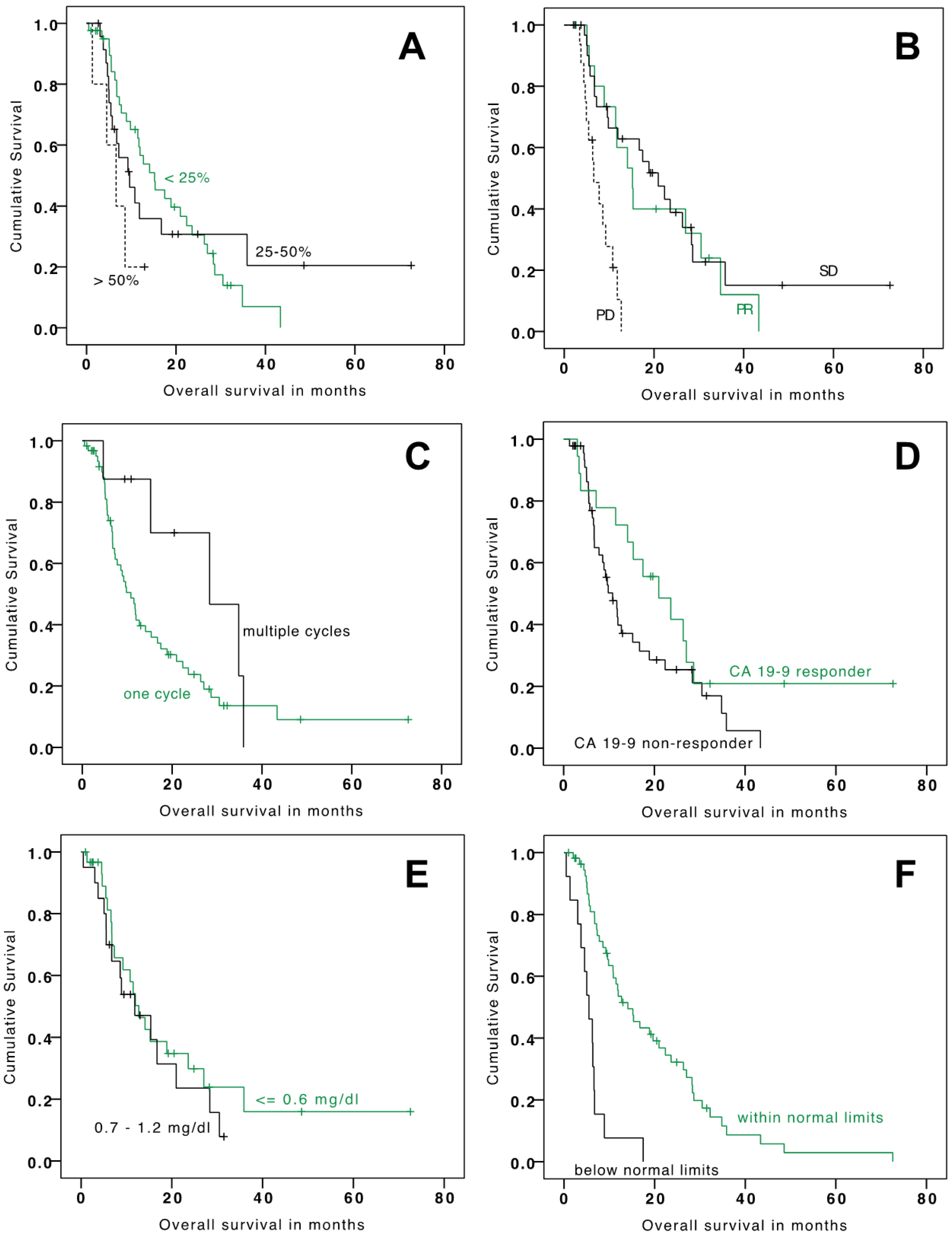
3.2. Outcome—Survival

The mean and median OS since TARE were 18.9 months (95% CI: 13.9–23.9) and 11.8 months (95% CI: 7.3–16.3), respectively (Table 3). Mean and median PFS were 10.1 months (95% CI: 7.9–12.2) and 6.4 months (95% CI: 5.2–7.6), respectively (Table 3). Factors that significantly prolonged median OS included tumor burden ≤ 25% at baseline versus tumor burden > 50%, a PR versus PD at 3 months, and baseline CHE ≥ 4.62 kU/L versus CHE < 4.62 kU/L (Table 3 and Figure 1). Factors that significantly prolonged median PFS included a PR versus PD at 3 months, baseline CHE ≥ 4.62 kU/L (lower limit of normal values) versus CHE < 4.62 kU/L and multiple treatment cycles versus one treatment cycle (Table 3 and Figure 2). It is of note that patients with repetitive TARE-sessions had a relevant prolonged overall survival (28.4 vs. 10.8 months). Progression-free survival was also relevantly better for patients with repetitive TARE (24.4 vs. 5.8 months).
Overall, 20 patients (27%) had a CA-19-9 response. A trend towards improved OS was observed for patients with a CA-19-9 response compared with those without a response (Table 3). At discharge CHE, albumin and thrombocyte levels decreased from baseline and subsequently increased (thrombocyte levels showed further decline) after 3 months but remained below baseline levels (Table 4). Bilirubin and CRP levels increased from baseline to time of discharge (Table 4). From discharge to month 3, the bilirubin value remained stable, whereas CRP decreased to levels closer to baseline (Table 4).

3.3. Safety

The procedure was associated with an acceptable toxicity profile (<Grade 4; no life-threatening events) including nausea and pain being the most frequent adverse events. No REILD was noted (Table 5). No adverse events attributed to the interventions themselves were observed.

4. Discussion

The main findings of this study are as follows: (i) trans-arterial radio-embolization (TARE) appears to improve overall survival in patients with irresectable intrahepatic cholangiocarcinoma (ICC) compared to the reported survival with standard care in the literature; (ii) CA-19-9, cholinesterase, and tumor burden have predictive value for survival in those patients treated with TARE; (iii) Multiple TARE treatments might further improve survival.

4.1. Outcomes, Previous Results, and Predictors

Effective treatment of irresectable ICC is clinically challenging, resulting in increased interest in TARE therapy in these patients [15,17,18,20]. Results from those studies highly implicate improved survival for patients treated with TARE. The results of our study exhibit an improved mean survival compared to standard care, which is also in line with previously reported outcomes [6,7,20]. Although current data suggest that TARE is a considerable treatment option, identification of patients that benefit from this specific treatment can be complicated. Previous papers have reported tumor burden as a predictive parameter in patients with TARE and ICC [16]. Similarly, in our study tumor burden was highly predictive and further cholinesterase exhibited good predictive value. Cholinesterase can be considered to be an inverse surrogate for tumor burden but also for the amount of functioning liver parenchyma. Previous chemotherapy, resection, ablation, or liver disease might also have reduced the amount of functioning parenchyma. As a result, doses for TARE might require relevant reduction below effective treatment levels to avoid radioembolization-induced liver disease, possibly also explaining the dependency of cholinesterase and survival with TARE next to other confounders. We observed improved survival (overall survival and progression-free) in patients with multiple TARE and sequential treatments for the same ICC lesions indicating a possible increased effectiveness of aggressive and sequential TARE treatment strategy. This finding has not been reported before and the effects are quite relevant in our study. However, we should not exclude the possibility that this effect is a result of the absence of treatment alternatives in the study population leading to increased numbers of TARE sessions in patients with longer survival. The causality remains somewhat unclear; patients with better survival might be treated more often because of their longer survival.
We further confirm the response of CA19-9 to prior chemotherapy to be a relevant predictor for survival in patients with ICC and TARE, which is comparable to results published before but in standard-care patients without TARE [23]. However, it can be speculated that CA19-9 response itself indicates, e.g., a certain genetic subtype of the tumor that is more susceptible to cytotoxic treatments in general and thus indicates whether the tumor might respond to TARE. In addition, tumor response according to RECIST criteria after 3 months was a factor associated with better survival. Other factors included in our analysis, such as patient age, sex, or bilirubin levels did not have an impact on patient outcome.
In this study, decreased CHE levels below reference values is a negative predictor for both OS and PFS. This is based on the assumption that CHE is a negatively correlated surrogate for the tumor burden. Large tumor masses with extensive liver infiltrations can impair liver function and as a consequence lead to a decrease in the unspecific liver synthesis parameter CHE.

4.2. Safety of TARE in ICC

Mouli et al. [24] reported about 46 patients (35% and 15% with previous chemotherapy or liver-directed therapies) who underwent TARE with glass spheres. Disease control (WHO imaging criteria) was reported in 98% of all treated patients, and >50% necrosis on follow-up imaging (EASL guidelines) in 73% of patients. Major post-treatment complications were fatigue (54%) and abdominal pain (28%), which is in line with our and other studies about TARE for hepatic tumors [13,25]. In this present study we observed no procedural complications; no life-threatening adverse events occurred (<Grade 4; no life-threatening events).

4.3. Treatment Alternatives

Other minimally invasive treatment options including RFA and TACE have been investigated, and RFA is known to be an effective treatment for focal liver lesions [26]. A recent study investigating RFA in ICC reports promising results and also prolonged survival compared to standard care [27]. In comparison to our results with TARE, complication rate and gravidity of the complications was higher for RFA. Several studies differing in quality have examined the use of TACE for patients with unresectable ICC [28,29,30]. In the prospective study of Kiefer et al. [28] with 62 ICC patients, PR, SD, and PD according to RECIST were reported in 11%, 64%, and 24% of patients, respectively. Five patients suffered from major complications. Post-embolization syndrome (CTCAE grade 1 or higher of post-procedural pain, fever, nausea, or vomiting) occurred in 65% of all patients. Median OS and median time to progression (TTP) after first chemoembolization were 15 months and 8 months, respectively. The potential procedural risk from catheterization for patients with TACE and TARE are similar. In comparison, per-session costs for TACE are considerably lower compared to TARE. However, TACE is not eligible for intermediate to increased tumor burden or diffuse tumor distribution.
TARE could be considered as an alternative to TACE in patients with increased tumor burden or in patients not suited to TACE [25].
A recent systematic review compared outcomes of different treatment methods in ICC. Even though the quality of available data is limited, ablation seems to be the best option in unresectable lesions. However, not all lesions are eligible for ablation. In tumors that are large or unfavorably located for ablation, trans-arterial therapies seem to play an important role [31].
Edeline et al. demonstrated improved survival for patients in ICC for a combination of TARE and chemotherapy in their prospective study and concluded that this combination is a valuable treatment option. This underlines that improved survival for patients with ICC requires an interdisciplinary and elaborated treatment strategy involving interventional radiologists, oncologists, and surgeons [17].

4.4. Limitations

This was a retrospective single-center study. As many patients received therapies before TARE (71% treated with chemotherapy, 31% treated with surgery, and 7% treated with RFA or TACE), attribution of effect sizes might by impaired. Subgroup analyses of OS and PFS for patients with different previous treatments might yield different or further results but has been waived because of the small sample sizes in the subgroups.

5. Conclusions

TARE with 90Y resin microspheres is a safe treatment option for unresectable ICC. Predictive factors for TARE in ICC are CA-19-9 response, tumor burden, and cholinesterase. Multiple TARE sessions might further improve overall survival; this has to be confirmed by further studies. Prospective randomized trials comparing TACE, TARE, RFA, and systemic chemotherapy will be necessary to further improve therapy for unresectable ICC.

Author Contributions

Conceptualization, P.M.P., and K.J.P.; methodology, K.J.P., P.M.P., and J.N.; software, M.I.; validation, T.G.; formal analysis, K.J.P., M.I., M.M., and F.G.; investigation, H.I.; M.M data curation, F.G.; writing—original draft preparation, J.N., A.T., and K.J.P.; writing—review and editing, J.N., T.G., H.I., and P.M.P.; visualization, A.T.; supervision, P.M.P. All authors have read and agreed to the published version of the manuscript.

Funding

This research received no external funding.

Institutional Review Board Statement

The study was conducted according to the guidelines of the Declaration of Helsinki, and approved by the Institutional Review Board (or Ethics Committee) of ETHIKKOMMISSION DER LMU MÜNCHEN on 09/2016 (ref. 617-16).

Informed Consent Statement

Informed consent was obtained from all subjects involved in the study.

Data Availability Statement

The data presented in this study are available on request from the corresponding author. The data are not publicly available due to data protection regulations.

Conflicts of Interest

The authors declare no conflict of interest.

References

  1. Jemal, A.; Siegel, R.; Ward, E.; Hao, Y.; Xu, J.; Thun, M.J. Cancer statistics, 2009. CA A Cancer J. Clin. 2009, 59, 225–249. [Google Scholar] [CrossRef]
  2. Patel, T. Increasing incidence and mortality of primary intrahepatic cholangiocarcinoma in the United States. Hepatology 2001, 33, 1353–1357. [Google Scholar] [CrossRef] [PubMed]
  3. Blechacz, B.; Gores, G.J. Cholangiocarcinoma: Advances in pathogenesis, diagnosis, and treatment. Hepatology 2008, 48, 308–321. [Google Scholar] [CrossRef] [Green Version]
  4. Yamamoto, J.; Kosuge, T.; Takayama, T.; Shimada, K.; Makuuchi, M.; Yoshida, J.; Sakamoto, M.; Hirohashi, S.; Yamasaki, S.; Hasegawa, H. Surgical treatment of intrahepatic cholangiocarcinoma: Four patients surviving more than five years. Surgery 1992, 111, 617–622. [Google Scholar] [PubMed]
  5. Lieser, M.J.; Barry, M.K.; Rowland, C.; Ilstrup, D.M.; Nagorney, D.M. Surgical management of intrahepatic cholangiocarcinoma: A 31-year experience. J. Hepato-Biliary-Pancreat. Surg. 1998, 5, 41–47. [Google Scholar] [CrossRef]
  6. Khan, S.A.; Thomas, H.C.; Davidson, B.R.; Taylor-Robinson, S.D. Cholangiocarcinoma. Lancet 2005, 366, 1303–1314. [Google Scholar] [CrossRef]
  7. Cunningham, S.C.; Choti, M.A.; Bellavance, E.C.; Pawlik, T.M. Palliation of hepatic tumors. Surg. Oncol. 2007, 16, 277–291. [Google Scholar] [CrossRef] [PubMed]
  8. Lamarca, A.; Ross, P.; Wasan, H.S.; Hubner, R.A.; McNamara, M.G.; Lopes, A.; Manoharan, P.; Palmer, D.; Bridgewater, J.; Valle, J.W. Advanced intrahepatic cholangiocarcinoma: Post hoc analysis of the ABC-01,-02, and-03 clinical trials. J. Natl. Cancer Inst. 2020, 112, 200–210. [Google Scholar] [CrossRef]
  9. Valle, J.; Wasan, H.; Palmer, D.H.; Cunningham, D.; Anthoney, A.; Maraveyas, A.; Madhusudan, S.; Iveson, T.; Hughes, S.; Pereira, S.P.; et al. Cisplatin plus gemcitabine versus gemcitabine for biliary tract cancer. N. Engl. J. Med. 2010, 362, 1273–1281. [Google Scholar] [CrossRef] [Green Version]
  10. Tepel, J.; Hinz, S.; Klomp, H.J.; Kapischke, M.; Kremer, B. Intraoperative radiofrequency ablation (RFA) for irresectable liver malignancies. Eur. J. Surg. Oncol. (EJSO) 2004, 30, 551–555. [Google Scholar] [CrossRef]
  11. Bruix, J.; Sherman, M. Practice Guidelines Committee, American Association for the Study of Liver Diseases. Management of hepatocellular carcinoma. Hepatology 2005, 42, 1208–1236. [Google Scholar] [CrossRef] [PubMed]
  12. Abdel-Rahman, O.; Elsayed, Z. External beam radiotherapy for unresectable hepatocellular carcinoma. Cochrane Database Syst. Rev. 2017, 3, CD011314. [Google Scholar] [CrossRef] [PubMed]
  13. Paprottka, K.J.; Schoeppe, F.; Ingrisch, M.; Rubenthaler, J.; Sommer, N.N.; De Toni, E.; Ilhan, H.; Zacherl, M.; Todica, A.; Paprottka, P.M. Pre-therapeutic factors for predicting survival after radioembolization: A single-center experience in 389 patients. Eur. J. Nucl. Med. Mol. Imaging 2017, 44, 1185–1193. [Google Scholar] [CrossRef]
  14. Fendler, W.P.; Lechner, H.; Todica, A.; Paprottka, K.J.; Paprottka, P.M.; Jakobs, T.F.; Michl, M.; Bartenstein, P.; Lehner, S.; Haug, A.R. Safety, Efficacy, and Prognostic Factors After Radioembolization of Hepatic Metastases from Breast Cancer: A Large Single-Center Experience in 81 Patients. J. Nucl. Med. Off. Publ. Soc. Nucl. Med. 2016, 57, 517–523. [Google Scholar] [CrossRef] [Green Version]
  15. Buettner, S.; Braat, A.J.; Margonis, G.A.; Brown, D.B.; Taylor, K.B.; Borgmann, A.J.; Kappadath, S.C.; Mahvash, A.; IJzermans, J.N.; Weiss, M.J. Yttrium-90 radioembolization in intrahepatic cholangiocarcinoma: A multicenter retrospective analysis. J. Vasc. Interv. Radiol. 2020, 31, 1035–1043. [Google Scholar] [CrossRef]
  16. Köhler, M.; Harders, F.; Lohöfer, F.; Paprottka, P.M.; Schaarschmidt, B.M.; Theysohn, J.; Herrmann, K.; Heindel, W.; Schmidt, H.H.; Pascher, A. Prognostic factors for overall survival in advanced intrahepatic cholangiocarcinoma treated with yttrium-90 radioembolization. J. Clin. Med. 2020, 9, 56. [Google Scholar] [CrossRef] [Green Version]
  17. Edeline, J.; Touchefeu, Y.; Guiu, B.; Farge, O.; Tougeron, D.; Baumgaertner, I.; Ayav, A.; Campillo-Gimenez, B.; Beuzit, L.; Pracht, M. Radioembolization plus chemotherapy for first-line treatment of locally advanced intrahepatic cholangiocarcinoma: A phase 2 clinical trial. JAMA Oncol. 2020, 6, 51–59. [Google Scholar] [CrossRef]
  18. Levillain, H.; Derijckere, I.D.; Ameye, L.; Guiot, T.; Braat, A.; Meyer, C.; Vanderlinden, B.; Reynaert, N.; Hendlisz, A.; Lam, M. Personalised radioembolization improves outcomes in refractory intra-hepatic cholangiocarcinoma: A multicenter study. Eur. J. Nucl. Med. Mol. Imaging 2019, 46, 2270–2279. [Google Scholar] [CrossRef]
  19. Ahmadzadehfar, H.; Biersack, H.J.; Ezziddin, S. Radioembolization of liver tumors with yttrium-90 microspheres. Semin. Nucl. Med. 2010, 40, 105–121. [Google Scholar] [CrossRef] [PubMed] [Green Version]
  20. Jakobs, T.F.; Hoffmann, R.T.; Tatsch, K.; Trumm, C.; Reiser, M.F.; Helmberger, T.K. Developments and perspectives in radioablative techniques. Der Radiol. 2007, 47, 1083–1088. [Google Scholar] [CrossRef]
  21. Eisenhauer, E.A.; Therasse, P.; Bogaerts, J.; Schwartz, L.H.; Sargent, D.; Ford, R.; Dancey, J.; Arbuck, S.; Gwyther, S.; Mooney, M. New response evaluation criteria in solid tumours: Revised RECIST guideline (version 1.1). Eur. J. Cancer 2009, 45, 228–247. [Google Scholar] [CrossRef] [PubMed]
  22. Health U.S. Department of Health and Human Services. Common Terminology Criteria for Adverse Events (CTCAE) Version 5.0; The National Cancer Institute: Washington, DC, USA, 2017.
  23. Hahn, F.; Müller, L.; Jungmann, F.; Mähringer-Kunz, A.; Tanyildizi, Y.; Düber, C.; Galle, P.R.; Weinmann, A.; Kloeckner, R. Survival prediction for patients with non-resectable intrahepatic cholangiocarcinoma undergoing chemotherapy: A retrospective analysis comparing the tumor marker CA 19-9 with cross-sectional imaging. J. Cancer Res. Clin. Oncol. 2020, 146, 1883–1890. [Google Scholar] [CrossRef] [Green Version]
  24. Mouli, S.; Memon, K.; Baker, T.; Benson, A.B., 3rd; Mulcahy, M.F.; Gupta, R.; Ryu, R.K.; Salem, R.; Lewandowski, R.J. Yttrium-90 radioembolization for intrahepatic cholangiocarcinoma: Safety, response, and survival analysis. J. Vasc. Interv. Radiol. 2013, 24, 1227–1234. [Google Scholar] [CrossRef] [PubMed] [Green Version]
  25. Salem, R.; Lewandowski, R.J.; Mulcahy, M.F.; Riaz, A.; Ryu, R.K.; Ibrahim, S.; Atassi, B.; Baker, T.; Gates, V.; Miller, F.H.; et al. Radioembolization for hepatocellular carcinoma using Yttrium-90 microspheres: A comprehensive report of long-term outcomes. Gastroenterology 2010, 138, 52–64. [Google Scholar] [CrossRef] [PubMed]
  26. Livraghi, T.; Meloni, F.; Di Stasi, M.; Rolle, E.; Solbiati, L.; Tinelli, C.; Rossi, S. Sustained complete response and complications rates after radiofrequency ablation of very early hepatocellular carcinoma in cirrhosis: Is resection still the treatment of choice? Hepatology 2008, 47, 82–89. [Google Scholar] [CrossRef]
  27. Brandi, G.; Rizzo, A.; Dall’Olio, F.G.; Felicani, C.; Ercolani, G.; Cescon, M.; Frega, G.; Tavolari, S.; Palloni, A.; De Lorenzo, S. Percutaneous radiofrequency ablation in intrahepatic cholangiocarcinoma: A retrospective single-center experience. Int. J. Hyperth. 2020, 37, 479–485. [Google Scholar] [CrossRef] [PubMed]
  28. Kiefer, M.V.; Albert, M.; McNally, M.; Robertson, M.; Sun, W.; Fraker, D.; Olthoff, K.; Christians, K.; Pappas, S.; Rilling, W.; et al. Chemoembolization of intrahepatic cholangiocarcinoma with cisplatinum, doxorubicin, mitomycin C, ethiodol, and polyvinyl alcohol: A 2-center study. Cancer 2011, 117, 1498–1505. [Google Scholar] [CrossRef] [PubMed]
  29. Burger, I.; Hong, K.; Schulick, R.; Georgiades, C.; Thuluvath, P.; Choti, M.; Kamel, I.; Geschwind, J.F. Transcatheter arterial chemoembolization in unresectable cholangiocarcinoma: Initial experience in a single institution. J. Vasc. Interv. Radiol. 2005, 16, 353–361. [Google Scholar] [CrossRef]
  30. Schicho, A.; Pereira, P.L.; Putzler, M.; Michalik, K.; Albrecht, T.; Nolte-Ernsting, C.; Stroszczynski, C.; Wiggermann, P. Degradable Starch Microspheres Transcatheter Arterial Chemoembolization (DSM-TACE) in Intrahepatic Cholangiocellular Carcinoma (ICC): Results from a National Multi-Center Study on Safety and Efficacy. Med. Sci. Monit. Int. Med. J. Exp. Clin. Res. 2017, 23, 796–800. [Google Scholar] [CrossRef] [Green Version]
  31. Edeline, J.; Lamarca, A.; McNamara, M.G.; Jacobs, T.; Hubner, R.A.; Palmer, D.; Koerkamp, B.G.; Johnson, P.; Guiu, B.; Valle, J.W. Locoregional therapies in patients with intrahepatic cholangiocarcinoma: A systematic review and pooled analysis. Cancer Treat. Rev. 2021, 99, 102258. [Google Scholar] [CrossRef]
Figure 1. Kaplan-Meier analysis for progression-free survival stratified by tumor burden at baseline (A), tumor response (B), number of treatment cycles (C), CA19-9 response (D), baseline levels of bilirubin (E), and baseline levels of cholinesterase (F).
Cancers 13 05399 g001
Figure 2. Kaplan-Meier analysis for overall survival stratified by tumor burden at baseline (A), RECIST response (B), number of treatment cycles (C), CA19-9 response (D), baseline levels of bilirubin (E), and baseline levels of cholinesterase (F).
Cancers 13 05399 g002
Table 1. Baseline characteristics.
VariableNo. of Patients%
Total cohort73100
Age
<65 years3142
≥65 years4258
Male4055
Previous therapies
Surgery2332
Radiofrequency ablation11
Prior trans-arterial chemoembolization45
Prior chemotherapy5271
Tumor characteristics
Extra-hepatic disease3751
Tumor burden
≤25%4460
26–50%2433
>50%57
Laboratory data
Bilirubin (absolute)
≤0.6 mg/dL4460
0.7–1.2 mg/dL2940
Cholinesterase (absolute)
Within normal limits (≥4.62 kU/L)5981
Below normal limits (<4.62 kU/L)1419
Table 2. Location of radioembolization and dosimetry (MBq).
Treatment Concept
(Patients)
n (%)Calculated DoseApplied Dose
Median DoseInterquartile RangeMedian DoseInterquartile Range
Overall731500933–19401373881–1848
Location
One lobe, single session25 (34%)1067412–25001050404–2157
Whole liver, single session25 (34%)1960750–25001887740–2475
Whole liver, two sessions17(23%)1000400–15001009390–1512
Multiple treatments6 (8%)966.5560–20001000.5564–1874
Table 3. Median OS and PFS in months.
Categoryn (%)Overall Survival (Months)Progression Free Survival (Months)
Median95% CIp-valueMedian95% CIp-value
All patients 11.827.32–16.32 6.415.20–7.61
Tumor burden 11.837.32–16.32 6.405.2–7.61
≤25%44 (60%)15.218.86–21.56(reference)6.933.67–10.19(reference)
26–50%24 (33%)9.624.54–14.700.9634.270.34–8.190.515
>50%5 (7%)6.602.16–11.040.0362.792.72–2.860.143
CA-19-9 response 11.957.53–16.38 6.735.58–7.88
Yes20 (27%)20.9610.44–31.48(reference)6.930.0–15.74(reference)
No53 (73%)10.817.74–13.880.0986.214.38–8.040.654
RECIST response 11.827.32–16.32 6.405.20–7.60
Partial response18 (25%)15.2110.64–19.77(reference)10.595.68–15.47(reference)
Stable disease36 (49%)20.9613.55–28.370.63710.056.54–13.560.634
Progressive disease19 (26%)6.603.99–9.21<0.0012.762.28–3.23<0.001
Bilirubin 11.467.94–14.98 6.204.67–7.74
<0.6 mg/dL44 (60%)11.958.75–15.16(reference)6.764.83–8.69(reference)
0.7–1.2 mg/dL29 (40%)8.9034.19–13.610.4195.554.40–6.700.967
Cholinesterase 10.877.88–13.86 6.405.08–7.73
≥4.62 kU/L59 (81%)14.08.59–19.59(reference)6.933.56–10.30(reference)
<4.62 kU/L14 (19%)5.523.51–7.52<0.0013.222.68–3.760.001
Treatment cycles 11.837.33–16.30 6.415.20–7.61
One67 (92%)10.817.78–13.84(reference)5.784.69–6.88(reference)
Multiple6 (8%)28.359.74–46.970.15624.440.0–52.0670.04
CI = Confidence interval; RECIST = response valuation criteria in solid tumors.
Table 4. Laboratory values at baseline, discharge, and 3 months after radioembolization.
Laboratory ValueBaselineFollow-Up
(Discharge)
Follow-Up
(3 Months Post Radioembolization)
MedianIQRMedianIQRChange from BaselineMedianIQRChange from Baseline
Abs.Rel. in %Abs.Rel. in %
Bilirubin (mg/dL)0.600.5–0.80.70.55–1.00.111.810.70.6–1.20.125
Serum cholinesterase (kU/L)6.625.27–7.585.794.55–6.85−0.85−12.85.904.54–6.89−0.87−11.03
Albumin (g/dL)4.254.0–4.53.83.5–4.0−0.40−9.644.13.3–4.4−0.2−4.13
Thrombocytes (Thousand/µL)197150.5–253.0160.0114.0–197.5−39.0−22.06147.5116.25–191.25−36.5−20.04
C-reactive protein (mg/dL)0.90.5–1.851.80.8–5.20.4501.250.6–2.70.336.67
Abs. = absolute; IQR = interquartile range; Rel. = relative.
Table 5. Adverse event acute and late toxicities.
SymptomsNo EventsGrade 1Grade 2Grade 3Grade 4Grade 5
AcuteNausea51 (70%)8 (11%)11 (15%)3 (4%)00
Vomiting65 (89%)8 (11%)0000
Pain52 (71%)11 (15%)8 (11%)2 (3%)00
Fever67 (92%)5 (7%)1 (1%)000
LateGastritis66 (90%)03 (4%)4 (5%)00
Pancreatitis71 (97%)2 (3%)0000
Cholecystits73 (100%)00000
Up to 3 months after radioembolizationREILD73 (100%)00000
Skin necrosis73 (100%)00000
REILD = radioembolization-induced liver disease.
Publisher’s Note: MDPI stays neutral with regard to jurisdictional claims in published maps and institutional affiliations.

