Article

Anticoagulation with Factor Xa Inhibitors Is Associated with Improved Overall Response and Progression-Free Survival in Patients with Metastatic Malignant Melanoma Receiving Immune Checkpoint Inhibitors—A Retrospective, Real-World Cohort Study

1
Department of Dermatology, University Medical Center of the Johannes-Gutenberg University Mainz, 55131 Mainz, Germany
2
Center for Thrombosis and Hemostasis, University Medical Center of the Johannes-Gutenberg University Mainz, 55131 Mainz, Germany
*
Author to whom correspondence should be addressed.
These authors contributed equally to this work.
Cancers 2021, 13(20), 5103; https://doi.org/10.3390/cancers13205103
Submission received: 28 August 2021 / Revised: 3 October 2021 / Accepted: 9 October 2021 / Published: 12 October 2021
(This article belongs to the Special Issue Tumor Vasculature and Immunity in Cancer)

Abstract

:

Simple Summary

The advent of immune checkpoint inhibitors (ICI) improved the prognosis for patients with advanced melanoma. However, many patients do not benefit from ICI therapy due to primary and acquired resistance. Observations in murine systems suggested that coagulation factor Xa impedes anti-tumor immunity and that the oral FXa-inhibitor (FXa-i) rivaroxaban might synergize with ICI. In this retrospective study, we could demonstrate that concomitant treatment with anticoagulants did not impact the objective response rate, progression-free survival, or overall survival of stage IV melanoma patients who were treated with ICI. Remarkably, however, patients receiving concomitant treatment with FXa-i during initial ICI therapy showed a significantly improved objective response rate and progression-free survival as compared to patients not receiving anticoagulation or patients treated with other anticoagulants, such as heparins or vitamin K antagonists. Hence, our data suggest that FXa-i may augment ICI therapy, while patients who received FXa-i were not more likely to encounter bleeding complications.

Abstract

Immune checkpoint inhibitors (ICI) significantly improved the prognosis of advanced melanoma patients. However, many patients do not derive long-term benefit from ICI therapy due to primary and acquired resistance. In this regard, it has been shown that coagulation factors contribute to cancer immune evasion and might therefore promote resistance to ICI. In particular, recent observations in murine systems demonstrated that myeloid-derived factor Xa (FXa) impedes anti-tumor immunity in the tumor microenvironment and that the oral FXa inhibitor (FXa-i) rivaroxaban synergizes with ICI. The synergistic effect of FXa inhibitors with clinical ICI therapy is unknown. We performed a retrospective study of 280 metastatic melanoma patients who were treated with ICI and stratified them for concomitant anticoagulation (AC) by medical chart review. Data on baseline patient characteristics, specific AC treatment, ICI therapy, other tumor-targeting therapies, and clinical outcomes were analyzed. Of 280 patients who received ICI, 76 received concomitant AC during initial ICI therapy. Patients on AC were treated either with heparins (n = 29), vitamin K antagonists (VKA) (n = 20), or FXa-i (n = 27). Patients requiring AC during ICI therapy showed no significantly reduced objective response rate (ORR) (p = 0.27), or progression-free (PFS; median PFS 4 vs. 4 months; p = 0.71) or overall survival (OS; median OS: 39 vs. 51 months; p = 0.31). Furthermore, patients who underwent AC did not show significantly more bleeding complications (p = 0.605) than those who were not anticoagulated. Remarkably, stratification of patients by the class of AC revealed that patients receiving FXa-i were more likely to obtain CR (26.9 vs. 12.6%, p = 0.037), and showed better ORR (69.2 vs. 36.4%, p = 0.005), PFS (median PFS: 12 months vs. 3 months; p = 0.006), and OS (median OS not reached vs. 42 months; p = 0.09) compared to patients not receiving FXa-i. Patient demographics and tumor characteristics in this patient subcohort did not significantly differ from patients not on FXa-i. In summary, our study provides first clinical evidence that the clinical application of FXa-i may enhance the efficacy of ICI therapy via the restoration of anti-tumor immunity, while patients who received FXa-i were not more likely to encounter bleeding complications.

Graphical Abstract

1. Introduction

Immunotherapy with immune checkpoint inhibitors (ICI) has emerged as a promising treatment for many different types of cancer [1] and demonstrated strong and durable tumor regressions even at advanced cancer stages [1]. Melanoma was the first tumor in which an ICI showed breakthrough success [2,3]. The efficacy of ICI has been documented both by an increase in the median survival and by the occurrence of long-term survivors [4,5,6]. Despite the observation of durable clinical responses for a subset of melanoma patients, the majority of cancer patients do not derive long-term benefit from ICI therapy, which has been attributed to an intrinsic or acquired resistance [7]. Hence, there is a vital need to delineate molecular characteristics of ICI resistance [8,9,10], and to identify new targets that augment the efficacy of ICI, optimize patient selection, and overcome mechanisms of ICI resistance.
In this context, immune evasion in the tumor microenvironment (TME) is considered a crucial barrier for effective immunotherapy. Recent studies suggest that coagulation factors contribute to tumor immune evasion and might promote resistance to ICI therapy [11,12]. The coagulation system is a major pathway of innate defense, limiting infections and supporting immunity during the restoration of tissue injuries. However, many aspects of the coagulation system can be exploited by tumor cells to promote tumor progression and metastasis [12]. In this regard, it has been shown that the upregulation of the coagulation-initiating tissue factor (TF) on cancer cells, the activation of platelets, and platelet-leukocyte interactions facilitate tumor cell survival and metastasis [12,13,14]. Due to the procoagulant properties of tumor cells, advanced malignancies are commonly associated with a higher risk of thrombotic diseases [15,16]. In addition to tumor cells increasing their survival by thrombin generation [17], the activation of protease-activated receptors (PAR), and specifically PAR2 via cancer cell-expressed TF and coagulation factor VIIa or coagulation factor Xa (FXa), directly contribute to tumor progression [18]. Consistent with these observations, recent preclinical data demonstrated that myeloid-cell-synthesized FXa impedes anti-tumor immunity in the TME via the activation of PAR2, which in turn directly promotes tumor progression independent of the blood clotting cascade [12,18]. Because rivaroxaban, a direct oral anticoagulant (DOAC) targeting FXa, enhanced the infiltration of cytotoxic T cells (CTL) and dendritic cells (DC) to the tumor site [12], it has been proposed that targeting this pathway by oral FXa inhibitors (FXa-i) provides synergistic effects with ICI therapy.
Aligning with the observations from Graf et al. indicating that anticoagulation (AC) with FXa-i might restore ICI efficacy [12], preclinical data from Metelli and coworkers [11] provided additional mechanisms for the clinical use of AC with thrombin inhibitors, which shape the TME towards favorable antitumor immunity and might also enhance ICI efficacy. In particular, the authors showed that thrombin inhibition with dabigatran restored anti-tumor immunity and increased efficacy of anti-programmed-cell-death-protein (PD)-1 therapy by blocking the release of transforming growth factor β (TGF-β) [11].
Despite the robust preclinical data, these findings have not been translated into the clinical setting. With few exceptions in tumor subtypes, AC, in general, does not appear to be associated with an improved outcome in tumor patients [19,20]. Investigating the role of AC in the outcome of patients with non-small-cell lung cancer (NSCLC) who have received anti-PD-1 or anti-PD-1-ligand (PD-L1) treatment, Nichetti et al. found no effect of AC on the progression-free survival (PFS) or overall survival (OS) [16]. AC at any time in combination with ICI also showed no benefit in a larger cohort of advanced cancer patients [19]. However, given the strong heterogeneity of patients in terms of the primary tumor localization, the question as to whether the use of AC in general and the application of FXa or thrombin inhibitors more specifically might impact the outcome of ICI treatment might be better addressed in defined tumor entities with unmet clinical needs in improvements of ICI therapy. The exigency to comprehensively analyze the clinical wirings between the use of (different) anticoagulants and ICI in patients with advanced melanoma is highlighted when taking into account the increased risk of thromboembolic events (TEE) in patients treated with ICI and the TEE-associated adverse survival [21].
In this study, we examined a real-world cohort of 280 stage IV melanoma patients who received ICI and stratified their treatment outcome to concomitant use of anticoagulants. We first compared the outcomes of patients who received concomitant AC during initial ICI therapy with patients who were not on AC, irrespective of the type of AC. Next, we compared the outcome of patients stratified by the different classes of AC, namely heparins, vitamin K antagonists (VKA), and FXa-DOAC. Last, we evaluated the risk of bleeding complications in patients who underwent AC or not during ICI therapy, since advanced cancer patients have an increased risk of bleeding complications [22,23].

2. Materials and Methods

2.1. Patient Population

We retrospectively examined 280 subsequent patients with stage IV malignant melanoma who were treated with one or more lines of ICI at the University Medical Center Mainz between 2011 and 2021. Immunotherapy comprised combined checkpoint inhibitor therapy (ipilimumab (IPI) + nivolumab (Nivo)) or monotherapy (Ipi, Nivo, pembrolizumab (Pembro)). Patients enrolled in blinded randomized controlled trials were excluded due to uncertainty of therapy assignment, as well as patients who received ICI in an adjuvant setting. Patients receiving ICI therapy in open-label or single-arm interventional trials were eligible for inclusion.
Data on baseline demographics, tumor specifics (i.e., BRAF-status, tumor thickness, ulceration status, AJCC stage at treatment initiation, and localization of metastases), laboratory results, treatment with anticoagulants, reasons for AC, primary and secondary clinical outcomes, as well as data on systemic pretreatments, and initial ICI therapy (i.e., ICI agents, treatment duration, tumor progression, treatment cessation due to adverse events, AEs), treatment regimens after discontinuation of initial ICI, and the status of the patient at the time of data lock (March 2021) were collected by electronic chart review. To allow for the identification of patients receiving concomitant AC while on ICI therapy, we searched the electronic medical record of these 280 patients for any of the following anticoagulants: heparin products (low molecular weight heparins, LMWH; unfractionated heparin, UFH; enoxaparin; fraxiparin), VKA, or FXa-i (apixaban, rivaroxaban, edoxaban). All patients who have received AC for at least 1 month in the course of initial ICI therapy were categorized as “on AC”, while those not receiving AC or receiving AC at a later time point (i.e., during subsequent antineoplastic therapy) have been categorized as “not on AC”. Furthermore, we excluded prophylactic, short-term AC from the analysis, which was defined as receiving LMWH for <14 days in the context of hospitalization. Clinical decisions regarding the prescription of AC were made independently of this study.

2.2. Primary Clinical Outcomes

We analyzed the impact of concomitant treatment with AC on the primary clinical outcomes of patients, which were defined as best overall response (BOR) to initial ICI therapy, objective response rate (ORR), disease control rate (DCR), PFS, and OS. BOR was defined as complete response (CR), partial response (PR), stable disease (SD), or progressive disease (PD) according to the revised Response Evaluation Criteria in Solid Tumors (RECIST) guidelines. ORR was defined as the proportion of patients with CR or PR, and DCR was the proportion of patients with CR, PR or SD.

2.3. Bleeding Complications

Secondary clinical outcomes included bleeding complications occurring in association with initial ICI therapy. Therefore, we identified all major and minor bleeding events that were documented in a time frame from the initiation of ICI until up to 6 months after ICI cessation, by electronic chart review. Bleeding events were categorized as major bleeding, which comprised fatal bleeding, symptomatic bleeding in a critical organ, and/or bleeding causing a decrease of hemoglobin level by 2 g/dL, or leading to a transfusion of two or more units of whole blood or red blood cells [24]; or minor clinically relevant bleeding [25]. Minor clinically relevant bleeding complications did not meet the criteria for a major bleed but prompted a clinical response, such as hospital admission, change of AC therapy, or medical intervention by a healthcare professional.

2.4. Statistical Analysis

Descriptive statistics were used to analyze the baseline characteristics of the study population. Treatment duration was calculated as the period between initial drug administration and treatment discontinuation. PFS was calculated from ICI start to the date of radiological or clinical disease progression, last follow-up, or death from any cause. OS was calculated from ICI start to the date of death or last follow-up. Chi-square test was used to assess the association between the AC status or the different classes of AC and BOR, ORR, and DCR. Confidence intervals (CI) of 95% for categorical variables were calculated using the Clopper–Pearson method. Testing for equality between patients on AC and patients not receiving AC was performed using student’s t-test or Chi-square test. Comparisons between continuous variables of the different AC classes were performed using ANOVA variance analysis. The association between bleeding complications and anticoagulant use was assessed using Fisher’s exact test.
We employed Kaplan–Meier survival plots to illustrate median OS and PFS probabilities and to explore the association between AC, PFS and OS. Survival curves were compared using log-rank tests. Median duration of follow-up was calculated using the reverse Kaplan-Meier method. Cox’s proportional hazards models were applied to identify the strongest predictors for survival analyses by adjusting for baseline characteristics, treatment regimen and AC status. Here, hazards ratios (HR) were provided with 95% confidence intervals (CI). Multivariate analysis was calculated for the significant (p ≤ 0.05) variables by the univariate test or by a priori selection for biological relevance to evaluate their conjoint, independent effects on PFS or OS. In all cases, two-tailed p-values were calculated and considered significant with values p < 0.05. SPSS (version 27, IBM, Ehningen, Germany), RStudio (Version 1.3.1093), and GraphPad PRISM (Version 5, San Diego, CA, USA) were used for all analyses.

3. Results

3.1. Patients’ Characteristics

A total of 280 patients (168 male and 112 female) who received at least one line of ICI therapy were included. The median age at initiation of ICI was 66 years. Overall, 116 patients (42.2%) were diagnosed with BRAF-mutant melanoma, while 114 patients received at least one line of systemic therapy before initial ICI administration. Systemic pretreatments comprised either chemotherapy, IFN-α-treatment, BRAF/MEK inhibitor (i) therapy or other (i.e., RNA vaccination). Median Breslow tumor thickness was 2.5 mm, and 51.1% of melanomas were ulcerated at primary diagnosis. Baseline LDH serum levels were elevated in 159 patients (66.8%) and 70 patients presented with LDH-levels elevated by 1.5-fold (29.4%).
Among 280 patients, 109 initially received combined ICI therapy, whereas 171 patients were given ICI monotherapy, which comprised nivolumab (n = 55), pembrolizumab (n = 93), or ipilimumab (n = 23). Notably, all patients showed distant metastasis at the time of initial ICI application. In 200 patients, ICI therapy was given in a first-line setting (71.4%). Median duration of initial ICI therapy was 4.0 months (95% CI: 1.0–24.9 months), with 67 patients ceasing ICI therapy due to severe AEs. Initial ICI treatment was ongoing at the time of database lock in 21 cases (7.5%). At the time of data analysis, 220 patients (78.5%) had shown tumor progression upon initial ICI treatment, with 91 patients developing melanoma brain metastases (MBM; 32.9%) and 92 patients presenting with new liver metastasis (32.9%) during follow-up. Tumor progression required the re-initiation of subsequent treatments in 146 patients (52.1%). These patients received a median of 1 subsequent treatment line (range: 0–4) after initial ICI therapy, which comprised BRAF/MEKi (n = 62), chemotherapy (n = 16), ICI therapy (n = 107), or other (i.e., study medications, n = 9). During the overall observation period, 114 patients died (40.6%). Median follow-up time of the study cohort was 28.0 months (95% CI: 23.4–32.6).
Overall, 76 patients underwent continuous anticoagulation during initial ICI therapy (27.0%), and 42 received platelet aggregation inhibition with aspirin (15.0%). Patients receiving AC were treated either with heparins (n = 29), VKA (Marcumar; n = 20), or oral FXa inhibitors (n = 27). The most common indications for AC were deep vein thrombosis (n = 25), atrial fibrillation (n = 26), or pulmonary embolisms (n = 11). Further details on baseline patient characteristics and the subgroups stratified by the application of AC during ICI therapy are provided in Table 1.
No significant differences in terms of clinical and biological characteristics were observed between patients receiving AC or not, except for the age at initiation of ICI, PTT serum levels, and the therapeutic agents applied during initial ICI therapy. Specifically, it has been found that patients who received concomitant AC during initial ICI therapy were significantly older (70 vs. 63 yrs, p < 0.001) and less often received combined ICI therapy as compared to patients not receiving AC (26.3 vs. 43.6%, p = 0.02).

3.2. Factors Associated with Disease Progression and Survival upon ICI Therapy

To allow for a comparison of the investigated cohort with melanoma cohorts described in previous studies, we used Cox regression analysis to identify clinical and biological factors affecting disease progression upon ICI therapy. The best predictors of disease progression in univariate analysis were elevated serum LDH levels, BRAF mutation, and the presence of MBM, while the application of ICI in a first-line setting, a longer treatment duration, objective response to ICI, and the absence of bleeding complications during ICI therapy were associated with a longer PFS (Supplementary Table S1). Cox regression analysis also revealed an association of elevated serum LDH levels and MBM with OS, whereas the response to initial ICI, treatment duration > 4 months, and the absence of bleeding events during ICI were associated with a better OS (Supplementary Table S2).
Given the number of target events and the biological rationale, we included BRAF-status, LDH-serum levels, the presence of MBM, and other covariates statistically associated either with PFS or OS in a multivariate analysis (Supplementary Table S3). This multivariate model confirmed an independent association of serum LDH levels (HR: 1.39, 95% CI: 1.03–1.88, p = 0.081), MBM (HR: 0.615, 95% CI: 0.45–0.85, p = 0.001), treatment duration (HR: 0.23, 95% CI: 0.16–0.33, p = 0.003), and objective response to initial ICI (HR: 0.19, 95% CI: 0.11–0.33. p < 0.001) with PFS. Of note, BRAF wildtype and ICI monotherapy only showed a non-significant trend for PFS. The multivariate model also confirmed a significant association of LDH serum levels, the presence of MBM, BOR to initial ICI therapy, and the event of bleeding complications during ICI with OS (Supplementary Table S4). Survival curves and results from log-rank tests for the variables affecting PFS and OS in this study cohort are shown in Supplementary Figures S1 and S2.

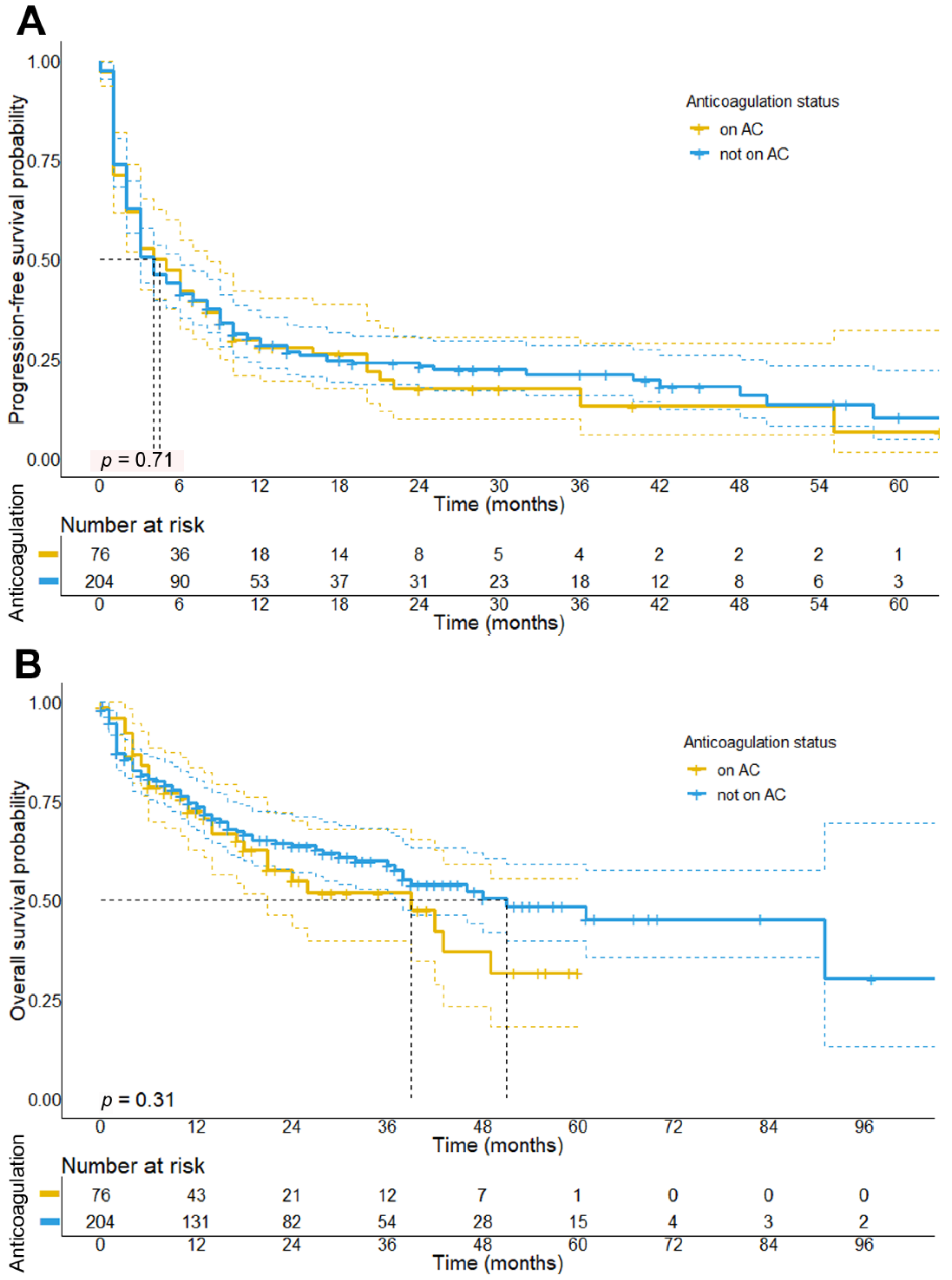
3.3. Impact of Concomitant Anticoagulation Treatment on Survival in Melanoma Patients Receiving ICI

Next, we investigated the impact of concomitant AC on the response to initial ICI therapy. Our analysis unveiled no significant differences in the BOR of patients who did or did not receive AC (p = 0.576). Moreover, we found no significant differences in the ORR (p = 0.270) or DCR (p = 0.946) between patients receiving AC and those who did not (Table 2). In particular, patients receiving AC had an ORR of 43.8% (95% CI: 32.2–55.9%), whereas those not on AC had an ORR of 38.4% (95% CI: 32.0–45.9%).
Aligning with the results from univariate Cox regression analysis (Supplementary Table S1), log-rank tests showed no significant association between PFS (median PFS: 4 vs. 4 months, p = 0.72, HR: 0.95, 95% CI: 0.71–1.28) or OS (median OS: 39 vs. 51 months, p = 0.31, HR: 0.81, 95% CI: 0.54–1.22) and concomitant anticoagulation (survival curves are shown in Figure 1). In a multivariate analysis, which has been performed after adjusting for potential confounders such as the higher age found in patients on AC, we also observed no association neither between PFS (HR: 0.86, 95% CI: 0.59–1.26, p = 0.44) or OS (HR: 0.78, 95% CI: 0.47–1.3, p = 0.34) and AC status (Supplementary Table S3). Due to the observation that patients receiving concomitant AC were a heterogeneous group in terms of clinical outcomes, we further distinguished patients into three categories stratified by the specific class of anticoagulant received. Here, regression analysis revealed that patients treated with FXa DOAC showed significantly better outcomes in terms of PFS compared to patients receiving other anticoagulants (i.e., heparins and VKA) (HR: 0.36, 95% CI: 0.20–0.64, p < 0.001), but also compared to all other patients not receiving FXa DOACs (HR: 0.44, 95% CI: 0.25–0.78, p = 0.0056).

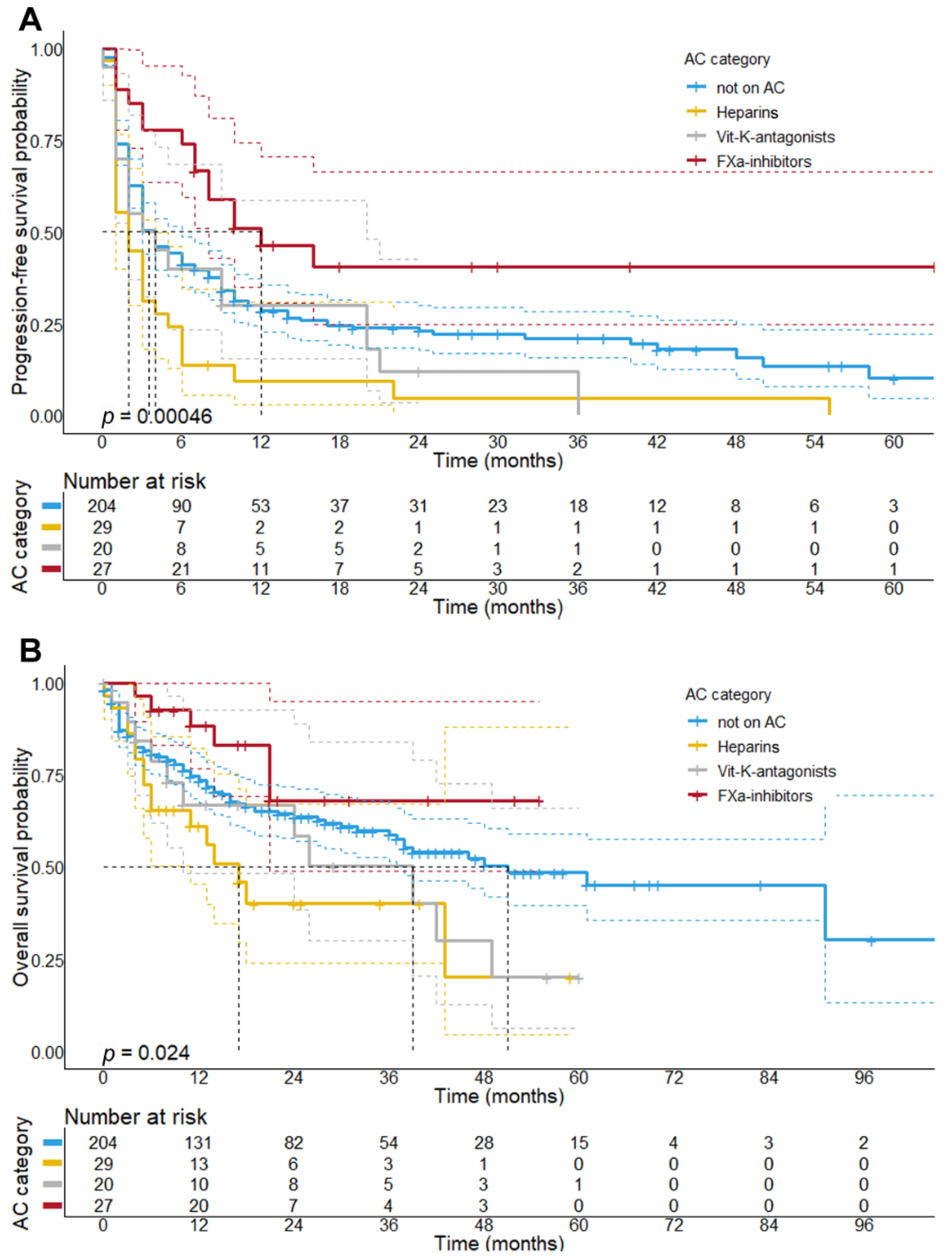
3.4. Concomitant Treatment with Factor-Xa Inhibitors Is Associated with a Better Response and Longer PFS upon Initial ICI Therapy

We then analyzed the treatment outcomes stratified by the class of AC received by patients while on initial ICI therapy. Using the Chi-square test, we observed that patients receiving FXa DOACs showed a significantly higher ORR (69.2%, p = 0.002) and DCR (84.6%, p = 0.004) as compared to patients receiving other anticoagulants (Table 3 and Figure 2). Notably, patients receiving FXa DOACs were more likely to obtain a CR (p = 0.035), and also showed a better ORR (p = 0.004) and DCR (p = 0.028) as compared to patients not receiving concomitant AC.
Given the preliminary associations of the AC categories with a better ORR, we further explored the impact of FXa DOACs on survival. With regard to the PFS, Kaplan–Meier analysis revealed that patients receiving FXa DOACs had a significantly longer PFS (median PFS: 12 months; 95% CI: 3.5–20.5 months, p < 0.001) compared to patients receiving other anticoagulants, such as heparins or VKA (median PFS: 2 months, 95% CI: 0.75–3.25 months). Patients on FXa DOACs also showed a longer PFS as compared to patients not on AC (median PFS: 4 months, 95% CI: 2.9–5.1 months, p = 0.016) (Figure 3A), whereas patients receiving heparins showed the shortest PFS (median PFS: 2 months) of all patient subcohorts.
Results from our survival analysis further showed that patients receiving concomitant FXa DOAC treatment presented with a longer OS (median OS: not reached, p = 0.007) as compared to patients given other AC, that is, heparins (median OS: 17 months, 95% CI: 10.6–23.4 months) and VKA (median OS: 39 months, 95% CI: 16.8–61.2 months). Despite an observed favorable trend, patients with concomitant FXa DOAC therapy did not present with a better OS as compared to patients not receiving AC (median OS: 51 months, 95% CI: 30.6–71.3 months, p = 0.152) (Figure 3B). This finding is consistent with Cox regression analysis, which demonstrated that patients receiving FXa DOACs had a smaller risk of death (HR: 0.49; 95% CI: 0.22–1.12); however, this association was below statistical significance both in univariate (p = 0.09) and multivariate analysis (p = 0.55) (Supplementary Tables S2 and S4). On the other hand, patients receiving heparins during initial ICI therapy showed a significantly shorter median OS as compared to patients not receiving AC (p = 0.003).

3.5. Patient Characteristics Receiving Concomitant FXa-Inhibitor Treatment

Patients receiving concomitant treatment with FXa DOACs were predominantly male (59.3%), of older age (median: 69 years), and BRAF-negative (63.0%), with 70.3% of patients showing elevated serum LDH levels at treatment initiation, and mainly received either combined ICI therapy (32.1%) or anti-PD-1 monotherapy (64.3%). Notably, patients on FXa DOACs therapy underwent systemic pretreatments in 32.1% of cases, developed MBM in 38.5%, and received ICI in the first-line setting in 85.2% of cases. Furthermore, our data revealed that there was an increased use of FXa DOACs over time (median: July 2018), despite no statistically significant differences in terms of the time of AC treatment initiation have been observed as compared to the other patient subcohorts (heparins: March 2017; VKA: February 2018; no anticoagulation: December 2017; p = 0.123). We next analyzed whether patient demographics, and tumor and treatment specifics of patients receiving concomitant FXa DOAC therapy differed from the other patient subcohorts. Our data revealed that patients on concomitant FXa DOACs did not significantly differ from other patients in terms of tumor and treatment specifics, except for being of older age (median age: 69 yrs vs. 63 yrs; p = 0.009) and having received initial ICI for a longer period; however, this association was below statistical significance (p = 0.063, Supplementary Table S5 and Supplementary Figure S3). Moreover, we could observe that patients on FXa DOAC therapy less often required treatment discontinuation until the time of data-lock (p = 0.013). In particular, patients were less likely to be withdrawn from initial ICI therapy due to PD (p = 0.016), but cessation due to AEs has not been reduced (Supplementary Table S6). Further, patients receiving FXa DOAC did not experience more immune-related AEs as compared to patients not on AC (33.3% vs. 30.4%). Last, patients with concomitant FXa-i received fewer lines of subsequent therapies as compared to patients prescribed with other anticoagulants or patients not receiving AC (Supplementary Table S5).

3.6. Treatment with Anticoagulants Does Not Increase the Event of Bleeding Complications in Melanoma Patients Treated with ICI
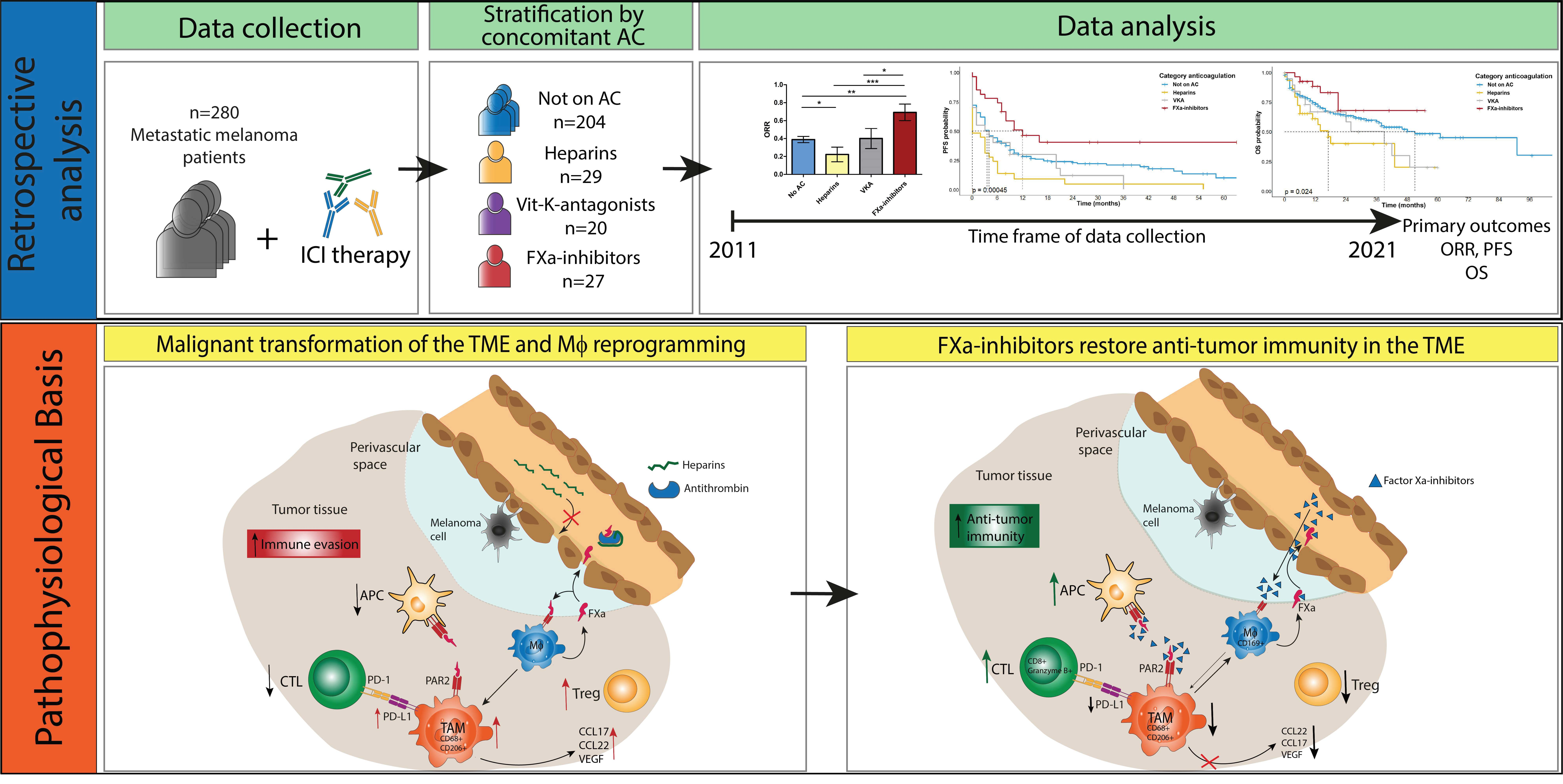
Major adverse effects of prophylactic and therapeutic AC are bleeding complications, which were identified to be significantly associated with PFS and OS of metastatic melanoma patients, as indicated by Cox regression analysis (Supplementary Tables S1, S2, and S4). Therefore, we compared the risk of bleeding complications between patients receiving concomitant AC and those not on AC. Results showed that patients receiving AC during ICI therapy were not more likely to suffer a major or clinically relevant minor bleeding complication at any point during initial ICI therapy as compared to patients not on AC (Table 4; 9.2% vs. 6.9%; p = 0.61). Overall, 21 bleeding complications were documented during the observation period of initial ICI therapy. Thereof, 7 patients (2.5%) experienced major bleeding (3 among patients with concomitant AC (4.0%) and 4 among patients not receiving AC (1.9%)), whereas 14 (3.9%) patients showed minor bleeding complications while receiving ICI (5.3% for patients with concomitant AC vs. 4.9% for patients not receiving AC). Notably, there was no significant difference in the incidence of major or clinically relevant minor bleeding complications between the different categories of AC (p = 0.661).

4. Discussion

The advent of ICI led to profound tumor responses in some patients with advanced melanoma [28]. However, only a subset of patients eventually showed a sustained clinical response upon receiving ICI [7]. Recent evidence suggests that coagulation factors contribute to tumor immune evasion and resistance to immunotherapy [11,12]. In particular, we and others could demonstrate in preclinical studies that FXa and thrombin contribute to immune evasion [11,12]. This is in accordance with the well-established link between the coagulation system and cancer progression [13,15]. More specifically, recent clinical observations indicate that the occurrence of TEE during ICI therapy might worsen the survival of melanoma patients. Therefore, it has been proposed that concomitant treatment with (specific types of) AC might synergize with ICI and thereby augment anti-tumor activity.
Here, we present the outcomes of 280 stage IV melanoma patients who were treated with ICI and analyzed the impact of concomitant anticoagulation therapy. To our knowledge, this is the first study to examine this issue specifically in a real-world cohort of patients with advanced melanoma. Here, we obtained several key findings that corroborate previous concepts of the pathophysiological role of the coagulation system in cancer progression.
First, our results revealed that concomitant AC (regardless of the type of AC treatment) did not improve response or survival (median PFS: 4 vs. 4 months, p = 0.72; median OS: 26 vs. 47 months, p = 0.31) for melanoma patients who simultaneously received initial ICI therapy. This finding was unexpected because randomized trials showed that the application of LMWH and DOACs reduced the risk of VTE in cancer patients and may thus also reduce the risk of VTE-associated mortality [29,30,31]. However, our data are consistent with previous studies and randomized phase III trials [32], which found no association of concomitant AC with prolonged survival in patients with NSCLC [16] or other advanced solid cancer entities [19,21]. Although the cancer-specific risk of VTE in the pre-ICI era was found to be smaller in melanoma patients [33], melanoma patients treated with ICI show similar rates of TEE- and VTE-associated mortality as compared to patients with other advanced cancers [27,34,35], providing a biological rationale for AC therapy [21].
In the absence of a biological rationale for our observation, we reasoned that patients receiving AC treatment might show worse prognostic properties in comparison to patients without AC. Aligning with previous studies our data unveiled that patients receiving AC were significantly older and less often received combined ICI therapy [16,19]. Moreover, patients receiving AC showed a trend towards a higher tumor burden as reflected by higher LDH serum levels upon ICI initiation. After adjusting for these potential confounders in multivariate analysis, our data yet revealed no significant differences in ICI therapy outcomes (PFS: HR: 1.12, p = 0.62; OS: HR: 0.91, 95%, p = 0.78) between patients receiving AC and those who did not, indicating that concomitant AC, in general, does not impact the efficacy of ICI in metastatic melanoma patients.
Second and most importantly, we demonstrated that the individual class of AC given during initial ICI therapy was significantly associated with treatment outcomes of metastatic melanoma patients. Owing to the strong heterogeneity in terms of treatment outcomes among patients receiving AC, we stratified the patients by the class of AC prescribed. Intriguingly, patients receiving FXa DOACs had a significantly better ORR (p = 0.005) and a longer PFS upon initial ICI therapy (median PFS: 12 vs. 3 months; p = 0.006) and showed a non-significant trend towards a better OS as compared to patients not receiving FXa-i (median OS: not reached vs. 42 months, p = 0.09). We find it remarkable that a statistically significant correlation between FXa inhibition and improved outcome of ICI therapy was apparent even with this relatively small patient cohort, which may suggest that the synergy between FXa DOACs and ICI is rather potent.
Notably, the beneficial effects of FXa DOACs with ICI were not seen in patients treated with VKA, which prevents the synthesis of functional prothrombin and FX in the liver and FX in macrophages [12]. This clinical evidence may suggest that additional Vitamin-K-dependent coagulation factors participate in the regulation of anti-tumor immunity.
Our finding is entirely consistent with results from preclinical studies, which demonstrated that specifically FXa DOACs augment anti-tumor immunity by promoting an accumulation of CTL and DC in the TME, while preventing the recruitment of regulatory T cells (Treg) and macrophage polarization towards an immunosuppressive phenotype [12]. Tumor-associated macrophages have been found to regulate angiogenesis in various mouse primary tumor models and correlate with microvessel density in human tumors [36]. The synergistic effect of FXa DOACs result from inhibiting the signaling function of macrophage-derived FXa that promotes immune evasion through PAR2. Only FXa DOACs with tissue penetrance reach FXa in this location and specifically target this cell-autonomous signaling pathway, whereas heparins, which depend on antithrombin for FXa inhibition, are largely restricted to the intravascular and perivascular space and were not shown to enhance ICI efficacy [12,37]. In accordance, patients receiving heparins apparently showed worse outcomes as compared to patients not receiving AC at all (median PFS 2 vs. 4 months, p = 0.003). Because these patients were more often prescribed heparins due to severe TEE, such as pulmonary embolisms, and had received more systemic pretreatments prior to initial ICI, our data do, however, indicate that these patients were in worse overall clinical condition in general. Due to the increased incidence of severe and/or fatal TEE in this patient subcohort, we reasoned that the occurrence of severe TEE might be an important confounder, particularly when interpretating the results of PFS analysis, since the occurrence of fatal TEE might potentially impact PFS irrespective of the biological response to ICI therapy. After adjusting for these potential confounders in multivariate analysis, we showed that the association between heparin treatment and the risk of disease progression was yet not significant (HR: 1.12, 95% CI: 0.72–1.73, p = 0.62) when compared to non-anticoagulated patients. This is consistent with recent data, which found no significant effect of heparin treatment on survival for advanced cancer patients treated with ICI [16,19].
Notably, patients given FXa DOACs did not significantly differ from patients receiving other classes of AC or patients not on AC, except for being of older age and showing a non-significant trend of having received ICI more often in a first-line setting. Therefore, it seems conceivable that the survival benefit found in this subcohort might even be underestimated, because patients with AC treatment in general, and FXa-i therapy in particular, tend to represent a cohort with negative prognostic properties as compared to patients not receiving AC, due to a higher rate of TEE and other comorbidities (i.e., atrial fibrillation, previous cardiovascular events, or VTE). However, patients receiving FXa-DOACs might yet show a favorable risk profile in terms of cardiovascular preconditions as compared to patients who have received heparins, since heparins were mainly used following acute, severe TEE, such as pulmonary embolism, whereas FXa DOACs were mainly used for atrial fibrillation or minor DVT events (see Supplementary Table S5). Furthermore, we have observed a yet non-significant trend towards an increased use of FXa DOACs over time, which might be considered an additional confounder, taking into account the introduction of more efficient treatment regimens over time.
Last, our results revealed that patients receiving concomitant AC did not show more bleeding complications (p = 0.61) or any immune-related AEs. These observations complement a recent retrospective study which observed no increased risk of intracranial hemorrhage during AC for patients with MBM [23], suggesting a potential safety of AC in melanoma patients receiving AC due to VTE. However, as for the limited number of bleeding events in our study, it has to be suspected that patients with bleeding events might have been primarily treated and documented elsewhere, which complicates the systematic documentation of bleeding events in our retrospective study. Therefore, the interpretation of our data on bleeding complications requires caution and needs further systematic investigation in prospective clinical studies.
Further limitations of our study are the retrospective, monocentric nature of the investigation, which adds an inherent selection bias within the cohort. Moreover, the heterogeneity in systemic pretreatments, the different classes of ICI initially administered, and subsequent treatment lines, as well as some missing data, such as tumor thickness or ulceration status for several cases, might have affected our results. We also acknowledge that patients in our retrospective analysis had worse outcomes as compared to previous ICI approval studies, which might be attributed to the frequent application of pretreatments and the fact that, in particular during the first years of our observation period, the administration of IPI was still a common scheme in the treatment of metastatic melanoma. Other limitations are the different schedules of AC treatment applied for melanoma patients (e.g., different durations of concomitant AC and changes in therapeutic and prophylactic dosages of AC) and the restriction of our analysis to initial ICI therapy.
In summary, we provide first clinical evidence for a synergy of FXa DOACs given during initial ICI therapy in patients with metastatic melanoma, which complements recent preclinical studies that demonstrated a synergy between ICI and FXa DOACs, but not heparin. This effect was restricted to FXa DOACs and not seen for other anticoagulants, suggesting that the inhibition of the prothrombotic effects of coagulation may not account for the observed effects of FXa DOACs. Although our study was exploratory in its nature and therefore does not allow for definitive conclusions, our data suggest a robust synergy between FXa DOACs and ICI and may therefore serve as a stimulus for further investigations in prospective studies. Based on our observations, such clinical studies investigating the efficacy of concomitant FXa DOAC treatment might be particularly relevant for patients who are at a high risk of VTE prior to the initiation of ICI-based immunotherapy, while these studies should further take into consideration the potential risk of bleeding complications. If these findings are confirmed, our observations may represent a valid option to prevent thrombotic events during ICI therapy and augment ICI efficacy.

5. Conclusions

This retrospective study including 280 stage IV melanoma patients provides first clinical evidence that the application of FXa-i may enhance the efficacy of ICI therapy via the restoration of anti-tumor immunity. In particular, we could show that concomitant treatment with FXa-i improved the objective response rate and progression-free survival upon first-line ICI therapy, while patients who received FXa-i were not more likely to encounter bleeding complications.

Supplementary Materials

The following are available online at https://www.mdpi.com/article/10.3390/cancers13205103/s1, Figure S1: Kaplan–Meier plots showing the progression-free (left) and overall survival probabilities of melanoma patients stratified by the BOR to initial ICI (top) and the duration of initial ICI therapy (bottom), Figure S2: Kaplan–Meier plots showing the progression-free (left) and overall survival probabilities (right) for melanoma patients stratified by BRAF status (A,B), the presence of MBM (C,D), the administration of ICI in a first-line setting (E,F), and LDH serum levels (G,H), Figure S3: Treatment duration of initial ICI therapy stratified by the different categories of concomitant AC, Table S1: Univariate Cox proportional hazards model for progression-free survival, Table S2: Univariate Cox proportional hazards model for overall survival, Table S3: Multivariate Cox proportional hazards models for progression-free survival, Table S4: Multivariate Cox proportional hazards models for overall survival, Table S5: Patient demographics, tumor characteristics, and specifics on concomitant AC treatment stratified by the class of AC received during initial ICI, Table S6: Status of ICI treatment at time of data cut-off and reasons for treatment discontinuation stratified by the different AC categories.

Author Contributions

Writing and editing of the manuscript, M.H., H.S., M.I.F., S.P., J.H., C.G., W.R., C.L. and S.G.; methodology, software, validation, formal analysis, funding acquisition, and illustration of tables and figures, M.H. and H.S.; conceptualization, H.S., M.H., W.R. and S.G.; project administration, M.H., H.S., C.L. and S.G.; supervision, S.G. and C.L.; data acquisition, J.H., S.P., H.S. and C.L. All authors have read and agreed to the published version of the manuscript.

Funding

H.S. and M.H. are supported by an intramural research funding of the UMC Mainz. M.H. is supported by the Clinician Scientist Fellowship “TransMed Jumpstart Program: 2019_A72” supported by the Else Kröner Fresenius Foundation. W.R. received funding from the German Research Foundation (SFB1292 TP10), and the German Federal Ministry of Education and Research (BMBF 01EO1003 and 01EO1503).

Institutional Review Board Statement

Ethical review and approval were waived due to the anonymized analysis of retrospective patient data, which have been routinely collected during the clinical treatment at the Skin Cancer Center of the Department of Dermatology (University Hospital Mainz). This procedure is regulated in Rhineland-Palatinate by the State Hospital Act (§ 36 and § 37). The analysis of the anonymized real-world patient data was conducted according to the guidelines of the Declaration of Helsinki. All patients gave their consent for treatment at the initiation of therapy.

Informed Consent Statement

Informed consent was not required due to the anonymized collection of retrospective patient data.

Data Availability Statement

All relevant data are within the manuscript and its supporting tables and figures.

Conflicts of Interest

M.H., H.S., J.H., M.I.F., S.P., C.G. and W.R. declare no conflicts of interest. S.G. declares honoraria for advisory boards, oral presentations, and travel expenses from Roche, Novartis, MSD, and BMS outside the submitted work. C.L. declares speakers, advisory board honoraria, and travel support from Bristol Myers Squibb, Merck Sharp and Dohme, Merck Serono, Novartis, Roche, Pierre Fabre, Sun Pharma, Kiowa Kirin, Sanofi, Biontech, and Almirall Hermal, outside the submitted work.

Abbreviations

ACAnticoagulation
AEAdverse event
AFAtrial fibrillation
BORBest overall response
BRAF/MEKiBRAF/MEK inhibitors
CIConfidence interval
cICICombined immune checkpoint inhibitor therapy
CRComplete response
CTLCytotoxic T-lymphocytes
DCDendritic cells
DCRDisease control rate
DOACDirect oral anticoagulant(s)
DVTDeep vein thrombosis
FXa-iFactor Xa-inhibitors
HRHazard ratio
ICIImmune checkpoint inhibitors
IFNInterferon
IPIIpilimumab
LMWHLow-molecular-weight heparins
MBMMelanoma brain metastases
Macrophage(s)
MRMixed response
NEDNo evidence of disease
NivoNivolumab
ORRObjective response rate
OSOverall survival
PARProtease activated receptors
PDProgressive disease
PD-1Programmed-cell-death protein 1
PD-L1Programmed-cell-death ligand 1
PEPulmonary embolism
PembroPembrolizumab
PFSProgression-free survival
PRPartial response
RECISTResponse Evaluation Criteria in Solid Tumors
SDStable disease
TEEThromboembolic events
TFTissue factor
TMETumor microenvironment
VKAVitamin K antagonists
VTEVenous thromboembolic events

References

  1. Dougan, M.; Dranoff, G. Immune therapy for cancer. Annu. Rev. Immunol. 2009, 27, 83–117. [Google Scholar] [CrossRef] [PubMed]
  2. Hodi, F.S.; O’Day, S.J.; McDermott, D.F.; Weber, R.W.; Sosman, J.A.; Haanen, J.B.; Gonzalez, R.; Robert, C.; Schadendorf, D.; Hassel, J.C.; et al. Improved survival with ipilimumab in patients with metastatic melanoma. N. Engl. J. Med. 2010, 363, 711–723. [Google Scholar] [CrossRef]
  3. Robert, C.; Thomas, L.; Bondarenko, I.; O’Day, S.; Weber, J.; Garbe, C.; Lebbe, C.; Baurain, J.F.; Testori, A.; Grob, J.J.; et al. Ipilimumab plus dacarbazine for previously untreated metastatic melanoma. N. Engl. J. Med. 2011, 364, 2517–2526. [Google Scholar] [CrossRef] [PubMed] [Green Version]
  4. Schmidberger, H.; Rapp, M.; Ebersberger, A.; Hey-Koch, S.; Loquai, C.; Grabbe, S.; Mayer, A. Long-term survival of patients after ipilimumab and hypofractionated brain radiotherapy for brain metastases of malignant melanoma: Sequence matters. Strahlenther. Onkol. 2018, 194, 1144–1151. [Google Scholar] [CrossRef] [Green Version]
  5. Kluger, H.M.; Chiang, V.; Mahajan, A.; Zito, C.R.; Sznol, M.; Tran, T.; Weiss, S.A.; Cohen, J.V.; Yu, J.; Hegde, U.; et al. Long-Term Survival of Patients With Melanoma With Active Brain Metastases Treated With Pembrolizumab on a Phase II Trial. J. Clin. Oncol. 2019, 37, 52–60. [Google Scholar] [CrossRef] [PubMed]
  6. Rahman, R.; Cortes, A.; Niemierko, A.; Oh, K.S.; Flaherty, K.T.; Lawrence, D.P.; Sullivan, R.J.; Shih, H.A. The impact of timing of immunotherapy with cranial irradiation in melanoma patients with brain metastases: Intracranial progression, survival and toxicity. J. Neurooncol. 2018, 138, 299–306. [Google Scholar] [CrossRef]
  7. Harder, N.; Schonmeyer, R.; Nekolla, K.; Meier, A.; Brieu, N.; Vanegas, C.; Madonna, G.; Capone, M.; Botti, G.; Ascierto, P.A.; et al. Automatic discovery of image-based signatures for ipilimumab response prediction in malignant melanoma. Sci. Rep. 2019, 9, 7449. [Google Scholar] [CrossRef]
  8. Mayer, A.; Haist, M.; Loquai, C.; Grabbe, S.; Rapp, M.; Roth, W.; Vaupel, P.; Schmidberger, H. Role of Hypoxia and the Adenosine System in Immune Evasion and Prognosis of Patients with Brain Metastases of Melanoma: A Multiplex Whole Slide Immunofluorescence Study. Cancers 2020, 12, 3753. [Google Scholar] [CrossRef]
  9. Chae, Y.K.; Oh, M.S.; Giles, F.J. Molecular Biomarkers of Primary and Acquired Resistance to T-Cell-Mediated Immunotherapy in Cancer: Landscape, Clinical Implications, and Future Directions. Oncologist 2018, 23, 410–421. [Google Scholar] [CrossRef] [Green Version]
  10. Franklin, C.; Livingstone, E.; Roesch, A.; Schilling, B.; Schadendorf, D. Immunotherapy in melanoma: Recent advances and future directions. Eur. J. Surg. Oncol. 2017, 43, 604–611. [Google Scholar] [CrossRef]
  11. Metelli, A.; Wu, B.X.; Riesenberg, B.; Guglietta, S.; Huck, J.D.; Mills, C.; Li, A.; Rachidi, S.; Krieg, C.; Rubinstein, M.P.; et al. Thrombin contributes to cancer immune evasion via proteolysis of platelet-bound GARP to activate LTGF-beta. Sci. Transl. Med. 2020, 12, eaay4860. [Google Scholar] [CrossRef]
  12. Graf, C.; Wilgenbus, P.; Pagel, S.; Pott, J.; Marini, F.; Reyda, S.; Kitano, M.; Macher-Goppinger, S.; Weiler, H.; Ruf, W. Myeloid cell-synthesized coagulation factor X dampens antitumor immunity. Sci. Immunol. 2019, 4, eaaw8405. [Google Scholar] [CrossRef]
  13. Ruf, W.; Disse, J.; Carneiro-Lobo, T.C.; Yokota, N.; Schaffner, F. Tissue factor and cell signalling in cancer progression and thrombosis. J. Thromb. Haemost. 2011, 9 (Suppl. 1), 306–315. [Google Scholar] [CrossRef] [PubMed] [Green Version]
  14. Haemmerle, M.; Stone, R.L.; Menter, D.G.; Afshar-Kharghan, V.; Sood, A.K. The Platelet Lifeline to Cancer: Challenges and Opportunities. Cancer Cell 2018, 33, 965–983. [Google Scholar] [CrossRef] [PubMed] [Green Version]
  15. Varki, A. Trousseau’s syndrome: Multiple definitions and multiple mechanisms. Blood 2007, 110, 1723–1729. [Google Scholar] [CrossRef] [PubMed]
  16. Nichetti, F.; Ligorio, F.; Zattarin, E.; Signorelli, D.; Prelaj, A.; Proto, C.; Galli, G.; Marra, A.; Apollonio, G.; Porcu, L.; et al. Is There an Interplay between Immune Checkpoint Inhibitors, Thromboprophylactic Treatments and Thromboembolic Events? Mechanisms and Impact in Non-Small Cell Lung Cancer Patients. Cancers 2019, 12, 67. [Google Scholar] [CrossRef] [Green Version]
  17. Ruf, W.; Mueller, B.M. Thrombin generation and the pathogenesis of cancer. Semin. Thromb. Hemost. 2006, 32 (Suppl. 1), 61–68. [Google Scholar] [CrossRef] [PubMed]
  18. Versteeg, H.H.; Schaffner, F.; Kerver, M.; Petersen, H.H.; Ahamed, J.; Felding-Habermann, B.; Takada, Y.; Mueller, B.M.; Ruf, W. Inhibition of tissue factor signaling suppresses tumor growth. Blood 2008, 111, 190–199. [Google Scholar] [CrossRef] [Green Version]
  19. Johannet, P.; Sawyers, A.; Gulati, N.; Donnelly, D.; Kozloff, S.; Qian, Y.; Floristan, A.; Hernando, E.; Zhong, J.; Osman, I. Treatment with therapeutic anticoagulation is not associated with immunotherapy response in advanced cancer patients. J. Transl. Med. 2021, 19, 47. [Google Scholar] [CrossRef]
  20. Zacharski, L.R.; Henderson, W.G.; Rickles, F.R.; Forman, W.B.; Cornell Jr, C.J.; Forcier, A.J.; Edwards, R.L.; Headley, E.; Kim, S.-H.; O’Donnell, J.F.; et al. Effect of warfarin anticoagulation on survival in carcinoma of the lung, colon, head and neck, and prostate: Final Report of VA cooperative study # 75. Cancer 1984, 53, 2046–2052. [Google Scholar] [CrossRef]
  21. Sussman, T.A.; Li, H.; Hobbs, B.; Funchain, P.; McCrae, K.R.; Khorana, A.A. Incidence of thromboembolism in patients with melanoma on immune checkpoint inhibitor therapy and its adverse association with survival. J. Immunother. Cancer 2021, 9, e001719. [Google Scholar] [CrossRef]
  22. Johnstone, C.; Rich, S.E. Bleeding in cancer patients and its treatment: A review. Ann. Palliat. Med. 2018, 7, 265–273. [Google Scholar] [CrossRef]
  23. Alvarado, G.; Noor, R.; Bassett, R.; Papadopoulos, N.E.; Kim, K.B.; Hwu, W.J.; Bedikian, A.; Patel, S.; Hwu, P.; Davies, M.A. Risk of intracranial hemorrhage with anticoagulation therapy in melanoma patients with brain metastases. Melanoma Res. 2012, 22, 310–315. [Google Scholar] [CrossRef]
  24. Schulman, S.; Kearon, C.; The Subcommittee on Control of Anticoagulation of the Scientific and Standardization Committee of the International Society on Thrombosis and Haemostasis. Definition of major bleeding in clinical investigations of antihemostatic medicinal products in non-surgical patients. J. Thromb. Haemost. 2005, 3, 692–694. [Google Scholar] [CrossRef]
  25. Kaatz, S.; Ahmad, D.; Spyropoulos, A.C.; Schulman, S.; Subcommittee on Control of, A. Definition of clinically relevant non-major bleeding in studies of anticoagulants in atrial fibrillation and venous thromboembolic disease in non-surgical patients: Communication from the SSC of the ISTH. J. Thromb. Haemost. 2015, 13, 2119–2126. [Google Scholar] [CrossRef]
  26. Couey, M.A.; Bell, R.B.; Patel, A.A.; Romba, M.C.; Crittenden, M.R.; Curti, B.D.; Urba, W.J.; Leidner, R.S. Delayed immune-related events (DIRE) after discontinuation of immunotherapy: Diagnostic hazard of autoimmunity at a distance. J. Immunother. Cancer 2019, 7, 165. [Google Scholar] [CrossRef] [Green Version]
  27. Moik, F.; Chan, W.E.; Wiedemann, S.; Hoeller, C.; Tuchmann, F.; Aretin, M.B.; Fuereder, T.; Zochbauer-Muller, S.; Preusser, M.; Pabinger, I.; et al. Incidence, risk factors, and outcomes of venous and arterial thromboembolism in immune checkpoint inhibitor therapy. Blood 2021, 137, 1669–1678. [Google Scholar] [CrossRef] [PubMed]
  28. Larkin, J.; Chiarion-Sileni, V.; Gonzalez, R.; Grob, J.J.; Cowey, C.L.; Lao, C.D.; Schadendorf, D.; Dummer, R.; Smylie, M.; Rutkowski, P.; et al. Combined Nivolumab and Ipilimumab or Monotherapy in Untreated Melanoma. N. Engl. J. Med. 2015, 373, 23–34. [Google Scholar] [CrossRef] [PubMed] [Green Version]
  29. Agnelli, G.; Gussoni, G.; Bianchini, C.; Verso, M.; Mandala, M.; Cavanna, L.; Barni, S.; Labianca, R.; Buzzi, F.; Scambia, G.; et al. Nadroparin for the prevention of thromboembolic events in ambulatory patients with metastatic or locally advanced solid cancer receiving chemotherapy: A randomised, placebo-controlled, double-blind study. Lancet Oncol. 2009, 10, 943–949. [Google Scholar] [CrossRef]
  30. Carrier, M.; Abou-Nassar, K.; Mallick, R.; Tagalakis, V.; Shivakumar, S.; Schattner, A.; Kuruvilla, P.; Hill, D.; Spadafora, S.; Marquis, K.; et al. Apixaban to Prevent Venous Thromboembolism in Patients with Cancer. N. Engl. J. Med. 2019, 380, 711–719. [Google Scholar] [CrossRef] [PubMed]
  31. Khorana, A.A.; Soff, G.A.; Kakkar, A.K.; Vadhan-Raj, S.; Riess, H.; Wun, T.; Streiff, M.B.; Garcia, D.A.; Liebman, H.A.; Belani, C.P.; et al. Rivaroxaban for Thromboprophylaxis in High-Risk Ambulatory Patients with Cancer. N. Engl. J. Med. 2019, 380, 720–728. [Google Scholar] [CrossRef] [PubMed]
  32. Ek, L.; Gezelius, E.; Bergman, B.; Bendahl, P.O.; Anderson, H.; Sundberg, J.; Wallberg, M.; Falkmer, U.; Verma, S.; Belting, M.; et al. Randomized phase III trial of low-molecular-weight heparin enoxaparin in addition to standard treatment in small-cell lung cancer: The RASTEN trial. Ann. Oncol. 2018, 29, 398–404. [Google Scholar] [CrossRef]
  33. Wun, T.; White, R.H. Epidemiology of cancer-related venous thromboembolism. Best Pract. Res. Clin. Haematol. 2009, 22, 9–23. [Google Scholar] [CrossRef] [Green Version]
  34. Ibrahimi, S.; Machiorlatti, M.; Vesely, S.K.; Malla, M.; Modhia, F.; Jones, S.A.; Roman, D.; Cherry, M.A. Incidence of Vascular Thromboembolic Events in Patients Receiving Immunotherapy: A Single Institution Experience. Blood 2017, 130, 4864. [Google Scholar] [CrossRef]
  35. Gutierrez-Sainz, L.; Martinez-Marin, V.; Vinal, D.; Martinez-Perez, D.; Pedregosa, J.; Garcia-Cuesta, J.A.; Villamayor, J.; Zamora, P.; Pinto, A.; Redondo, A.; et al. Incidence of venous thromboembolic events in cancer patients receiving immunotherapy: A single-institution experience. Clin. Transl. Oncol. 2021, 23, 1245–1252. [Google Scholar] [CrossRef]
  36. Lewis, C.E.; Harney, A.S.; Pollard, J.W. The Multifaceted Role of Perivascular Macrophages in Tumors. Cancer Cell 2016, 30, 18–25. [Google Scholar] [CrossRef] [Green Version]
  37. Carlson, T.H.; Simon, T.L.; Atencio, A.C. In vivo behavior of human radioiodinated antithrombin III: Distribution among three physiologic pools. Blood 1985, 66, 13–19. [Google Scholar] [CrossRef] [Green Version]
Figure 1. Progression-free and overall survival in patients stratified by the concomitant treatment with anticoagulants. Results show that patients receiving AC during initial ICI therapy did not have a significantly shorter PFS compared to patients not on AC (median PFS 4.0; 95% CI: 1.7–6.3 months vs. 4.0 months; 95% CI: 2.9–5.1 months; p = 0.705) (A). Neither did patients on AC show a significantly worse OS as compared to patients not on AC (median OS: 39.0; 95% CI: 18.4–59.6 months vs. 51.0 months; 95% CI: 30.6–71.3 months; p = 0.31), although there was a trend towards a shorter OS in this cohort of patients (B). The bold is used for the title of the figures.
Cancers 13 05103 g001
Figure 2. Bar chart showing the objective response rate (A) and disease control rate upon initial ICI therapy (B) stratified by the category of concomitant AC treatment. Patients receiving concomitant treatment with FXa inhibitors showed a significantly higher ORR and DCR as compared to other AC agents, such as heparins or vitamin K antagonists (VKA), as well as patients not receiving AC. Notably, patients on heparin treatment presented with the worst ORR and DCR as compared to the other categories. Abbreviations: * p < 0.05, ** p < 0.005, *** p < 0.001.
Cancers 13 05103 g002
Figure 3. Progression-free and overall survival of patients stratified by the category of concomitant anticoagulation. Results revealed that PFS (p = 0.00046) and OS (p = 0.024) of patients undergoing initial ICI therapy significantly differed depending on the class of concomitant AC (p-values indicate statistically significant differences between the groups tested in this model). Particularly, patients receiving FXa-inhibitors (FXa-i) presented with the best PFS (median: 12 months) as compared to patients given heparins or VKA (median PFS: 2 and 3 months) during initial ICI therapy (p < 0.001). Further, patients not on AC did show a longer PFS compared to patients treated with heparins (p = 0.003), but a worse PFS as compared to patients receiving FXa-i (p = 0.016) (A). Log-rank test comparing the OS of patients stratified by AC category revealed a significantly shorter OS of patients receiving heparin treatment (median OS: 17.0 months, 95% CI: 10.6–23.4 months, p = 0.03), whereas concomitant treatment with VKA (median OS: 39 months; 95% CI: 16.8–61.2 months, p = 0.19) or FXa-i (median OS: not reached, p = 0.15) did not differ from patients not on AC (median OS: 51 months; 95% CI: 30.6–71.3 months) (B).The bold has been used for the image caption title.
Cancers 13 05103 g003
Table 1. Baseline patient characteristics and treatment outcomes.
Clinicopathological FeaturesNot on AnticoagulationOn Anticoagulationp-Value
Total number of patients20476
Median age at initiation of ICI63.070.00.0006
Gender 0.784
Female121 (59.3%)47 (61.8%)
Male83 (40.7%)29 (38.2%)
Primary Tumor and Metastases
Median Breslow thickness (95% CI) 12.5 (0.7–12.7 mm)2.4 (0.7–6.8 mm)0.185
Ulceration 267/132 (50.8%)28/54 (51.8%)0.511
BRAF status 388/201 (43.8%)28/74 (37.8%)0.411
Elevated serum LDH levels (>245 U/L) 4115/173 (66.5%)44/65 (67.7%)0.269
PTT at initiation of ICI 528.45 s30.89 s<0.0001
Systemic pretreatments prior to ICI
-
BRAF/MEKi therapy
-
IFNα therapy
-
Chemotherapy
-
Other
86 (42.1%)
48
37
11
5
28 (36.8%)
17
12
3
3
0.494
Melanoma brain metastases65 (32.3%)26 (34.6%)0.774
Liver metastases67 (32.8%)25 (33.3%)0.523
Treatments
Initial ICI therapy 0.020
Ipilimumab + nivolumab
Nivolumab
Pembrolizumab
Ipilimumab
89 (43.6%)
39 (19.1%)
58 (28.4%)
18 (8.8%)
20 (26.3%)
16 (21.1%)
35 (46.1%)
5 (6.6%)
Treatment line 0.883
-
First
-
Not first
145 (70.4%)
59 (42.6%)
55 (72.4%)
21 (36.9%)
Median treatment duration (range)
Ongoing treatment
Number of patients
Cessation due to toxicity
Number of patients
Cessation due to PD
Number of patients
3.0 months (0–41)
14 months (7–41)
13 (6.4%)
3 months (1–23)
51 (25.1%)
2 months (0–8)
80 (39.2%)
4.0 months (0–38)
10 months (7–28)
8 (10.5%)
2 months (1–21)
16 (21.1%)
3 months (0–10)
28 (36.8%)
0.261
BOR to initial ICI therapy 6 0.569
-
CR
-
PR
-
SD
-
PD
-
Could not be evaluated
30 (14.7%)
48 (23.5%)
45 (22.1%)
80 (39.2%)
1
9 (11.8%)
23 (30.3%)
13 (17.1%)
28 (36.8%)
3
TP upon initial ICI159 (77.9%)61 (80.2%)0.674
Median progression-free survival (95% CI)4.0 months (2.9–5.1)4.0 months (1.7–6.2)0.705
VTE during initial ICI therapy 79 (4.4%)26 (36.8%)<0.001
ATE during initial ICI therapy 86 (2.9%)6 (7.9%)0.094
Subsequent treatments
Patients receiving subsequent therapy
Number of post-treatments
Re-induction of ICI 9
ORR
TP
(Re)-induction of BRAF ± MEKi 10
ORR
TP
103
168
78
33 (42.3%)
66 (84.6%)
45
18 (40.0%)
39 (86.6%)
43
68
29
11 (38.0%)
22 (75.9%)
19
6 (31.5%)
17 (89.5%)
0.900
0.673
0.587
0.764
0.611
0.614
Follow-up
Median follow-up upon ICI initiation (95% CI)30 months (24.4–35.6)22 months (13.6–30.4)0.055
Median overall survival (95% CI)51 months (30.6–71.4)39 months (18.4–59.6)0.313
Deceased81 (39.8%)33 (43.4%)0.587
Abbreviations: CR = complete response; PR = partial response; SD = stable disease; PD = progressive disease, ORR = objective response rate (CR + PR); ICI = immune checkpoint inhibitors; CI = confidence interval; TP = tumor progression; PTT = partial thromboplastin time; VTE = venous thromboembolic events; ATE = arterial thromboembolic events, including myocardial infarction, ischemic stroke, or acute arterial occlusion. 1,2,3,4,5 Statistics based on the total number of patients with known Breslow thickness (n = 207), ulceration status (n = 186), BRAF-status (n = 275), LDH serum levels (n = 238), and PTT at ICI initiation (n = 229). 6 Statistics based on the total number of patients with known BOR to initial ICI (n = 276); 7,8 The observation period for VTE/ATE started at the first day of initial ICI therapy and terminated either with the initiation of subsequent antineoplastic therapies or 3 months after the last cycle of initial ICI therapy, which is in accordance with clinical trials investigating ICI with a median time of reporting adverse events after the last cycle of 90 days [26,27]. 9,10 Statistics based on the total number of patients who received a re-induction of ICI therapy (n = 107) or BRAF/MEKi therapy (n = 64). The p-value is indicated in bold numbers when statistically significant. Bold is used in order to indicate titles or subtitles.
Table 2. Response to immune checkpoint inhibitor therapy based on the status of concomitant anticoagulation.
OutcomeNot on AnticoagulationOn Anticoagulationp-Value
Best overall response—no. (%) 0.576
Complete response (CR)30 (14.7%)9 (12.3%)
Partial response (PR)48 (23.6%)23 (31.5%)
Stable disease (SD)45 (22.2%)13 (17.8%)
Progressive disease (PD)80 (39.4%)28 (38.4%)
Objective response rate 1 0.270
No. (%)78/203 (38.4%)32/73 (43.8%)
95% CI 132.0–45.9%32.2–55.9%
Disease control rate 2 0.946
No. (%)123/203 (60.5%)45/73 (61.6%)
95% CI 354.1–68.0%49.5–72.8%
Progress during initial ICI 0.647
No. (%)159 (77.9%)61 (80.2%)
95% CI 371.6–83.4%69.5–88.5%
Abbreviations: Objective response rate was defined as the percentage of patients who obtained CR or PR; disease control rate was defined as the percentage of patients who obtained CR, PR, or SD. 1,2 ORR and DCR based on the total number of patients with known BOR to initial ICI (n = 203 for patients not on AC and n = 73 for patients receiving AC). 3 The 95% confidence intervals were calculated using the Clopper–Pearson method.
Table 3. Response to immune checkpoint inhibitor therapy stratified by the class of the anticoagulants used.
OutcomeNot on ACHeparinsVKAFXa DOACChi-Square Test
Best overall response—no. (%) 0.037
Complete response (CR)30 (14.7%)1 (3.7%)1 (5.0%)7 (26.9%)
Partial response (PR)48 (23.6%)5 (18.5%)7 (35.0%)11 (42.3%)
Stable disease (SD)45 (22.2%)5 (18.5%)4 (20.0%)4 (15.4%)
Progressive disease (PD)80 (39.4%)16 (55.2%)8 (40.0%)4 (15.4%)
Could not be evaluated3201
Objective response rate 1 0.005
No. (%)78 (38.2%)6 (22.2%)8 (40.0%)18 (69.2%)
95% CI 332.0–45.6%8.6–42.3%18.1–63.9%48.2–85.7%
Disease control rate 2 0.011
No. (%)123 (60.3%)11 (40.7%)12 (60%)22 (84.6%)
95% CI 354.1–68.0%22.4–61.2%36.1–80.9%65.1–95.6%
Progress during initial ICI 0.001
No. (%)159 (77.9%)28 (96.6%)18 (90.0%)15 (55.6%)
95% CI 371.6–83.4%82.2–99.9%68.3–98.8%35.3–74.5%
Abbreviations: Objective response rate was defined as the percentage of patients who obtained CR or PR; disease control rate was defined as the percentage of patients who obtained CR, PR, or SD. VKA = vitamin K antagonists; FXa DOAC = Factor Xa dual oral anticoagulants. 1,2 ORR and DCR based on the total number of patients with known BOR to initial ICI (n = 203 for patients not on AC, n = 27 for patients receiving heparins, n = 20 for patients on VKA, and n = 26 for patients on FXa DOAC). 3 The 95% confidence intervals were calculated using the Clopper–Pearson method. The p-value is indicated in bold numbers when statistically significant.
Table 4. Summary of bleeding complications during initial ICI treatment stratified by the concomitant use of anticoagulants. Fisher’s exact test revealed that the risk of bleeding events during concomitant AC was not significantly higher compared to patients not receiving AC. Notably, the event of bleeding complications did not differ significantly between the particular agents of AC (heparin: n = 3; VKA: n = 2; FXa-i: n = 2, p = 0.923).
Secondary OutcomeNot on AnticoagulationOn Anticoagulationp-Value
Bleeding complications 0.610
No. (%)14 (6.9%)7 (9.2%)
95% CI3.8–11.2%3.8–18.1%
Abbreviations: Bleeding outcomes included major and clinically relevant minor bleeding.
Publisher’s Note: MDPI stays neutral with regard to jurisdictional claims in published maps and institutional affiliations.

