
Multiparametric Magnetic Resonance Imaging-Ultrasound Fusion Transperineal Prostate Biopsy: Diagnostic Accuracy from a Single Center Retrospective Study

 ,
,  and
and
Abstract
:Simple Summary
Abstract
1. Introduction
2. Materials and Methods
3. Results
4. Discussion
5. Conclusions
Supplementary Materials
Author Contributions
Funding
Institutional Review Board Statement
Data Availability Statement
Acknowledgments
Conflicts of Interest
References
- Kohaar, I.; Petrovics, G.; Srivastava, S. A Rich Array of Prostate Cancer Molecular Biomarkers: Opportunities and Challenges. Int. J. Mol. Sci. 2019, 20, 1813. [Google Scholar] [CrossRef] [Green Version]
- Ahmed, H.U.; El-Shater Bosaily, A.; Brown, L.C.; Gabe, R.; Kaplan, R.; Parmar, M.K.; Collaco-Moraes, Y.; Ward, K.; Hindley, R.G.; Freeman, A.; et al. Diagnostic accuracy of multi-parametric MRI and TRUS biopsy in prostate cancer (PROMIS): A paired validating confirmatory study. Lancet 2017, 389, 815–822. [Google Scholar] [CrossRef] [Green Version]
- Drost, F.H.; Osses, D.F.; Nieboer, D.; Steyerberg, E.W.; Bangma, C.H.; Roobol, M.J.; Schoots, I.G. Prostate MRI, with or without MRI-targeted biopsy, and systematic biopsy for detecting prostate cancer. Cochrane Database Syst. Rev. 2019, 4, CD012663. [Google Scholar] [CrossRef] [PubMed]
- Kasivisvanathan, V.; Rannikko, A.S.; Borghi, M.; Panebianco, V.; Mynderse, L.A.; Vaarala, M.H.; Briganti, A.; Budaus, L.; Hellawell, G.; Hindley, R.G.; et al. MRI-Targeted or Standard Biopsy for Prostate-Cancer Diagnosis. N. Engl. J. Med. 2018, 378, 1767–1777. [Google Scholar] [CrossRef] [PubMed]
- Rouviere, O.; Puech, P.; Renard-Penna, R.; Claudon, M.; Roy, C.; Mege-Lechevallier, F.; Decaussin-Petrucci, M.; Dubreuil-Chambardel, M.; Magaud, L.; Remontet, L.; et al. Use of prostate systematic and targeted biopsy on the basis of multiparametric MRI in biopsy-naive patients (MRI-FIRST): A prospective, multicentre, paired diagnostic study. Lancet. Oncol. 2019, 20, 100–109. [Google Scholar] [CrossRef]
- van der Leest, M.; Cornel, E.; Israel, B.; Hendriks, R.; Padhani, A.R.; Hoogenboom, M.; Zamecnik, P.; Bakker, D.; Setiasti, A.Y.; Veltman, J.; et al. Head-to-head Comparison of Transrectal Ultrasound-guided Prostate Biopsy Versus Multiparametric Prostate Resonance Imaging with Subsequent Magnetic Resonance-guided Biopsy in Biopsy-naive Men with Elevated Prostate-specific Antigen: A Large Prospective Multicenter Clinical Study. Eur. Urol. 2019, 75, 570–578. [Google Scholar] [CrossRef] [PubMed] [Green Version]
- Weinreb, J.C.; Barentsz, J.O.; Choyke, P.L.; Cornud, F.; Haider, M.A.; Macura, K.J.; Margolis, D.; Schnall, M.D.; Shtern, F.; Tempany, C.M.; et al. PI-RADS Prostate Imaging—Reporting and Data System: 2015, Version 2. Eur. Urol. 2016, 69, 16–40. [Google Scholar] [CrossRef] [PubMed]
- Marra, G.; Marquis, A.; Tappero, S.; D’Agate, D.; Oderda, M.; Calleris, G.; Falcone, M.; Faletti, R.; Molinaro, L.; Zitella, A.; et al. Transperineal Free-hand mpMRI Fusion-targeted Biopsies Under Local Anesthesia: Technique and Feasibility From a Single-center Prospective Study. Urology 2020, 140, 122–131. [Google Scholar] [CrossRef] [PubMed]
- Marra, G.; Ploussard, G.; Futterer, J.; Valerio, M.; Party, E.-Y.P.C.W. Controversies in MR targeted biopsy: Alone or combined, cognitive versus software-based fusion, transrectal versus transperineal approach? World J. Urol. 2019, 37, 277–287. [Google Scholar] [CrossRef]
- Marra, G.; Zhuang, J.; Beltrami, M.; Calleris, G.; Zhao, X.; Marquis, A.; Kan, Y.; Oderda, M.; Huang, H.; Faletti, R.; et al. Transperineal freehand multiparametric MRI fusion targeted biopsies under local anaesthesia for prostate cancer diagnosis: A multicentre prospective study of 1014 cases. BJU Int. 2021, 127, 122–130. [Google Scholar] [CrossRef]
- Mottet, N.; van den Bergh, R.C.N.; Briers, E.; Van den Broeck, T.; Cumberbatch, M.G.; De Santis, M.; Fanti, S.; Fossati, N.; Gandaglia, G.; Gillessen, S.; et al. EAU-EANM-ESTRO-ESUR-SIOG Guidelines on Prostate Cancer-2020 Update. Part 1: Screening, Diagnosis, and Local Treatment with Curative Intent. Eur. Urol. 2021, 79, 243–262. [Google Scholar] [CrossRef]
- Epstein, J.I.; Egevad, L.; Amin, M.B.; Delahunt, B.; Srigley, J.R.; Humphrey, P.A.; Grading, C. The 2014 International Society of Urological Pathology (ISUP) Consensus Conference on Gleason Grading of Prostatic Carcinoma: Definition of Grading Patterns and Proposal for a New Grading System. Am. J. Surg. Pathol. 2016, 40, 244–252. [Google Scholar] [CrossRef]
- Epstein, J.I.; Zelefsky, M.J.; Sjoberg, D.D.; Nelson, J.B.; Egevad, L.; Magi-Galluzzi, C.; Vickers, A.J.; Parwani, A.V.; Reuter, V.E.; Fine, S.W.; et al. A Contemporary Prostate Cancer Grading System: A Validated Alternative to the Gleason Score. Eur. Urol. 2016, 69, 428–435. [Google Scholar] [CrossRef] [PubMed] [Green Version]
- Mischinger, J.; Kaufmann, S.; Russo, G.I.; Harland, N.; Rausch, S.; Amend, B.; Scharpf, M.; Loewe, L.; Todenhoefer, T.; Notohamiprodjo, M.; et al. Targeted vs systematic robot-assisted transperineal magnetic resonance imaging-transrectal ultrasonography fusion prostate biopsy. BJU Int. 2018, 121, 791–798. [Google Scholar] [CrossRef] [Green Version]
- Borkowetz, A.; Hadaschik, B.; Platzek, I.; Toma, M.; Torsev, G.; Renner, T.; Herout, R.; Baunacke, M.; Laniado, M.; Baretton, G.; et al. Prospective comparison of transperineal magnetic resonance imaging/ultrasonography fusion biopsy and transrectal systematic biopsy in biopsy-naive patients. BJU Int. 2018, 121, 53–60. [Google Scholar] [CrossRef] [PubMed] [Green Version]
- Hakozaki, Y.; Matsushima, H.; Murata, T.; Masuda, T.; Hirai, Y.; Oda, M.; Kawauchi, N.; Yokoyama, M.; Kume, H. Detection rate of clinically significant prostate cancer in magnetic resonance imaging and ultrasonography-fusion transperineal targeted biopsy for lesions with a prostate imaging reporting and data system version 2 score of 3–5. Int. J. Urol. Off. J. Jpn. Urol. Assoc. 2019, 26, 217–222. [Google Scholar] [CrossRef]
- Hansen, N.L.; Barrett, T.; Kesch, C.; Pepdjonovic, L.; Bonekamp, D.; O’Sullivan, R.; Distler, F.; Warren, A.; Samel, C.; Hadaschik, B.; et al. Multicentre evaluation of magnetic resonance imaging supported transperineal prostate biopsy in biopsy-naive men with suspicion of prostate cancer. BJU Int. 2018, 122, 40–49. [Google Scholar] [CrossRef]
- Baco, E.; Rud, E.; Eri, L.M.; Moen, G.; Vlatkovic, L.; Svindland, A.; Eggesbo, H.B.; Ukimura, O. A Randomized Controlled Trial To Assess and Compare the Outcomes of Two-core Prostate Biopsy Guided by Fused Magnetic Resonance and Transrectal Ultrasound Images and Traditional 12-core Systematic Biopsy. Eur. Urol. 2016, 69, 149–156. [Google Scholar] [CrossRef] [Green Version]
- Maxeiner, A.; Kittner, B.; Blobel, C.; Wiemer, L.; Hofbauer, S.L.; Fischer, T.; Asbach, P.; Haas, M.; Penzkofer, T.; Fuller, F.; et al. Primary magnetic resonance imaging/ultrasonography fusion-guided biopsy of the prostate. BJU Int. 2018, 122, 211–218. [Google Scholar] [CrossRef] [PubMed] [Green Version]
- Leyten, G.H.; Hessels, D.; Jannink, S.A.; Smit, F.P.; de Jong, H.; Cornel, E.B.; de Reijke, T.M.; Vergunst, H.; Kil, P.; Knipscheer, B.C.; et al. Prospective multicentre evaluation of PCA3 and TMPRSS2-ERG gene fusions as diagnostic and prognostic urinary biomarkers for prostate cancer. Eur. Urol. 2014, 65, 534–542. [Google Scholar] [CrossRef]
- Marks, L.S.; Fradet, Y.; Deras, I.L.; Blase, A.; Mathis, J.; Aubin, S.M.; Cancio, A.T.; Desaulniers, M.; Ellis, W.J.; Rittenhouse, H.; et al. PCA3 molecular urine assay for prostate cancer in men undergoing repeat biopsy. Urology 2007, 69, 532–535. [Google Scholar] [CrossRef] [PubMed]
- Van Neste, L.; Hendriks, R.J.; Dijkstra, S.; Trooskens, G.; Cornel, E.B.; Jannink, S.A.; de Jong, H.; Hessels, D.; Smit, F.P.; Melchers, W.J.; et al. Detection of High-grade Prostate Cancer Using a Urinary Molecular Biomarker-Based Risk Score. Eur. Urol. 2016, 70, 740–748. [Google Scholar] [CrossRef] [PubMed]
- Busetto, G.M.; Del Giudice, F.; Maggi, M.; De Marco, F.; Porreca, A.; Sperduti, I.; Magliocca, F.M.; Salciccia, S.; Chung, B.I.; De Berardinis, E.; et al. Prospective assessment of two-gene urinary test with multiparametric magnetic resonance imaging of the prostate for men undergoing primary prostate biopsy. World J. Urol. 2021, 39, 1869–1877. [Google Scholar] [CrossRef] [PubMed]
- Maggi, M.; Salciccia, S.; Del Giudice, F.; Busetto, G.M.; Falagario, U.G.; Carrieri, G.; Ferro, M.; Porreca, A.; Di Pierro, G.B.; Fasulo, V.; et al. A Systematic Review and Meta-Analysis of Randomized Controlled Trials With Novel Hormonal Therapies for Non-Metastatic Castration-Resistant Prostate Cancer: An Update From Mature Overall Survival Data. Front. Oncol. 2021, 11, 700258. [Google Scholar] [CrossRef]
- McKiernan, J.; Donovan, M.J.; O’Neill, V.; Bentink, S.; Noerholm, M.; Belzer, S.; Skog, J.; Kattan, M.W.; Partin, A.; Andriole, G.; et al. A Novel Urine Exosome Gene Expression Assay to Predict High-grade Prostate Cancer at Initial Biopsy. Jama Oncol. 2016, 2, 882–889. [Google Scholar] [CrossRef] [PubMed] [Green Version]
- McKiernan, J.; Donovan, M.J.; Margolis, E.; Partin, A.; Carter, B.; Brown, G.; Torkler, P.; Noerholm, M.; Skog, J.; Shore, N.; et al. A Prospective Adaptive Utility Trial to Validate Performance of a Novel Urine Exosome Gene Expression Assay to Predict High-grade Prostate Cancer in Patients with Prostate-specific Antigen 2–10 ng/mL at Initial Biopsy. Eur. Urol. 2018, 74, 731–738. [Google Scholar] [CrossRef] [Green Version]
- Sartori, D.A.; Chan, D.W. Biomarkers in prostate cancer: What’s new? Curr. Opin. Oncol. 2014, 26, 259–264. [Google Scholar] [CrossRef] [Green Version]
- Eifler, J.B.; Feng, Z.; Lin, B.M.; Partin, M.T.; Humphreys, E.B.; Han, M.; Epstein, J.I.; Walsh, P.C.; Trock, B.J.; Partin, A.W. An updated prostate cancer staging nomogram (Partin tables) based on cases from 2006 to 2011. BJU Int. 2013, 111, 22–29. [Google Scholar] [CrossRef]
- Rigau, M.; Morote, J.; Mir, M.C.; Ballesteros, C.; Ortega, I.; Sanchez, A.; Colas, E.; Garcia, M.; Ruiz, A.; Abal, M.; et al. PSGR and PCA3 as biomarkers for the detection of prostate cancer in urine. Prostate 2010, 70, 1760–1767. [Google Scholar] [CrossRef]
- Rigau, M.; Ortega, I.; Mir, M.C.; Ballesteros, C.; Garcia, M.; Llaurado, M.; Colas, E.; Pedrola, N.; Montes, M.; Sequeiros, T.; et al. A three-gene panel on urine increases PSA specificity in the detection of prostate cancer. Prostate 2011, 71, 1736–1745. [Google Scholar] [CrossRef]


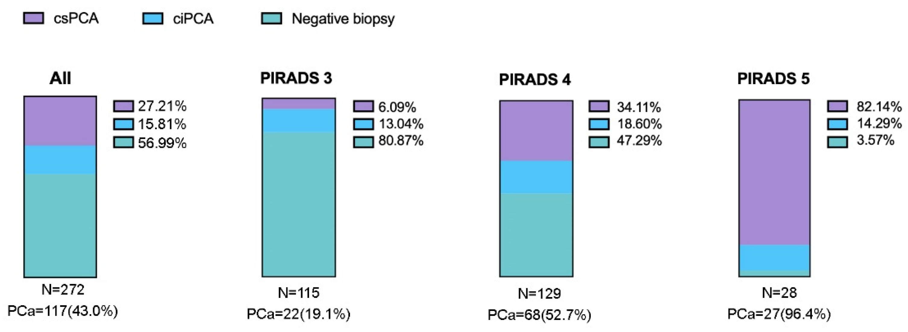
| Characteristics | All (n = 272) | Biopsy Naïve (n = 190) | Prior Negative Biopsy (n = 82) |
|---|---|---|---|
| Age (years), median (IQR 1) | 68 (62–74) | 68 (62–74) | 68 (63–72) |
| PSA (ng/mL), median (IQR) | 7.2 (4.8–10.1) | 7.1 (4.8–9.6) | 7.3 (4.9–11.7) |
| PIRADS n, (%) | |||
| 3 | 115 (42.3%) | 73 (26.8%) | 42 (15.4%) |
| 4 | 129 (47.4%) | 96 (35.3%) | 33 (12.1%) |
| 5 | 28 (10.3%) | 21 (7.7%) | 7 (2.6%) |
| Cases | Biopsy Negative | csPCa (ISUP ≥ 2) | ciPCa (ISUP < 1) |
|---|---|---|---|
| Biopsy naïve 190/272 (69.9%) | 99 (52.1%; 59.1–45.0) | 60 (31.6%; 38.5–25.4) | 31 (16.3%; 22.2–11.7) |
| Prior biopsy 82/272 (30.1%) | 56 (68.3%; 77.4–57.6) | 14 (17.1%; 26.6–10.4) | 12 (14.6%; 23.9–8.6) |
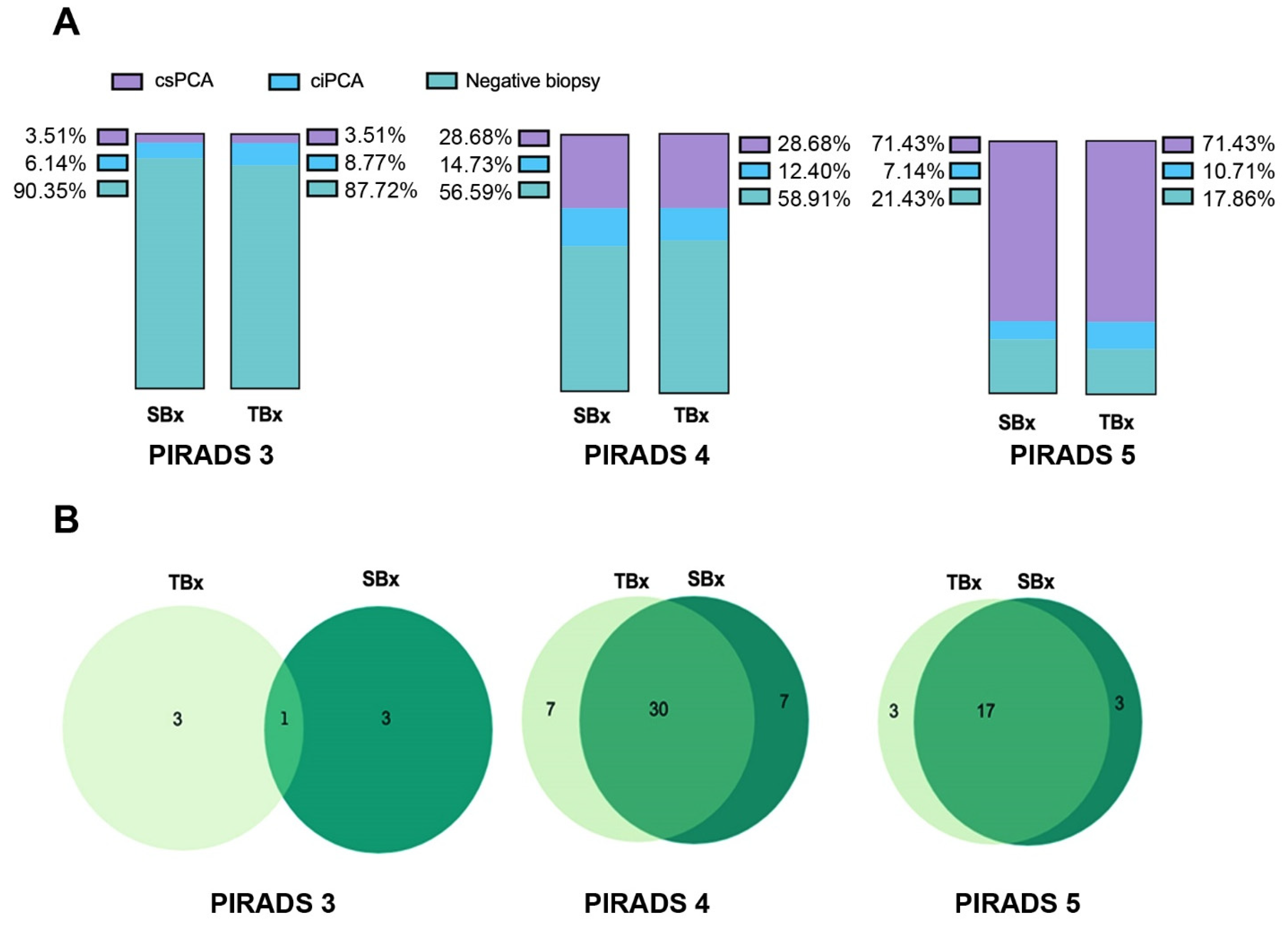
| Cases | All n (%) | TBx n (%) | SBx n (%) | TBx/SBx n (%) |
|---|---|---|---|---|
| 1 Biopsy negative | 155 (57.0%) | 183 (67.3%) | 182 (66.9%) | 155 (57.0%) |
| 1 CDR for ciPCa | 43 (15.8%) | 28 (10.3%) | 29 (10.7%) | 43 (15.8%) |
| 1 CDR for csPCa | 74 (27.2%) | 61 (22.4%) | 61 (22.4%) | 74 (27.2%) |
| 1 False negative PCa | 28 (10.3%) | 27 (9.9%) | 0 (0%) | |
| 2 Sensitivity for csPCa | 82.4% | 82.4% | 100% | |
| Specificity for csPCa | 85.% | 85.8% | 78.3% |
Publisher’s Note: MDPI stays neutral with regard to jurisdictional claims in published maps and institutional affiliations. |
© 2021 by the authors. Licensee MDPI, Basel, Switzerland. This article is an open access article distributed under the terms and conditions of the Creative Commons Attribution (CC BY) license (https://creativecommons.org/licenses/by/4.0/).
Share and Cite
Fulco, A.; Chiaradia, F.; Ascalone, L.; Andracchio, V.; Greco, A.; Cappa, M.; Scarcia, M.; Ludovico, G.M.; Pagliarulo, V.; Palmieri, C.; et al. Multiparametric Magnetic Resonance Imaging-Ultrasound Fusion Transperineal Prostate Biopsy: Diagnostic Accuracy from a Single Center Retrospective Study. Cancers 2021, 13, 4833. https://doi.org/10.3390/cancers13194833
Fulco A, Chiaradia F, Ascalone L, Andracchio V, Greco A, Cappa M, Scarcia M, Ludovico GM, Pagliarulo V, Palmieri C, et al. Multiparametric Magnetic Resonance Imaging-Ultrasound Fusion Transperineal Prostate Biopsy: Diagnostic Accuracy from a Single Center Retrospective Study. Cancers. 2021; 13(19):4833. https://doi.org/10.3390/cancers13194833
Chicago/Turabian StyleFulco, Andrea, Francesco Chiaradia, Luigi Ascalone, Vincenzo Andracchio, Antonio Greco, Manlio Cappa, Marcello Scarcia, Giuseppe Mario Ludovico, Vincenzo Pagliarulo, Camillo Palmieri, and et al. 2021. "Multiparametric Magnetic Resonance Imaging-Ultrasound Fusion Transperineal Prostate Biopsy: Diagnostic Accuracy from a Single Center Retrospective Study" Cancers 13, no. 19: 4833. https://doi.org/10.3390/cancers13194833
APA StyleFulco, A., Chiaradia, F., Ascalone, L., Andracchio, V., Greco, A., Cappa, M., Scarcia, M., Ludovico, G. M., Pagliarulo, V., Palmieri, C., & Alba, S. (2021). Multiparametric Magnetic Resonance Imaging-Ultrasound Fusion Transperineal Prostate Biopsy: Diagnostic Accuracy from a Single Center Retrospective Study. Cancers, 13(19), 4833. https://doi.org/10.3390/cancers13194833