Review

Chimeric Antigen Receptor T cell Therapy and the Immunosuppressive Tumor Microenvironment in Pediatric Sarcoma

1
Children’s Cancer Institute, Randwick 2031, Australia
2
School of Women and Children’s Health, University of New South Wales, Randwick 2052, Australia
3
Cancer Immunology Program, Peter MacCallum Cancer Centre, Melbourne 3000, Australia
4
Sir Peter MacCallum Department of Oncology, University of Melbourne, Melbourne 3000, Australia
5
Inserm, Université de Paris, U976 HIPI Unit, Institut de Recherche Saint-Louis, 75475 Paris, France
6
Translational Innate Immunotherapy, University of Queensland Diamantina Institute (UQDI), Brisbane 4102, Australia
7
Oncology Services Group, Queensland Children Hospital, Brisbane 4101, Australia
8
Kids Cancer Centre, Sydney Children’s Hospital, Randwick 2145, Australia
9
Murdoch Children’s Research Institute, Royal Children’s Hospital, Melbourne 3052, Australia
*
Author to whom correspondence should be addressed.
These authors contributed equally to the manuscript.
Cancers 2021, 13(18), 4704; https://doi.org/10.3390/cancers13184704
Submission received: 23 August 2021 / Revised: 15 September 2021 / Accepted: 15 September 2021 / Published: 20 September 2021
(This article belongs to the Special Issue Targeted Therapies for Pediatric Solid Tumors)

Abstract

:

Simple Summary

This review explores the current trials using cellular immunotherapies in pediatric sarcoma and describes examples of promising new CAR T targets in sarcoma that are in preclinical development. We provide insights into the ways in which the immunosuppressive tumor immune microenvironment can impact on CAR T cell therapy, highlighting specific mechanisms by which the tumor microenvironment may limit CAR T efficacy. Appreciation of these mechanisms may lead to rational combinations of immunotherapies, for example, the combination of CAR T cells with checkpoint inhibitor drugs. We also describe innovations in CAR T cell generation and combination therapies that may pave the way to better clinical outcomes for these patients.

Abstract

Sarcomas are a diverse group of bone and soft tissue tumors that account for over 10% of childhood cancers. Outcomes are particularly poor for children with refractory, relapsed, or metastatic disease. Chimeric antigen receptor T (CAR T) cells are an exciting form of adoptive cell therapy that potentially offers new hope for these children. In early trials, promising outcomes have been achieved in some pediatric patients with sarcoma. However, many children do not derive benefit despite significant expression of the targeted tumor antigen. The success of CAR T cell therapy in sarcomas and other solid tumors is limited by the immunosuppressive tumor microenvironment (TME). In this review, we provide an update of the CAR T cell therapies that are currently being tested in pediatric sarcoma clinical trials, including those targeting tumors that express HER2, NY-ESO, GD2, EGFR, GPC3, B7-H3, and MAGE-A4. We also outline promising new CAR T cells that are in pre-clinical development. Finally, we discuss strategies that are being used to overcome tumor-mediated immunosuppression in solid tumors; these strategies have the potential to improve clinical outcomes of CAR T cell therapy for children with sarcoma.

1. Introduction

Sarcomas are rare cancers of the connective tissue that can arise anywhere in the body. There are over 100 different histological subtypes that occur in children, with osteosarcomas, Ewing’s sarcomas, and rhabdomyosarcomas being the most prevalent [1,2]. Together, they account for over 10% of childhood cancers [3]. This diversity of histology means that any one subtype of sarcoma is rare, and there has been little progress made towards new treatments in decades [4]. Current standard chemotherapies are toxic and have long-term side effects [5]. Furthermore, children with recurrent, relapsed, or metastatic disease have only a 20–30% chance of survival despite intensive chemotherapy, radiotherapy, and surgery [6].
There is now considerable interest in the potential for cellular immunotherapies to drive advances in treatment outcomes for children with sarcoma [7]. Chimeric antigen receptor (CAR) T cells are a promising type of adoptive cell therapy, where a patient’s own T cells are engineered to express a single-chain CAR, that typically comprises the extracellular binding domain of an antibody that recognizes a tumor cell surface antigen linked via a hydrophobic transmembrane domain to intracellular signaling motifs for T cell receptor (TCR) CD3zeta and a co-stimulatory molecule such as CD28 or 4-1BB. While normal T cell activation occurs when the TCR binds to peptide antigen presented on major histocompatibility complexes (MHC), CAR T cells can directly bind to tumor antigen, independently of MHC, and kill the tumor cells [8]. First generation CAR T cells contained the CD3zeta intracellular domain only and were largely ineffective, but second generation constructs that included a second, co-stimulatory signal, necessary for more effective T cell activation and persistence, have proven far more durable and effective in vivo and are now, by far, the most common format used clinically [9]. Third generation CAR T cells now encode two co-stimulatory domains, while the fourth generation, also known as “TRUCKS”, also incorporates a third (soluble) stimulatory signal—typically the local release of a pro-inflammatory cytokine such as IL-12, IL-15, IL-18, or IL-21 [10].
CAR T cell therapy has had, by far, its greatest success to date in treating hematological malignancies, particularly B cell cancers [11,12]. In 2017, the FDA approved CD19 CAR T cells to treat children with relapsed or refractory acute lymphoblastic leukemia and adolescents/adults with relapsed or refractory large B cell lymphoma [13]. However, despite extensive research, CAR T cell therapy has not yet had significant impact in sarcomas or, indeed, in any other solid tumor type. Of relevance to this review, only a small number of sarcoma patients have gained clinical benefit from a CAR T cell therapy despite apparently strong target antigen expression in some cases. One of the key contributors to this lack of efficacy is the intensely immunosuppressive tumor microenvironment (TME) inherent to most sarcomas [14]. In this review, we will discuss the CAR T cell therapies that are currently being tested in children with sarcoma, as well as new targets that are in pre-clinical development. We further discuss the strategies that are being investigated to overcome immunosuppression in the TME, which we propose have the potential to significantly improve CAR T cell therapy for children with sarcoma.

2. CAR T Cells in Clinical Trials for Pediatric Sarcomas

There are several clinical trials in progress testing CAR T cells in children with sarcoma, directed against different types of sarcoma antigen (Table 1). A number are membrane-bound receptor tyrosine kinases (RTKs), which represent one attractive class of CAR T targets in pediatric sarcomas, as many are recurrently overexpressed, and in some instances, are also driving oncogenes in these tumors [15,16]. Of all RTKs, CAR T cells directed against the human epidermal growth factor receptor 2 (HER2) have gained most clinical interest in pediatric sarcomas. HER2 is not considered a pediatric sarcoma driver. However, HER2 expression has been reported in several different subtypes, including osteosarcoma (~50%), Ewing’s sarcoma (16%), rhabdomyosarcoma (~33%), and desmoplastic small round cell tumor (DSRCT). The availability of HER2 CAR T cells developed for other, primarily adult malignancies, prompted trials in pediatric sarcomas [16,17,18]. In the first phase I/II trial testing HER2 CAR T in sarcomas, 19 patients with HER2-positive sarcomas were treated (NCT00902044). Although there were some concerns regarding safety after the death of an adult patient with colon cancer who received 1 × 10¹⁰ cells in another study [19], in this study, there were no evident toxicities. Promisingly, 3 out of 16 patients with recurrent or refractory osteosarcoma achieved stable disease (SD) for 12 to 15 weeks. Furthermore, after having their residual tumor removed, these three patients have remained in remission [17]. The single DSRCT patient in the study also achieved SD for 14 months. In the same trial, another group of patients was treated with HER2 CAR T cells after lymphodepletion, including five patients with osteosarcoma, three with rhabdomyosarcoma, and one patient each with Ewing’s sarcoma or synovial sarcoma. Excitingly, one of the five osteosarcoma patients who presented with lung metastases has so far maintained a CR for >32 months. A second patient with metastatic rhabdomyosarcoma showed a complete response (CR) for 12 months but subsequently relapsed. Upon receiving a further CAR T cell infusion, the patient achieved a CR that has been maintained for >15 months. A further three patients had SD after treatment. The 10 patients included had detectable CAR T at 6 weeks by qPCR [20]. A further report on this same trial described tumor remission and endogenous immune reactivity after administration of HER2 CAR T cells combined with pembrolizumab in a child with metastatic rhabdomyosarcoma, with a CR at 6 months [21]. If proven to be reproducible, such promising results in a heavily pre-treated cohort, with otherwise extreme prognosis, are quite exceptional and indicate that, for a small number of sarcoma patients, this treatment holds significant promise.
Clinical trials, in which patients with pediatric sarcoma were treated with CAR T cells. The table lists the trial registration number, CAR T cell antigen target, sarcoma subtype, clinical disease stage, patient age range, CAR T cell dose range, lymphodepletion regimen, and the trial status.
The epidermal growth factor (EGFR) RTK is also being explored as a CAR T target. EGFR is expressed in a range of sarcoma subtypes, including soft tissue sarcomas (~78%) and osteosarcomas (57%) [22,23]. A phase I clinical trial is currently recruiting children with EGFR-positive solid tumors (NCT03618381). Although it is still too early to draw conclusions, interim reports indicate that the therapy has promise as 23% of the trial subjects expressed EGFR on IHC, and over 60% of these EGFR-positive tumors were sarcomas, including osteosarcoma, Ewing’s sarcoma, rhabdomyosarcoma, and synovial sarcoma patients. No dose-limiting toxicities have been reported, so far, using the EGFR806-specific CAR alone, and its combination with a CD19-specific “driver” CAR is currently ongoing (NCT03618381). The main hypothesis is that CD19+ B cells serve in their normal role as antigen presenting cells to T cells and will therefore promote the expansion and persistence of the CAR T cells. However, a potential complication is that co-targeting CD19 is likely to deplete normal B cells.
Another CAR T target that has yielded promising clinical results in the treatment of children with sarcoma is NY-ESO-1, a cancer/testis antigen expressed in 47–80% of synovial sarcomas, 85% of myxoid liposarcomas, and 6–33% of conventional chondrosarcomas [24]. As a cell surface antigen, NY-ESO-1 can be targeted by CAR T cells, but also through adoptive immunotherapies that utilize TCR-transduced T cells that detect peptide antigen. This approach has been found to be promising for NY-ESO-1+ synovial sarcoma [25], as objective responses including tumor regression occurred in four out of six HLA-A*0201 patients with advanced disease treated with anti-NY-ESO-1 T cell receptor transduced T cells. Long-term follow-up confirmed objective responses in 11 out of 18 synovial sarcoma patients, including two long-lasting complete responses [26,27]. A subsequent multi-cohort phase I trial, with an updated protocol, has recently tested NY-ESO-1 CAR T cells in patients with synovial sarcomas following lymphodepletion. Patients that responded to the therapy for >3 months were treated with a second dose of NY-ESO CAR T cells. Promisingly, 1 patient achieved a CR after infusion, while 14 patients showed a PR. A further 25 patients showed SD after NY-ESO CAR T cell infusion, with 3 patients experiencing PD (NCT01343043) [28,29]. Of note, in early 2016, the FDA granted breakthrough therapy designation for the affinity enhanced T cell therapy, targeting NY-ESO-1 for advanced synovial sarcoma expressing NY-ESO-1 and certain specified HLA-A2 alleles—alleles expressed in ~79% (Peru) to only 12.7% (Myanmar) of the population, depending on the country studied [25]. A number of NY-ESO-1 directed T cell therapies have been initiated [30].
Similarly, the melanoma antigen gene (MAGE)-A4, a cancer/testis antigen expressed in many solid tumors, was targeted with specific peptide enhanced affinity receptor (SPEAR) T cells in phase I (NCT03132922) and phase II clinical trials (NCT04044768). Of 25 evaluable HLA-A2+ patients with inoperable or metastatic MAGE-A4+ synovial sarcoma or myxoid/round cell liposarcoma, 2 had a complete response, and 8 had a partial response. Comparable preliminary results were demonstrated in the prior phase I [31].
The ganglioside GD2 is also being explored as a CAR T cell target for children with sarcoma. GD2 expression has been reported in most osteosarcomas, as well as some Ewing’s sarcomas and rhabdomyosarcomas [32,33,34]. A phase I clinical trial testing dose escalation and expansion of a third generation GD2 CAR T cells was recently completed; however, the results are not yet available (NCT02107963). Three additional trials are active (NCT1953900), recruiting (NCT03635632), or not yet recruiting (NCT04539366). Although no clinical results have been published yet in pediatric sarcomas, GD2-directed CAR T cells, in combination with either a hepatocyte growth factor (HGF)-targeted neutralizing antibody or doxorubicin, were effective in preclinical Ewing’s sarcoma and osteosarcoma models [35,36]. This supports ongoing clinical trials in pediatric sarcoma directed against GD2 and highlights its potential for combination therapies.
Erythropoietin-producing hepatocellular receptor tyrosine class A2 (EphA2) is highly expressed in both osteosarcoma and Ewing’s, contrasting with its low expression in normal bone, and is reported to reveal targetable peptide epitopes in malignant tissue unavailable for binding in normal epithelial tissues. Anti-EphA2 CAR T intravenously infused in osteosarcoma, and Ewing’s sarcoma-bearing mice significantly improved their survival and eradicated metastatic deposits in liver and lungs in an aggressive metastatic osteosarcoma murine model [37]. These CAR T cells have not yet been tested in sarcoma patients but have demonstrated acceptable tolerance and transient clinical efficacy in recurrent glioblastoma [38].
B7-H3 (CD276) CAR T cells are also being tested for the treatment of pediatric sarcomas. B7-H3 is a member of the immunoglobulin superfamily, which may play a key role in suppressing T-cell responses to solid tumors. Expression of B7-H3 protein is described in a range of pediatric sarcomas, including osteosarcomas, rhabdomyosarcomas, Ewing’s sarcomas, synovial sarcomas, and desmoplastic small round cell tumors [39,40]. A phase I clinical trial is currently recruiting patients with solid tumors that express B7-H3 (NCT04483778). Several different CAR T cell constructs, targeting B7-H3, have been developed and showed tumor regression in preclinical mouse xenograft models osteosarcoma and Ewing’s sarcoma [41].
Glypican 3 (GPC3) is another promising target for CAR T cell therapy. GPC3 expression is reported in rhabdomyosarcoma and undifferentiated soft tissue sarcomas [39,40,42,43]. Two phase I clinical trials that are not yet recruiting, will be open to young patients with GPC3 tumors (NCT04377932; NCT04715191).

3. CAR T Cells in Pre-Clinical Development for Pediatric Sarcoma

In addition to the CAR T cell therapies in clinical trial, there are also several promising CAR T targets that have been tested against cell lines, in vitro and in animal models, of pediatric sarcomas (Table 2). CAR T cells have been developed to directly target the Receptor Tyrosine Kinases (RTK) signaling networks, activated by tyrosine kinase orphan-like receptor 1 (ROR1), 2 (ROR2), insulin-like growth factor 1 receptor (IGF-1R), vascular endothelial growth factor 2 (VEGF2), EPHA2 [37,44,45], and FGFR4 [42,43]. Other targets include the unique fusion protein, EWS-FLI-1, overexpressed antigens such as interleukin 11 receptor alpha subunit (IL11Rα), developmental/cancer testis antigens such as fetal acetylcholine receptor (fAChR), and costimulatory proteins such as NK cell activating receptor group2-member D (NKG2D) [44,45,46,47,48,49]. These CAR T cell approaches are in pre-clinical development but are yet to be trialed for safety or efficacy in patients.
Shown are the new CAR T cell targets being investigated in a range of pediatric sarcoma subtypes and the summary key data from each study.
Ewings Sarcoma-Friend leukemia integration 1 transcription factor (EWS-FLI-1), ephrin type-A receptor 2 (EPHA2), fetal Acetyl Choline Receptor (fAchR), Insulin-like Growth Factor-1 Receptor (IGF-1R), Interleukin-11 Receptor Alpha (IL11Ra), Natural Killer Group D (NKG2D), Pregnancy Associated Plasma Protein A (PAPPA), Platelet Derived Growth Factor Receptor Alpha (PDGFRA), Receptor tyrosine kinase-like orphan receptor 1 (ROR1), Vascular Endothelial Growth Factor Receptor 2 (VEGFR2).

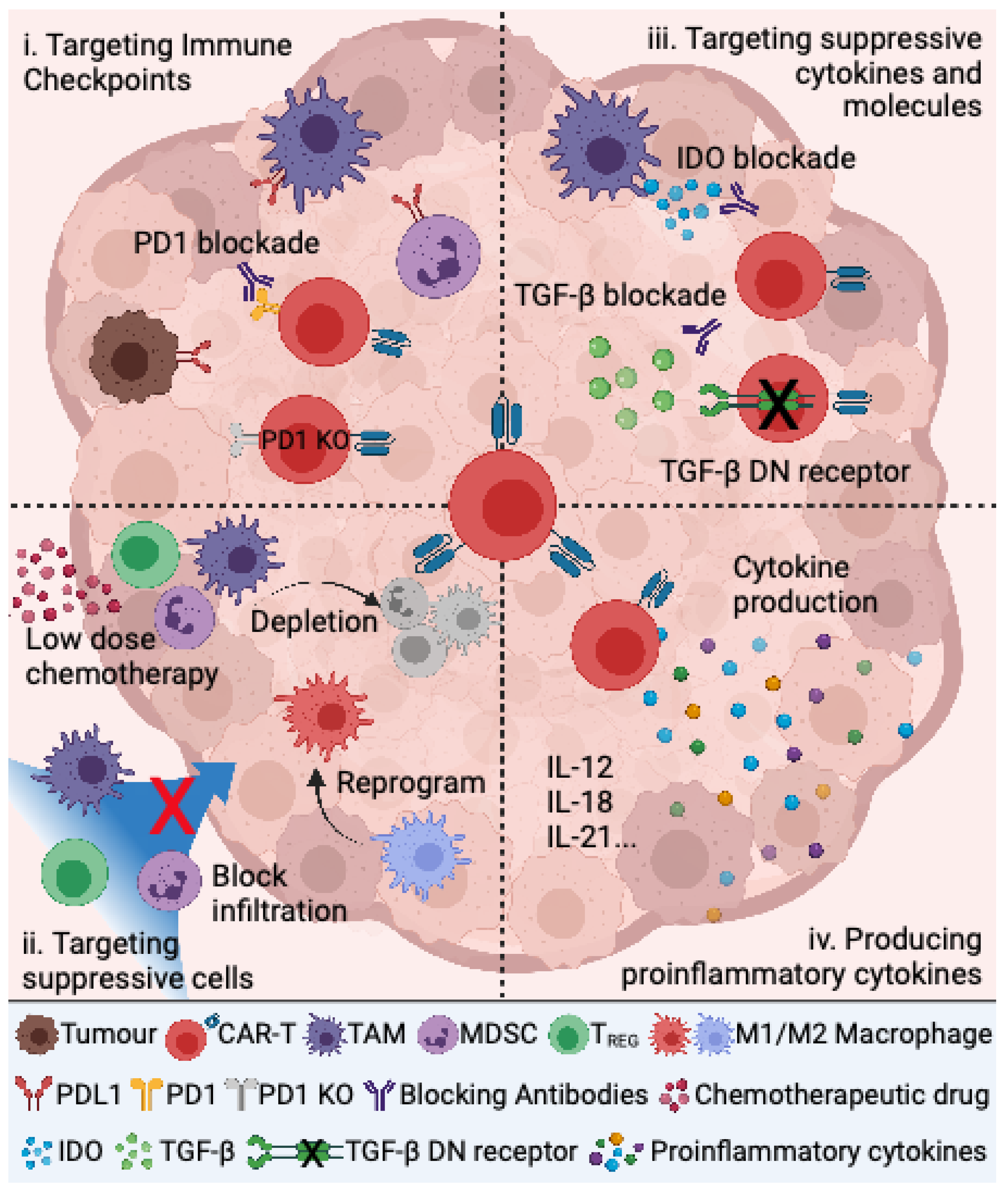
4. Targeting the Immunosuppressive TME to Improve CAR T Cell Therapy for Pediatric Sarcomas

In the limited trials to date, the majority of pediatric sarcoma patients fail to show any clinical response, notwithstanding the impressive responses in a small number of patients. The question is why do some patients respond, whilst others do not despite apparent target antigen expression? One significant contributing factor to this problem is the immunosuppressive nature of the solid tumor TME. This severely limits CAR T cell recruitment and infiltration into the tumor, CAR T cell survival, proliferation, and anti-tumor activity. In the following sections, we discuss potential approaches that involve modulation of the TME, or the CAR T cells themselves, to overcome mechanisms of immunosuppression in solid tumors that could be applied to improve the efficacy of CAR T cell therapy in sarcomas. These strategies are summarized in Figure 1.

4.1. Targeting Immune Checkpoints

Immune checkpoints are regulators of immune cell activation that suppress T cell-mediated responses in many cancer types [54]. PD-L1 is expressed by tumor cells and immunosuppressive immune cell populations, such as tumor-associated macrophages (TAMs) and myeloid-derived suppressor cells (MDSCs); PD-L1 inhibits T cell activation by binding to PD-1 expressed by T cells [55]. Combination therapy with anti-PD-1 can boost the efficacy of CAR T cell therapy [56] by limiting tumor-mediated CAR T cell exhaustion [57]. PD-1 blockade in combination with HER2 CAR T cell therapy has so far achieved promising results in one child with rhabdomyosarcoma. This patient was first successfully treated with HER2 CAR T cells alone, but after disease relapse at 6 months off therapy, additional HER2 CAR T infusions were given together with pembrolizumab. This patient is now 20 months post T cell infusions with no detectable disease [21]. Another approach to mitigating the PD-1/PD-L1 axis is to knockout PD-1 expression by CAR T cells. This strategy is currently being tested in over 20 clinical trials in adult cancers, including those of the breast, lung, and prostate [58]. The potential of this is exemplified by several studies. Rupp et al. demonstrated, using CD19+ PD-L1+ K562 cells, that PD-1 disrupted CAR T cells significantly enhanced anti-tumor efficacy in vitro and in vivo (tumor clearance rate of 17% for conventional anti-CD19 CAR T cells increased to 100% in animals receiving PD-1 edited CAR T cells at 28 days post tumor implant) [58,59]. Hu et al. showed that PD-1 disruption improved the in vitro and in vivo anti-tumor activity of CD133-specific CAR T cells in an orthotopic mouse model of glioma, without apparent toxicity [60].
Other approaches include the use of dominant-negative PD-1 receptor CAR T cells and triple inhibitory receptor CAR T cells that downregulate PD-1, as well as two other immune checkpoint molecules (TIM-3 and LAG-3). Genetic blockade of these three checkpoint inhibitory receptors resulted in high expression of CD56 on CAR T cells, and enhanced inhibition of tumor growth [61]. A high expression of PD-L1 was detected in alveolar rhabdomyosarcoma (86%), Ewing’s sarcoma (57%), embryonal rhabdomyosarcoma (50%), and osteosarcoma (47%) [62]. In addition, there are a multitude of immune checkpoints beyond the PD-1/PD-L1 axis [63], which may be more relevant to target in different subsets of pediatric sarcomas, in conjunction with CAR T cell therapy. For instance, a higher expression of CTL-4 in circulating CD4+ and CD8+ T cells was detected in children affected by aggressive sarcomas [64]. Recently, the largest screen of B7-H3 expression in pediatric tumors showed a high and homogeneous expression in rhabdomyosarcoma and Ewing’s sarcoma [41,65]. In advanced osteosarcoma, TIGIT+ T cells are highly present supporting the potential clinical application of TIGIT blockade in this tumor [66]. Although further research is needed to pinpoint the most appropriate targets, the examples of superior efficacy of combined PD-1 blockade, together with CAR T cell therapy, indicates the potential promise of CAR T cell modifications. Extending this to other immune checkpoint targets more specific to pediatric sarcomas could improve clinical response rates.

4.2. Targeting Suppressive Immune Cells

The TME is a heterogenous network of immune cell populations, including immune cell subsets that support tumor growth, survival, and suppress cytotoxic T cell responses, such as tumor-associated macrophages (TAMs), myeloid-derived suppressor cells (MDSCs), and regulatory T cells (Treg) [67]. Several approaches that inhibit these cell types in the TME are being tested. Low dose chemotherapy, with agents such as 5-fluorouracil, paclitaxel, cisplatin, and gemcitabine deplete MDSCs [68] and could potentially be used in combination with CAR T cell therapy to boost anti-tumor responses. Low dose chemotherapy was also found to deplete Treg cells [69]. Indeed, low dose chemotherapy added to the conditioning regimen has been shown to boost CAR T cell activity in a lung tumor model by activating tumor macrophages to express T-cell recruiting chemokines [70].
Blockade of the chemokines that TAMs, MDSCs, and Treg cells use to traffic to the tumor is another approach to deplete these immunosuppressive subtypes from the TME. Antibodies that block the CCL2/CCR2 axis have shown promising results in depleting myeloid cells from the TME in several pre-clinical models [71]. The CXCR4/CXCL12 pathway is another target for inhibition that could be combined with CAR T cell therapy to reduce the number of immunosuppressive myeloid cells within the TME [72]. The cytokine Colony Stimulating Factor 1 (CSF-1) is an important regulator of myeloid cell recruitment. Blockade of the receptor of this cytokine (CSF1R) has been shown not only to reduce the recruitment of myeloid cells into the tumor but to also preferentially promote differentiation into an M1 pro-inflammatory phenotype, as opposed to an M2 anti-inflammatory phenotype [73]. The chemokine receptor CCR4 is highly expressed on T regulatory cells and blockade of the CCR4/CCL22 axis could be used to reduce the number of these cells in the TME [74]. Several tyrosine kinase inhibitors have also been found to reduce MDSC [75,76] and regulatory T cells [77] in the TME and could, potentially, be used in conjunction with CAR T cell therapy in sarcoma.
Low dose radiotherapy has been shown to reprogram TAM into M1 pro-inflammatory macrophages in several studies [78,79] and to reduce the numbers of T regs in the TME [80]. There are also some promising results using toll-like receptor agonists to promote M1 macrophage polarization in the TME [72]. RNA-based therapies are emerging that promote M1 macrophage differentiation, by downregulating the expression of M2 genes [81,82]. Targeting the BTK–PI3Kγ pathway has been shown to reprogram TAMs into M1 macrophages in several studies [83,84,85] and has the potential to be combined with CAR T cell therapy to improve their efficacy. However, first generation BTK inhibitors suppress NK-cell cytotoxicity so could potentially inhibit CAR T cell function. This was not observed with second generation BTK inhibitors, highlighting the need for careful consideration of the most appropriate combination therapies when using this class of drug with CAR T therapy [86].

4.3. Targeting Immunosuppressive Cytokines and Other Molecules

Tumor cells and immunosuppressive immune cells in the TME produce a range of anti-inflammatory cytokines that suppress CAR T cell proliferation and activity. Blockade of transforming growth factor beta (TGFβ) signaling has been shown to reduce tumor growth and promote T cell-driven anti-tumor responses in vivo [87]. Another approach is to engineer CAR T cells with a TGFβ dominant negative receptor to mitigate the suppressive effect of TGFβ signaling on CAR T cell responses. This has been shown to increase the efficacy of these CAR T cells in models of prostate cancer [88]. Indoleamine 2,3, dioxygenase (IDO1) is another anti-inflammatory molecule produced by TAM and other immunosuppressive immune cells in the TME, which suppresses T cell activation and function [89]. Blockade of IDO significantly improved the efficacy of CAR T therapy in a pre-clinical model of colon cancer [90].

4.4. Production of Proinflammatory Cytokines

Another way to boost the efficacy of CAR T cells is to directly engineer them to express pro-inflammatory cytokines (TRUCKs or ARMOURED CARs) [91]. IL-12 enhances T cell cytotoxicity and induces the production of IFNγ driving the Th1 response. IFNγ in turn activates monocytes and macrophages to further increase IL-12 production [92]. CAR T cells producing IL-12 have shown increased anti-tumor efficacy [93]. Furthermore, IL-23 antagonists improve the IL-12: IL-13 ratio in favor of IL-12, with the potential to be used in combination with CAR T cells [94]. IL-18 promotes both Th1 and Th2 responses and, in conjunction with IFNγ production, recruits and activates innate immune cells and enhances further IFNγ production [95]. CAR T cells that constitutively express IL-18 have also shown increased efficacy in a model of melanoma [96]. IL-21 is a pleiotropic cytokine that is produced by T cell subsets and enhances the survival and anti-tumor activity of CD8+ T cells [97]. Indeed, CAR T cells, producing IL-21, have shown increased efficacy against tumor cells in vivo [98].

5. Understanding the Immunosuppressive TME Is Key to Improving CAR T Cell Therapy

There are many different methods that can be deployed to target the immunosuppressive TME of solid tumors. However, it is currently unclear which method should be applied to which subsets of pediatric sarcomas. Indeed, the astonishing diversity of histological and molecular subtypes of sarcoma might suggest that there will be many immunosuppressive pathways operating in these entities. A detailed characterization of the immune TME of pediatric sarcomas will provide the necessary insights into the immunosuppressive mechanisms that should be targeted in different sarcoma subtypes. This detailed characterization of the immune TME is mostly lacking [99]. Indeed, even at a basic level, there is little data to indicate whether subsets of pediatric sarcomas are immune-inflamed and may respond to CAR T cell therapy, as well as which subsets would require specific co-therapies to boost CAR T cell efficacy. A better understanding of the TME of different sarcoma subtypes will undoubtedly allow for more specific targeting of the immunosuppressive mechanisms that limit CAR T cell recruitment and migration into the tumor, as well as their proliferation and persistence [100].
Solid tumors can be classified into three subtypes based on their immune microenvironments. Immune-inflamed “hot” tumors have significant numbers of CD8+ T cells in the parenchyma and express pro-inflammatory cytokines, chemokines, and immune checkpoints [101]. These patients could be anticipated to be the best responders to CAR T cell therapy as they already permit endogenous T cell infiltration into the TME [102]. As they also express immune checkpoint molecules, they may also benefit from co-therapy with immune checkpoint inhibitors.
Immune-altered “intermediate” tumors are characterized by the restriction of T cell infiltrate to the surrounding stroma, with production of chemokines and tumor-derived inhibitory factors, such as TGFβ [101]. To improve CAR T cell therapy, these tumors could potentially benefit from co-therapies that target immunosuppressive cytokines, such as TGFβ inhibitors, or by treatment with fourth generation CAR T cells that express inflammatory cytokines.
Immune desert “cold” tumors completely lack CD8+ T cells and are populated with immunosuppressive immune cell populations such as TAMs, MDSCs, and regulatory T cells [101]. The cytokine milieu in these tumors is that of immune suppression and tolerance. These tumors would be expected to be the poorest responders to CAR T cell therapy as the same mechanisms that exclude endogenous T cell infiltrate into the TME would be expected to limit CAR T cell recruitment [102]. These tumors present the most challenging TME for immunotherapies. Co-treatment with low dose chemotherapy, radiotherapy, or targeted therapy, which significantly modify the TME and reduce immunosuppressive immune cell populations, may have the greatest impact on efficacy. Cisplatin is one of the main chemotherapies used in standard protocols to treat osteosarcoma [103]. Similarly, radiotherapy is standard of care for both rhabdomyosarcoma and Ewing’s sarcoma [104]. CAR T cell therapy could be combined with standard protocols to treat certain subtypes of refractory sarcoma.
There are also intrinsic features of CAR T cells which may limit their efficacy, but which may, nonetheless, be amenable to intervention. This has been reviewed elsewhere [105]. However, some interesting lessons from the application of CAR T therapies in leukemia may be relevant to sarcoma immunotherapy. One recent line of investigation is the capacity of bromodomain inhibitors to maintain expression of the CAR and prolong CAR T cell persistence [106,107]. Epigenetic mechanisms appear to play a significant role in diminishing CAR expression over time, perhaps a result of the epigenetic profile of the parent T-cells from which the CAR T cells in the final vaccine product are derived [106], as well as the non-random sites of lentiviral integration during CAR T cell manufacture [107,108]. Treatment of CAR T cells, during the in vitro expansion phase with the BET inhibitor drug JQ1, can maintain a memory-cell phenotype, prolonging expression of the CAR and persistence of the CAR T cells. The wider application of CAR T therapy in hematological malignancies also raises questions about the possibility of high-quality donor cells that may be derived from sources other than the individual patient (so-called non-autologous, “off-the-shelf” CAR T cells). Here, the balance is between using T cells, which are more readily available, and the risks of eliciting graft versus host disease. At least one study has shown that deletion of the endogenous TCR in such CAR T cells greatly reduces their alloreactivity, but at some cost to efficacy [109].

6. Conclusions

CAR T cell therapy harnesses a patient’s own immune system to recognize and kill cancer cells. It has the potential to improve clinical outcomes for children with sarcomas that are resistant to standard therapies or who have relapsed. There are currently several clinical trials open to children with tumors that express HER2, NY-ESO, GD2, B7-H3, EGFR, and GPC3, and some promising results have already been reported, notably using HER2 CAR T cells, in which complete responses have been achieved in patients with osteosarcoma and rhabdomyosarcoma [20] and with NY-ESO-1+ synovial sarcomas treated with NY-ESO CAR T cells [28,29]. However, there are many other promising options for the future development of cell-based immunotherapies, including CAR T cells directed against new, promising targets such as EPHA2. EPHA 2 CAR T cells have very recently entered a first-in-human clinical trial in adult glioblastoma [38]. Although optimization will be required, these exciting developments pave the road towards further testing of EPHA2 CAR T cell-directed therapies in childhood sarcoma patients, ideally in combination with strategies that tackle the immunosuppressive barriers in these tumors.
One of the main limitations in CAR T trials, in children with solid tumors, is the recruitment of patients based solely on tumor antigen expression rather than on the subtype of their cancer. As a result, patients with different types of cancer may receive the same CAR T product without considering the features of their specific TME, which is a key contributor to failed CAR T cell responses [110]. At this time, we do not routinely screen for sarcomas that are immune-inflamed and may permit CAR T cell infiltration into the TME or distinguish them from those that are not inflamed and are likely to exclude or lack CAR T cell infiltrates in the TME. However, tackling this immunosuppressive barrier, for example, with a PD-1 blockade, can increase efficacy of CAR T cell mediated therapy. This clearly mandates a deeper investigation into the unique TME of pediatric sarcomas. Identification of checkpoint inhibitors, or other TME components specific to pediatric sarcomas, holds the promise to increase efficacy of CAR T cells and tailored CAR T cell-focused combination therapies.
Pediatric sarcomas present several obstacles for CAR T cell efficacy. These obstacles can be divided into four main CAR T cell combination treatment strategies by (1) targeting immune checkpoint molecules and ligands, (2) targeting immune suppressor cells in the tumor microenvironment, (3) targeting suppressive cytokines and molecules, and (4) producing pro-inflammatory cytokines.

Author Contributions

R.L.T. and D.M. conceptualized the review with P.J.N. and P.G.E., and wrote the initial draft of the manuscript. All authors R.L.T., D.M., E.D.G.F., C.M., J.Z., N.O., D.S.Z., M.H., P.K.D., J.A.T., P.J.N., and P.G.E. contributed specialist knowledge to the writing, review and editing of the manuscript. All authors have read and agreed to the published version of the manuscript.

Funding

This review was funded by Tour de Cure Pioneering Research Grant Scheme, grant number RSP-00042-19/20 and ANZSA Sarcoma Research Grant, grant number 43.

Acknowledgments

R.L.T. and P.G.E. acknowledge the support of the Australian and New Zealand Sarcoma Association. R.L.T. and P.G.E. acknowledge the support of Tour de Cure. D.M. acknowledges the support of Plan Cancer, Fondation de France and Fondation Nuovo Soldati.

Conflicts of Interest

The authors declare no conflict of interest.

References

  1. Steliarova-Foucher, E.; Colombet, M.; Ries, L.A.G.; Moreno, F.; Dolya, A.; Bray, F.; Hesseling, P.; Shin, H.Y.; Stiller, C.A.; Bouzbid, S.; et al. International incidence of childhood cancer, 2001–2010: A population-based registry study. Lancet Oncol. 2017, 18, 719–731. [Google Scholar] [CrossRef]
  2. Ward, E.; DeSantis, C.; Robbins, A.; Kohler, B.; Jemal, A. Childhood and adolescent cancer statistics, 2014. CA Cancer J. Clin. 2014, 64, 83–103. [Google Scholar] [CrossRef]
  3. Howlader, N.; Noone, A.M.; Krapcho, M.; Miller, D.; Brest, A.; Yu, M.; Ruhl, J.; Tatalovich, Z.; Mariotto, A.; Lewis, D.R.; et al. SEER Cancer Statistics Review, 1975–2017; National Cancer Institute: Bethesda, MD, USA, 2020; Based on November 2019 SEER data submission, posted to the SEER web site, April 2020. [Google Scholar]
  4. Lewis, D.R.; Siembida, E.J.; Seibel, N.L.; Smith, A.W.; Mariotto, A.B. Survival outcomes for cancer types with the highest death rates for adolescents and young adults, 1975–2016. Cancer 2021. [Google Scholar] [CrossRef]
  5. Bhakta, N.; Liu, Q.; Ness, K.K.; Baassiri, M.; Eissa, H.; Yeo, F.; Chemaitilly, W.; Ehrhardt, M.J.; Bass, J.; Bishop, M.W.; et al. The cumulative burden of surviving childhood cancer: An initial report from the St Jude Lifetime Cohort Study (SJLIFE). Lancet 2017, 390, 2569–2582. [Google Scholar] [CrossRef]
  6. Keegan, T.H.; Ries, L.A.; Barr, R.D.; Geiger, A.M.; Dahlke, D.V.; Pollock, B.H.; Bleyer, W.A. National Cancer Institute Next Steps for Adolescent and Young Adult Oncology Epidemiology Working Group Comparison of cancer survival trends in the United States of adolescents and young adults with those in children and older adults. Cancer 2016, 122, 1009–1016. [Google Scholar] [CrossRef] [PubMed]
  7. Dyson, K.A.; Stover, B.D.; Grippin, A.; Mendez-Gomez, H.R.; Lagmay, J.; Mitchell, D.A.; Sayour, E.J. Emerging trends in immunotherapy for pediatric sarcomas. J. Hematol. Oncol. 2019, 12, 78. [Google Scholar] [CrossRef]
  8. June, C.H.; O’Connor, R.S.; Kawalekar, O.U.; Ghassemi, S.; Milone, M.C. CAR T cell immunotherapy for human cancer. Science 2018, 359, 1361–1365. [Google Scholar] [CrossRef] [PubMed] [Green Version]
  9. Moeller, M.; Haynes, N.M.; Trapani, J.A.; Teng, M.W.; Jackson, J.T.; Tanner, J.E.; Cerutti, L.; Jane, S.M.; Kershaw, M.H.; Smyth, M.J.; et al. A functional role for CD28 costimulation in tumor recognition by single-chain receptor-modified T cells. Cancer Gene Ther. 2004, 11, 371–379. [Google Scholar] [CrossRef]
  10. Petersen, C.T.; Krenciute, G. Next Generation CAR T Cells for the Immunotherapy of High-Grade Glioma. Front. Oncol. 2019, 9, 69. [Google Scholar] [CrossRef] [Green Version]
  11. Cao, J.; Wang, G.; Cheng, H.; Wei, C.; Qi, K.; Sang, W.; Zhenyu, L.; Shi, M.; Li, H.; Qiao, J.; et al. Potent anti-leukemia activities of humanized CD19-targeted Chimeric antigen receptor T (CAR-T) cells in patients with relapsed/refractory acute lymphoblastic leukemia. Am. J. Hematol. 2018, 93, 851–858. [Google Scholar] [CrossRef] [PubMed] [Green Version]
  12. Kochenderfer, J.N.; Somerville, R.P.T.; Lu, T.; Yang, J.C.; Sherry, R.M.; Feldman, S.A.; McIntyre, L.; Bot, A.; Rossi, J.; Lam, N.; et al. Long-Duration Complete Remissions of Diffuse Large B Cell Lymphoma after Anti-CD19 Chimeric Antigen Receptor T Cell Therapy. Mol. Ther. 2017, 25, 2245–2253. [Google Scholar] [CrossRef] [Green Version]
  13. Chavez, J.C.; Bachmeier, C.; Kharfan-Dabaja, M.A. CAR T-cell therapy for B-cell lymphomas: Clinical trial results of available products. Ther. Adv. Hematol. 2019, 10, 2040620719841581. [Google Scholar] [CrossRef] [PubMed] [Green Version]
  14. Tesfaye, M.; Savoldo, B. Adoptive Cell Therapy in Treating Pediatric Solid Tumors. Curr. Oncol. Rep. 2018, 20, 73. [Google Scholar] [CrossRef]
  15. Fleuren, E.D.; Versleijen-Jonkers, Y.M.; Boerman, O.C.; van der Graaf, W.T. Targeting receptor tyrosine kinases in osteosarcoma and Ewing sarcoma: Current hurdles and future perspectives. Biochim. Biophys. Acta 2014, 1845, 266–276. [Google Scholar] [CrossRef]
  16. Scotlandi, K.; Manara, M.C.; Hattinger, C.M.; Benini, S.; Perdichizzi, S.; Pasello, M.; Bacci, G.; Zanella, L.; Bertoni, F.; Picci, P.; et al. Prognostic and therapeutic relevance of HER2 expression in osteosarcoma and Ewing’s sarcoma. Eur. J. Cancer 2005, 41, 1349–1361. [Google Scholar] [CrossRef]
  17. Ahmed, N.; Brawley, V.S.; Hegde, M.; Robertson, C.; Ghazi, A.; Gerken, C.; Liu, E.; Dakhova, O.; Ashoori, A.; Corder, A.; et al. Human Epidermal Growth Factor Receptor 2 (HER2) -Specific Chimeric Antigen Receptor-Modified T Cells for the Immunotherapy of HER2-Positive Sarcoma. J. Clin. Oncol. 2015, 33, 1688–1696. [Google Scholar] [CrossRef]
  18. Tabak, S.A.; Khalifa, S.E.; Fathy, Y. HER-2 Immunohistochemical Expression in Bone Sarcomas: A New Hope for Osteosarcoma Patients. Open Access Maced. J. Med. Sci. 2018, 6, 1555–1560. [Google Scholar] [CrossRef] [PubMed] [Green Version]
  19. Morgan, R.A.; Yang, J.C.; Kitano, M.; Dudley, M.E.; Laurencot, C.M.; Rosenberg, S.A. Case report of a serious adverse event following the administration of T cells transduced with a chimeric antigen receptor recognizing ERBB2. Mol. Ther. 2010, 18, 843–851. [Google Scholar] [CrossRef] [PubMed]
  20. Navai, S.; Derenzo, C.; Joseph, S.; Sanber, K.; Byrd, T.; Zhang, H.; Mata, M.; Gerken, C.; Shree, A.; Mathew, P.; et al. Abstract LB-147: Administration of HER2-CAR T Cells after Lymphodepletion Safely Improves T Cell Expansion and Induces Clinical Responses in Patients with Advanced Sarcomas; 2019; p. LB-147. [Google Scholar]
  21. Hegde, M.; Joseph, S.K.; Pashankar, F.; DeRenzo, C.; Sanber, K.; Navai, S.; Byrd, T.T.; Hicks, J.; Xu, M.L.; Gerken, C.; et al. Tumor response and endogenous immune reactivity after administration of HER2 CAR T cells in a child with metastatic rhabdomyosarcoma. Nat. Commun. 2020, 11, 3549. [Google Scholar] [CrossRef] [PubMed]
  22. Wen, Y.H.; Koeppen, H.; Garcia, R.; Chiriboga, L.; Tarlow, B.D.; Peters, B.A.; Eigenbrot, C.; Yee, H.; Steiner, G.; Greco, M.A. Epidermal growth factor receptor in osteosarcoma: Expression and mutational analysis. Hum. Pathol. 2007, 38, 1184–1191. [Google Scholar] [CrossRef] [PubMed]
  23. Yang, J.L.; Hannan, M.T.; Russell, P.J.; Crowe, P.J. Expression of HER1/EGFR protein in human soft tissue sarcomas. Eur. J. Surg. Oncol. 2006, 32, 466–468. [Google Scholar] [CrossRef] [PubMed]
  24. Smith, S.M.; Iwenofu, O.H. NY-ESO-1: A promising cancer testis antigen for sarcoma immunotherapy and diagnosis. Chin. Clin. Oncol. 2018, 7, 44. [Google Scholar] [CrossRef] [PubMed]
  25. Desar, I.M.E.; Fleuren, E.D.G.; van der Graaf, W.T.A. Systemic Treatment for Adults with Synovial Sarcoma. Curr. Treat. Options Oncol. 2018, 19, 13. [Google Scholar] [CrossRef] [Green Version]
  26. Robbins, P.F.; Kassim, S.H.; Tran, T.L.; Crystal, J.S.; Morgan, R.A.; Feldman, S.A.; Yang, J.C.; Dudley, M.E.; Wunderlich, J.R.; Sherry, R.M.; et al. A pilot trial using lymphocytes genetically engineered with an NY-ESO-1-reactive T-cell receptor: Long-term follow-up and correlates with response. Clin. Cancer Res. 2015, 21, 1019–1027. [Google Scholar] [CrossRef] [PubMed] [Green Version]
  27. Robbins, P.F.; Morgan, R.A.; Feldman, S.A.; Yang, J.C.; Sherry, R.M.; Dudley, M.E.; Wunderlich, J.R.; Nahvi, A.V.; Helman, L.J.; Mackall, C.L.; et al. Tumor regression in patients with metastatic synovial cell sarcoma and melanoma using genetically engineered lymphocytes reactive with NY-ESO-1. J. Clin. Oncol. 2011, 29, 917–924. [Google Scholar] [CrossRef]
  28. D’Angelo, S.P.; Melchiori, L.; Merchant, M.S.; Bernstein, D.; Glod, J.; Kaplan, R.; Grupp, S.; Tap, W.D.; Chagin, K.; Binder, G.K.; et al. Antitumor Activity Associated with Prolonged Persistence of Adoptively Transferred NY-ESO-1 (c259)T Cells in Synovial Sarcoma. Cancer Discov. 2018, 8, 944–957. [Google Scholar] [CrossRef] [Green Version]
  29. Ramachandran, I.; Lowther, D.E.; Dryer-Minnerly, R.; Wang, R.; Fayngerts, S.; Nunez, D.; Betts, G.; Bath, N.; Tipping, A.J.; Melchiori, L.; et al. Systemic and local immunity following adoptive transfer of NY-ESO-1 SPEAR T cells in synovial sarcoma. J. Immunother. Cancer 2019, 7, 276. [Google Scholar] [CrossRef] [PubMed]
  30. Keung, E.Z.; Tawbi, H.A. Engineered T Cells in Synovial Sarcoma: Persistence Pays Off! Cancer Discov. 2018, 8, 914–917. [Google Scholar] [CrossRef] [Green Version]
  31. Van Tine, B.A.; Butler, M.O.; Araujo, D.; Johnson, M.L.; Clarke, J.; Liebner, D.; Odunsi, K.; Olszanski, A.J.; Basu, S.; Brophy, F.; et al. ADP-A2M4 (MAGE-A4) in patients with synovial sarcoma. Ann. Oncol. 2019, 30, v684–v685. [Google Scholar] [CrossRef]
  32. Dobrenkov, K.; Ostrovnaya, I.; Gu, J.; Cheung, I.Y.; Cheung, N.K. Oncotargets GD2 and GD3 are highly expressed in sarcomas of children, adolescents, and young adults. Pediatr. Blood Cancer 2016, 63, 1780–1785. [Google Scholar] [CrossRef] [Green Version]
  33. Nazha, B.; Inal, C.; Owonikoko, T.K. Disialoganglioside GD2 Expression in Solid Tumors and Role as a Target for Cancer Therapy. Front. Oncol. 2020, 10, 1000. [Google Scholar] [CrossRef]
  34. Poon, V.I.; Roth, M.; Piperdi, S.; Geller, D.; Gill, J.; Rudzinski, E.R.; Hawkins, D.S.; Gorlick, R. Ganglioside GD2 expression is maintained upon recurrence in patients with osteosarcoma. Clin. Sarcoma Res. 2015, 5, 4. [Google Scholar] [CrossRef] [Green Version]
  35. Charan, M.; Dravid, P.; Cam, M.; Audino, A.; Gross, A.C.; Arnold, M.A.; Roberts, R.D.; Cripe, T.P.; Pertsemlidis, A.; Houghton, P.J.; et al. GD2-directed CAR-T cells in combination with HGF-targeted neutralizing antibody (AMG102) prevent primary tumor growth and metastasis in Ewing sarcoma. Int. J. Cancer 2020, 146, 3184–3195. [Google Scholar] [CrossRef] [PubMed]
  36. Chulanetra, M.; Morchang, A.; Sayour, E.; Eldjerou, L.; Milner, R.; Lagmay, J.; Cascio, M.; Stover, B.; Slayton, W.; Chaicumpa, W.; et al. GD2 chimeric antigen receptor modified T cells in synergy with sub-toxic level of doxorubicin targeting osteosarcomas. Am. J. Cancer Res. 2020, 10, 674–687. [Google Scholar] [PubMed]
  37. Hsu, K.; Middlemiss, S.; Saletta, F.; Gottschalk, S.; McCowage, G.B.; Kramer, B. Chimeric Antigen Receptor-modified T cells targeting EphA2 for the immunotherapy of paediatric bone tumours. Cancer Gene Ther. 2021, 28, 321–334. [Google Scholar] [CrossRef]
  38. Lin, Q.; Ba, T.; Ho, J.; Chen, D.; Cheng, Y.; Wang, L.; Xu, G.; Xu, L.; Zhou, Y.; Wei, Y.; et al. First-in-Human Trial of EphA2-Redirected CAR T-Cells in Patients With Recurrent Glioblastoma: A Preliminary Report of Three Cases at the Starting Dose. Front. Oncol. 2021, 11, 694941. [Google Scholar] [CrossRef] [PubMed]
  39. Modak, S.; Kramer, K.; Gultekin, S.H.; Guo, H.F.; Cheung, N.K. Monoclonal antibody 8H9 targets a novel cell surface antigen expressed by a wide spectrum of human solid tumors. Cancer Res. 2001, 61, 4048–4054. [Google Scholar] [PubMed]
  40. Wang, L.; Zhang, Q.; Chen, W.; Shan, B.; Ding, Y.; Zhang, G.; Cao, N.; Liu, L.; Zhang, Y. B7-H3 is overexpressed in patients suffering osteosarcoma and associated with tumor aggressiveness and metastasis. PLoS ONE 2013, 8, e70689. [Google Scholar] [CrossRef]
  41. Majzner, R.G.; Theruvath, J.L.; Nellan, A.; Heitzeneder, S.; Cui, Y.; Mount, C.W.; Rietberg, S.P.; Linde, M.H.; Xu, P.; Rota, C.; et al. CAR T Cells Targeting B7-H3, a Pan-Cancer Antigen, Demonstrate Potent Preclinical Activity Against Pediatric Solid Tumors and Brain Tumors. Clin. Cancer Res. 2019, 25, 2560–2574. [Google Scholar] [CrossRef]
  42. Alijaj, N.; Moutel, S.; Gouveia, Z.L.; Gray, M.; Roveri, M.; Dzhumashev, D.; Weber, F.; Meier, G.; Luciani, P.; Rossler, J.K.; et al. Novel FGFR4-Targeting Single-Domain Antibodies for Multiple Targeted Therapies against Rhabdomyosarcoma. Cancers 2020, 12, 3313. [Google Scholar] [CrossRef] [PubMed]
  43. Khan, J.; Wei, J.S.; Ringner, M.; Saal, L.H.; Ladanyi, M.; Westermann, F.; Berthold, F.; Schwab, M.; Antonescu, C.R.; Peterson, C.; et al. Classification and diagnostic prediction of cancers using gene expression profiling and artificial neural networks. Nat. Med. 2001, 7, 673–679. [Google Scholar] [CrossRef]
  44. Evans, C.H.; Liu, F.; Porter, R.M.; O’Sullivan, R.P.; Merghoub, T.; Lunsford, E.P.; Robichaud, K.; Van Valen, F.; Lessnick, S.L.; Gebhardt, M.C.; et al. EWS-FLI-1-targeted cytotoxic T-cell killing of multiple tumor types belonging to the Ewing sarcoma family of tumors. Clin. Cancer Res. 2012, 18, 5341–5351. [Google Scholar] [CrossRef] [Green Version]
  45. Gattenlohner, S.; Marx, A.; Markfort, B.; Pscherer, S.; Landmeier, S.; Juergens, H.; Muller-Hermelink, H.K.; Matthews, I.; Beeson, D.; Vincent, A.; et al. Rhabdomyosarcoma lysis by T cells expressing a human autoantibody-based chimeric receptor targeting the fetal acetylcholine receptor. Cancer Res. 2006, 66, 24–28. [Google Scholar] [CrossRef] [PubMed] [Green Version]
  46. Huang, G.; Yu, L.; Cooper, L.J.; Hollomon, M.; Huls, H.; Kleinerman, E.S. Genetically modified T cells targeting interleukin-11 receptor alpha-chain kill human osteosarcoma cells and induce the regression of established osteosarcoma lung metastases. Cancer Res. 2012, 72, 271–281. [Google Scholar] [CrossRef] [PubMed] [Green Version]
  47. Huang, X.; Park, H.; Greene, J.; Pao, J.; Mulvey, E.; Zhou, S.X.; Albert, C.M.; Moy, F.; Sachdev, D.; Yee, D.; et al. IGF1R- and ROR1-Specific CAR T Cells as a Potential Therapy for High Risk Sarcomas. PLoS ONE 2015, 10, e0133152. [Google Scholar] [CrossRef]
  48. Simon-Keller, K.; Paschen, A.; Hombach, A.A.; Strobel, P.; Coindre, J.M.; Eichmuller, S.B.; Vincent, A.; Gattenlohner, S.; Hoppe, F.; Leuschner, I.; et al. Survivin blockade sensitizes rhabdomyosarcoma cells for lysis by fetal acetylcholine receptor-redirected T cells. Am. J. Pathol. 2013, 182, 2121–2131. [Google Scholar] [CrossRef] [Green Version]
  49. Fernandez, L.; Metais, J.Y.; Escudero, A.; Vela, M.; Valentin, J.; Vallcorba, I.; Leivas, A.; Torres, J.; Valeri, A.; Patino-Garcia, A.; et al. Memory T Cells Expressing an NKG2D-CAR Efficiently Target Osteosarcoma Cells. Clin. Cancer Res. 2017, 23, 5824–5835. [Google Scholar] [CrossRef]
  50. Lehner, M.; Gotz, G.; Proff, J.; Schaft, N.; Dorrie, J.; Full, F.; Ensser, A.; Muller, Y.A.; Cerwenka, A.; Abken, H.; et al. Redirecting T cells to Ewing’s sarcoma family of tumors by a chimeric NKG2D receptor expressed by lentiviral transduction or mRNA transfection. PLoS ONE 2012, 7, e31210. [Google Scholar] [CrossRef] [PubMed]
  51. Kirschner, A.; Thiede, M.; Grunewald, T.G.; Alba Rubio, R.; Richter, G.H.; Kirchner, T.; Busch, D.H.; Burdach, S.; Thiel, U. Pappalysin-1 T cell receptor transgenic allo-restricted T cells kill Ewing sarcoma in vitro and in vivo. Oncoimmunology 2017, 6, e1273301. [Google Scholar] [CrossRef] [Green Version]
  52. Xiao, W.; Wang, J.; Wen, X.; Xu, B.; Que, Y.; Yu, K.; Xu, L.; Zhao, J.; Pan, Q.; Zhou, P.; et al. Chimeric antigen receptor-modified T-cell therapy for platelet-derived growth factor receptor alpha-positive rhabdomyosarcoma. Cancer 2020, 126 (Suppl. 9), 2093–2100. [Google Scholar] [CrossRef] [PubMed] [Green Version]
  53. Englisch, A.; Altvater, B.; Kailayangiri, S.; Hartmann, W.; Rossig, C. VEGFR2 as a target for CAR T cell therapy of Ewing sarcoma. Pediatr. Blood Cancer 2020, 67, e28313. [Google Scholar] [CrossRef]
  54. He, X.; Xu, C. Immune checkpoint signaling and cancer immunotherapy. Cell Res. 2020, 30, 660–669. [Google Scholar] [CrossRef] [PubMed]
  55. Sharpe, A.H.; Pauken, K.E. The diverse functions of the PD1 inhibitory pathway. Nat. Rev. Immunol. 2018, 18, 153–167. [Google Scholar] [CrossRef] [PubMed]
  56. John, L.B.; Devaud, C.; Duong, C.P.; Yong, C.S.; Beavis, P.A.; Haynes, N.M.; Chow, M.T.; Smyth, M.J.; Kershaw, M.H.; Darcy, P.K. Anti-PD-1 antibody therapy potently enhances the eradication of established tumors by gene-modified T cells. Clin. Cancer Res. 2013, 19, 5636–5646. [Google Scholar] [CrossRef] [PubMed] [Green Version]
  57. Xu, J.; Zhang, Q.; Tian, K.; Wang, H.; Yin, H.; Zheng, J. Current status and future prospects of the strategy of combining CART with PD1 blockade for antitumor therapy (Review). Mol. Med. Rep. 2018, 17, 2083–2088. [Google Scholar] [CrossRef] [PubMed] [Green Version]
  58. McGowan, E.; Lin, Q.; Ma, G.; Yin, H.; Chen, S.; Lin, Y. PD-1 disrupted CAR-T cells in the treatment of solid tumors: Promises and challenges. Biomed. Pharmacother. 2020, 121, 109625. [Google Scholar] [CrossRef]
  59. Rupp, L.J.; Schumann, K.; Roybal, K.T.; Gate, R.E.; Ye, C.J.; Lim, W.A.; Marson, A. CRISPR/Cas9-mediated PD-1 disruption enhances anti-tumor efficacy of human chimeric antigen receptor T cells. Sci. Rep. 2017, 7, 737. [Google Scholar] [CrossRef] [Green Version]
  60. Hu, B.; Zou, Y.; Zhang, L.; Tang, J.; Niedermann, G.; Firat, E.; Huang, X.; Zhu, X. Nucleofection with Plasmid DNA for CRISPR/Cas9-Mediated Inactivation of Programmed Cell Death Protein 1 in CD133-Specific CAR T Cells. Hum. Gene Ther. 2019, 30, 446–458. [Google Scholar] [CrossRef]
  61. Zou, F.; Lu, L.; Liu, J.; Xia, B.; Zhang, W.; Hu, Q.; Liu, W.; Zhang, Y.; Lin, Y.; Jing, S.; et al. Engineered triple inhibitory receptor resistance improves anti-tumor CAR-T cell performance via CD56. Nat. Commun. 2019, 10, 4109. [Google Scholar] [CrossRef]
  62. Majzner, R.G.; Simon, J.S.; Grosso, J.F.; Martinez, D.; Pawel, B.R.; Santi, M.; Merchant, M.S.; Geoerger, B.; Hezam, I.; Marty, V.; et al. Assessment of programmed death-ligand 1 expression and tumor-associated immune cells in pediatric cancer tissues. Cancer 2017, 123, 3807–3815. [Google Scholar] [CrossRef] [Green Version]
  63. Marin-Acevedo, J.A.; Kimbrough, E.O.; Lou, Y. Next generation of immune checkpoint inhibitors and beyond. J. Hematol. Oncol. 2021, 14, 45. [Google Scholar] [CrossRef]
  64. Hingorani, P.; Maas, M.L.; Gustafson, M.P.; Dickman, P.; Adams, R.H.; Watanabe, M.; Eshun, F.; Williams, J.; Seidel, M.J.; Dietz, A.B. Increased CTLA-4(+) T cells and an increased ratio of monocytes with loss of class II (CD14(+) HLA-DR(lo/neg)) found in aggressive pediatric sarcoma patients. J. Immunother. Cancer 2015, 3, 35. [Google Scholar] [CrossRef] [PubMed] [Green Version]
  65. Cocco, C.; Morandi, F.; Airoldi, I. Immune Checkpoints in Pediatric Solid Tumors: Targetable Pathways for Advanced Therapeutic Purposes. Cells 2021, 10, 927. [Google Scholar] [CrossRef] [PubMed]
  66. Zhou, Y.; Yang, D.; Yang, Q.; Lv, X.; Huang, W.; Zhou, Z.; Wang, Y.; Zhang, Z.; Yuan, T.; Ding, X.; et al. Single-cell RNA landscape of intratumoral heterogeneity and immunosuppressive microenvironment in advanced osteosarcoma. Nat. Commun. 2020, 11, 6322. [Google Scholar] [CrossRef] [PubMed]
  67. Labani-Motlagh, A.; Ashja-Mahdavi, M.; Loskog, A. The Tumor Microenvironment: A Milieu Hindering and Obstructing Antitumor Immune Responses. Front. Immunol. 2020, 11, 940. [Google Scholar] [CrossRef] [PubMed]
  68. Law, A.M.K.; Valdes-Mora, F.; Gallego-Ortega, D. Myeloid-Derived Suppressor Cells as a Therapeutic Target for Cancer. Cells 2020, 9, 561. [Google Scholar] [CrossRef] [Green Version]
  69. Ghiringhelli, F.; Larmonier, N.; Schmitt, E.; Parcellier, A.; Cathelin, D.; Garrido, C.; Chauffert, B.; Solary, E.; Bonnotte, B.; Martin, F. CD4+CD25+ regulatory T cells suppress tumor immunity but are sensitive to cyclophosphamide which allows immunotherapy of established tumors to be curative. Eur. J. Immunol. 2004, 34, 336–344. [Google Scholar] [CrossRef]
  70. Srivastava, S.; Furlan, S.N.; Jaeger-Ruckstuhl, C.A.; Sarvothama, M.; Berger, C.; Smythe, K.S.; Garrison, S.M.; Specht, J.M.; Lee, S.M.; Amezquita, R.A.; et al. Immunogenic Chemotherapy Enhances Recruitment of CAR-T Cells to Lung Tumors and Improves Antitumor Efficacy when Combined with Checkpoint Blockade. Cancer Cell 2021, 39, 193–208.e110. [Google Scholar] [CrossRef]
  71. Argyle, D.; Kitamura, T. Targeting Macrophage-Recruiting Chemokines as a Novel Therapeutic Strategy to Prevent the Progression of Solid Tumors. Front. Immunol. 2018, 9, 2629. [Google Scholar] [CrossRef]
  72. Anfray, C.; Ummarino, A.; Andon, F.T.; Allavena, P. Current Strategies to Target Tumor-Associated-Macrophages to Improve Anti-Tumor Immune Responses. Cells 2019, 9, 46. [Google Scholar] [CrossRef] [Green Version]
  73. Clappaert, E.J.; Murgaski, A.; Van Damme, H.; Kiss, M.; Laoui, D. Diamonds in the Rough: Harnessing Tumor-Associated Myeloid Cells for Cancer Therapy. Front. Immunol. 2018, 9, 2250. [Google Scholar] [CrossRef] [PubMed] [Green Version]
  74. Sugiyama, D.; Nishikawa, H.; Maeda, Y.; Nishioka, M.; Tanemura, A.; Katayama, I.; Ezoe, S.; Kanakura, Y.; Sato, E.; Fukumori, Y.; et al. Anti-CCR4 mAb selectively depletes effector-type FoxP3+CD4+ regulatory T cells, evoking antitumor immune responses in humans. Proc. Natl. Acad. Sci. USA 2013, 110, 17945–17950. [Google Scholar] [CrossRef] [PubMed] [Green Version]
  75. Guislain, A.; Gadiot, J.; Kaiser, A.; Jordanova, E.S.; Broeks, A.; Sanders, J.; van Boven, H.; de Gruijl, T.D.; Haanen, J.B.; Bex, A.; et al. Sunitinib pretreatment improves tumor-infiltrating lymphocyte expansion by reduction in intratumoral content of myeloid-derived suppressor cells in human renal cell carcinoma. Cancer Immunol. Immunother. 2015, 64, 1241–1250. [Google Scholar] [CrossRef] [PubMed]
  76. Stiff, A.; Trikha, P.; Wesolowski, R.; Kendra, K.; Hsu, V.; Uppati, S.; McMichael, E.; Duggan, M.; Campbell, A.; Keller, K.; et al. Myeloid-Derived Suppressor Cells Express Bruton’s Tyrosine Kinase and Can Be Depleted in Tumor-Bearing Hosts by Ibrutinib Treatment. Cancer Res. 2016, 76, 2125–2136. [Google Scholar] [CrossRef] [Green Version]
  77. Yang, Y.; Liu, C.; Peng, W.; Lizee, G.; Overwijk, W.W.; Liu, Y.; Woodman, S.E.; Hwu, P. Antitumor T-cell responses contribute to the effects of dasatinib on c-KIT mutant murine mastocytoma and are potentiated by anti-OX40. Blood 2012, 120, 4533–4543. [Google Scholar] [CrossRef] [Green Version]
  78. Klug, F.; Prakash, H.; Huber, P.E.; Seibel, T.; Bender, N.; Halama, N.; Pfirschke, C.; Voss, R.H.; Timke, C.; Umansky, L.; et al. Low-dose irradiation programs macrophage differentiation to an iNOS(+)/M1 phenotype that orchestrates effective T cell immunotherapy. Cancer Cell 2013, 24, 589–602. [Google Scholar] [CrossRef] [Green Version]
  79. Nadella, V.; Singh, S.; Jain, A.; Jain, M.; Vasquez, K.M.; Sharma, A.; Tanwar, P.; Rath, G.K.; Prakash, H. Low dose radiation primed iNOS + M1macrophages modulate angiogenic programming of tumor derived endothelium. Mol. Carcinog. 2018, 57, 1664–1671. [Google Scholar] [CrossRef]
  80. Liu, R.; Xiong, S.; Zhang, L.; Chu, Y. Enhancement of antitumor immunity by low-dose total body irradiationis associated with selectively decreasing the proportion and number of T regulatory cells. Cell. Mol. Immunol. 2010, 7, 157–162. [Google Scholar] [CrossRef] [PubMed] [Green Version]
  81. Cai, X.; Yin, Y.; Li, N.; Zhu, D.; Zhang, J.; Zhang, C.Y.; Zen, K. Re-polarization of tumor-associated macrophages to pro-inflammatory M1 macrophages by microRNA-155. J. Mol. Cell Biol. 2012, 4, 341–343. [Google Scholar] [CrossRef]
  82. Zhang, F.; Parayath, N.N.; Ene, C.I.; Stephan, S.B.; Koehne, A.L.; Coon, M.E.; Holland, E.C.; Stephan, M.T. Genetic programming of macrophages to perform anti-tumor functions using targeted mRNA nanocarriers. Nat. Commun. 2019, 10, 3974. [Google Scholar] [CrossRef]
  83. Gunderson, A.J.; Kaneda, M.M.; Tsujikawa, T.; Nguyen, A.V.; Affara, N.I.; Ruffell, B.; Gorjestani, S.; Liudahl, S.M.; Truitt, M.; Olson, P.; et al. Bruton Tyrosine Kinase-Dependent Immune Cell Cross-talk Drives Pancreas Cancer. Cancer Discov. 2016, 6, 270–285. [Google Scholar] [CrossRef] [Green Version]
  84. Kaneda, M.M.; Messer, K.S.; Ralainirina, N.; Li, H.; Leem, C.J.; Gorjestani, S.; Woo, G.; Nguyen, A.V.; Figueiredo, C.C.; Foubert, P.; et al. PI3Kgamma is a molecular switch that controls immune suppression. Nature 2016, 539, 437–442. [Google Scholar] [CrossRef] [Green Version]
  85. Locatelli, S.L.; Careddu, G.; Serio, S.; Consonni, F.M.; Maeda, A.; Viswanadha, S.; Vakkalanka, S.; Castagna, L.; Santoro, A.; Allavena, P.; et al. Targeting Cancer Cells and Tumor Microenvironment in Preclinical and Clinical Models of Hodgkin Lymphoma Using the Dual PI3Kdelta/gamma Inhibitor RP6530. Clin. Cancer Res. 2019, 25, 1098–1112. [Google Scholar] [CrossRef] [PubMed] [Green Version]
  86. Flinsenberg, T.W.H.; Tromedjo, C.C.; Hu, N.; Liu, Y.; Guo, Y.; Thia, K.Y.T.; Noori, T.; Song, X.; Aw Yeang, H.X.; Tantalo, D.G.; et al. Differential effects of BTK inhibitors ibrutinib and zanubrutinib on NK-cell effector function in patients with mantle cell lymphoma. Haematologica 2020, 105, e76–e79. [Google Scholar] [CrossRef] [PubMed] [Green Version]
  87. Park, J.; Wrzesinski, S.H.; Stern, E.; Look, M.; Criscione, J.; Ragheb, R.; Jay, S.M.; Demento, S.L.; Agawu, A.; Licona Limon, P.; et al. Combination delivery of TGF-beta inhibitor and IL-2 by nanoscale liposomal polymeric gels enhances tumour immunotherapy. Nat. Mater. 2012, 11, 895–905. [Google Scholar] [CrossRef] [PubMed]
  88. Kloss, C.C.; Lee, J.; Zhang, A.; Chen, F.; Melenhorst, J.J.; Lacey, S.F.; Maus, M.V.; Fraietta, J.A.; Zhao, Y.; June, C.H. Dominant-Negative TGF-beta Receptor Enhances PSMA-Targeted Human CAR T Cell Proliferation And Augments Prostate Cancer Eradication. Mol. Ther. 2018, 26, 1855–1866. [Google Scholar] [CrossRef] [PubMed] [Green Version]
  89. Munn, D.H.; Mellor, A.L. IDO in the Tumor Microenvironment: Inflammation, Counter-Regulation, and Tolerance. Trends Immunol. 2016, 37, 193–207. [Google Scholar] [CrossRef] [PubMed] [Green Version]
  90. Huang, Q.; Xia, J.; Wang, L.; Wang, X.; Ma, X.; Deng, Q.; Lu, Y.; Kumar, M.; Zhou, Z.; Li, L.; et al. miR-153 suppresses IDO1 expression and enhances CAR T cell immunotherapy. J. Hematol. Oncol. 2018, 11, 58. [Google Scholar] [CrossRef] [Green Version]
  91. Rafiq, S.; Hackett, C.S.; Brentjens, R.J. Engineering strategies to overcome the current roadblocks in CAR T cell therapy. Nat. Rev. Clin. Oncol. 2020, 17, 147–167. [Google Scholar] [CrossRef]
  92. Trinchieri, G. Interleukin-12: A proinflammatory cytokine with immunoregulatory functions that bridge innate resistance and antigen-specific adaptive immunity. Annu. Rev. Immunol. 1995, 13, 251–276. [Google Scholar] [CrossRef] [PubMed]
  93. Pegram, H.J.; Purdon, T.J.; van Leeuwen, D.G.; Curran, K.J.; Giralt, S.A.; Barker, J.N.; Brentjens, R.J. IL-12-secreting CD19-targeted cord blood-derived T cells for the immunotherapy of B-cell acute lymphoblastic leukemia. Leukemia 2015, 29, 415–422. [Google Scholar] [CrossRef] [Green Version]
  94. Kansara, M.; Thomson, K.; Pang, P.; Dutour, A.; Mirabello, L.; Acher, F.; Pin, J.P.; Demicco, E.G.; Yan, J.; Teng, M.W.L.; et al. Infiltrating Myeloid Cells Drive Osteosarcoma Progression via GRM4 Regulation of IL23. Cancer Discov. 2019, 9, 1511–1519. [Google Scholar] [CrossRef] [PubMed] [Green Version]
  95. Nakanishi, K.; Yoshimoto, T.; Tsutsui, H.; Okamura, H. Interleukin-18 regulates both Th1 and Th2 responses. Annu. Rev. Immunol. 2001, 19, 423–474. [Google Scholar] [CrossRef] [PubMed]
  96. Hu, B.; Ren, J.; Luo, Y.; Keith, B.; Young, R.M.; Scholler, J.; Zhao, Y.; June, C.H. Augmentation of Antitumor Immunity by Human and Mouse CAR T Cells Secreting IL-18. Cell Rep. 2017, 20, 3025–3033. [Google Scholar] [CrossRef] [Green Version]
  97. Spolski, R.; Leonard, W.J. Interleukin-21: A double-edged sword with therapeutic potential. Nat. Rev. Drug Discov. 2014, 13, 379–395. [Google Scholar] [CrossRef]
  98. Markley, J.C.; Sadelain, M. IL-7 and IL-21 are superior to IL-2 and IL-15 in promoting human T cell-mediated rejection of systemic lymphoma in immunodeficient mice. Blood 2010, 115, 3508–3519. [Google Scholar] [CrossRef] [Green Version]
  99. Terry, R.L.; Meyran, D.; Ziegler, D.S.; Haber, M.; Ekert, P.G.; Trapani, J.A.; Neeson, P.J. Immune profiling of pediatric solid tumors. J. Clin. Investig. 2020, 130, 3391–3402. [Google Scholar] [CrossRef] [PubMed]
  100. Ramakrishna, S.; Barsan, V.; Mackall, C. Prospects and challenges for use of CAR T cell therapies in solid tumors. Expert Opin. Biol. Ther. 2020, 20, 503–516. [Google Scholar] [CrossRef]
  101. Galon, J.; Bruni, D. Approaches to treat immune hot, altered and cold tumours with combination immunotherapies. Nat. Rev. Drug Discov. 2019, 18, 197–218. [Google Scholar] [CrossRef]
  102. Trujillo, J.A.; Sweis, R.F.; Bao, R.; Luke, J.J. T Cell-Inflamed versus Non-T Cell-Inflamed Tumors: A Conceptual Framework for Cancer Immunotherapy Drug Development and Combination Therapy Selection. Cancer Immunol. Res. 2018, 6, 990–1000. [Google Scholar] [CrossRef] [Green Version]
  103. Bielack, S.S.; Smeland, S.; Whelan, J.S.; Marina, N.; Jovic, G.; Hook, J.M.; Krailo, M.D.; Gebhardt, M.; Papai, Z.; Meyer, J.; et al. Methotrexate, Doxorubicin, and Cisplatin (MAP) Plus Maintenance Pegylated Interferon Alfa-2b Versus MAP Alone in Patients With Resectable High-Grade Osteosarcoma and Good Histologic Response to Preoperative MAP: First Results of the EURAMOS-1 Good Response Randomized Controlled Trial. J. Clin. Oncol. 2015, 33, 2279–2287. [Google Scholar] [CrossRef]
  104. Gaspar, N.; Hawkins, D.S.; Dirksen, U.; Lewis, I.J.; Ferrari, S.; Le Deley, M.C.; Kovar, H.; Grimer, R.; Whelan, J.; Claude, L.; et al. Ewing Sarcoma: Current Management and Future Approaches Through Collaboration. J. Clin. Oncol. 2015, 33, 3036–3046. [Google Scholar] [CrossRef] [PubMed]
  105. Meyran, D.; Terry, R.L.; Zhu, J.J.; Haber, M.; Ziegler, D.S.; Ekert, P.G.; Trapani, J.A.; Darcy, P.K.; Neeson, P.J. Early-phenotype CAR-T cells for the treatment of pediatric cancers. Ann. Oncol. 2021. [Google Scholar] [CrossRef] [PubMed]
  106. Kagoya, Y.; Nakatsugawa, M.; Yamashita, Y.; Ochi, T.; Guo, T.; Anczurowski, M.; Saso, K.; Butler, M.O.; Arrowsmith, C.H.; Hirano, N. BET bromodomain inhibition enhances T cell persistence and function in adoptive immunotherapy models. J. Clin. Investig. 2016, 126, 3479–3494. [Google Scholar] [CrossRef] [Green Version]
  107. Kong, W.; Dimitri, A.; Wang, W.; Jung, I.-Y.; Ott, C.J.; Fasolino, M.; Wang, Y.; Kulikovskaya, I.; Gupta, M.; Yoder, T.; et al. BET bromodomain protein inhibition reverses chimeric antigen receptor extinction and reinvigorates exhausted T cells in chronic lymphocytic leukemia. J. Clin. Investig. 2021, 131, 1–16. [Google Scholar] [CrossRef] [PubMed]
  108. Nobles, C.L.; Sherrill-Mix, S.; Everett, J.K.; Reddy, S.; Fraietta, J.A.; Porter, D.L.; Frey, N.; Gill, S.I.; Grupp, S.A.; Maude, S.L.; et al. CD19-targeting CAR T cell immunotherapy outcomes correlate with genomic modification by vector integration. J. Clin. Investig. 2020, 130, 673–685. [Google Scholar] [CrossRef] [PubMed]
  109. Stenger, D.; Stief, T.A.; Kaeuferle, T.; Willier, S.; Rataj, F.; Schober, K.; Vick, B.; Lotfi, R.; Wagner, B.; Grünewald, T.G.P.; et al. Endogenous TCR promotes in vivo persistence of CD19-CAR-T cells compared to a CRISPR/Cas9-mediated TCR knockout CAR. Blood 2020, 136, 1407–1418. [Google Scholar] [CrossRef]
  110. Martinez, M.; Moon, E.K. CAR T Cells for Solid Tumors: New Strategies for Finding, Infiltrating, and Surviving in the Tumor Microenvironment. Front. Immunol. 2019, 10, 128. [Google Scholar] [CrossRef] [Green Version]
Figure 1. Strategies for addressing tumor-induced immune suppression in combination with CAR T cells to treat pediatric sarcoma.
Cancers 13 04704 g001
Table 1. CAR T cell clinical trials in pediatric sarcoma.
Trial NumberTarget AntigenSarcoma SubtypePhaseAge (Years)DoseLymphodepletionStatus
NCT00902044HER2All sarcomaI/IIAll1 × 104–1 × 10⁸/m2Fludarabine, CyclophosphamideCompleted, with results
NCT00889954HER2All cancersI3+1 × 10⁴–1 × 10⁸/m2NoCompleted
NCT01343043NY-ESO-1SynovialI4+>40 kg: 1 × 10⁹–6 × 10⁹
<40 kg: 0.025 × 10⁹ cells/kg
Fludarabine, CyclophosphamideCompleted, with results
NCT03638206NY-ESO-1All cancersI/II4–70Not specifiedCyclophosphamide or FludarabineRecruiting
NCT02107963GD2AllI<361 × 105–1 × 10⁷ cells/kgCyclophosphamideCompleted
NCT1953900GD2OsteosarcomaI 1 × 10⁶–1 × 10⁹Fludarabine, CyclophosphamideActive
NCT03635632GD2AllI1–741 × 107–1 × 10⁸Fludarabine, CyclophosphamideRecruiting
NCT04539366GD2OsteosarcomaI<36Not specifiedFludarabine, CyclophosphamideNot yet recruiting
NCT03721068GD2OsteosarcomaI1.5–180.5 × 10⁶–1.5 × 10⁶Fludarabine, CyclophosphamideRecruiting
NCT04483778B7-H3AllI0–27Not specifiedNot specifiedRecruiting
NCT04897321B7-H3AllI0–21Not specifiedFludarabine, CyclophosphamideNot yet recruiting
NCT04864821B7-H3AllI1–70Not specifiedNot specifiedNot yet recruiting
NCT03618381EGFRAllI1–26Not specifiedNot specifiedRecruiting
NCT04377932GPC3AllI1–213 × 107–3 × 10⁸/m2Fludarabine, CyclophosphamideNot yet recruiting
NCT04715191GPC3AllI1–211 × 108–1 × 10⁹/m2Fludarabine, CyclophosphamideNot yet recruiting
Table 2. New CAR T cell targets for pediatric sarcoma currently under development in pre-clinical models.
Target AntigenSarcoma SubtypeIn Vitro and In Vivo ResponsesReference
EWS-FLI-1Ewing’s sarcoma
Cytotoxicity against 8/11 Ewing’s sarcoma cell lines Increased survival of mice bearing Ewing’s sarcoma cell line TC-71
[44]
EPHA2Osteosarcoma and Ewing’s sarcoma
Cytotoxicity against osteosarcoma and Ewing’s sarcoma cell lines
Produced high levels of IFN-γ and IL-2
Tumor regression in mice bearing osteosarcoma cell line 143B and Ewing’s sarcoma cell line A673
[37]
fAchRRhabdomyosarcoma
Production of IFN and granzyme B in co-cultures with rhabdomyosarcoma cell line TE-671 cells transfected with fetal AChr
Cytotoxicity against rhabdomyosarcoma cell line TE-671 cells transfected with fetal AChR
[45]
fAchRRhabdomyosarcoma
Delayed tumor growth in mice bearing rhabdomyosarcoma cell line RD6
Tumor growth further delayed by co-treatment with the surviving inhibitor Shepherdin
[48]
FGFR1
CAR T developed using specific single domain antibody
Cytotoxicity against FGFR4-expressing rhabdomyosarcoma cell lines (RH4) in vitro
Specificity demonstrated by FGFR4 gene-deletion
[42]
IGF-1RRhabdomyosarcoma, Osteosarcoma, Ewing’s sarcoma, Fibrosarcoma
Cytotoxicity against a large panel of rhabdomyosarcoma, Ewing’s sarcoma, fibrosarcoma and osteosarcoma cell lines
Produced high levels of Th1 cytokines (IFN-γ and TNF-α)
Delayed tumor growth in mice bearing osteosarcoma cell line SaOS2
[47]
IL11RaOsteosarcoma
Cytotoxicity against 4 osteosarcoma cell lines in vitro
Reduced lung metastases in mice bearing osteosarcoma cell line KRIB
[46]
NKG2DEwing’s sarcoma
Cytotoxicity against Ewing’s sarcoma cell lines
[50]
PAPPAEwing’s sarcoma
Cytotoxicity against A673 cell line
Delayed tumor growth in mice bearing Ewing’s sarcoma cell line A673
[51]
PDGFRARhabdomyosarcoma
Cytotoxicity against rhabdomyosarcoma cell line RD
Produced high levels of pro-inflammatory cytokines (IL-2, IL-13, GM-CSF, MIP-1α, IFN-γ, TNF-α)
Delayed tumor growth and increased survival in mice bearing rhabdomyosarcoma cell line RD
[52]
ROR1Osteosarcoma
Cytotoxicity against osteosarcoma cell line SaOS2
Produced high levels of Th1 cytokines (IFN-γ and TNF-α)
Delayed tumor growth in mice bearing SaOS2
[47]
VEGFR2Ewing sarcoma
VEGFR2 expressed on tumor-associated blood vessels
Specific cytotoxicity of VEGFR2-expressing targets
[53]
Publisher’s Note: MDPI stays neutral with regard to jurisdictional claims in published maps and institutional affiliations.

