
Vitreoretinal Lymphoma

 , ,
, ,  and
and
Simple Summary
Abstract
1. Introduction and Nomenclature
The Relationship of Vitreoretinal Lymphoma and Primary Central Nervous System Lymphoma
2. Epidemiology and Pathogenesis
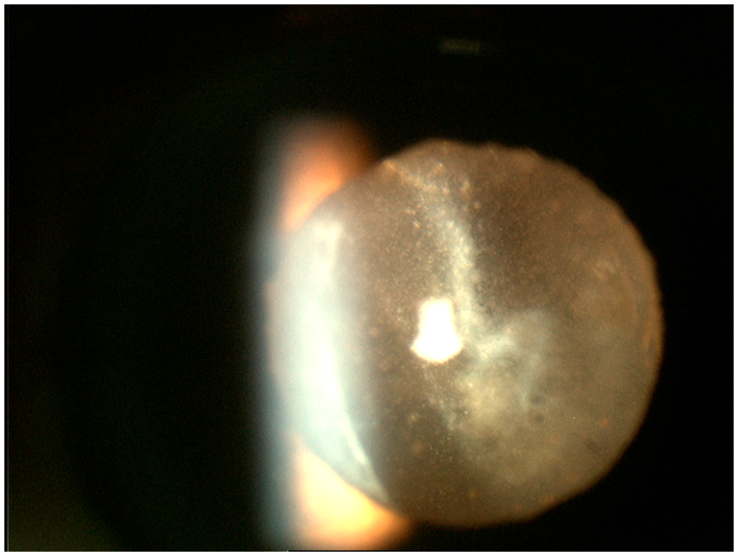
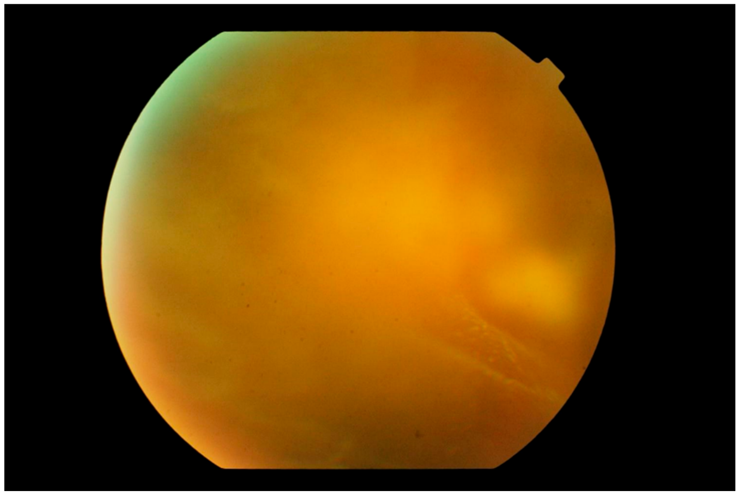
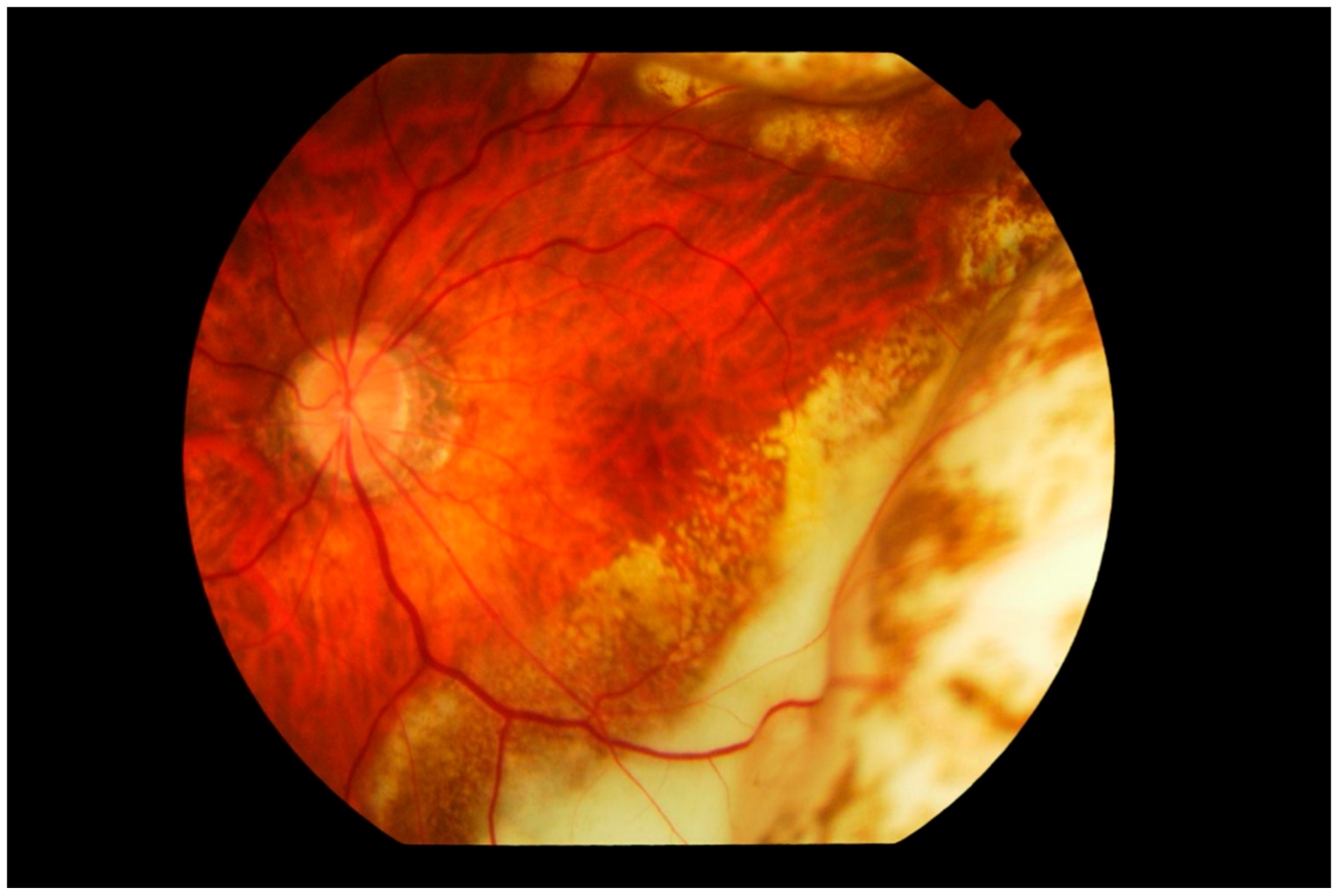
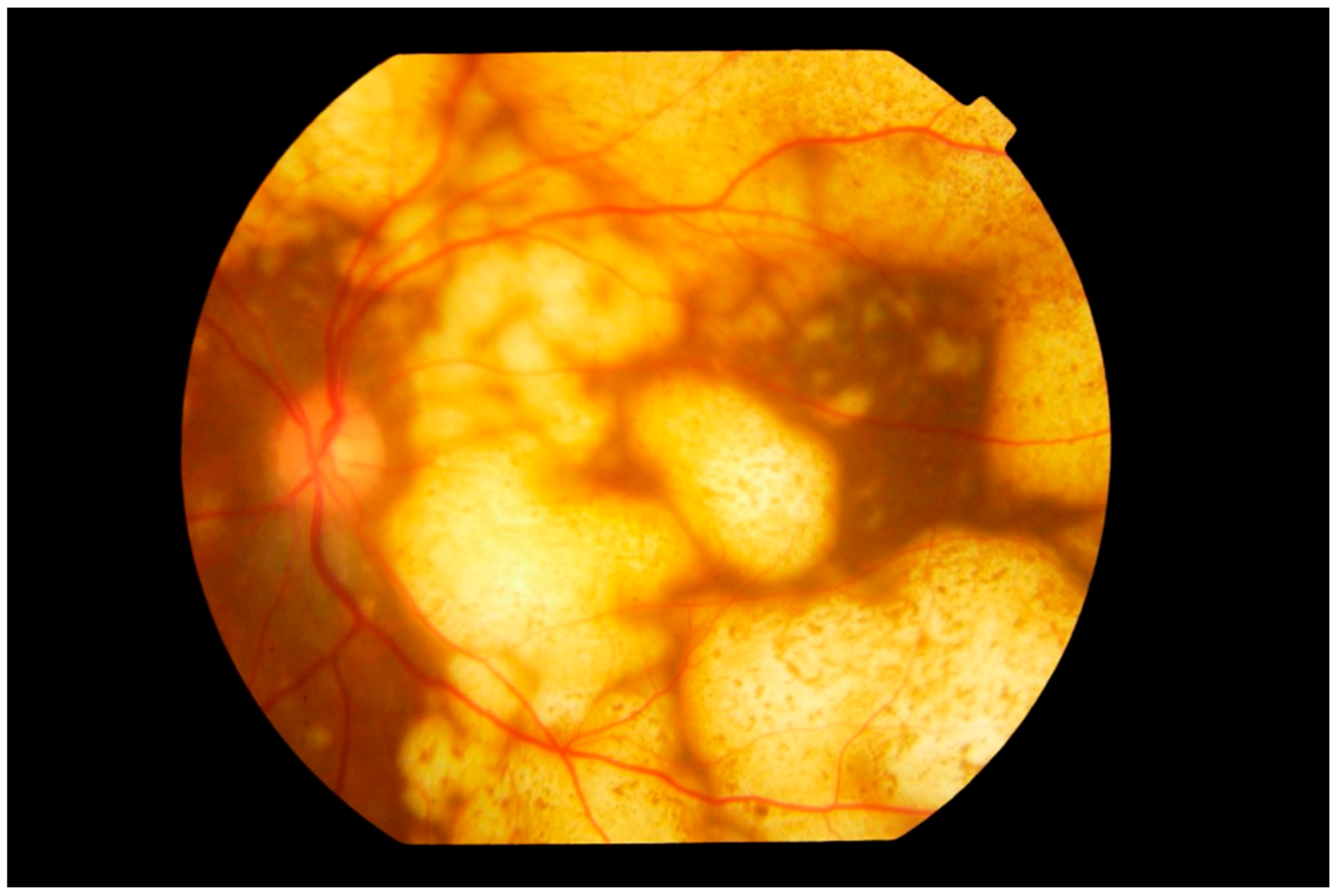
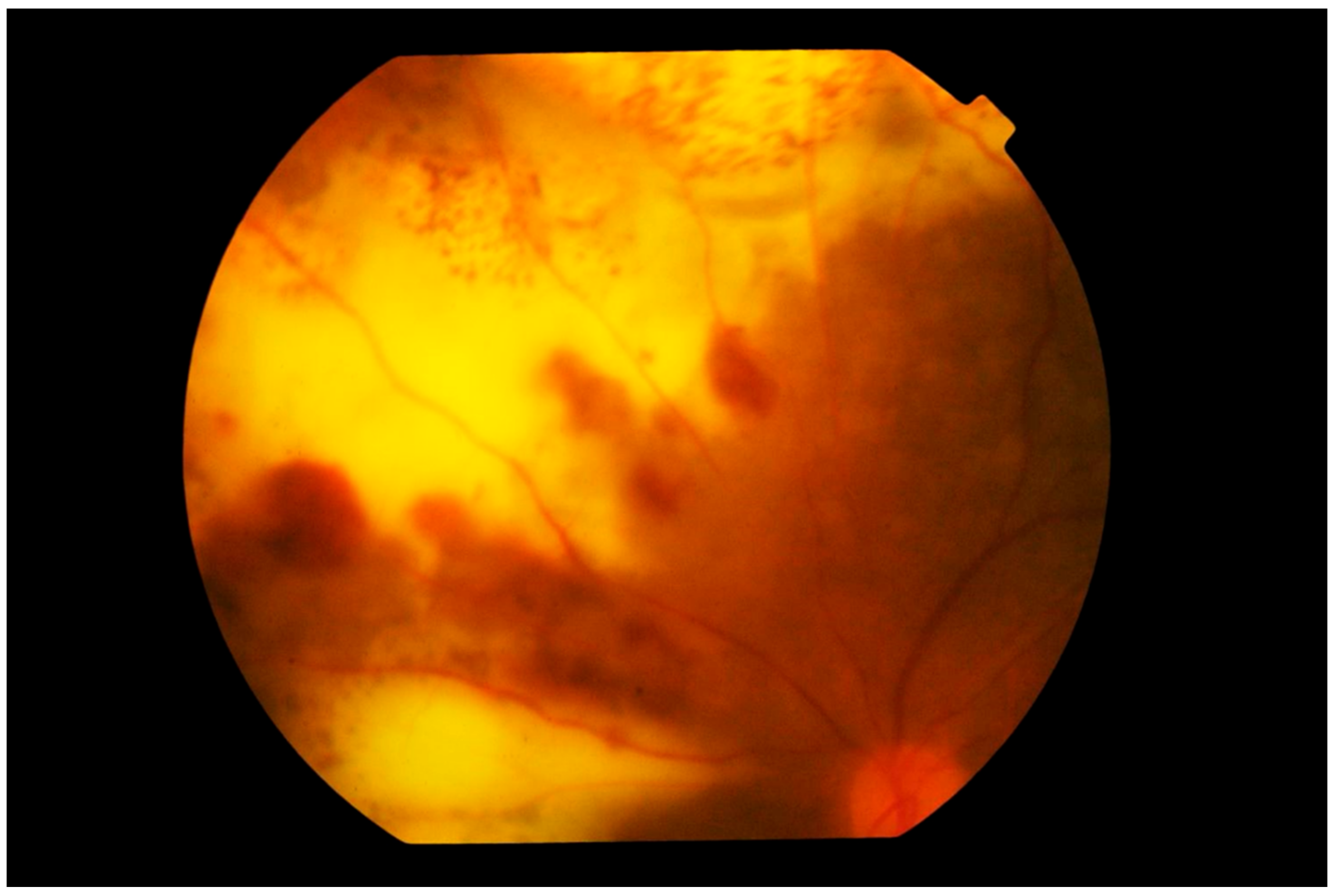
3. Clinical Features
Differential Diagnosis
4. Diagnostics
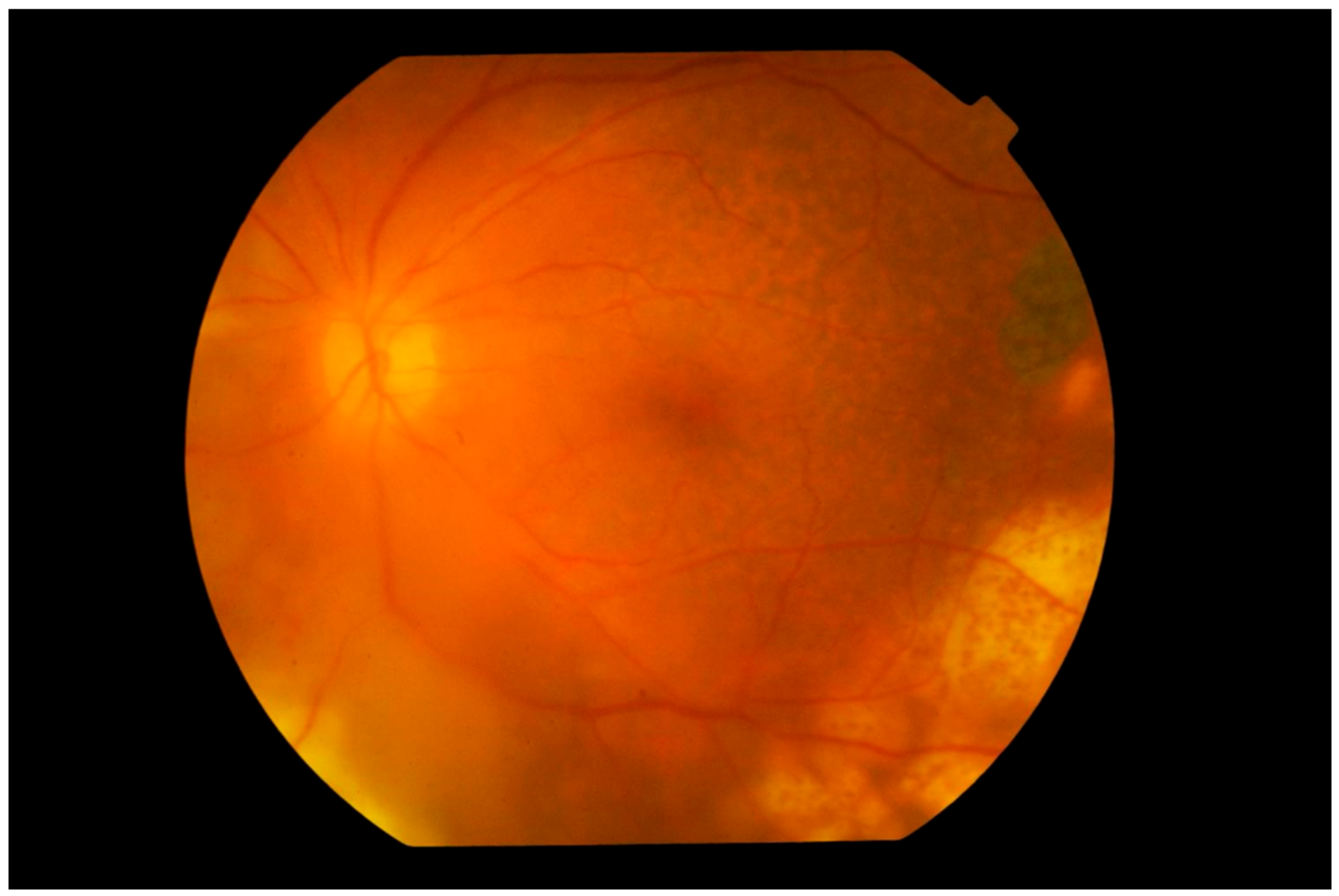
4.1. Imaging
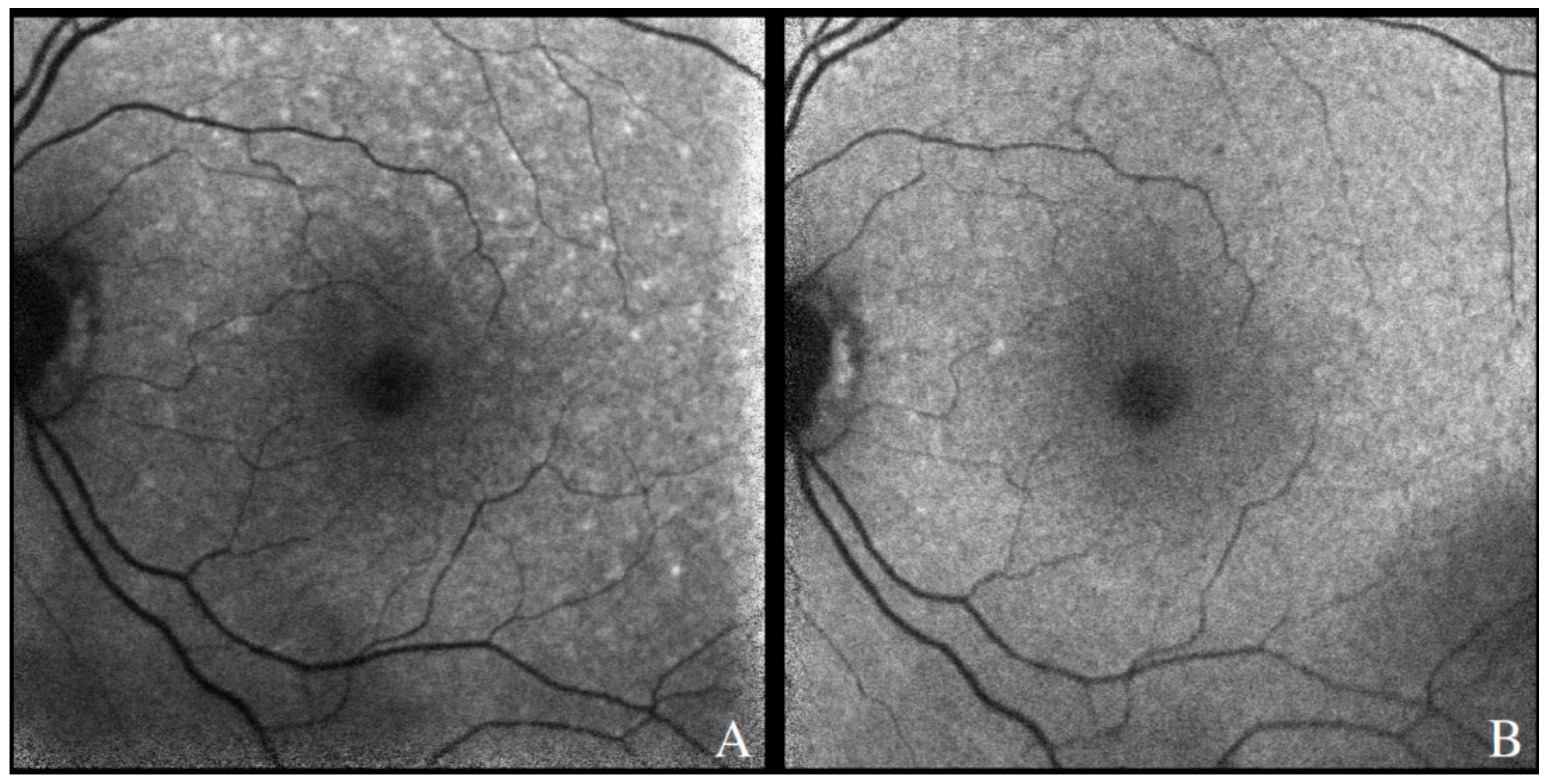
4.1.1. Fundus Autofluorescence
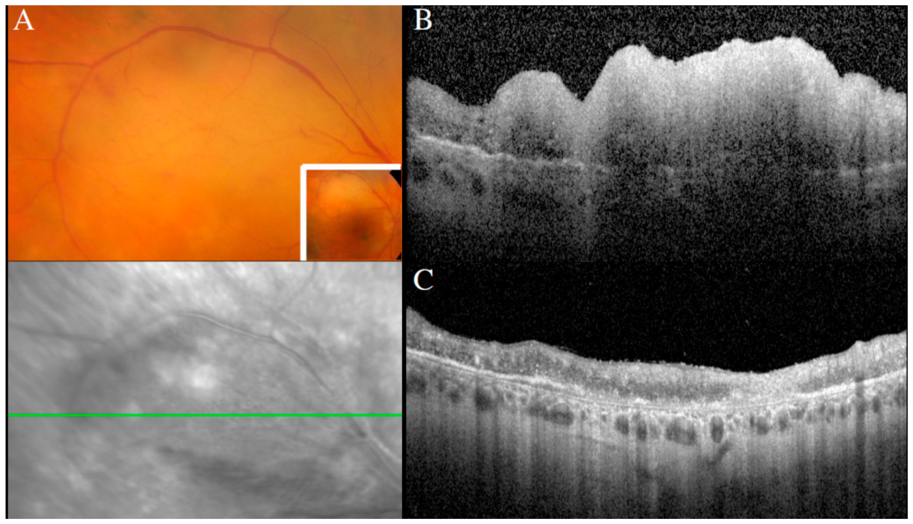
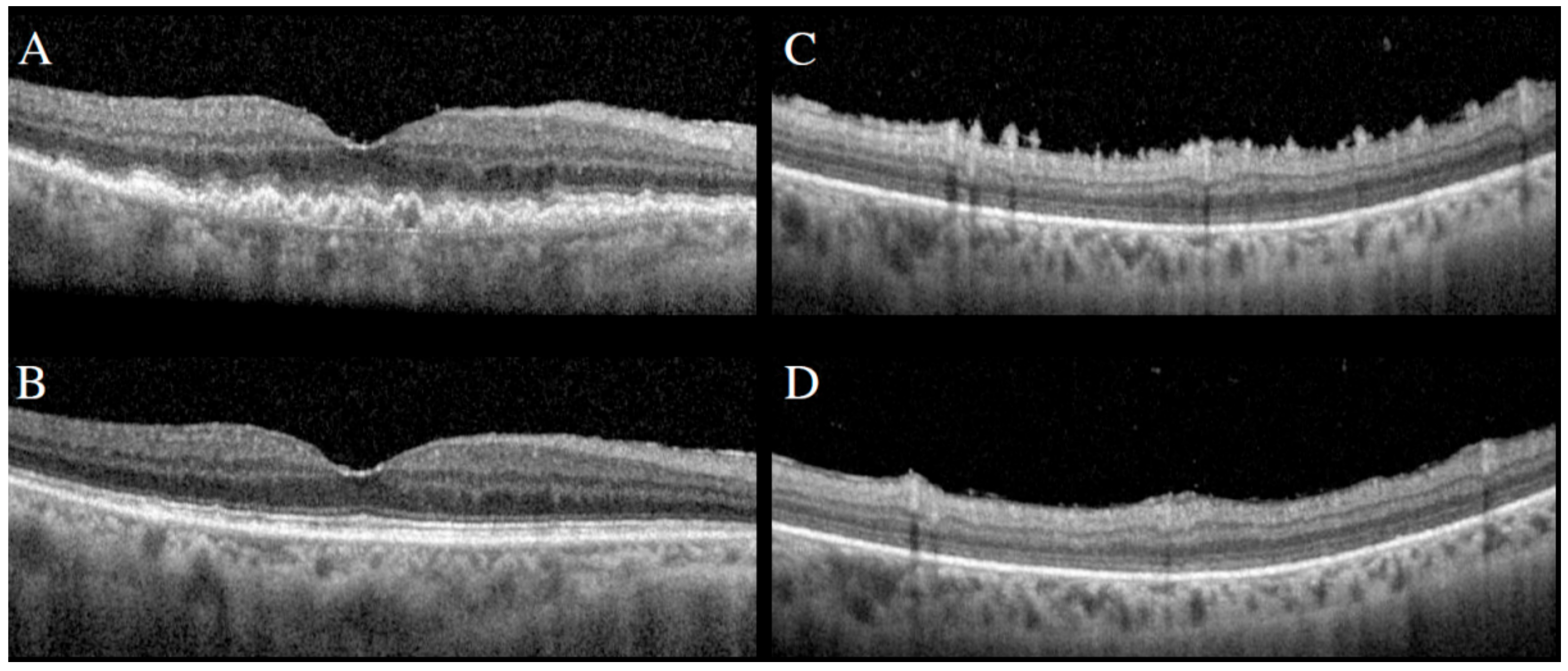
4.1.2. Spectral Domain Optical Coherence Tomography
4.1.3. Fluorescein Angiography
4.1.4. Indocyanine Green Angiography
4.2. Ocular Fluid- and Tissue-Based Diagnostics
4.2.1. Cytology and Immunophenotyping
4.2.2. Cytokine Levels
4.2.3. Determination of Clonality by Immunoglobulin Gene Rearrangement Studies
4.2.4. Mutational Analysis
4.2.5. Future Opportunities in Testing
5. Treatment
5.1. Management of Vitreoretinal Lymphoma
5.2. Local Ocular Therapy for PVRL
5.3. Systemic Therapy of PVRL
Author Contributions
Funding
Acknowledgments
Conflicts of Interest
References
- Cao, X.; Shen, D.; Callanan, D.G.; Mochizuki, M.; Chan, C.-C. Diagnosis of systemic metastatic retinal lymphoma. Acta Ophthalmol. 2011, 89, e149–e154. [Google Scholar] [CrossRef]
- Tiu Teo, H.M.; Çiftçi, S.; Elner, V.M.; Demirci, H. Systemic anti-CD20 (rituximab) as primary treatment for symptomatic primary uveal lymphoma. Am. J. Ophthalmol. Case Rep. 2019, 15, 100484. [Google Scholar] [CrossRef]
- Salomão, D.R.; Pulido, J.S.; Johnston, P.B.; Canal-Fontcuberta, I.; Feldman, A. Vitreoretinal Presentation of Secondary Large B-Cell Lymphoma in Patients with Systemic Lymphoma. JAMA Ophthalmol. 2013, 131, 1151–1158. [Google Scholar] [CrossRef]
- Jahnke, K.; Korfel, A.; Komm, J.; Bechrakis, N.E.; Stein, H.; Thiel, E.; Coupland, S.E. Intraocular lymphoma 2000–2005: Results of a retrospective multicentre trial. Graefe’s Arch. Clin. Exp. Ophthalmol. 2006, 244, 663–669. [Google Scholar] [CrossRef] [PubMed]
- Fonkem, E.; Lok, E.; Robison, D.; Gautam, S.; Wong, E.T. The natural history of intravascular lymphomatosis. Cancer Med. 2014, 3, 1010–1024. [Google Scholar] [CrossRef]
- Coupland, S.E.; Damato, B. Understanding intraocular lymphomas. Clin. Exp. Ophthalmol. 2008, 36, 564–578. [Google Scholar] [CrossRef]
- Fend, F.; Ferreri, A.J.M.; Coupland, S. How we diagnose and treat vitreoretinal lymphoma. Br. J. Haematol. 2016, 173, 680–692. [Google Scholar] [CrossRef] [PubMed]
- Chan, C.-C.; Sen, H.N. Current concepts in diagnosing and managing primary vitreoretinal (intraocular) lymphoma. Discov. Med. 2013, 15, 93–100. [Google Scholar] [PubMed]
- Swerdlow, S.H.; Campo, E.; Pileri, S.A.; Harris, N.L.; Stein, H.; Siebert, R.; Advani, R.; Ghielmini, M.; Salles, G.A.; Zelenetz, A.D.; et al. The 2016 revision of the World Health Organization classification of lymphoid neoplasms. Blood 2016, 127, 2375–2390. [Google Scholar] [CrossRef] [PubMed]
- Alizadeh, A.A.; Eisen, M.B.; Davis, R.E.; Ma, C.; Lossos, I.S.; Rosenwald, A.; Boldrick, J.C.; Sabet, H.; Tran, T.; Yu, X.; et al. Distinct types of diffuse large B-cell lymphoma identified by gene expression profiling. Nat. Cell Biol. 2000, 403, 503–511. [Google Scholar] [CrossRef]
- Gündüz, K.; Pulido, J.S.; McCannel, C.; O’Neill, B.P. Ocular manifestations and treatment of central nervous system lymphomas. Neurosurg. Focus 2006, 21, 1–7. [Google Scholar] [CrossRef]
- Alqahtani, A.; Touitou, V.; Cassoux, N.; Aknin, C.; Merle-Beral, H.; Bodaghi, B.; LeHoang, P. More Than a Masquerade Syndrome: Atypical Presentations of Vitreoretinal Lymphomas. Ocul. Immunol. Inflamm. 2014, 22, 189–196. [Google Scholar] [CrossRef]
- Coupland, S.E.; Anastassiou, G.; Bornfeld, N.; Hummel, M.; Stein, H. Primary intraocular lymphoma of T-cell type: Report of a case and review of the literature. Graefe’s Arch. Clin. Exp. Ophthalmol. 2004, 243, 189–197. [Google Scholar] [CrossRef]
- Cimino, L.; Chan, C.-C.; Shen, D.; Masini, L.; Ilariucci, F.; Masetti, M.; Asioli, S.; Sartori, A.; Cappuccini, L. Ocular involvement in nasal natural killer T-cell lymphoma. Int. Ophthalmol. 2009, 29, 275–279. [Google Scholar] [CrossRef]
- Bonzheim, I.; Giese, S.; Deuter, C.; Süsskind, D.; Zierhut, M.; Waizel, M.; Szurman, P.; Federmann, B.; Schmidt, J.; Quintanilla-Martinez, L.; et al. High frequency of MYD88 mutations in vitreoretinal B-cell lymphoma: A valuable tool to improve diagnostic yield of vitreous aspirates. Blood 2015, 126, 76–79. [Google Scholar] [CrossRef] [PubMed]
- Cani, A.K.; Hovelson, D.H.; Demirci, H.; Johnson, M.W.; Tomlins, S.A.; Rao, R.C. Next generation sequencing of vitreoretinal lymphomas from small-volume intraocular liquid biopsies: New routes to targeted therapies. Oncotarget 2016, 8, 7989–7998. [Google Scholar] [CrossRef] [PubMed]
- Coupland, S.E.; Hummel, M.; Müller, H.-H.; Stein, H. Molecular Analysis of Immunoglobulin Genes in Primary Intraocular Lymphoma. Investig. Opthalmol. Vis. Sci. 2005, 46, 3507–3514. [Google Scholar] [CrossRef]
- Hiemcke-Jiwa, L.S.; Loon, N.H.T.D.-V.; Leguit, R.J.; Nierkens, S.; Norel, J.O.-V.; De Boer, J.H.; Roholl, F.F.; De Weger, R.A.; Huibers, M.M.H.; De Groot-Mijnes, J.D.F.; et al. Potential Diagnosis of Vitreoretinal Lymphoma by Detection ofMYD88Mutation in Aqueous Humor with Ultrasensitive Droplet Digital Polymerase Chain Reaction. JAMA Ophthalmol. 2018, 136, 1098–1104. [Google Scholar] [CrossRef] [PubMed]
- Kakkassery, V.; Schroers, R.; Coupland, S.E.; Wunderlich, M.-I.; Schargus, M.; Heinz, C.; Wasmuth, S.; Heiligenhaus, A.; Ahle, G.; Lenoble, P.; et al. Vitreous microRNA levels as diagnostic biomarkers for vitreoretinal lymphoma. Blood 2017, 129, 3130–3133. [Google Scholar] [CrossRef]
- Lee, J.; Kim, B.; Lee, H.; Park, H.; Byeon, S.H.; Choi, J.R.; Lee, S.C.; Lee, S.-T.; Lee, C.S. Whole exome sequencing identifies mutational signatures of vitreoretinal lymphoma. Haematology 2020, 105, e458–e460. [Google Scholar] [CrossRef] [PubMed]
- Malumbres, R.; Davis, J.; Ruiz, P.; Lossos, I.S. Somatically mutated immunoglobulinIGHV@genes without intraclonal heterogeneity indicate a postgerminal centre origin of primary intraocular diffuse large B-cell lymphomas. Br. J. Haematol. 2007, 138, 749–755. [Google Scholar] [CrossRef] [PubMed]
- Merle-Béral, H.; Davi, F.; Cassoux, N.; Baudet, S.; Colin, C.; Gourdet, T.; Bodaghi, B.; LeHoang, P. Biological diagnosis of primary intraocular lymphoma. Br. J. Haematol. 2004, 124, 469–473. [Google Scholar] [CrossRef] [PubMed]
- Pulido, J.S.; Salomao, D.R.; Frederick, L.A.; Viswanatha, D.S. Myd-88 L265p Mutations are Present in Some Cases of Vitreoretinal Lymphoma. Retina 2015, 35, 624–627. [Google Scholar] [CrossRef]
- Raja, H.; Salomão, D.R.; Viswanatha, D.S.; Pulido, J.S. Prevalence of Myd88 L265p Mutation in Histologically Proven, Diffuse Large B-Cell Vitreoretinal Lymphoma. Retina 2016, 36, 624–628. [Google Scholar] [CrossRef]
- Shi, H.; Zhou, X.; Chen, B.; Xiao, J.; Li, Y.; Zhou, X.; Zhou, Q.; Chen, K.; Wang, Q. Clinical Relevance of the High Prevalence of MYD88 L265P Mutated Vitreoretinal Lymphoma Identified by Droplet Digital Polymerase Chain Reaction. Ocul. Immunol. Inflamm. 2019, 2019, 1–8. [Google Scholar] [CrossRef]
- Wang, L.; Sato-Otsubo, A.; Sugita, S.; Takase, H.; Mochizuki, M.; Usui, Y.; Goto, H.; Koyama, T.; Akiyama, H.; Miura, O.; et al. High-resolution genomic copy number profiling of primary intraocular lymphoma by single nucleotide polymorphism microarrays. Cancer Sci. 2014, 105, 592–599. [Google Scholar] [CrossRef]
- Yonese, I.; Takase, H.; Yoshimori, M.; Onozawa, E.; Tsuzura, A.; Miki, T.; Mochizuki, M.; Miura, O.; Arai, A. CD79B mutations in primary vitreoretinal lymphoma: Diagnostic and prognostic potential. Eur. J. Haematol. 2019, 102, 191–196. [Google Scholar] [CrossRef]
- Chapuy, B.; Stewart, C.; Dunford, A.J.; Kim, J.; Kamburov, A.; Redd, R.A.; Lawrence, M.S.; Roemer, M.G.M.; Li, A.J.; Ziepert, M.; et al. Molecular subtypes of diffuse large B cell lymphoma are associated with distinct pathogenic mechanisms and outcomes. Nat. Med. 2018, 24, 679–690. [Google Scholar] [CrossRef] [PubMed]
- Schmitz, R.; Wright, G.W.; Huang, D.W.; Johnson, C.A.; Phelan, J.D.; Wang, J.Q.; Roulland, S.; Kasbekar, M.; Young, R.M.; Shaffer, A.L.; et al. Genetics and Pathogenesis of Diffuse Large B-Cell Lymphoma. N. Engl. J. Med. 2018, 378, 1396–1407. [Google Scholar] [CrossRef]
- Chapuy, B.; Roemer, M.G.M.; Stewart, C.; Tan, Y.; Abo, R.P.; Zhang, L.; Dunford, A.J.; Meredith, D.M.; Thorner, A.R.; Jordanova, E.S.; et al. Targetable genetic features of primary testicular and primary central nervous system lymphomas. Blood 2016, 127, 869–881. [Google Scholar] [CrossRef]
- Kraan, W.; Horlings, H.M.; Van Keimpema, M.; Schildertol, E.J.M.; Oud, M.E.C.M.; Scheepstra, C.; Kluin, P.M.; Kersten, M.J.; Spaargaren, M.; Pals, S.T. High prevalence of oncogenic MYD88 and CD79B mutations in diffuse large B-cell lymphomas presenting at immune-privileged sites. Blood Cancer J. 2013, 3, e139. [Google Scholar] [CrossRef] [PubMed]
- Belhouachi, N.; Xochelli, A.; Boudjoghra, M.; Lesty, C.; Cassoux, N.; Fardeau, C.; Tran, T.H.C.; Choquet, S.; Sarker, B.; Houillier, C.; et al. Primary vitreoretinal lymphomas display a remarkably restricted immunoglobulin gene repertoire. Blood Adv. 2020, 4, 1357–1366. [Google Scholar] [CrossRef] [PubMed]
- Coupland, S.E.; Chan, C.C.; Smith, J. Pathophysiology of Retinal Lymphoma. Ocul. Immunol. Inflamm. 2009, 17, 227–237. [Google Scholar] [CrossRef]
- Hong, J.T.; Chae, J.B.; Lee, J.Y.; Kim, J.-G.; Yoon, Y.H. Ocular involvement in patients with primary CNS lymphoma. J. Neuro Oncol. 2010, 102, 139–145. [Google Scholar] [CrossRef]
- Sjö, L.D. Ophthalmic lymphoma: Epidemiology and pathogenesis. Acta Ophthalmol. 2009, 87, 1–20. [Google Scholar] [CrossRef] [PubMed]
- Haldorsen, I.S.; Krossnes, B.K.; Aarseth, J.H.; Scheie, D.; Johannesen, T.B.; Mella, O.; Espeland, A. Increasing incidence and continued dismal outcome of primary central nervous system lymphoma in Norway 1989–2003: Time trends in a 15-year national survey. Cancer 2007, 110, 1803–1814. [Google Scholar] [CrossRef] [PubMed]
- Corn, B.W.; Marcus, S.M.; Topham, A.; Hauck, W.; Curran, W.J., Jr. Will primary central nervous system lymphoma be the most frequent brain tumor diagnosed in the year 2000? Cancer 1997, 79, 2409–2413. [Google Scholar] [CrossRef]
- Levasseur, S.D.; Wittenberg, L.A.; White, V.A. Vitreoretinal Lymphoma: A 20-year review of incidence, clinical and cytologic features, treatment, and outcomes. JAMA Ophthalmol. 2013, 131, 50–55. [Google Scholar] [CrossRef]
- Chan, C.; Rubenstein, J.L.; Coupland, S.; Davis, J.L.; Harbour, J.W.; Johnston, P.B.; Cassoux, N.; Touitou, V.; Smith, J.; Batchelor, T.T.; et al. Primary Vitreoretinal Lymphoma: A Report from an International Primary Central Nervous System Lymphoma Collaborative Group Symposium. Oncology 2011, 16, 1589–1599. [Google Scholar] [CrossRef]
- Sagoo, M.S.; Mehta, H.; Swampillai, A.J.; Cohen, V.; Amin, S.Z.; Plowman, P.N.; Lightman, S. Primary intraocular lymphoma. Surv. Ophthalmol. 2014, 59, 503–516. [Google Scholar] [CrossRef] [PubMed]
- Meunier, J.; Rouic, L.L.-L.; Vincent-Salomon, A.; Dendale, R.; Asselain, B.; Arnaud, P.; Fourquet, A.; Desjardins, L.; Plancher, C.; Validire, P.; et al. Ophthalmologic and intraocular non-Hodgkin’s lymphoma: A large single centre study of initial characteristics, natural history, and prognostic factors. Hematol. Oncol. 2004, 22, 143–158. [Google Scholar] [CrossRef] [PubMed]
- Rothova, A.; Ooijman, F.; Kerkhoff, F.; Van der Lelij, A.; Lokhorst, H.M. Uveitis masquerade syndromes. Ophthalmology 2001, 108, 386–399. [Google Scholar] [CrossRef]
- Akpek, E.K.; Ahmed, I.; Hochberg, F.H.; Soheilian, M.; Dryja, T.P.; A Jakobiec, F.; Foster, C. Intraocular-central nervous system lymphoma: Clinical features, diagnosis, and outcomes. Ophthalmology 1999, 106, 1805–1810. [Google Scholar] [CrossRef]
- Pe’Er, J.; Hochberg, F.H.; Foster, C.S. Clinical Review: Treatment of Vitreoretinal Lymphoma. Ocul. Immunol. Inflamm. 2009, 17, 299–306. [Google Scholar] [CrossRef]
- Gerstner, E.R.; Batchelor, T.T. Primary Central Nervous System Lymphoma. Arch. Neurol. 2010, 67, 291–297. [Google Scholar] [CrossRef] [PubMed]
- Kimura, K.; The Japanese Intraocular Lymphoma Study Group; Usui, Y.; Goto, H. Clinical features and diagnostic significance of the intraocular fluid of 217 patients with intraocular lymphoma. Jpn. J. Ophthalmol. 2012, 56, 383–389. [Google Scholar] [CrossRef]
- Chan, C.-C.; Shen, D.F.; Whitcup, S.M.; Nussenblatt, R.B.; LeHoang, P.; Roberge, F.G.; Cassoux, N.; Herbort, C.; Zhuang, Z. Detection of Human Herpesvirus-8 and Epstein-Barr Virus DNA in Primary Intraocular Lymphomas. Blood 1999, 93, 2749–2751. [Google Scholar] [CrossRef]
- Chan, C.-C.; Buggage, R.R.; Nussenblatt, R.B. Intraocular lymphoma. Curr. Opin. Ophthalmol. 2002, 13, 411–418. [Google Scholar] [CrossRef]
- Tuaillon, N.; Chan, C.C. Molecular analysis of primary central nervous system and primary intraocular lymphomas. Curr. Mol. Med. 2001, 1, 259–272. [Google Scholar] [CrossRef]
- Cote, T.R.; Manns, A.; Hardy, C.R.; Yellin, F.J.; Hartge, P.; AIDS/Cancer Study Group. Epidemiology of Brain Lymphoma Among People with or without Acquired Immunodeficiency Syndrome. J. Natl. Cancer Inst. 1996, 88, 675–679. [Google Scholar] [CrossRef]
- Davis, J.L. Intraocular lymphoma: A clinical perspective. Eye 2013, 27, 153–162. [Google Scholar] [CrossRef]
- Cassoux, N.; Merle-Béral, H.; Leblond, V.; Bodaghi, B.; Miléa, D.; Gerber, S.; Fardeau, C.; Reux, I.; Xuan, K.H.; Chan, C.-C.; et al. Ocular and central nervous system lymphoma: Clinical features and diagnosis. Ocul. Immunol. Inflamm. 2000, 8, 243–250. [Google Scholar] [CrossRef]
- Schabet, M. Epidemiology of primary CNS lymphoma. J. Neuro Oncol. 1999, 43, 199–201. [Google Scholar] [CrossRef]
- Llorenç, V.; Fuster, C.; Alba-Linero, C.; Moll-Udina, A.; Serrano, A.; Solé, M.; De La Maza, M.S.; Aldecoa, I.; Adán, A. Clinical Features of Primary and Systemic Metastatic Intraocular Lymphomas in Spanish Patients. J. Ophthalmol. 2019, 2019, 1–9. [Google Scholar] [CrossRef]
- Carbonell, D.; Mahajan, S.; Chee, S.-P.; Sobolewska, B.; Agrawal, R.; Bülow, T.; Gupta, V.; Jones, N.P.; Accorinti, M.; Agarwal, M.; et al. Consensus Recommendations for the Diagnosis of Vitreoretinal Lymphoma. Ocul. Immunol. Inflamm. 2021, 1–14. [Google Scholar] [CrossRef]
- Freeman, L.N.; Schachat, A.P.; Knox, D.L.; Michels, R.G.; Green, W.R. Clinical Features, Laboratory Investigations, and Survival in Ocular Reticulum Cell Sarcoma. Ophthalmology 1987, 94, 1631–1639. [Google Scholar] [CrossRef]
- Fardeau, C.; Lee, C.P.; Merle-Béral, H.; Cassoux, N.; Bodaghi, B.; Davi, F.; LeHoang, P. Retinal Fluorescein, Indocyanine Green Angiography, and Optic Coherence Tomography in Non-Hodgkin Primary Intraocular Lymphoma. Am. J. Ophthalmol. 2009, 147, 886.e1–894.e1. [Google Scholar] [CrossRef]
- Chan, C.C.; Gonzalez, J.A. Primary Intraocular Lymphoma; World Scientific Publishing Co. Pte Ltd: Hackensack, NJ, USA, 2007. [Google Scholar]
- Marchese, A.; Miserocchi, E.; Giuffrè, C.; Cicinelli, M.V.; Querques, G.; Bandello, F.; Modorati, G. Aurora borealis and string of pearls in vitreoretinal lymphoma: Patterns of vitreous haze. Br. J. Ophthalmol. 2019, 103, 1656–1659. [Google Scholar] [CrossRef]
- Dean, J.M.; Novak, M.A.; Chan, C.-C.; Green, W.R. Tumor Detachments of the Retinal Pigment Epithelium in Ocular/Central Nervous System Lymphoma. Retina 1996, 16, 47–56. [Google Scholar] [CrossRef] [PubMed]
- Goto, H.; Murase, K.; Usui, M. Case of spontaneous regression of intraocular lymphoma demonstrated by subretinal biopsy. Nippon Ganka Gakkai Zasshi 2006, 110, 226–231. [Google Scholar] [PubMed]
- Mantopoulos, D.; Cebulla, C.M. Multimodal Imaging of Spontaneously Shifting Primary Vitreoretinal Lymphoma. Ocul. Oncol. Pathol. 2015, 1, 237–240. [Google Scholar] [CrossRef] [PubMed]
- Kase, S.; Namba, K.; Jin, X.-H.; Kubota, K.C.; Ishida, S. Spontaneous Regression of Intraocular Lymphoma. Ophthalmology 2012, 119, 1083.e2–1084.e2. [Google Scholar] [CrossRef] [PubMed]
- Gill, M.K.; Jampol, L.M. Variations in the Presentation of Primary Intraocular Lymphoma: Case Reports and a Review. Surv. Ophthalmol. 2001, 45, 463–471. [Google Scholar] [CrossRef]
- Rajagopal, R.; Harbour, J.W. Diagnostic Testing and Treatment Choices in Primary Vitreoretinal Lymphoma. Retina 2011, 31, 435–440. [Google Scholar] [CrossRef]
- Michelson, J.B.; Michelson, P.E.; Bordin, G.M.; Chisari, F.V. Ocular Reticulum Cell Sarcoma. Presentation as retinal detachment with demonstration of monoclonal immunoglobulin light chains on the vitreous cells. Arch. Ophthalmol. 1981, 99, 1409–1411. [Google Scholar] [CrossRef]
- Chaput, F.; Amer, R.; Baglivo, E.; Touitou, V.; Kozyreff, A.; Bron, D.; Bodaghi, B.; LeHoang, P.; Bergstrom, C.; Grossniklaus, H.E.; et al. Intraocular T-cell Lymphoma: Clinical Presentation, Diagnosis, Treatment, and Outcome. Ocul. Immunol. Inflamm. 2016, 25, 644–653. [Google Scholar] [CrossRef] [PubMed]
- Gass, J.D.M.; Trattler, H.L. Retinal Artery Obstruction and Atheromas Associated with Non-Hodgkin’s Large Cell Lymphoma (Reticulum Cell Sarcoma). Arch. Ophthalmol. 1991, 109, 1134–1139. [Google Scholar] [CrossRef]
- Lee, J.; Kim, S.W.; Kim, H.; Lee, C.; Kim, M.; Lee, S.C. Differential Diagnosis for Vitreoretinal Lymphoma with Vitreoretinal Findings, Immunoglobulin Clonality Tests, and Interleukin Levels. Retina 2019, 39, 1165–1176. [Google Scholar] [CrossRef]
- Russell, J.F.; Pichi, F.; Scott, N.L.; Hartley, M.J.; Bell, D.; Agarwal, A.; Leong, B.; Holland, G.N.; Freund, K.B.; Sarraf, D. Masqueraders of multiple evanescent white dot syndrome (MEWDS). Int. Ophthalmol. 2020, 40, 627–638. [Google Scholar] [CrossRef] [PubMed]
- Chan, N.S.; Chee, S.P. Keratic precipitates: The under-utilised diagnostic clue. Ocul. Immunol. Inflamm. 2020, in press. [Google Scholar]
- Hoffman, P.M.; McKelvie, P.; Hall, A.J.; Stawell, R.J.; Santamaria, J.D. Intraocular lymphoma: A series of 14 patients with clinicopathological features and treatment outcomes. Eye 2003, 17, 513–521. [Google Scholar] [CrossRef]
- Velez, G.; de Smet, M.; Whitcup, S.M.; Robinson, M.; Nussenblatt, R.B.; Chan, C.-C. Iris Involvement in Primary Intraocular Lymphoma: Report of Two Cases and Review of the Literature. Surv. Ophthalmol. 2000, 44, 518–526. [Google Scholar] [CrossRef]
- Lobo, A.; Larkin, G.; Clark, B.J.; A Towler, H.M.; Lightman, S. Pseudo-hypopyon as the presenting feature in B-cell and T-cell intraocular lymphoma. Clin. Exp. Ophthalmol. 2003, 31, 155–158. [Google Scholar] [CrossRef]
- Gonzales, J.A.; Chan, C.-C. Biopsy techniques and yields in diagnosing primary intraocular lymphoma. Int. Ophthalmol. 2007, 27, 241–250. [Google Scholar] [CrossRef] [PubMed]
- Faia, L.J.; Chan, C.-C. Primary intraocular lymphoma. Arch. Pathol. Lab. Med. 2009, 133, 1228–1232. [Google Scholar] [CrossRef]
- Grimm, S.A.; McCannel, C.; Omuro, A.; Ferreri, A.J.; Blay, J.-Y.; Neuwelt, E.A.; Siegal, T.; Batchelor, T.; Jahnke, K.; Shenkier, T.N.; et al. Primary CNS lymphoma with intraocular involvement: International PCNSL Collaborative Group Report. Neurology 2008, 71, 1355–1360. [Google Scholar] [CrossRef] [PubMed]
- Hormigo, A.; Abrey, L.; Heinemann, M.-H.; DeAngelis, L. Ocular presentation of primary central nervous system lymphoma: Diagnosis and treatment. Br. J. Haematol. 2004, 126, 202–208. [Google Scholar] [CrossRef]
- Davis, J.L. Diagnosis of intraocular lymphoma. Ocul. Immunol. Inflamm. 2004, 12, 7–16. [Google Scholar] [CrossRef]
- Tan, S.Z.; Steeples, L.R.; Chhabra, R.; Jones, N.P. An unusual case report of primary vitreoretinal lymphoma. BMC Ophthalmol. 2018, 18, 223. [Google Scholar] [CrossRef]
- Ono, K. Clinical significance of natural killing activity in patients with advanced lymphoma. J. Clin. Immunol. 1998, 18, 132–141. [Google Scholar] [CrossRef]
- Citterio, G.; Calimeri, T.; Ferreri, A.J.M. Challenges and prospects in the diagnosis and treatment of primary central nervous system lymphoma. Expert Rev. Neurother. 2018, 18, 379–393. [Google Scholar] [CrossRef]
- Peterson, K.; Gordon, K.B.; Heinemann, M.-H.; DeAngelis, L.M. The clinical spectrum of ocular lymphoma. Cancer 1993, 72, 843–849. [Google Scholar] [CrossRef]
- Raparia, K.; Chang, C.-C.J.; Chévez-Barrios, P. Intraocular lymphoma: Diagnostic approach and immunophenotypic findings in vitrectomy specimens. Arch. Pathol. Lab. Med. 2009, 133, 1233–1237. [Google Scholar] [CrossRef]
- Sen, H.N.; Bodaghi, B.; Le Hoang, P.; Nussenblatt, R. Primary Intraocular Lymphoma: Diagnosis and Differential Diagnosis. Ocul. Immunol. Inflamm. 2009, 17, 133–141. [Google Scholar] [CrossRef]
- Casady, M.; Faia, L.; Nazemzadeh, M.; Nussenblatt, R.; Chan, C.-C.; Sen, H.N. Fundus Autofluorescence Patterns in Primary Intraocular Lymphoma. Retina 2014, 34, 366–372. [Google Scholar] [CrossRef]
- Ishida, T.; Ohno-Matsui, K.; Kaneko, Y.; Tobita, H.; Shimada, N.; Takase, H.; Mochizuki, M. Fundus Autofluorescence Patterns in Eyes with Primary Intraocular Lymphoma. Retina 2010, 30, 23–32. [Google Scholar] [CrossRef] [PubMed]
- Lee, J.; Goldstein, D.A. The diverse multi-modal imaging findings of recurrent primary vitreoretinal lymphoma. Am. J. Ophthalmol. Case Rep. 2020, 20, 100936. [Google Scholar] [CrossRef]
- Barry, R.J.; Tasiopoulou, A.; Murray, P.; Patel, P.J.; Sagoo, M.; Denniston, A.K.; A Keane, P. Characteristic optical coherence tomography findings in patients with primary vitreoretinal lymphoma: A novel aid to early diagnosis. Br. J. Ophthalmol. 2018, 102, 1362–1366. [Google Scholar] [CrossRef]
- Liu, T.Y.A.; Ibrahim, M.; Bittencourt, M.; Sepah, Y.J.; Do, D.V.; Nguyen, Q.D. Retinal optical coherence tomography manifestations of intraocular lymphoma. J. Ophthalmic Inflamm. Infect. 2012, 2, 215–218. [Google Scholar] [CrossRef] [PubMed]
- Deák, G.G.; Goldstein, D.A.; Zhou, M.; Fawzi, A.A.; Jampol, L.M. Vertical Hyperreflective Lesions on Optical Coherence Tomography in Vitreoretinal Lymphoma. JAMA Ophthalmol. 2019, 137, 194–198. [Google Scholar] [CrossRef] [PubMed]
- Shields, C.L.; Manalac, J.; Das, C.; Saktanasate, J.; A Shields, J. Review of spectral domain enhanced depth imaging optical coherence tomography of tumors of the choroid. Indian J. Ophthalmol. 2015, 63, 117–121. [Google Scholar] [CrossRef]
- Velez, G.; Chan, C.-C.; Csaky, K.G. Fluorescein Angiographic Findings in Primary Intraocular Lymphoma. Retina 2002, 22, 37–43. [Google Scholar] [CrossRef]
- Lavine, J.A.; Singh, A.D.; Sharma, S.; Baynes, K.; Lowder, C.Y.; Srivastava, S.K. Ultra-widefield multimodal imaging of primary vitreoretinal lymphoma. Retina 2019, 39, 1861–1871. [Google Scholar] [CrossRef] [PubMed]
- Mehta, M.; Rasheed, R.A.; Duker, J.; Reichel, E.; Feinberg, E.; Husain, D.; Foster, C.S.; Laver, N.V. Vitreous evaluation: A diagnostic challenge. Ophthalmology 2015, 122, 531–537. [Google Scholar] [CrossRef]
- Raja, H.; Snyder, M.R.; Johnston, P.B.; O’Neill, B.P.; Caraballo, J.N.; Balsanek, J.G.; Peters, B.E.; Decker, P.A.; Pulido, J.S. Effect of Intravitreal Methotrexate and Rituximab on Interleukin-10 Levels in Aqueous Humor of Treated Eyes with Vitreoretinal Lymphoma. PLoS ONE 2013, 8, e65627. [Google Scholar] [CrossRef]
- Karma, A.; Von Willebrand, E.O.; Tommila, P.V.; Paetau, A.E.; Oskala, P.S.; Immonen, I.J. Primary Intraocular Lymphoma: Improving the Diagnostic Procedure. Ophthalmology 2007, 114, 1372–1377. [Google Scholar] [CrossRef]
- Tang, P.H.; Karkhur, S.; Nguyen, Q.D. Obtaining undiluted vitreous sample using small gauge pars plana vitrectomy and air infusion. Am. J. Ophthalmol. Case Rep. 2020, 19, 100768. [Google Scholar] [CrossRef] [PubMed]
- Trivedi, D.; Denniston, A.; I Murray, P. Safety profile of anterior chamber paracentesis performed at the slit lamp. Clin. Exp. Ophthalmol. 2011, 39, 725–728. [Google Scholar] [CrossRef] [PubMed]
- Smith, J.R.; Pe’er, J.; Belfort, R.N.; Cardoso, F.; Carvajal, R.D.; Carvalho, C.; Coupland, S.E.; Desjardins, L.; Francis, J.H.; Gallie, B.L. Proceedings of the Association for Research in Vision and Ophthalmology and Champalimaud Foundation Ocular Oncogenesis and Oncology Conference. Transl. Vis. Sci. Technol. 2019, 8, 9. [Google Scholar] [CrossRef]
- Dawson, A.C.; A Williams, K.; Appukuttan, B.; Smith, J.R. Emerging diagnostic tests for vitreoretinal lymphoma: A review. Clin. Exp. Ophthalmol. 2018, 46, 945–954. [Google Scholar] [CrossRef] [PubMed]
- Davis, J.L.; Miller, D.M.; Ruiz, P. Diagnostic Testing of Vitrectomy Specimens. Am. J. Ophthalmol. 2005, 140, 822.e2–829.e2. [Google Scholar] [CrossRef]
- Dalal, M.; Casady, M.; Moriarty, E.; Faia, L.; Nussenblatt, R.; Chan, C.-C.; Sen, H.N. Diagnostic Procedures in Vitreoretinal Lymphoma. Ocul. Immunol. Inflamm. 2013, 22, 270–276. [Google Scholar] [CrossRef] [PubMed]
- Arcinue, C.A.; Hochberg, F.; Neumann, R.; Foster, C.S. Diagnostic Criteria for Primary Ocular Lymphoma. Ophthalmology 2013, 120, 646.e2. [Google Scholar] [CrossRef] [PubMed]
- Dunn, J.P. Interleukins in the Diagnosis of Intraocular Lymphoma: Do We Still Need Histologic Confirmation? Retina 2018, 38, 647–649. [Google Scholar] [CrossRef]
- Sugita, S.; Takase, H.; Sugamoto, Y.; Arai, A.; Miura, O.; Mochizuki, M. Diagnosis of intraocular lymphoma by polymerase chain reaction analysis and cytokine profiling of the vitreous fluid. Jpn. J. Ophthalmol. 2009, 53, 209–214. [Google Scholar] [CrossRef]
- Coupland, S.E. Analysis of Intraocular Biopsies. In Current Concepts in Uveal Melanoma; Jager, M.J., Desjardins, L., Kivelä, T., Damato, B.E., Eds.; Karger Publishers: Basel, Switzerland, 2012; Volume 49, pp. 96–116. [Google Scholar]
- Kanno-Okada, H.; Takakuwa, E.; Tagawa, Y.; Kase, S.; Hatanaka, K.C.; Namba, K.; Mitsuhashi, T.; Matsuno, Y. Cytopathologic findings of cell block materials from the vitreous: Diagnostic distinction between intraocular lymphoma and non-lymphomatous diseases. Pathol. Int. 2017, 67, 342–349. [Google Scholar] [CrossRef]
- Kase, S.; Namba, K.; Iwata, D.; Mizuuchi, K.; Kitaichi, N.; Tagawa, Y.; Okada-Kanno, H.; Matsuno, Y.; Ishida, S. Diagnostic efficacy of cell block method for vitreoretinal lymphoma. Diagn. Pathol. 2016, 11, 29. [Google Scholar] [CrossRef]
- Char, D.H.; Ljung, B.-M.; Miller, T.; Phillips, T. Primary Intraocular Lymphoma (ocular reticulum cell sarcoma) Diagnosis and Management. Ophthalmology 1988, 95, 625–630. [Google Scholar] [CrossRef]
- Gooi, P.; Farmer, J.; Hurley, B.; Brodbaker, E. Cytomegalovirus retinitis mimicking intraocular lymphoma. Clin. Ophthalmol. 2008, 2, 969–971. [Google Scholar] [CrossRef] [PubMed][Green Version]
- Wang, Y.; Shen, D.; Wang, V.M.; Sen, H.N.; Chan, C.-C. Molecular Biomarkers for the Diagnosis of Primary Vitreoretinal Lymphoma. Int. J. Mol. Sci. 2011, 12, 5684–5697. [Google Scholar] [CrossRef] [PubMed]
- Frenkel, S.; Pe’Er, J.; Kaufman, R.; Maly, B.; Habot-Wilner, Z. The importance of cytokines analysis in the diagnosis of vitreoretinal lymphoma. Acta Ophthalmol. 2020, 98, e668–e673. [Google Scholar] [CrossRef]
- Missotten, T.; Tielemans, D.; Bromberg, J.E.; van Hagen, P.M.; van Lochem, E.G.; van Dongen, J.J.; Baarsma, G.S.; Langerak, A.W. Multicolor Flowcytometric Immunophenotyping Is a Valuable Tool for Detection of Intraocular Lymphoma. Ophthalmology 2013, 120, 991–996. [Google Scholar] [CrossRef] [PubMed]
- Davis, J.L.; Ruiz, P.; Shah, M.; Mandelcorn, E.D. Evaluation of the Reactive T-Cell Infiltrate in Uveitis and Intraocular Lymphoma with Flow Cytometry of Vitreous Fluid (An American Ophthalmological Society Thesis). Trans. Am. Ophthalmol. Soc. 2012, 110, 117–129. [Google Scholar] [PubMed]
- Chan, C.-C.; Whitcup, S.M.; Solomon, D.; Nussenblatt, R.B. Interleukin-10 in the Vitreous of Patients with Primary Intraocular Lymphoma. Am. J. Ophthalmol. 1995, 120, 671–673. [Google Scholar] [CrossRef]
- Blay, J.Y.; Burdin, N.; Rousset, F.; Lenoir, G.; Biron, P.; Philip, T.; Banchereau, J.; Favrot, M.C. Serum interleukin-10 in non-Hodgkin’s lymphoma: A prognostic factor. Blood 1993, 82, 2169–2174. [Google Scholar] [CrossRef]
- Bonacini, M.; Soriano, A.; Cimino, L.; De Simone, L.; Bolletta, E.; Gozzi, F.; Muratore, F.; Nicastro, M.; Belloni, L.; Zerbini, A.; et al. Cytokine Profiling in Aqueous Humor Samples from Patients with Non-Infectious Uveitis Associated with Systemic Inflammatory Diseases. Front. Immunol. 2020, 11, 358. [Google Scholar] [CrossRef]
- Park, Y.G.; Park, W.-K.; Kim, R.-Y.; Kim, M. Serial changes in the aqueous IL-10 level after intravitreal methotrexate injection as an indicator of primary vitreoretinal lymphoma recurrence. Sci. Rep. 2020, 10, 15992. [Google Scholar] [CrossRef] [PubMed]
- Pochat-Cotilloux, C.; Bienvenu, J.; Nguyen, A.-M.; Ohanessian, R.; Ghesquières, H.; Sève, P.; Garnier, L.; Kodjikian, L. Use of a Threshold of Interleukin-10 and Il-10/Il-6 Ratio in Ocular Samples for The Screening of Vitreoretinal Lymphoma. Retina 2018, 38, 773–781. [Google Scholar] [CrossRef]
- Costopoulos, M.; Touitou, V.; Golmard, J.-L.; Darugar, A.; Fisson, S.; Bonnemye, P.; Le Lez, M.-L.; Soussain, C.; Cassoux, N.; Lamy, T.; et al. ISOLD: A New Highly Sensitive Interleukin Score for Intraocular Lymphoma Diagnosis. Ophthalmology 2016, 123, 1626–1628. [Google Scholar] [CrossRef]
- Kuo, D.E.; Wei, M.M.; Knickelbein, J.E.; Armbrust, K.; Yeung, I.Y.; Lee, A.Y.; Chan, C.-C.; Sen, H.N. Logistic Regression Classification of Primary Vitreoretinal Lymphoma versus Uveitis by Interleukin 6 and Interleukin 10 Levels. Ophthalmology 2020, 127, 956–962. [Google Scholar] [CrossRef]
- Hardy, R.R.; Hayakawa, K. B cell development pathways. Annu. Rev. Immunol. 2001, 19, 595–621. [Google Scholar] [CrossRef] [PubMed]
- Katai, N.; Kuroiwa, S.; Fujimori, K.; Yoshimura, N. Diagnosis of intraocular lymphoma by polymerase chain reaction. Graefe’s Arch. Clin. Exp. Ophthalmol. 1997, 235, 431–436. [Google Scholar] [CrossRef]
- Choi, S.; Shin, S.; Lee, S.-T.; Lee, J.; Lee, J.S.; Kim, B.; Lee, C.S. Serial Detection of MYD88 L265P Mutation in the Aqueous Humor of a Patient with Vitreoretinal Lymphoma for Disease Monitoring. Ocul. Immunol. Inflamm. 2020, 2020, 1–5. [Google Scholar] [CrossRef]
- Tan, W.J.; Wang, M.M.; Ricciardi-Castagnoli, P.; Tang, T.; Chee, S.P.; Lim, T.S.; Chan, A.S.Y. Single-cell MYD88 sequencing of isolated B cells from vitreous biopsies aids vitreoretinal lymphoma diagnosis. Blood 2019, 134, 709–712. [Google Scholar] [CrossRef] [PubMed]
- Tan, W.J.; Wang, M.M.; Castagnoli, P.R.; Tang, T.; Chan, A.S.Y.; Lim, T.S. Single B-Cell Genomic Analyses Differentiate Vitreoretinal Lymphoma from Chronic Inflammation. Ophthalmology 2021, 128, 1079–1090. [Google Scholar] [CrossRef] [PubMed]
- Davis, J.L.; Solomon, D.; Nussenblatt, R.B.; Palestine, A.G.; Chan, C.-C. Immunocytochemical Staining of Vitreous Cells. Ophthalmology 1992, 99, 250–256. [Google Scholar] [CrossRef]
- Arai, A.; Takase, H.; Yoshimori, M.; Yamamoto, K.; Mochizuki, M.; Miura, O. Gene expression profiling of primary vitreoretinal lymphoma. Cancer Sci. 2020, 111, 1417–1421. [Google Scholar] [CrossRef]
- Bartel, D.P. MicroRNAs: Genomics, Biogenesis, Mechanism, and Function. Cell 2004, 116, 281–297. [Google Scholar] [CrossRef]
- Tuo, J.; Shen, D.; Yang, H.H.; Chan, C.-C. Distinct MicroRNA-155 Expression in the Vitreous of Patients with Primary Vitreoretinal Lymphoma and Uveitis. Am. J. Ophthalmol. 2014, 157, 728–734. [Google Scholar] [CrossRef] [PubMed]
- Minezaki, T.; Usui, Y.; Asakage, M.; Takanashi, M.; Shimizu, H.; Nezu, N.; Narimatsu, A.; Tsubota, K.; Umazume, K.; Yamakawa, N.; et al. High-Throughput MicroRNA Profiling of Vitreoretinal Lymphoma: Vitreous and Serum MicroRNA Profiles Distinct from Uveitis. J. Clin. Med. 2020, 9, 1844. [Google Scholar] [CrossRef]
- Takeda, A.; Hasegawa, E.; Nakao, S.; Ishikawa, K.; Murakami, Y.; Hisatomi, T.; Arima, M.; Yawata, N.; Oda, Y.; Kimura, K.; et al. Vitreous levels of interleukin-35 as a prognostic factor in B-cell vitreoretinal lymphoma. Sci. Rep. 2020, 10, 15715. [Google Scholar] [CrossRef]
- Smith, J.R.; Braziel, R.M.; Paoletti, S.; Lipp, M.; Uguccioni, M.; Rosenbaum, J.T. Expression of B-cell-attracting chemokine 1 (CXCL13) by malignant lymphocytes and vascular endothelium in primary central nervous system lymphoma. Blood 2003, 101, 815–821. [Google Scholar] [CrossRef]
- Rubenstein, J.L.; Wong, V.S.; Kadoch, C.; Gao, H.-X.; Barajas, R.; Chen, L.; Josephson, S.A.; Scott, B.; Douglas, V.; Maiti, M.; et al. CXCL13 plus interleukin 10 is highly specific for the diagnosis of CNS lymphoma. Blood 2013, 121, 4740–4748. [Google Scholar] [CrossRef]
- Kuiper, J.J.W.; Beretta, L.; Nierkens, S.; Van Leeuwen, R.; Loon, N.H.T.D.-V.; Norel, J.O.-V.; Bartels, M.C.; De Groot-Mijnes, J.D.F.; Schellekens, P.; De Boer, J.H.; et al. An Ocular Protein Triad Can Classify Four Complex Retinal Diseases. Sci. Rep. 2017, 7, 41595. [Google Scholar] [CrossRef]
- Nezu, N.; Usui, Y.; Saito, A.; Shimizu, H.; Asakage, M.; Yamakawa, N.; Tsubota, K.; Wakabayashi, Y.; Narimatsu, A.; Umazume, K.; et al. Machine Learning Approach for Intraocular Disease Prediction Based on Aqueous Humor Immune Mediator Profiles. Ophthalmology 2021, 128, 1197–1208. [Google Scholar] [CrossRef]
- Farrall, A.L.; Smith, J.R. Eye involvement in primary central nervous system lymphoma. Surv. Ophthalmol. 2020, 65, 548–561. [Google Scholar] [CrossRef] [PubMed]
- Wang, Y.; Cheung, D.S.; Chan, C.-C. Case 01-2017—Primary vitreoretinal lymphoma (PVRL): Report of a case and update of literature from 1942 to 2016. Ann. Eye Sci. 2018, 2, 32. [Google Scholar] [CrossRef] [PubMed]
- Pulido, J.S.; Johnston, P.B.; Nowakowski, G.S.; Castellino, A.; Raja, H. The diagnosis and treatment of primary vitreoretinal lymphoma: A review. Int. J. Retin. Vitr. 2018, 4, 18. [Google Scholar] [CrossRef]
- Cooper, E.L.; Riker, J.L. Malignant Lymphoma of the Uveal Tract⋆. Am. J. Ophthalmol. 1951, 34, 1153–1158. [Google Scholar] [CrossRef]
- Klingele, T.G.; Hogan, M.J. Ocular Reticulum Cell Sarcoma. Am. J. Ophthalmol. 1975, 79, 39–47. [Google Scholar] [CrossRef]
- Grimm, S.A.; Pulido, J.S.; Jahnke, K.; Schiff, D.; Hall, A.J.; Shenkier, T.N.; Siegal, T.; Doolittle, N.D.; Batchelor, T.; Herrlinger, U.; et al. Primary intraocular lymphoma: An International Primary Central Nervous System Lymphoma Collaborative Group Report. Ann. Oncol. 2007, 18, 1851–1855. [Google Scholar] [CrossRef]
- Riemens, A.A.; Bromberg, J.J.; Touitou, V.V.; Sobolewska, B.B.; Missotten, T.T.; Baarsma, S.S.; Hoyng, C.C.; Cordero-Coma, M.M.; Tomkins-Netzer, O.; Rozalski, A.A.; et al. Treatment Strategies in Primary Vitreoretinal Lymphoma. A 17-center European Collaborative Study. JAMA Ophthalmol. 2015, 133, 191–197. [Google Scholar] [CrossRef]
- Cheah, C.Y.; Milgrom, S.; Chihara, D.; Gombos, D.S.; Pinnix, C.C.; Dabaja, B.S.; Fowler, N.H. Intensive chemoimmunotherapy and bilateral globe irradiation as initial therapy for primary intraocular lymphoma. Neuro-Oncology 2015, 18, 575–581. [Google Scholar] [CrossRef]
- Fishburne, B.C.; Wilson, D.J.; Rosenbaum, J.T.; Neuwelt, E.A. Intravitreal Methotrexate as an Adjunctive Treatment of Intraocular Lymphoma. Arch. Ophthalmol. 1997, 115, 1152–1156. [Google Scholar] [CrossRef]
- De Smet, M.; Vancs, V.S.; Kohler, D.; Solomon, D.; Chan, C.C. Intravitreal chemotherapy for the treatment of recurrent intraocular lymphoma. Br. J. Ophthalmol. 1999, 83, 448–451. [Google Scholar] [CrossRef]
- Smith, J.R.; Rosenbaum, J.T.; Wilson, D.J.; Doolittle, N.D.; Siegal, T.; Neuwelt, E.A.; Pe’er, J. Role of intravitreal methotrexate in the management of primary central nervous system lymphoma with ocular involvement. Ophthalmology 2002, 109, 1709–1716. [Google Scholar] [CrossRef]
- Frenkel, S.; Hendler, K.; Siegal, T.; Shalom, E.; Pe’Er, J. Intravitreal methotrexate for treating vitreoretinal lymphoma: 10 years of experience. Br. J. Ophthalmol. 2008, 92, 383–388. [Google Scholar] [CrossRef] [PubMed]
- Sou, R.; Ohguro, N.; Maeda, T.; Saishin, Y.; Tano, Y. Treatment of primary intraocular lymphoma with intravitreal methotrexate. Jpn. J. Ophthalmol. 2008, 52, 167–174. [Google Scholar] [CrossRef]
- Kitzmann, A.S.; Pulido, J.S.; Mohney, B.G.; Baratz, K.H.; Grube, T.; Marler, R.J.; Donaldson, M.; O’Neill, B.P.; Johnston, P.B.; Johnson, K.M.; et al. Intraocular use of rituximab. Eye 2007, 21, 1524–1527. [Google Scholar] [CrossRef] [PubMed]
- Soussain, C.; Malaise, D.; Cassoux, N. Primary vitreoretinal lymphoma: A diagnostic and management challenge. Blood 2021. [Google Scholar] [CrossRef] [PubMed]
- Hashida, N.; Nakai, K.; Saitoh, N.; Nishida, K. Association between ocular findings and preventive therapy with onset of central nervous system involvement in patients with primary vitreoretinal lymphoma. Graefe’s Arch. Clin. Exp. Ophthalmol. 2014, 252, 687–693. [Google Scholar] [CrossRef] [PubMed]
- Lam, M.; Touitou, V.; Choquet, S.; Cassoux, N.; Ghesquières, H.; Kodjikian, L.; Schmitt, A.; Gattoussi, S.; Tabouret, É.; Sampo, M.; et al. Intravenous high-dose methotrexate based systemic therapy in the treatment of isolated primary vitreoretinal lymphoma: An LOC network study. Am. J. Hematol. 2021, 96, 823–833. [Google Scholar] [CrossRef]
- Akiyama, H.; Takase, H.; Kubo, F.; Miki, T.; Yamamoto, M.; Tomita, M.; Mochizuki, M.; Miura, O.; Arai, A. High-dose methotrexate following intravitreal methotrexate administration in preventing central nervous system involvement of primary intraocular lymphoma. Cancer Sci. 2016, 107, 1458–1464. [Google Scholar] [CrossRef] [PubMed]
- Kaburaki, T.; Taoka, K.; Matsuda, J.; Yamashita, H.; Matsuda, I.; Tsuji, H.; Tanaka, R.; Nakazaki, K.; Nakamura, F.; Kamiya, K.; et al. Combined intravitreal methotrexate and immunochemotherapy followed by reduced-dose whole-brain radiotherapy for newly diagnosed B-cell primary intraocular lymphoma. Br. J. Haematol. 2017, 179, 246–255. [Google Scholar] [CrossRef] [PubMed]


|
|
|
|
|
|
|
|
|
|
|
|
Publisher’s Note: MDPI stays neutral with regard to jurisdictional claims in published maps and institutional affiliations. |
© 2021 by the authors. Licensee MDPI, Basel, Switzerland. This article is an open access article distributed under the terms and conditions of the Creative Commons Attribution (CC BY) license (https://creativecommons.org/licenses/by/4.0/).
Share and Cite
Sobolewska, B.; Chee, S.-P.; Zaguia, F.; Goldstein, D.A.; Smith, J.R.; Fend, F.; Mochizuki, M.; Zierhut, M. Vitreoretinal Lymphoma. Cancers 2021, 13, 3921. https://doi.org/10.3390/cancers13163921
Sobolewska B, Chee S-P, Zaguia F, Goldstein DA, Smith JR, Fend F, Mochizuki M, Zierhut M. Vitreoretinal Lymphoma. Cancers. 2021; 13(16):3921. https://doi.org/10.3390/cancers13163921
Chicago/Turabian StyleSobolewska, Bianka, Soon-Phaik Chee, Fatma Zaguia, Debra Anne Goldstein, Justine R. Smith, Falko Fend, Manabu Mochizuki, and Manfred Zierhut. 2021. "Vitreoretinal Lymphoma" Cancers 13, no. 16: 3921. https://doi.org/10.3390/cancers13163921
APA StyleSobolewska, B., Chee, S.-P., Zaguia, F., Goldstein, D. A., Smith, J. R., Fend, F., Mochizuki, M., & Zierhut, M. (2021). Vitreoretinal Lymphoma. Cancers, 13(16), 3921. https://doi.org/10.3390/cancers13163921