Article

Characterization and Clinical Utility of BRAFV600 Mutation Detection Using Cell-Free DNA in Patients with Advanced Melanoma

1
Maria Sklodowska-Curie National Research Institute of Oncology, 00-001 Warsaw, Poland
2
Department of Pathology, Antwerp University Hospital, 2650 Edegem, Belgium
3
Centre for Oncological Research (CORE), Antwerp University, 2610 Wilrijk, Belgium
4
Institut Jules Bordet, Université Libre de Bruxelles (ULB), 1000 Brussels, Belgium
5
Biocartis, 2800 Mechelen, Belgium
6
F. Hoffmann–La Roche, 4070 Basel, Switzerland
7
Department of Medical Oncology, Vrije Universiteit Brussel (VUB), Universitair Ziekenhuis Brussel (UZ Brussel), 1090 Brussels, Belgium
*
Author to whom correspondence should be addressed.
Cancers 2021, 13(14), 3591; https://doi.org/10.3390/cancers13143591
Submission received: 19 May 2021 / Revised: 9 July 2021 / Accepted: 14 July 2021 / Published: 17 July 2021
(This article belongs to the Special Issue Prevention, Diagnosis and Treatment of Skin Cancer)

Abstract

:

Simple Summary

The choice of cancer drug(s) for the treatment of advanced melanoma is based on the types of gene alterations that are present in the patient’s tumor(s). Sometimes, the tumor sample that is obtained from surgery may be degraded, and the test does not provide a reliable result, leading to the selection of the wrong treatment, and, consequently, poor outcomes for the patient. Surgery to obtain fresh tumor samples is inconvenient. In recent years, scientists have learned that fragments of genes from dying cells, including tumors, are constantly being released into the blood. This study shows that the presence of altered genes can be reliably determined using easy-to-obtain blood samples. The study also shows that, while there is a small rate of error with the commonly used tests based on the tumor tissue sample, retests using blood samples may be a less invasive and rapid alternative for identifying the BRAF mutation status and selecting the right treatment for these patients.

Abstract

Tissue-based tests for BRAFV600 mutation-positive melanoma involve invasive biopsy procedures, and can lead to an erroneous diagnosis when the tumor samples degrade. Herein, we explored a minimally invasive, cell-free deoxyribonucleic acid (cfDNA)-based platform, to retest patients for BRAFV600 mutations. This phase 2 study enrolled adult patients with unresectable/metastatic melanoma. A prescreening testing phase evaluated the concordance between a prior tissue-based BRAFV600 mutation test result and a subsequent plasma cfDNA-based test result. A treatment phase evaluated the patients who were confirmed as BRAFV600 mutation-positive, and were treated with cobimetinib plus vemurafenib. It was found that 35/54 patients (64.8%) with a mutant BRAF status by prior tissue test had a positive BRAFV600 mutation with the cfDNA test. Further, 7/118 patients (5.9%) with a wild-type BRAF status had a positive BRAFV600 mutation cfDNA test; tissue retests on archival samples confirmed BRAFV600 mutation positivity in 5/7 patients (71.4%). One of these patients received BRAF pathway-targeted therapy (cobimetinib plus vemurafenib), and had progression-free survival commensurate with previous experience. In the overall cobimetinib plus vemurafenib-treated population, 29/36 patients (80.6%) had an objective response. The median progression-free survival was 13.6 months (95% confidence interval, 9.5–16.5). Cell-free DNA–based tests may be a fast and convenient option to identify BRAF mutation status in melanoma patients, and help inform treatment decisions.

1. Introduction

Approximately half of all patients with cutaneous melanoma harbor BRAF mutations [1,2,3,4]. BRAF pathway-targeted therapies are an optimal treatment option among patients with advanced disease [5,6], and also as an adjuvant treatment for high-risk patients [7,8,9]. The eligibility for BRAF pathway-targeted therapy is predicated on the detection of a BRAFV600 mutation in the tumor [10,11,12]. Currently, multiple diagnostic methodologies are used to detect the presence of BRAF mutations in tumor tissue, including high-resolution melt polymerase chain reaction (PCR); the Cobas 4800 BRAF V600 mutation test (Roche Diagnostics, Indianapolis, IN, USA); real-time allele-specific amplification PCR; next-generation sequencing; digital droplet PCR; immunohistochemistry, which is restricted to the BRAFV600E mutation; and Idylla, an automated, PCR-based, molecular platform (Biocartis, Jersey City, NJ, USA) [1,11,13,14,15]. All of these techniques have varying degrees of inherent sensitivity and specificity [1,2,16].
Sample quality is a key factor underlying the reliability of the BRAF mutation tests [17]. Traditionally, the samples used for the detection of BRAF mutations in patients with melanoma, are formalin-fixed paraffin-embedded tissue biopsies [17,18]. The inherent limitations of formalin-fixed paraffin-embedded tissue samples are at least two-fold. First, the presence of formaldehyde degrades nucleic acids and denatures protein epitopes, which reduces the detectable signal in the sample [19]. Second, discrepancies have been reported between the analysis of primary versus metastatic lesions, especially when there has been a long interval between both the diagnoses [20]. Both the preceding situations can lead to false-negative results that are potentially deleterious for the patient, because they would lead to the selection of the wrong treatment. The other obvious challenge with tumor tissue biopsies is that they constitute an invasive procedure that cannot always be safely repeated. One approach to overcome the limitation of a tissue biopsy-based approach is to use circulating plasma DNA as the test sample [21]. Small fragments of DNA from tumor cells are constantly being released into the lymph and blood. Therefore, the circulating cell-free DNA (cfDNA) has tumor DNA that is representative of all the lesions present in an individual patient. Further, blood sampling is far less invasive procedure than tumor biopsy, and can be easily repeated in an outpatient setting [21]. Thus, plasma cfDNA-based tests could allow for easy rechecking of the BRAF mutation status in patients with advanced melanoma and correct diagnoses.
Accordingly, we designed a multicenter phase 2 study to assess the ability of the Idylla platform to detect the presence of BRAFV600 mutations in cfDNA in the plasma of patients with advanced melanoma, and to determine the clinical benefit derived from the subsequent test-informed treatment decision.

2. Materials and Methods

2.1. Study Design

This single-arm, open-label, multicenter, phase 2 study enrolled adult patients with unresectable or metastatic melanoma. The study comprised two sequential phases. The first, the prescreening testing phase, provided a preliminary estimate of the concordance between a prior tissue-based BRAFV600 mutation test result and a subsequent plasma cfDNA-based test result. The second, the treatment phase, evaluated clinical outcomes in all patients confirmed to have BRAFV600 mutation-positive tumors (either by a prior tissue test, or confirmed by tissue retest for patients who had a positive plasma cfDNA test result following a prior negative tissue test). Within the treatment phase, a substudy (n = 8), to evaluate the correlation between the duration of response (DOR), progression-free survival (PFS), and concentration of the BRAFV600 mutation in patient plasma, was also conducted. To accomplish this substudy evaluation, additional plasma cfDNA tests were performed on plasma samples that were collected prior to the first treatment dose of cobimetinib and vemurafenib, on day 15 of the first cycle, prior to the first dose of each subsequent treatment cycle, and upon disease progression.

2.2. Patients

Adult patients with a diagnosis of stage IIIC unresectable locally advanced or stage IV metastatic cutaneous melanoma (as per American Joint Committee on Cancer 7th Edition TNM Staging System) [22], and a tissue test result for the BRAFV600 mutation, were eligible to enroll in the prescreening phase. The patients entering the treatment phase could have received prior systemic treatment for metastatic melanoma—with the exception of prior BRAF/MEK pathway-inhibitor treatment. Any adverse events (AEs) from prior therapy should have been resolved or be of ≤grade 1 severity per the common terminology criteria for adverse events, version 4.03 [23]. Additional eligibility criteria for the treatment phase included an Eastern Cooperative Oncology Group performance status of 0–2 and adequate renal, hepatic, and end-organ function (see Supplementary Materials Appendix for full eligibility criteria).

2.3. Samples and Mutation Analysis

Prior to entering the prescreening phase of the study, the patients had documented BRAFV600 mutation testing performed by Cobas® or routine PCR testing. Once enrolled, patient plasma was prepared from 10 mL of venous blood collected in an EDTA (ethylenediaminetetraacetic acid) tube for cfDNA analysis using the Idylla ctBRAF mutation assay on the Idylla platform (see Supplementary Materials for additional details) [15,20].
For patients with a prior BRAF wild-type tissue test result and for whom a BRAFV600 mutation was detected using plasma cfDNA, tissue samples were obtained to perform a new tissue analysis. If tissue material from the prior test was still available, it was retested using the Idylla platform, by means of the Idylla BRAF mutation assay. If tissue was not available, but it was possible to obtain a new tissue sample from a recent metastasis, this was tested using the Idylla platform. If it was not possible to obtain a new tissue sample from a recent metastasis, the patient was discontinued from the study. To assess the concentration of BRAFV600 and NRAS mutant cfDNA in samples collected during treatment, an Idylla ctNRAS-BRAF mutation assay was performed retrospectively once all samples for each individual patient included in the substudy were collected (Figure 1).
All the patients who satisfied all the inclusion criteria for the prescreening testing phase of the study were also tested for the presence of the NRAS mutation using plasma cfDNA. This NRAS/BRAF mutation testing on serial liquid biopsies was performed as an exploratory post hoc analysis; the results of these analyses did not influence treatment decisions.

2.4. Treatments

The patients with tumors that were BRAFV600 mutation-positive, based on a tumor tissue sample, received oral cobimetinib 60 mg once daily for 21 days, followed by a 7-day rest period in each 28-day cycle and oral vemurafenib 960 mg twice daily. The eligible patients were treated until disease progression as per investigator assessment, unacceptable toxicity, or consent withdrawal—whichever occurred first.

2.5. Outcomes and Assessments

Medical history obtained within 28 days of cycle 1 day 1, included confirmation of melanoma, clinically significant diseases within the previous 3 years, major surgeries, and cancer history. The progression of disease during screening was documented.
All measurable and non-measurable lesions were documented at screening, within 28 days prior to cycle 1 day 1. On-treatment tumor assessments were performed every 8 weeks until investigator-determined progression or death, and evaluation of tumor response was conducted—conforming to response evaluation criteria in solid tumors, version 1.1. All patients had a brain screening by computed tomography or magnetic resonance imaging within 28 days prior to cycle 1 day 1, to assess for brain metastases, and subsequently as per local standard of care and as clinically indicated.
Efficacy assessments included objective response rates as per RECIST version 1.1 and Kaplan–Meier estimates for DOR, PFS, and overall survival. For the substudy, the association of these efficacy endpoints with the level of BRAF mutation was analyzed.
Safety assessments consisted of monitoring and recording AEs, including serious AEs and nonserious AEs of special interest, performing protocol-specified safety laboratory assessments, measuring protocol-specified vital signs, and conducting other protocol-specified tests that were deemed critical to the safety evaluation of the study. Adverse event severity was assessed using common terminology criteria for adverse events (CTCAE), version 4.03.

2.6. Statistical Considerations

Testing of the BRAFV600 mutation using plasma cfDNA was considered to be of clinical interest if ≥10% of the patient tumors designated as BRAF wild-type based on a prior tissue test were subsequently shown to be BRAFV600 mutation-positive based on cfDNA and confirmed in a new tissue sample or retest of the archival tissue using the Idylla platform. The plasma test was considered not to be of clinical value if ≤3% of BRAF wild-type tumors were reclassified as BRAFV600 mutation-positive tumors.
Based on a Fleming one-stage design with a one-sided α of 0.05 and a power of 0.90, a sample size of ≥104 patients for whom the prior tissue test indicates wild-type status should be identified. Assuming an equal number of patients with BRAFV600 mutation and wild-type BRAF mutation based on the prior tissue test, a total of 208 patients were to be enrolled in the prescreening phase of the study. For 104 patients, the power to show that the frequency of reclassifying tumors as BRAFV600 mutation-positive exceeds 3% is 84%, 75%, and 63% for frequencies of interest of 9%, 8%, and 7%, respectively.
The modified intention-to-treat (mITT) population comprised all enrolled patients with a documented BRAFV600 tissue test result on melanoma tumor tissue at study entry and an available result of the plasma cfDNA test. The study treatment intention-to-treat (STITT) population comprised all enrolled patients with a plasma cfDNA test who received ≥1 dose of cobimetinib or vemurafenib. The analysis of the primary objective was performed on all patients in the mITT population with BRAFV600 wild-type status based on a prior tissue test.
The analysis of the secondary and the exploratory objectives was performed on all patients in the mITT population. The analysis of the secondary objectives related to clinical outcome (tumor response) was performed on the STITT population. Patients in the substudy were a subset of patients from the STITT population.

3. Results

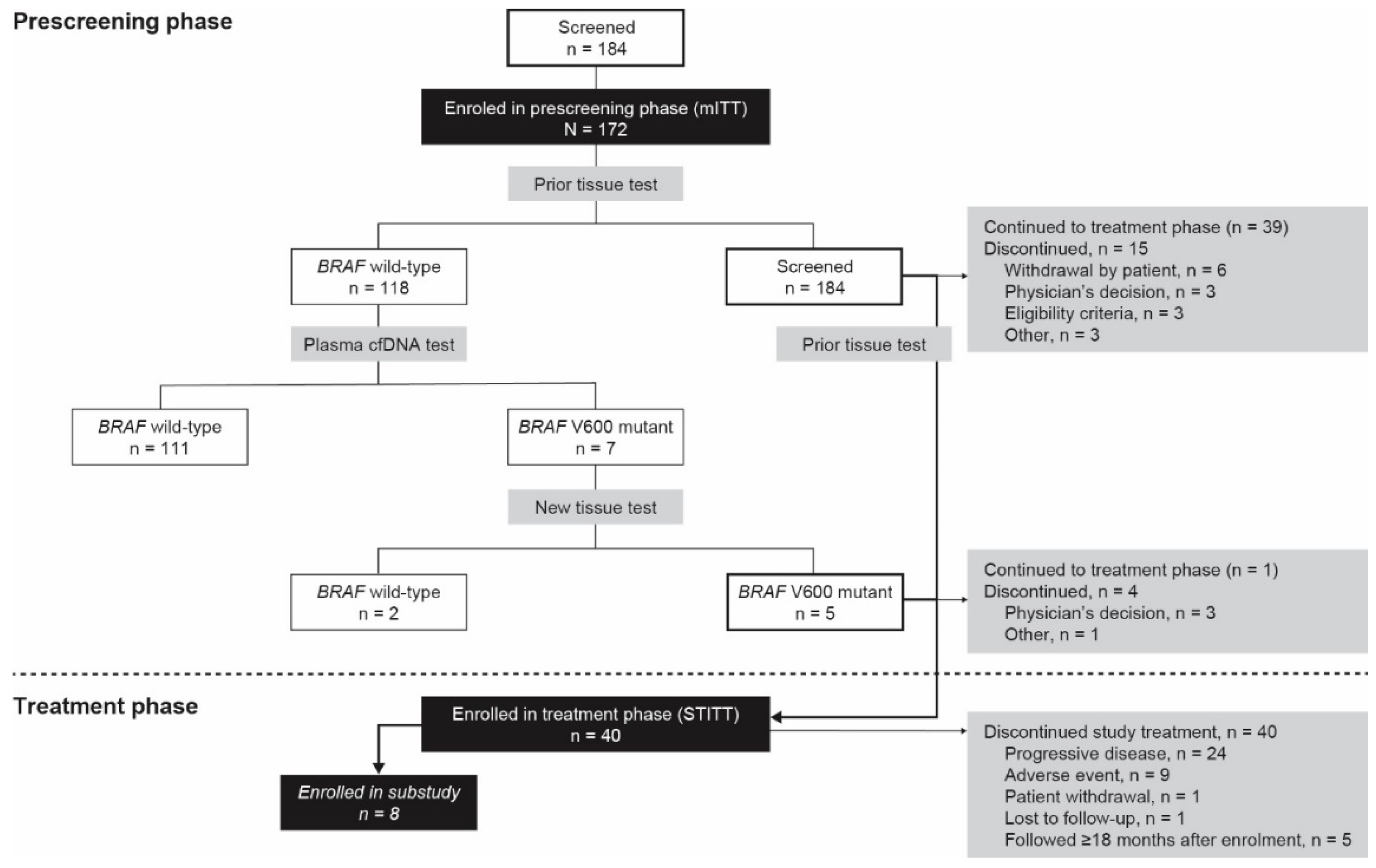
3.1. Patient Disposition

A total of 184 patients consented, and were evaluated for eligibility for the prescreening phase. Of these patients, 10 did not have documented BRAFV600 mutation test results on melanoma tissue upon study entry, and two had no plasma cfDNA test results (Figure 1). The remaining 172 patients represented the mITT population. Of these, 118 (68.6%) had a wild-type BRAF status and 54 (31.4%) had a mutant BRAFV600 status. Among the 118 patients with an initial wild-type BRAF status, the tumors in five of these patients were confirmed to be BRAFV600 mutant in the tissue retests, and one patient continued into the treatment phase. Of the 54 patients with a mutant BRAFV600 status, based on the prior tissue test, 39 continued into the treatment phase. The baseline characteristics of the patients are shown in Table 1.

3.2. Prescreening Phase

3.2.1. Primary Endpoint

Among the 118 patients with a wild-type BRAF status, seven (5.9%) had a positive BRAFV600 mutation test result, based on the plasma cfDNA test. Tissue retests on archival samples confirmed the BRAFV600 mutation in five (71.4%) of the seven patients. Thus, for 4.2% of the patients with a wild-type BRAF status, the presence of a BRAFV600 mutation was confirmed in a tissue retest; the one-sided test, comparing this frequency with the a priori hypothesis of 3%, was not statistically significant (p = 0.215).

3.2.2. Secondary Endpoint

The plasma cfDNA test was concordant with the prior tissue test in 35 of 54 of the patients who were classified as BRAFV600 mutation-positive, and in 111 of 118 of the patients with a wild-type status (Table 2). Relative to tissue testing, the plasma test demonstrated 64.8% sensitivity and 94.1% specificity. Of the 42 patients with a BRAFV600 mutant plasma cfDNA test result, 35 had a mutant status, based on the prior tissue test. The positive predictive value (i.e., the likelihood that the mutation detected with the plasma test corresponds to the tissue test) was 83.3%. Of the 130 patients with a wild-type plasma test result, 111 had a wild-type status, based on the prior tissue test. The negative predictive value (i.e., the likelihood that the wild-type status detected with the plasma test corresponds to the tissue test) was 85.4%.

3.2.3. Exploratory Endpoint

Of the 172 patients in the mITT population, 171 (99.4%) had an available NRAS mutation test result. Of these patients, 149 (87.1%) had an NRAS wild-type status and 22 (12.9%) had a mutant status. A summary of the relationship of NRAS mutant status and BRAFV600 mutant plasma test results is presented in Table 2. The mutations in BRAF and NRAS were mutually exclusive.

3.3. Treatment Phase

3.3.1. Efficacy Outcomes

In the STITT population, 29 of the 36 patients (80.6%) with target lesions and measurable disease, had either a complete response (three patients) or a partial response (26 patients) (Table 3). One patient (2.8%) had progressive disease, and two patients (5.6%) had stable disease. The median PFS (95% confidence interval (CI)) was 13.6 months (9.5–16.5). Of the 29 patients who had a complete or partial response, the median DOR (95% CI) was 11.0 months (9.2—not estimable). The median overall survival time was not estimable at the time of the analysis.

3.3.2. Correlation of Mutation Testing and Clinical Outcome

The BRAFV600 mutation was detected in the plasma at prescreening for 26 of the 40 treated patients. The median quantitation cycle (Cq) for the BRAF mutation was 40.25. The median PFS was 12.8 months and 10.8 months for the patients below and above the median mutation Cq, respectively; however, the Cq was not significantly associated with PFS duration (p = 0.653). Of these 26 patients, 22 had a complete or partial response. No significant relationship was found between mutation Cq and the DOR (p = 0.409). The median DOR was similar for these patients, regardless of whether the mutation Cq was above or below the median (10.7 vs. 10.9 months, respectively).
The clinical outcome was analyzed separately for patients with a BRAFV600 mutation, based on detection of the mutation in the plasma test at the prescreening. The median PFS was 14.8 months for the patients with a BRAF wild-type plasma test result, and 12.8 months for the patients with a BRAFV600 mutation plasma test result (p = 0.286). The median DOR was not evaluable for the patients with a BRAF wild-type plasma test result, and was 10.9 months for the patients with a BRAFV600 mutation plasma test result (p = 0.220).

3.3.3. Substudy

The concentration of the BRAFV600 mutation before the first dose was evaluated as a prognostic factor/metric for PFS duration in the eight patients who were participating in the substudy. The BRAFV600 mutation was observed at the start of cycle 1 for five of the eight patients (62.5%). The frequency of detection of the BRAFV600 mutation then decreased to 28.6% at the last assessment. All of these mutations were BRAFV600E/E2/D, except for one instance of BRAFV600K/R/W. No correlation between the plasma concentration of the mutation (average Cq for mutation) at the start of cycle 1, and the PFS duration, was observed (Spearman’s rank correlation coefficient: −0.100; p = 0.873).

3.3.4. Exposure and Safety

The mean duration of the treatment with both cobimetinib and vemurafenib was 11.2 ± 9.6 months (median, 7.9 months; range, 0.4–36.8 months). The median number of cycles was 9.0 for both cobimetinib and vemurafenib. The most frequently reported AEs were rash (47.5%), blood creatinine phosphokinase level increase (32.5%), diarrhea (32.5%), photosensitivity reaction (27.5%), pyrexia (27.5%), arthralgia (27.5%), and maculopapular rash (22.5%) (Table 4).

4. Discussion

The current study aimed to evaluate the clinical utility of a plasma-derived cfDNA test for the detection of BRAFV600 mutations in patients with metastatic melanoma, in whom the original testing using tumor biopsy falsely diagnosed the BRAFV600 wild-type status. It was observed that ~6% of the patients who were originally classified as having BRAF wild-type tumors tested positive for BRAF mutations using the cfDNA test. The sensitivity of the plasma cfDNA testing was 64.8%, and its positive and negative predictive values were 83.3% and 85.4%, respectively. No significant difference was observed between the median PFS and the median DOR for the patients with a BRAF wild-type versus BRAFV600 mutation-positive test result using cfDNA testing.
Previous studies have shown that cfDNA testing may be a surrogate for determining BRAF mutations in patients with melanoma [24,25,26,27,28]. In our study, the presence of BRAF mutations was confirmed by a subsequent tissue-based retest in five of the seven patients who showed BRAF mutations using cfDNA testing. The remaining two patients showed BRAF wild-type tumors upon the retest, which could potentially be a result of the archival tissue sample being degraded. Although the study did not meet its predefined criteria for the clinical significance of cfDNA testing (≥10% of patients designated as BRAF wild-type based on a prior tissue test to subsequently show BRAFV600 mutations on cfDNA testing), ~6% of BRAF wild-type patients were subsequently shown to be BRAFV600-mutation-positive, based on cfDNA tests, suggesting that cfDNA testing may have a place in the clinical setting. Furthermore, one of the five BRAF mutation-positive patients went on to receive BRAF-targeted therapy and responded to treatment with a PFS that was in a range that was observed in previous experience [29]. These results suggest that cfDNA testing may be a quick and easy alternative to identify BRAF mutations in patients for whom an archival tumor tissue sample may not be available in sufficient quality to allow for a retesting population, so they can benefit from the early initiation of BRAF-targeted therapy. However, it should be noted that cfDNA testing demonstrated a sensitivity of 64.8% in the present study. Therefore, although the cfDNA technique may be beneficial in the initial screening for the BRAF mutation status (owing to the ease of obtaining a blood sample versus a tumor re-biopsy, and the possibility of loss of the archival sample due to degradation, as seen in two patients in this study), retesting of the archival tumor tissue, if available, should be the primary choice for confirmation of the BRAF mutation status.
This study also evaluated any potential correlation with the cell-free BRAF mutation load of the plasma and treatment outcomes, as suggested by previous reports [29,30,31]; however, no appreciable differences were observed in either the median PFS or DOR among patients in whom the Cq for the BRAF mutation was above or below the median Cq of 40.25. The substudy, which tested BRAF mutation load as a metric of treatment response, showed that there was a decrease in the frequency of detection of the BRAFV600 mutation during the treatment, and the assay failed to detect the BRAFV600 mutation in a number of patients as the treatment progressed. Overall, the treatment outcomes and the observed safety profile with combination treatment with cobimetinib plus vemurafenib, were consistent with the pivotal phase 3 data in a population of patients with advanced melanoma and BRAF mutations [29,32].
A key limitation of the study was the relatively small number of patients who were included. In addition, only one patient whose tumor was originally classified as BRAF wild-type, and subsequently flagged as BRAF mutation-positive by the cell-free assay, went on to receive and benefit from targeted therapy with cobimetinib plus vemurafenib. The strengths of the study include its prospective nature, and that consecutive eligible and consenting patients at the study centers were enrolled (i.e., there was no selection bias). Furthermore, the study confirmed that a high percentage of patients that are designated as having BRAF-mutated tumors, using tissue-based tests, have positive results in cfDNA testing.

5. Conclusions

The study shows that plasma testing using cfDNA is a less invasive and rapid alternative to confirm the presence of BRAF mutations versus tumor biopsies, and may help in the early identification of patients who are more likely to benefit from treatment with BRAF inhibitors. However, cfDNA testing should be used with caution in patients undergoing BRAF inhibitor therapy, owing to its decreased sensitivity with treatment in these patients. No significant correlation between the use of cfDNA testing, and PFS and DOR could be determined among the patients receiving cobimetinib plus vemurafenib treatment. Additional studies in a larger patient population are warranted to confirm these results.

Supplementary Materials

The following is available online at https://www.mdpi.com/article/10.3390/cancers13143591/s1, Supplementary material appendix: patient eligibility criteria.

Author Contributions

Conceptualization, P.R., P.P., J.K., G.M., V.G. and B.N.; data curation, V.G., A.T. and K.d.B.; formal analysis, B.J., V.G., A.T. and K.d.B.; investigation, P.R. and J.K.; methodology, P.R., P.P., J.K., G.M., V.G. and B.N.; project administration, V.G., A.T. and K.d.B.; resources, B.J. and G.M.; supervision, V.G., A.T. and K.d.B.; validation, V.G., A.T. and K.d.B.; visualization, V.G., A.T. and K.d.B.; writing—original draft, P.R. and B.N.; writing—review and editing, P.R., P.P., B.J., V.G., A.T., K.d.B. and B.N. All authors have read and agreed to the published version of the manuscript.

Funding

This study was sponsored by F. Hoffmann–La Roche Ltd.; Genentech, Inc., a member of the Roche Group; and Biocartis Group NV.

Institutional Review Board Statement

The study was registered with ClinicalTrials.gov (NCT02768207) and EudraCT (2015-001731-20). The study was conducted in accordance with the good clinical practice guidelines, and the principles of the Declaration of Helsinki. The protocol was approved by an independent ethics committee at each site.

Informed Consent Statement

Written informed consent was obtained for all the patients before performing any study-specific prescreening test or evaluation. A second written informed consent form was obtained prior to the conduct of any study-related procedures in relation to the treatment phase. The patients who participated in the translational substudy signed a separate (third) informed consent form.

Data Availability Statement

Qualified researchers may request access to individual patient-level data through the clinical study data request platform (https://vivli.org/, accessed on 19 January 2021). Further details on Roche’s criteria for eligible studies are available here (https://vivli.org/members/ourmembers/, accessed on 19 January 2021). For further details on Roche’s global policy on the sharing of clinical information and how to request access to related clinical study documents, see here (https://www.roche.com/research_and_development/who_we_are_how_we_work/clinical_trials/our_commitment_to_data_sharing.htm, accessed on 19 January 2021).

Acknowledgments

We thank the patients and investigators who participated in the study, and Jerome Sah (ApotheCom, San Francisco, CA, USA) for providing medical writing and editorial support, which was funded by F. Hoffmann—La Roche Ltd.

Conflicts of Interest

Piotr Rutkowski reports consultancy fees from Novartis, Bristol Myers Squibb, Merck Sharp & Dohme Pierre Fabre, and Blueprint Medicines; research grants to his institution and/or himself, from Pfizer and Bristol Myers Squibb; and fees for speaking engagements from Novartis, Roche, Pierre Fabre, Bristol Myers Squibb, Merck Sharp & Dohme, Sanofi, Merck, and Pfizer. Patrick Pauwels reports consultancy fees from AstraZeneca, Pfizer, Roche, Merck Sharp & Dohme, Bristol Myers Squibb, Takeda, Biocartis, and Novartis; and grants from AstraZeneca, Roche, and Biocartis. Joseph Kerger has no conflicts to disclose. Bart Jacobs reports employment with Biocartis and Novartis. Geert Maertens reports employment with, patents with, and royalties from, Biocartis. Valerie Gadeyne, Anne Thielemans, and Katrien de Backer report employment with F. Hoffmann—La Roche. Bart Neyns reports the following paid monies to his institution: consultancy fees from Novartis, and research grants and payments for speaking engagements from Novartis, Pfizer, Bristol Myers Squibb, and Merck Sharp & Dohme.

References

  1. Bisschop, C.; Ter, E.A.; Bosman, L.J.; Platteel, I.; Jalving, M.; van den Berg, A.; Diepstra, A.; van Hemel, B.; Diercks, G.F.H.; Hospers, G.A.P.; et al. Rapid BRAF mutation tests in patients with advanced melanoma: Comparison of immunohistochemistry, Droplet Digital PCR, and the Idylla Mutation Platform. Melanoma Res. 2018, 28, 96–104. [Google Scholar] [CrossRef] [Green Version]
  2. Bruno, W.; Martinuzzi, C.; Andreotti, V.; Pastorino, L.; Spagnolo, F.; Dalmasso, B.; Cabiddu, F.; Gualco, M.; Ballestrero, A.; Bianchi-Scarrà, G.; et al. Heterogeneity and frequency of BRAF mutations in primary melanoma: Comparison between molecular methods and immunohistochemistry. Oncotarget 2017, 8, 8069–8082. [Google Scholar] [CrossRef] [Green Version]
  3. Davies, H.; Bignell, G.R.; Cox, C.; Stephens, P.; Edkins, S.; Clegg, S.; Teague, J.; Woffendin, H.; Garnett, M.J.; Bottomley, W.; et al. Mutations of the BRAF gene in human cancer. Nature 2002, 417, 949–954. [Google Scholar] [CrossRef]
  4. Li, Y.; Umbach, D.M.; Li, L. Putative genomic characteristics of BRAF V600K versus V600E cutaneous melanoma. Melanoma Res. 2017, 27, 527–535. [Google Scholar] [CrossRef]
  5. Keilholz, U.; Ascierto, P.A.; Dummer, R.; Robert, C.; Lorigan, P.; van Akkooi, A.; Arance, A.; Blank, C.U.; Sileni, V.C.; Donia, M.; et al. ESMO consensus conference recommendations on the management of metastatic melanoma: Under the auspices of the ESMO Guidelines Committee. Ann. Oncol. 2020, 31, 1435–1448. [Google Scholar] [CrossRef] [PubMed]
  6. Swetter, S.M.; Thompson, J.A.; Albertini, M.R.; Barker, C.A.; Boland, G.; Carson, W.E. National Comprehensive Cancer Network® (NCCN®) Clinical Practice Guidelines in Oncology—Cutaneous Melanoma Version 4.2020. 2020. Available online: https://www.nccn.org/professionals/physician_gls/pdf/cutaneous_melanoma.pdf (accessed on 16 September 2020).
  7. Cohen, J.V.; Buchbinder, E.I. The evolution of adjuvant therapy for melanoma. Curr. Oncol. Rep. 2019, 21, 106. [Google Scholar] [CrossRef] [PubMed]
  8. Dimitriou, F.; Long, G.V.; Menzies, A.M. Novel adjuvant options for cutaneous melanoma. Ann. Oncol. 2021, 32, 854–865. [Google Scholar] [CrossRef] [PubMed]
  9. Blankenstein, S.A.; van Akkooi, A.C.J. Adjuvant systemic therapy in high-risk melanoma. Melanoma Res. 2019, 29, 358–364. [Google Scholar] [CrossRef]
  10. Noor, R.; Trinh, V.; Kim, K.; Hwu, W.J. BRAF-targeted therapy for metastatic melanoma: Rationale, clinical activity and safety. Clin. Investig. 2011, 1, 1127–1139. [Google Scholar] [CrossRef]
  11. Cheng, L.; Lopez-Beltran, A.; Massari, F.; MacLennan, G.T.; Montironi, R. Molecular testing for BRAF mutations to inform melanoma treatment decisions: A move toward precision medicine. Mod. Pathol. 2018, 31, 24–38. [Google Scholar] [CrossRef]
  12. Cheng, L.Y.; Haydu, L.E.; Song, P.; Nie, J.; Tetzlaff, M.T.; Kwong, L.N.; Gershenwald, J.E.; Davies, M.A.; Zhang, D.Y. High sensitivity sanger sequencing detection of BRAF mutations in metastatic melanoma FFPE tissue specimens. Sci. Rep. 2021, 11, 9043. [Google Scholar] [CrossRef]
  13. Harle, A.; Salleron, J.; Franczak, C.; Dubois, C.; Filhine-Tressarieu, P.; Leroux, A.; Merlin, J.-L. Detection of BRAF mutations using a fully automated platform and comparison with high resolution melting, real-time allele specific amplification, immunohistochemistry and next generation sequencing assays, for patients with metastatic melanoma. PLoS ONE 2016, 11, e0153576. [Google Scholar] [CrossRef]
  14. Huang, W.; Kuo, T.T.; Wu, C.E.; Cheng, H.-Y.; Hsieh, C.-H.; Hsieh, J.-J.; Shen, Y.-C.; Hou, M.-M.; Hsu, T.; Chang, J.W.-C. A comparison of immunohistochemical and molecular methods used for analyzing the BRAFV600E gene mutation in malignant melanoma in Taiwan. Asia Pac. J. Clin. Oncol. 2016, 12, 403–408. [Google Scholar] [CrossRef] [PubMed]
  15. Janku, F.; Claes, B.; Huang, H.J.; Falchook, G.S.; Devogelaere, B.; Kockx, M.; Bempt, I.V.; Reijans, M.; Naing, A.; Fu, S.; et al. BRAF mutation testing with a rapid, fully integrated molecular diagnostics system. Oncotarget 2015, 6, 26886–26894. [Google Scholar] [CrossRef] [Green Version]
  16. Alrabadi, N.; Gibson, N.; Curless, K.; Cheng, L.; Kuhar, M.; Chen, S.; Warren, S.J.P.; Alomari, A.K. Detection of driver mutations in BRAF can aid in diagnosis and early treatment of dedifferentiated metastatic melanoma. Mod. Pathol. 2019, 32, 330–337. [Google Scholar] [CrossRef]
  17. Kong, B.Y.; Carlino, M.S.; Menzies, A.M. Biology and treatment of BRAF mutant metastatic melanoma. Melanoma Manag. 2016, 3, 33–45. [Google Scholar] [CrossRef]
  18. Vanni, I.; Tanda, E.T.; Spagnolo, F.; Andreotti, V.; Bruno, W.; Ghiorzo, P. The current state of molecular testing in the BRAF-mutated melanoma landscape. Front. Mol. Biosci. 2020, 7, 113. [Google Scholar] [CrossRef] [PubMed]
  19. Donadio, E.; Giusti, L.; Cetani, F.; Da Valle, Y.; Ciregia, F.; Giannaccini, G.; Pardi, E.; Saponaro, F.; Torregrossa, L.; Basolo, F.; et al. Evaluation of formalin-fixed paraffin-embedded tissues in the proteomic analysis of parathyroid glands. Proteome Sci. 2011, 9, 29. [Google Scholar] [CrossRef] [PubMed] [Green Version]
  20. Janku, F.; Huang, H.J.; Claes, B.; Falchook, G.S.; Fu, S.; Hong, D.; Ramzanali, N.M.; Nitti, G.; Cabrilo, G.; Tsimberidou, A.M.; et al. BRAF mutation testing in cell-free DNA from the plasma of patients with advanced cancers using a rapid, automated molecular diagnostics system. Mol. Cancer Ther. 2016, 15, 1397–1404. [Google Scholar] [CrossRef] [Green Version]
  21. Calapre, L.; Warburton, L.; Millward, M.; Ziman, M.; Gray, E.S. Circulating tumour DNA (ctDNA) as a liquid biopsy for melanoma. Cancer Lett. 2017, 404, 62–69. [Google Scholar] [CrossRef]
  22. Balch, C.M.; Gershenwald, J.E.; Soong, S.J.; Thompson, J.F.; Atkins, M.B.; Byrd, D.R.; Buzaid, A.C.; Cochran, A.J.; Coit, D.G.; Ding, S.; et al. Final version of 2009 AJCC melanoma staging and classification. J. Clin. Oncol. 2009, 27, 6199–6206. [Google Scholar] [CrossRef] [Green Version]
  23. National Institutes of Health. Common Terminology Criteria for Adverse Events (CTCAE) v4.03. 2009. Available online: https://www.eortc.be/services/doc/ctc/CTCAE_4.03_2010-06-14_QuickReference_5x7.pdf (accessed on 16 September 2020).
  24. Long-Mira, E.; Ilie, M.; Chamorey, E.; Leduff-Blanc, F.; Montaudié, H.; Tanga, V.; Allégra, M.; Lespinet-Fabre, V.; Bordone, O.; Bonnetaud, C.; et al. Monitoring BRAF and NRAS mutations with cell-free circulating tumor DNA from metastatic melanoma patients. Oncotarget 2018, 9, 36238–36249. [Google Scholar] [CrossRef] [Green Version]
  25. Molina-Vila, M.A.; de-Las-Casas, C.M.; Bertran-Alamillo, J.; Jordana-Ariza, N.; González-Cao, M.; Rosell, R. cfDNA analysis from blood in melanoma. Ann. Transl. Med. 2015, 3, 309. [Google Scholar] [CrossRef]
  26. Burjanivova, T.; Malicherova, B.; Grendar, M.; Minarikova, E.; Dusenka, R.; Vanova, B.; Bobrovska, M.; Pecova, T.; Homola, I.; Lasabova, Z.; et al. Detection of BRAFV600E mutation in melanoma patients by digital PCR of circulating DNA. Genet. Test. Mol. Biomark. 2019, 23, 241–245. [Google Scholar] [CrossRef]
  27. Knol, A.C.; Vallée, A.; Herbreteau, G.; Nguyen, J.M.; Varey, E.; Gaultier, A.; Théoleyre, S.; Saint-Jean, M.; Peuvrel, L.; Brocard, A.; et al. Clinical significance of BRAF mutation status in circulating tumor DNA of metastatic melanoma patients at baseline. Exp. Dermatol. 2016, 25, 783–788. [Google Scholar] [CrossRef]
  28. Kozak, K.; Kowalik, A.; Gos, A.; Wasag, B.; Lugowska, I.; Jurkowska, M.; Krawczynska, N.; Kosela-Paterczyk, H.; Switaj, T.; Teterycz, P.; et al. Cell-free DNA BRAF V600E measurements during BRAF inhibitor therapy of metastatic melanoma: Long-term analysis. Tumori 2020. [Google Scholar] [CrossRef]
  29. Larkin, J.; Ascierto, P.A.; Dreno, B.; Atkinson, V.; Liszkay, G.; Maio, M.; Mandalà, M.; Demidov, L.; Stroyakovskiy, D.; Thomas, L.; et al. Combined vemurafenib and cobimetinib in BRAF-mutated melanoma. N. Engl. J. Med. 2014, 371, 1867–1876. [Google Scholar] [CrossRef] [PubMed] [Green Version]
  30. Schreuer, M.; Jansen, Y.; Planken, S.; Chevolet, I.; Seremet, T.; Kruse, V.; Neyns, B. Combination of dabrafenib plus trametinib for BRAF and MEK inhibitor pretreated patients with advanced BRAF(V600)-mutant melanoma: An open-label, single arm, dual-centre, phase 2 clinical trial. Lancet Oncol. 2017, 18, 464–472. [Google Scholar] [CrossRef]
  31. Schreuer, M.; Meersseman, G.; Van Den Herrewegen, S.; Jansen, Y.; Chevolet, I.; Bott, A.; Wilgenhof, S.; Seremet, T.; Jacobs, B.; Buyl, R.; et al. Quantitative assessment of BRAFV600 mutant circulating cell-free tumor DNA as a tool for therapeutic monitoring in metastatic melanoma patients treated with BRAF/MEK inhibitors. J. Transl. Med. 2016, 14, 95. [Google Scholar] [CrossRef] [PubMed] [Green Version]
  32. Ascierto, P.A.; McArthur, G.A.; Dréno, B.; Atkinson, V.; Liszkay, G.; Di Giacomo, A.M.; Mandalà, M.; Demidov, L.; Stroyakovskiy, D.; Thomas, L.; et al. Cobimetinib combined with vemurafenib in advanced BRAF(V600)-mutant melanoma (coBRIM): Updated efficacy results from a randomised, double-blind, phase 3 trial. Lancet Oncol. 2016, 17, 1248–1260. [Google Scholar] [CrossRef]
Figure 1. Patient disposition. mITT, modified intention-to-treat; STITT, study treatment intention-to-treat.
Cancers 13 03591 g001
Table 1. Baseline demographic, disease, biopsy, and mutation characteristics.
mITTSTITT
n = 172n = 40
Age, median years (range)62.5 (20–93)56.5 (26–82)
Sex, n (%)
 Male84 (48.8)18 (45.0)
 Female88 (51.2)22 (55.0)
ECOG score, n (%)
 0NE21 (52.5)
 1NE16 (40.0)
 2NE3 (7.5)
Time since diagnosis of metastases, median months (range)19.3 (0.1–260.7)12.4 (0.8–260.7)
Age at diagnosis of metastases, median years (range)59.1 (18–91)53.5 (26–83)
Disease stage at study entry, n (%)
 Unresectable stage IIIC16 (9.3)1 (2.5)
 Stage IV156 (90.7)29 (97.5)
Measurable disease at study entry, n (%)147 (85.5)37 (92.5)
Number of target lesions, n (%)
 0–3NE22 (55.0)
 >3NE18 (45.0)
Type of tissue material, n (%)
 Archival140 (81.4)30 (75.0)
 Recent32 (18.6)10 (25.0)
Prior tissue BRAF mutation test result, n (%)
BRAF wild-type118 (68.6)1 (2.5)
BRAFV600 mutation54 (31.4)39 (97.5)
Prior therapy, n (%)
 Immunotherapy9 (33)
 Targeted therapy0
 Other systemic therapy7 (17.5)
 Investigational treatment1 (2.5)
 Radiotherapy8 (20.0)
ECOG, Eastern Cooperative Oncology Group; mITT, modified intention-to-treat; NE, not evaluated; STITT, study treatment intention-to-treat.
Table 2. Comparison of tissue-based and cfDNA-based mutation tests. (A) Comparison of plasma and tissue BRAFV600 mutation test results, and (B) comparison of BRAF and NRAS plasma mutation tests results in the mITT population. Grey-shaded cells designate the discordance between the respective tests. cfDNA, cell-free DNA; mITT, modified intention-to-treat.
A. BRAFPlasma cfDNA Test Result
Wild-TypeMutant
Tissue test resultWild-type1117Patients with wild-type tissue test = 118
Mutant1935Patients with mutant tissue test = 54
Patients with wild-type plasma test = 130Patients with mutant plasma test = 42
B. PlasmaBRAF cfDNA test result
Wild-typeMutant
NRAS cfDNA test resultWild-type10742Patients with wild-type NRAS = 149
Mutant220Patients with mutant NRAS = 22
Patients with wild-type plasma test = 129Patients with mutant plasma test = 42
Table 3. Objective response rate and PFS (STITT).
STITT
n = 40
Archival Tissue *
n = 29
Recent Tissue
n = 10
Objective response rate, ‡ n (%)
 CR3 (8.3)3 (11.5)
 PR26 (72.2)20 (76.9)6 (60.0)
 SD2 (5.6)2 (20.0)
 PD1 (2.8)1 (10.0)
 Other nonresponders4 (11.1)3 (11.5)1 (10.0)
Responders (CR + PR), n/n′ (%)29/36 (80.6)23/26 (88.5)6/10 (60.0)
95% CI64.0–91.869.8–97.626.2–87.8
Duration of Response, n′ ‡2923n′ = 6
Median, months (95% CI)11.0 (9.2–NE)11.0 (9.2–NE)8.8 (3.6–NE)
Progression-free survival
Median months (95% CI)13.6 (9.5–16.5)14.5 (10.8–NE)6.2 (3.6–NE)
CI, confidence interval; CR, complete response; n′, number of patients considered in the analysis; percentages calculated based on n′; NE, not estimable; PD, progressive disease; PR, partial response; SD, stable disease; STITT, study treatment intention-to-treat. * Patients for whom treatment was based on the mutation identified by prior tissue test and for whom archival tissue was used. Patients for whom treatment was based on the mutation identified by prior tissue test and for whom recent tissue was used. For patients with measurable disease at screening having responded during the study (CR or PR).
Table 4. Common treatment-emergent adverse events (incidence ≥10% by preferred term).
System Organ Class/Preferred Termn = 40
Any treatment-emergent adverse event, n (%)39 (97.5)
Skin and subcutaneous tissue disorders, n (%)33 (82.5)
 Rash19 (47.5)
 Photosensitivity reaction11 (27.5)
 Maculopapular rash9 (22.5)
 Alopecia4 (10.0)
Investigations, n (%)22 (55.0)
 Blood creatinine phosphokinase increased13 (32.5)
 Gamma-glutamyltransferase increased5 (12.5)
 C-reactive protein increased4 (10.0)
General disorders and administration site conditions, n (%)21 (52.5)
 Pyrexia11 (27.5)
 Fatigue8 (20.0)
 Oedema peripheral5 (12.5)
Infections and infestations, n (%)21 (52.5)
 Upper respiratory tract infection8 (20.0)
 Conjunctivitis6 (15.0)
 Urinary tract infection4 (10.0)
Gastrointestinal disorders, n (%)20 (50.0)
 Diarrhea13 (32.5)
 Vomiting6 (15.0)
 Nausea5 (12.5)
Musculoskeletal and connective tissue disorders, n (%)14 (35.0)
 Arthralgia11 (27.5)
 Musculoskeletal pain4 (10.0)
 Myalgia4 (10.0)
 Pain in extremity4 (10.0)
Eye disorders, n (%)12 (30.0)
 Vision blurred6 (15.0)
 Chorioretinopathy4 (10.0)
Nervous system disorders, n (%)12 (30.0)
 Headache6 (15.0)
Metabolism and nutrition disorders, n (%)7 (17.5)
 Decreased appetite4 (10.0)
 Hypokalemia4 (10.0)
 Hypertension5 (12.5)
Publisher’s Note: MDPI stays neutral with regard to jurisdictional claims in published maps and institutional affiliations.

