Review

Breaching the Blood–Brain Tumor Barrier for Tumor Therapy

by
Fabrizio Marcucci
1,*,
Angelo Corti
2,3,*,† and
Andrés J. M. Ferreri
4,†
1
Department of Pharmacological and Biomolecular Sciences, University of Milan, 20132 Milan, Italy
2
Division of Experimental Oncology, Tumor Biology and Vascular Targeting Unit, IRCCS San Raffaele Scientific Institute, 20132 Milan, Italy
3
Faculty of Medicine and Surgery, Università Vita-Salute San Raffaele, 20132 Milan, Italy
4
Lymphoma Unit, Department of Onco-Hematology, IRCCS San Raffaele Scientific Institute, 20132 Milan, Italy
*
Authors to whom correspondence should be addressed.
These authors contributed equally (co-last authors).
Cancers 2021, 13(10), 2391; https://doi.org/10.3390/cancers13102391
Submission received: 15 April 2021 / Revised: 6 May 2021 / Accepted: 11 May 2021 / Published: 15 May 2021
(This article belongs to the Section Cancer Therapy)

Abstract

:

Simple Summary

The blood–brain tumor barrier (BBTB) represents a major obstacle for the delivery of anticancer drugs to tumors of the central nervous system. Various approaches have been so far developed for overcoming this obstacle and for increasing anti-cancer drug concentrations in tumor tissues. This review is focused on the latest clinical advances and achievements in breaching the BBTB for primary and secondary brain tumor therapy.

Abstract

Tumors affecting the central nervous system (CNS), either primary or secondary, are highly prevalent and represent an unmet medical need. Prognosis of these tumors remains poor, mostly due to the low intrinsic chemo/radio-sensitivity of tumor cells, a meagerly known role of the microenvironment and the poor CNS bioavailability of most used anti-cancer agents. The BBTB is the main obstacle for anticancer drugs to achieve therapeutic concentrations in the tumor tissues. During the last decades, many efforts have been devoted to the identification of modalities allowing to increase drug delivery into brain tumors. Until recently, success has been modest, as few of these approaches reached clinical testing and even less gained regulatory approval. In recent years, the scenario has changed, as various conjugates and drug delivery technologies have advanced into clinical testing, with encouraging results and without being burdened by a heavy adverse event profile. In this article, we review the different approaches aimed at increasing drug delivery to brain tumors, with particular attention to new, promising approaches that increase the permeability of the BBTB or exploit physiological transport mechanisms.

1. Introduction

Despite some recent progress, the overall prognosis of tumors that primarily arise in or metastasize to the central nervous system (CNS) remains largely unsatisfactory. The poor CNS bioavailability of most anti-cancer agents plays a crucial role in this regard. This is due to the presence of the blood–brain barrier (BBB), which protects the CNS from potentially harmful blood-borne substances, including anticancer drugs, and regulates influx and efflux of molecules. Patients with gliomas, or other neoplasms of the CNS, such as brain metastases or lymphomas, would greatly profit from enhanced access of drugs into the CNS. Thus, overcoming this barrier in brain tumors (referred to as the blood–brain tumor barrier, BBTB) with innovative strategies has become an important therapeutic goal. Literature on the biological and molecular characteristics of the BBTB is rapidly growing, and progress in gaining knowledge on the physiology and pathophysiology of this barrier has provided the background for the development of new approaches that have been addressed in a number of clinical studies. This article is not focused on the biological and molecular details of the BBTB, which can be found in excellent recent reviews [1,2,3,4]. Instead, we review the available literature on novel treatments for primary or secondary brain cancers that overcome the BBTB and discuss in details some recent approaches that hold considerable promise. The molecular bases, targets, and drug delivery systems, as well as the design and results of clinical trials, are critically analyzed, and future perspectives are discussed.

2. The Blood–Brain Barrier (BBB) and the Blood–Brain Tumor Barrier (BBTB)—Some Fundamentals

Different cell types contribute to the BBB: endothelial cells, pericytes, astrocytes, microglial cells and neurons, which, together, constitute the neurovascular unit (NVU) [3]. Endothelial cells are the main component of the BBB and display some unique properties compared to endothelial cells in other compartments: they have continuous intercellular tight junctions (TJ), lack fenestrations and undergo very low rates of transcytosis, thereby minimizing any kind of paracellular and transcellular transport through the endothelial cell layer [1]. Moreover, low expression of leukocyte adhesion molecules keeps at a minimum immune cell infiltration into the brain. Likewise, astrocytes, pericytes, microglia cells and neurons exhibit important specializations, and recent investigations on single-cell components of the NVU have confirmed the organotypic molecular signatures of these cells [5,6]. In addition, the basement membrane, produced by the different cell types of the NVU through the secretion of extracellular matrix (ECM) molecules, contributes to the BBB, by keeping the different cell types in place and regulating their intercellular crosstalk [3]. The BBB is so efficient in sealing off the CNS that only small, lipophilic molecules, with an Mr < 400 Da, can enter the CNS through passive diffusion across the lipid bilayer membranes of endothelial cells [7]. Otherwise, exchange of molecules between the blood and brain parenchyma is the task of specific transporters that regulate the translocation of selected molecules across the endothelium [8,9].
The BBB is not an immutable entity, experiencing significant changes in response to pathologies affecting the CNS. In the presence of a primary or secondary brain tumor, for example, it is now customary to refer to a BBTB rather than to a BBB, to evidence the differences between the two [4]. Some of these differences are the consequence of the abnormal angiogenesis that characterizes tumor formation, with neoangiogenic tumor vessels expressing markers that are not present in quiescent endothelial cells [10]. Brain tumorigenesis is also accompanied, similarly to other tumors arising outside the CNS, by inflammatory changes that are closely intertwined with neoangiogenic changes [11]. Moreover, existing vessels may be compressed by the growing tumor, thereby impairing blood flow [12], or they may be co-opted by the tumor [13]. Altogether, these and other alterations lead to increased leakiness of the BBTB, which, however, is very heterogeneous even within individual malignant foci [14] and, therefore, inappropriate to be exploited for homogeneous drug delivery within affected brain areas. Furthermore, tumor cells can be detected by histopathologic analysis not only in areas characterized by high permeability and detected by neuroimaging techniques, but also far from these areas. In the present article, which has its main focus on the therapy of brain tumors, we will always utilize the term BBTB unless we refer to studies regarding non-neoplastic diseases. In these cases, the term BBB will be used.
Overall, the BBB/BBTB represents an especially difficult barrier to overcome within the more general context of delivering drugs out of the circulation into diseased tissues, whether affected by cancer or other pathologies [15,16].

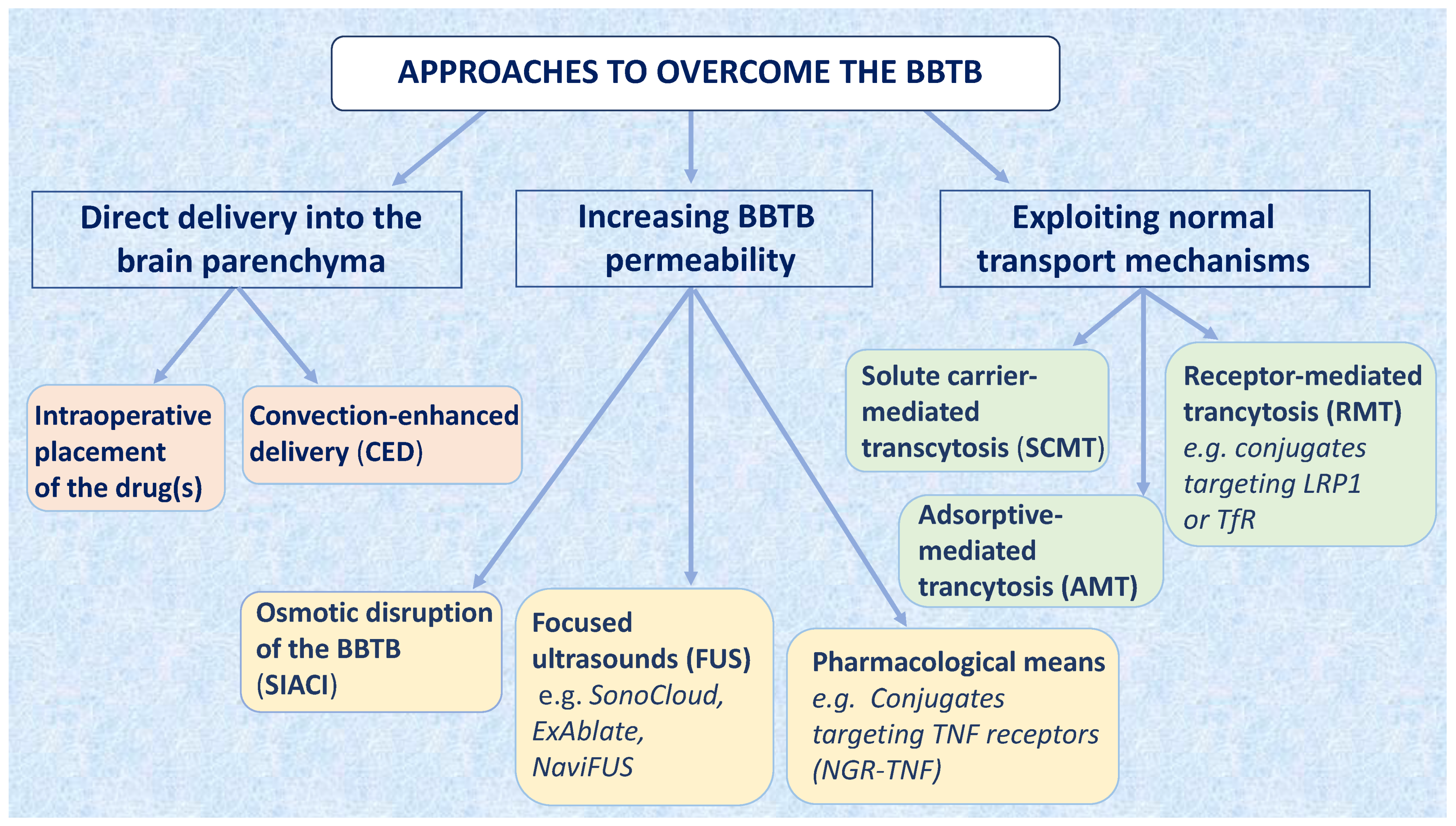
3. Approaches to Overcome the BBTB for Brain Tumor Therapy

The BBTB allows to deliver to tumor tissues only a few highly lipophilic compounds of low Mr (e.g., thiotepa and temozolomide). Other drugs are delivered at high doses, allowing sufficient tumor penetration by passive diffusion following a concentration gradient between intra- and extra-vascular spaces, but at the expense of higher side effects [17,18]. The development of strategies to overcome the BBTB could expand the spectrum of usable drugs, with a potential improvement in therapeutic efficacy and tolerability (Figure 1). The implementation of these strategies is expected to improve the delivery of drugs both against primary brain tumors as well as tumors that metastasize to the brain [19,20,21].

3.1. Delivering Drugs Directly into the Brain

3.1.1. Wafers Impregnated with Anticancer Drugs

A first approach rests on delivering the desired compound(s) directly into the tumor parenchyma. This can be achieved through placement of a drug in the resection cavity created during a surgical intervention of tumor debulking. For this purpose, polymer wafers impregnated with a chemotherapeutic drug are used and placed in the cavity [22]. Wafers embedded with carmustine (e.g., Gliadel® wafers), which allow a sustained drug release for at least 5 days while the wafer is degraded in 2–3 weeks, have received regulatory approval for the treatment of glioblastoma by the US Food and Drug Administration (FDA) and other regulatory authorities in 2003. In general, efficacy studies have demonstrated modest survival advantages [23,24], but a controlled, propensity-matched analysis has shown a significant prolongation of progression-free survival (PFS) in a multicenter cohort of patients with newly diagnosed glioblastoma treated with Gliadel® wafer implantation followed by standard chemoradiotherapy [25]. An ongoing randomized phase III trial (JCOG1703) is addressing the contribution of Gliadel® to maximal tumor resection followed by chemoradiotherapy in patients with glioblastoma [26]. Clinical trials with Gliadel®, retrievable from the clinicaltrials.gov website, are reported in Table 1. Research in this setting is focused on the use of new impregnated drugs and the development of novel materials and device technologies [27].

3.1.2. Convection-Enhanced Delivery (CED)

CED represents a valid alternative to infuse a compound directly in the tumor parenchyma. Here, a syringe pump and a cannula implanted into the tumor creates a pressure gradient, thereby allowing distribution of the compound throughout contiguous brain areas [28]. Several clinical studies have demonstrated the safety and feasibility of a variety of agents injected by CED (e.g., recombinant fusion proteins, oncolytic viruses, monoclonal antibodies (mAbs) and liposomal drugs); however, the large variability in the techniques, infusates and hardware used make it difficult to draw reliable conclusions on antitumor efficacy. Despite these limitations, encouraging results have been reported in some early-phase clinical studies. Diffuse intrinsic pontine glioma, a universally fatal tumor of pediatric patients, is a suitable candidate for CED-based treatment because it is constrained within a limited anatomical compartment, without the tissue inhomogeneities typically observed in other brain tumors managed with cytoreductive resection. The CED of 124I-labeled anti-B7-H3 mAb 8H9 has been performed in children with diffuse intrinsic glioma in a dose-escalation phase I trial, showing a good safety profile and high intratumoral dosing with negligible systemic exposure [29]. A recently reported phase I trial has demonstrated that nimustine delivered by CED in combination with temozolomide was safe and active in patients with recurrent diffuse intrinsic pontine glioma (n = 11) and other brainstem gliomas (n = 5) [30]. Most treated patients experienced worsening of transient symptoms due to local edema; tumor size reduction was recorded in most patients who received the highest assessed dose.
The CED of recombinant proteins consisting of a bacterial toxin, such as the Pseudomonas aeruginosa exotoxin, fused to a cytokine (such as transforming growth factor (TGF), interleukin (IL)-4 or IL-13) has been investigated in patients with recurrent high-grade gliomas [31,32,33]. Phase I trials have demonstrated that the CED of these compounds is associated with grade 3–4 side effects in one-third of patients, with no deaths due to toxicity [31,32]. In the PRECISE randomized phase III trial [34], the IL-13-Pseudomonas exotoxin A conjugate (termed IL13-PE38QQR) has produced results similar to those of the Gliadel® wafer, in 296 patients with recurrent glioblastoma, as a result of which the clinical development of this compound was discontinued. The CED of recombinant, nonpathogenic polio-rhinovirus chimera (PVSRIPO), a virus that recognizes CD155 in tumors cells and the microenvironment, has been safe in 61 patients with recurrent glioblastoma [35], with a 3-year OS of 21%, which is better than the survival rates reported in historical controls; these results deserve confirmation in more advanced trials.
The CED of chemotherapeutic drugs is an effective way to overcome the BBTB, which could result in higher antitumor activity as well as in higher toxicity. The CED of paclitaxel is a good example of this balance [36,37]. Paclitaxel is an anticancer drug inefficacious in patients with recurrent glioblastoma, as it crosses the BBTB to a minimal degree. When administered by CED, this taxane has been associated with an overall response rate (ORR) of 73% in these patients but, at the same time, with important complications, such as chemical meningitis, infectious complications, transient neurological impairment, and skin necrosis. Thus, optimization of this approach to reduce related toxicity should be explored further.
Therapeutic failures with CED, mostly those reported in the PRECISE trial [34], have been attributed to ineffective drug infusion resulting from suboptimal catheter positioning [38]. In fact, all criteria of the catheter positioning guidelines were met only in half of the treated patients, which represents an important limitation in a multicenter setting. Despite these limitations, CED remains the most intensively investigated approach to bypass the BBTB, with several ongoing clinical trials (Table 1).
It should be noted, however, that this method, as the other methods herein described based on local drug delivery to a specific brain area, will deliver the chemotherapy only to the tumor bulk and not to distant, migrating tumor cells (which may still reside behind the BBTB).

3.2. Increasing the Permeability of the BBTB

Some strategies have been designed to transiently disrupt the BBTB in order to increase its permeability, allowing the extravasation of drugs into the brain tumor parenchyma.

3.2.1. Osmotic Disruption of the BBTB

This approach rests on the temporary disruption of the BBTB by carotid or vertebral artery injection of a hyperosmotic solution, generally a highly concentrated solution of mannitol, just before drug administration by the same route [39]. The administration of the hyperosmotic solution causes water to be withdrawn from the endothelial cells lining the BBTB, with the consequent shrinkage and opening of the inter-endothelial junctions, which, in turn, leads to increased transport of systemically administered drugs across the BBTB. Osmotic disruption of the BBTB is transient and reversible [40]. This approach is referred to as superselective intraarterial cerebral infusion (SIACI). The locoregional administration protocol is required in order to avoid systemic effects. However, given the invasiveness of the procedure, it comes to no surprise that it requires hospitalization, patient sedation and can be accompanied by adverse events (AEs), such as neurological deficits, strokes, seizures and new tumor-nodule formation [41], mostly due to a transient edema that ensues the increased bulk fluid influx.
SIACI has been used to deliver mAbs. SIACI of cetuximab, an anti-epidermal growth factor receptor (EGFR) mAb, has been safe and well tolerated in patients with recurrent glioblastoma in a phase I/II trial, with no dose-limiting toxicity (DLT) up to 250 mg/m2 [42]; a phase II trial is ongoing (Table 2). When administered by SIACI, bevacizumab, an anti-vascular endothelial growth factor (VEGF) mAb, has been associated with a good safety profile and some cases of tumor regression in patients with recurrent gliomas [43,44].
Reversible BBTB disruption by intra-arterial infusion of mannitol followed by intra-arterial methotrexate has been investigated in patients with newly diagnosed or relapsed primary CNS lymphoma, in a few institutions with adequate expertise. This strategy has been associated with a 58% complete response (CR) rate, 5-year PFS of 31% and acceptable morbidity and neurotoxicity [45]. Overall, BBTB disruption shows an efficacy similar to that of standard high-dose-methotrexate-based treatments, but it is a procedurally intensive treatment, requiring monthly intravascular interventions under general anesthesia over the course of 1 year. Osmotic disruption by SIACI is currently being investigated in several clinical trials in combination with various drugs in patients with primary brain tumors or brain metastases (Table 2).

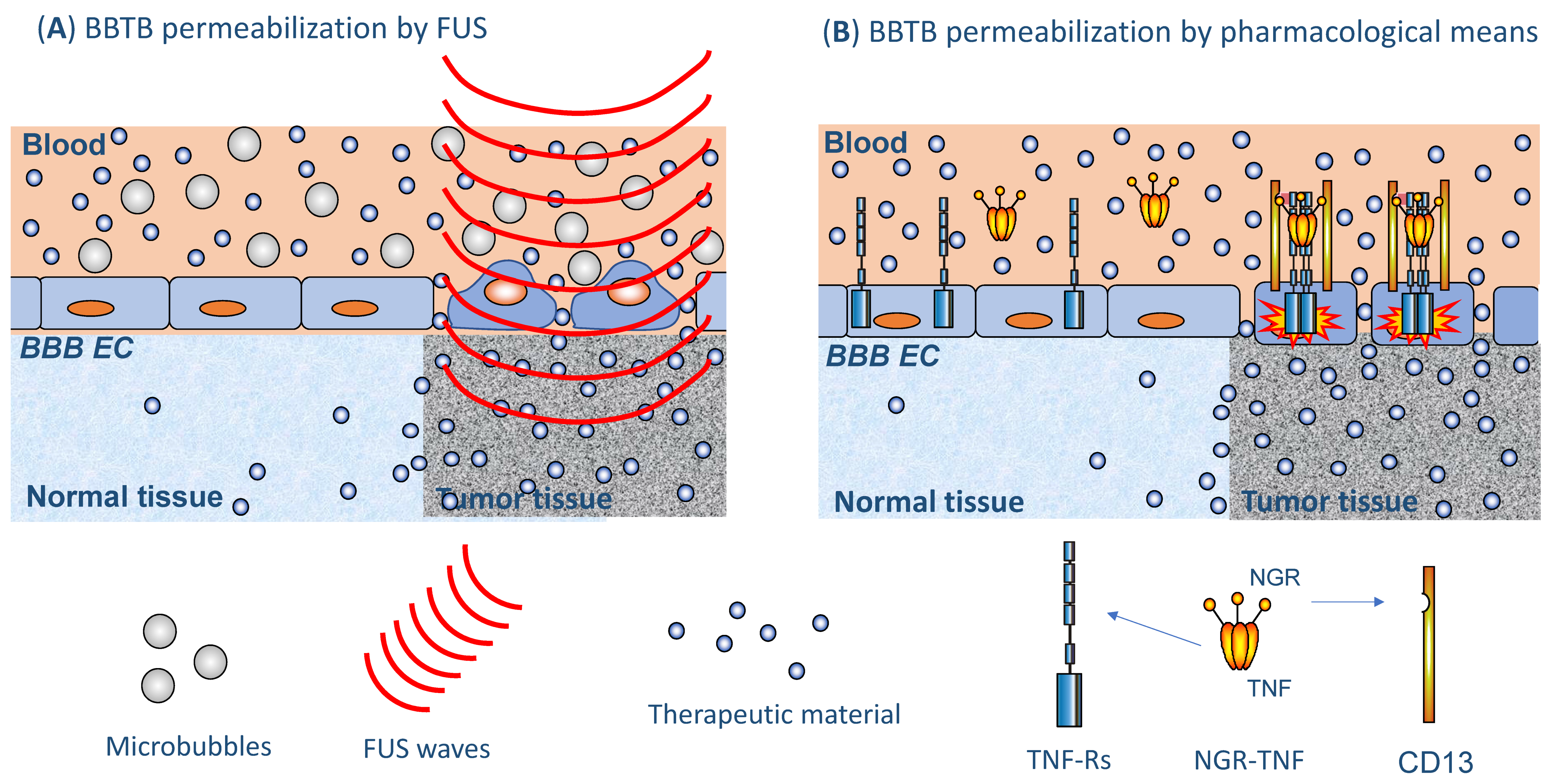
3.2.2. Ultrasound/Focused Ultrasound

Low-intensity pulsed ultrasound (US) can be directed to discrete areas of the brain by different means and is, therefore, referred to as focused US (FUS) [7,46]. This technique allows targeting and destroying desired cell clusters by thermocoagulation while sparing adjacent cells and tissues. FUS is combined with intravenous injection of microbubbles (1–10 μm diameter), which consist of lipid or albumin shells that encapsulate gaseous material [7] (Figure 2A). In the selected area of the brain, the microbubbles vibrate in response to the US waves, leading to the transient (4–6 h) disruption and increased permeability of the BBTB [47]. There are different classes of commercially available microbubbles but none appears to be clearly superior to another [48]. Furthermore, there are different FUS devices for BBTB breaching that are being investigated in clinical trials. A first approach foresees the implantation of a transducing device in the skull (SonoCloud®, CarThera) in order to minimize a major problem of US technology in BBTB opening; i.e., the thickness of the skull bone in humans, which distorts and absorbs much US energy. SonoCloud is, in fact, an unfocused, low-intensity, pulsed US system that allows for diffuse, transient opening of the BBTB. It is actually the site of implantation of the device and the sonicated area that allow it be considered as a focused approach. The device can be activated repeatedly allowing the repetitive administration of drugs into the desired area of the brain. It has been shown to be safe and afford efficient, transient opening of the BBB in nonhuman primates over a 3-month period without behavioral, immunological or neurological consequences [49,50]. SonoCloud followed by intravenous carboplatin injection has been tested in 21 patients with recurrent glioblastoma enrolled in a phase I/II trial; the BBTB has been disrupted monthly with pulsed US in combination with microbubbles, with transient and manageable AEs [51]. No carboplatin-related neurotoxicity was observed. Patients with no or poor BBTB disruption visible on magnetic resonance imaging (MRI) had a median PFS of 3 months and a median overall survival (OS) of 9 months. Interestingly, patients with clear BBTB disruption had a median PFS of 4 months and a median OS of 13 months [52]. These latter results compare favorably with historical data. In order to improve these outcomes, the authors intend to develop a device with a larger sonication area allowing to extend drug delivery to the largest possible tumor region. SonoCloud is being investigated in several other clinical trials for BBTB opening (Table 2). The results obtained with this device appear promising, but on the downside is the need to implant it in the skull of the patients—an apparently well-tolerated but nevertheless invasive procedure.
A second approach foresees the use of an extracorporeal fixed MRI-guided US-emitting device (ExAblate®, InSightec). The device is embedded within a diagnostic MRI scanner, which guides and monitors the USs to the desired area of the brain [53,54]. ExAblate® has been investigated successfully for several clinical indications, beyond BBTB disruption. It has been successfully tested in several animal models and the results of two clinical studies published on this approach, performed in patients with non-neoplastic CNS diseases, have shown that the BBB was safely, reversibly and repeatedly opened in all patients [55,56]. No AEs have been detected post-sonication and patients have tolerated up to 17 FUS treatments. BBB opening with an average of 95% of the targeted FUS volume has been achieved and this has been followed by BBB closure within 24 h. The safety and feasibility of this procedure in patients with brain tumors are currently addressed in several clinical trials (Table 2), whereas efficacy studies are not yet ongoing.
Interestingly, some clinical studies, not reported in Table 2 because they are beyond of the scope of the article, describe the use of FUS to induce direct antitumor effects on brain tumors independently of its effects on the BBTB. This is not surprising since ExAblate® is being investigated for direct antitumor effects on many extracranial tumors and has received FDA approval for the treatment of uterine fibroids [57]. This suggests the possibility of using FUS both for BBTB opening as well as for inducing direct antitumor effects, most likely by tailoring key parameters such as frequency and acoustic pressure for each of the two objectives, which could be pursued contemporarily.
A third FUS-based approach uses a frameless neuronavigation-guided device (NaviFUS®), which generates FUS treating units for covering the desired brain volume (e.g., tumor volume). The location of the treating units is determined using software from CT/MRI images of the patient that are collected before treatment. Guidance of FUS energy to the selected treatment area is achieved by means of a high-precision neuro-navigation tracking system [58]. The NaviFUS® system has been investigated in one clinical trial for BBTB opening [59].
Overall, US-based approaches seem of considerable promise. They are less invasive than CED and osmotic BBTB disruption, exhibit the possibility of precisely targeting discrete regions of the brain and appear fully reversible [49,52,60,61,62,63]. While the safety of these approaches looks very promising, more studies are required to evaluate the risks associated with repeated treatments, with or without delivery of a therapeutic agent [63]. Based on these encouraging features, results of ongoing clinical studies (Table 2) are expected to promote this strategy to more advanced levels.

3.2.3. Increasing BBTB Permeability by Pharmacological Means

Historically, this is one of the earliest approaches that have been pursued to increase permeability of the BBTB. In particular, bradykinin and adenosine agonists have been extensively studied for this purpose and some of these compounds have progressed into clinical trials. This is the case of the bradykinin B2 receptor agonist cereport (RMP-7, lobradimil) [64], which has been used to transiently open the BBTB to different chemotherapeutics. After initial encouraging results [65,66], a phase II, randomized, controlled trial has shown that the addition of cereport (300 ng/kg) had no effect on the pharmacokinetics, toxicity or efficacy of carboplatin in patients with recurrent glioblastoma [67]. Subsequent pharmacokinetic studies suggested that higher doses of cereport may be required to increase carboplatin delivery to the tumor. However, a single-arm phase II trial failed to demonstrate any antitumor activity of the combination of 600 ng/kg of cereport and carboplatin in 38 children with varied brain tumors [68], and no further clinical trials with cereport in brain tumors have been reported.
Regadenoson (CVT-3146, Lexiscan) is an adenosine 2A receptor agonist that increased the BBB permeability in murine models in a dose-dependent and time-limited manner [69]. Clinical studies have failed to demonstrate the capacity of regadenoson (400 μg) to increase permeability of the integral BBB [70]. Likewise, a study aimed at enhancing regadenoson-induced increase of intratumoral temozolomide concentrations in glioblastoma patients yielded negative results [71].
The most likely explanation for the negative results obtained with these compounds in the clinics is their lack of specificity for the BBTB endothelium and their short biological half-life (e.g., 2–3 min for regadenoson). Increasing the dose administered may eventually yield the desired effect but at the possible expense of an undesirable, generalized increase of endothelial permeability. Despite these concerns, a clinical dose-finding study (from 50 to 1400 μg) has been scheduled to find the optimal regadenoson dose to increase BBB permeability (NCT03971734).
Tumor necrosis factor-alpha (TNF) is an inflammatory cytokine that has the potential to enhance the BBTB permeability. Indeed, preclinical studies on murine models of brain metastases have shown that systemic administration of TNF can permeabilize the BBTB to the anti-human epidermal growth factor receptor (HER) 2 mAb trastuzumab at tumor sites [72]. However, systemic administration of TNF is precluded because of its prohibitive systemic toxicity [73]. This may be bypassed by fusing TNF through its N-terminus to CNGRCG, a tumor vasculature-homing peptide that recognizes an isoform of membrane-bound aminopeptidase N (CD13) that is upregulated in the neovasculature of many solid tumors [74,75], including the vasculature of glioblastoma and primary CNS lymphoma [75,76,77]. The tumor vasculature-homing properties of the CNGRCG-TNF fusion protein (called NGR-TNF) allows the targeted delivery of extremely low, yet pharmacologically active doses of TNF to tumor blood vessels, thereby avoiding systemic toxicity (Figure 2B). Studies in various murine models of solid tumors have shown that picogram doses of NGR-TNF are sufficient to target the tumor vasculature, increase barrier permeability and, consequently, enhance drug penetration and efficacy [75,78,79]. On these bases, low-dose NGR-TNF has been used, alone or in combination with various chemotherapeutic agents, in patients with different types of solid tumors, with evidence of therapeutic efficacy and an excellent safety profile [73,80]. Relevant to the present context are the results of a phase II trial on 28 patients with relapsed or refractory primary CNS lymphoma, showing that NGR-TNF could alter the BBTB in a transient and reversible manner and increase the efficacy of R-CHOP (rituximab, cyclophosphamide, doxorubicin, vincristine and prednisone), a chemotherapy regimen consisting of drugs that very poorly cross the BBTB [76,77]. NGR-TNF-induced alteration of the BBTB was no longer detectable before the subsequent R-CHOP cycle three weeks later, thereby demonstrating the reversibility of its effects on the BBTB permeability. The effect of NGR-TNF on tumor vasculature permeability, which has been demonstrated by dynamic contrast-enhanced (DCE)-MRI and single-photon emission CT (SPECT) with diethylenetriaminepentaacetic acid tagged with 99mTc (99mTc-DTPA-SPECT), has been more evident in lymphomatous and peritumoral areas [76]. Moreover, CD13, the target of the NGR domain of this conjugate, has been found to be expressed by endothelial cells and pericytes of the brain tumor vasculature [76,77]. NGR-TNF has not impaired R-CHOP tolerability and no patient has interrupted treatment or needed dose reduction because of toxicity. The primary endpoint of the trial has been achieved, with an ORR of 75% and a median disease-free survival of 11 months (range 6–25+) [77]. These encouraging results contrast with limited benefit achieved with regadenoson and cereport, which may be explained, at least in part, by the widespread distribution of receptors to the latter drugs throughout the endothelial lining of the organism, whereas CD13 expression is more restricted to angiogenic/inflamed endothelia, including tumor blood vessels. While the use of this targeted version of TNF has yielded very positive results, a note of caution regarding the use of TNF in the CNS should be sounded in consideration of some recent results showing the negative effects of TNF on neurogenesis [81]. It is doubtful, however, that this may represent a major concern for NGR-TNF, which is delivered at very low doses to the brain tumor vasculature rather than to the neuronal components of the CNS. Furthermore, after a median follow-up of 2-years, no signs of neurotoxicity were observed in patients treated with NGR-TNF [77].

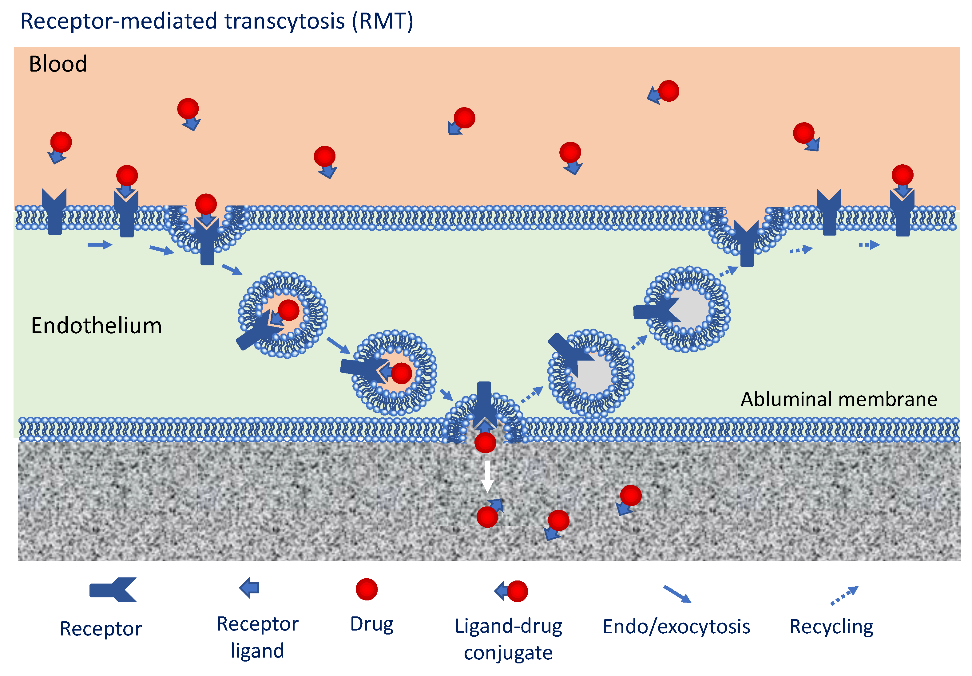
3.3. Approaches to Overcome the BBTB Exploiting Intrinsic Transport Mechanisms

Enhancing drug delivery to the brain taking advantage of normal transport mechanisms through the BBTB is another approach that has been pursued. For this purpose, drugs are conjugated to molecules that use these transport mechanisms, without altering, even temporarily, the BBTB. Several routes have been investigated for this purpose, in particular adsorptive-mediated transcytosis (AMT), solute carrier-mediated transcytosis (SCMT) and receptor-mediated transcytosis (RMT). AMT is triggered by an electrostatic interaction between a cationic ligand and anionic proteoglycans expressed on the surface of the BBTB endothelium [7]. Cationic proteins [82] or cell-penetrating peptides [83,84] can be delivered with this approach. However, the main drawback of this approach is related to its inherent nonspecificity, owing to the fact target molecules are present also in normal vascular endothelia, and not exclusively in the BBTB. SCMT exploits the function of solute carriers as bidirectional transporters that carry their substrates down their concentration gradient from the luminal side to the abluminal side of the endothelial lining and vice versa [85]. Glucose transporters, large neutral amino acid transporters, organic anion-transporting polypeptides are examples of solute carriers that have been investigated to facilitate BBTB crossing by drugs [86,87,88]. AMT and SCMT are far away from being used in clinical neuro-oncology. Conversely, RMT is certainly the most deeply investigated and, theoretically, the most sophisticated route for shuttling drugs or candidate drugs into the brain. In RMT, a ligand is transported across the BBTB upon interaction with its specific receptor (Figure 3). This transport mechanism is not restricted by the size of the ligand and is suitable also for large molecules such as mAbs [89]. However, its transport capacity may be constrained by the number of receptors expressed per endothelial cell and this may not be balanced by the capacity of these receptors to recycle. The initial step occurring during RMT is a ligand-receptor interaction on the luminal side of the BBTB (Figure 3). This is followed by endocytosis of the ligand–receptor complex, endosome-mediated transport to the abluminal side of the BBTB and eventual exocytosis of the ligand [89]. Examples of receptors of this category are the insulin receptor [90], the transferrin receptor (TfR) [91], the low-density lipoprotein receptor (LDLR) [92], the diphtheria toxin receptor [93] and the nicotinic acetylcholine receptor [94]. The TfR has been intensively investigated because it is not only expressed on the endothelial cells of the BBTB but is also overexpressed on tumor cells, whether primary or metastatic tumors [95].
So far, results from clinical studies using the RMT route had disappointed the expectations, but, in recent times, two drug conjugates have progressed into clinical trials. The first uses Angiopep-2 as the RMT ligand. This is a 19-amino acid peptide that traverses the capillary endothelium of the BBTB upon interaction with the LDLR-related protein 1 (LRP1) [96]. LRP1 is expressed in a wide range of tissues and in multiple tumors [97]. Conditions of cellular stress, such as hypoxia and acidosis, upregulate its expression [97]. Functionally, LRP1 acts as a multifunctional scavenger receptor and contributes to the malignant phenotype of tumor cells [98]. ANG1005 (also termed GRN1005) comprises Angiopep-2 conjugated via cleavable ester bonds to three molecules of paclitaxel [99]. Systemically administered ANG1005 penetrated the brain parenchyma more rapidly and to a greater extent than unconjugated paclitaxel [99] and localized to orthotopic gliomas in mice expressing high levels of LRP1 [97]. In a phase I study, 36 patients with recurrent malignant glioma have received ANG1005 every 3 weeks [100]. Therapy was well tolerated with mucositis and neutropenia as DLTs. One CR, two partial responses (PR) and 8 stable diseases (SD), which lasted a median of 51 days, were recorded. Very recently, results of an open-label phase II study in adult patients with brain metastases from both HER2-positive or HER2-negative breast cancer, with or without leptomeningeal carcinomatosis, have been reported [101]. ANG1005 was administered intravenously at 600 mg/m2 every 3 weeks. Patient benefit (SD or better) was seen in intracranial and extracranial disease in, respectively, 77% and 86% of the evaluated patients, with an intracranial ORR of 15%. This result has not met the preset rule, but, when compared to historical control, symptom and OS improvement has been seen in patients with leptomeningeal carcinomatosis. ANG1005 is now investigated in phase III trials in a similar patient population with brain metastases from HER2-negative breast cancer (Table 3). Three other Angiopep-2-based conjugates for cancer therapy have been generated: ANG1007, carrying doxorubicin; ANG1009, carrying etoposide [102]; and ANG4043, carrying an anti-HER2 mAb [103]. ANG1007 and ANG4043 are now in preclinical development (company website inspected as of 2021/03/25).
A TfR-targeted construct designed to cross the BBTB is SGT-53, a cationic liposome formulation encapsulating a plasmid with the human tumor suppressor gene TP53. TfR is targeted by a single chain mAb fragment [104]. This nanoparticulate was shown to cross the BBTB and target intracranial glioblastoma xenografts. The combination of SGT-53 and temozolomide has limited development of resistance to this alkylating drug and had a prolonged antitumor effect in a model of temozolomide-resistant glioblastoma [105,106]. Results of a phase II study (NCT02340156) addressing the combination of SGT-53 and temozolomide in patients with recurrent glioblastoma are pending (Table 3). Interestingly, SGT-53 seems to boost antitumor immunity and sensitize glioblastoma to anti-PD-1 (anti-programmed cell death protein 1) therapy by upregulating PD-L1 expression [107], while reducing immune-related adverse events [108].
As a promising approach, a platform technology referred to as a BBB transport vehicle, consisting of a human IgG1 Fc engineered to bind to RMT targets expressed on the surface of brain endothelial cells, was recently developed [109,110]. TfR has been used as a BBB receptor target for proof-of-concept work to successfully deliver iduronate 2-sulfatase, the lysosomal enzyme deficient in mucopolysaccharidosis type II, to the brain in a preclinical model. The use of this ligand could likely be extended to primary or secondary brain tumors upon conjugation to antitumor drugs.

4. Conclusions

Overall, the last few years have represented a turning point in the development of efficacious approaches for traversing the BBTB and facilitating drug delivery to brain tumors. It seems likely that, in the near future, the more invasive approaches (e.g., CED, SACI, etc.) will be replaced by more patient-friendly and efficacious means. Enhancers of BBTB permeability, such as the US-based approaches or NGR-TNF, or refined approaches exploiting RMT for drug delivery have yielded very promising results in preclinical studies and, in some cases, also in early-stage clinical studies in patients with brain tumors. It is tempting to speculate that the combination of some of these approaches may give rise to even better additive or synergistic therapeutic effects.

Author Contributions

F.M., A.C. and A.J.M.F. performed literature research, data interpretation, clinical trials analysis, figures, and wrote and approved the submitted manuscript. All authors have read and agreed to the published version of the manuscript.

Funding

This research was funded by Associazione Italiana per la Ricerca sul Cancro (AIRC) under IG 2019–ID. 23470 project–P.I. Angelo Corti.

Institutional Review Board Statement

Not applicable.

Informed Consent Statement

Not applicable.

Data Availability Statement

Published studies were identified through a search of PubMed using the reported keywords alone or combined. No filters have been added regarding publication dates, article types (i.e., review, meta-analysis, clinical trial) and text availability (abstract or full text). However, the reviewed literature was limited to studies published in the English language in peer-reviewed, international journals.

Conflicts of Interest

A.C. and A.J.M.F. are inventors of patents on the use of NGR-TNF and S-NGR-TNF in patients with relapsed/refractory primary CNS lymphoma and other brain tumors (Patent numbers 19209484.5 and 20164689.0).

References

  1. Obermeier, B.; Daneman, R.; Ransohoff, R.M. Development, maintenance and disruption of the blood-brain barrier. Nat. Med. 2013, 19, 1584–1596. [Google Scholar] [CrossRef] [PubMed]
  2. Zhao, Z.; Nelson, A.R.; Betsholtz, C.; Zlokovic, B.V. Establishment and dysfunction of the blood-brain barrier. Cell 2015, 163, 1064–1078. [Google Scholar] [CrossRef] [PubMed] [Green Version]
  3. Daneman, R.; Prat, A. The blood-brain barrier. Cold Spring Harb. Perspect. Biol. 2015, 7, a020412. [Google Scholar] [CrossRef] [PubMed] [Green Version]
  4. Arvanitis, C.D.; Ferraro, G.B.; Jain, R.K. The blood-brain barrier and blood-tumour barrier in brain tumours and metastases. Nat. Rev. Cancer 2020, 20, 26–41. [Google Scholar] [CrossRef] [PubMed]
  5. Vanlandewijck, M.; He, L.; Mäe, M.A.; Andrae, J.; Ando, K.; Del Gaudio, F.; Nahar, K.; Lebouvier, T.; Laviña, B.; Gouveia, L. A molecular atlas of cell types and zonation in the brain vasculature. Nature 2018, 554, 475–480. [Google Scholar] [CrossRef] [Green Version]
  6. Saunders, A.; Macosko, E.Z.; Wysoker, A.; Goldman, M.; Krienen, F.M.; de Rivera, H.; Bien, E.; Baum, M.; Bortolin, L.; Wang, S. Molecular diversity and specializations among the cells of the adult mouse brain. Cell 2018, 174, 1015–1030. [Google Scholar] [CrossRef] [Green Version]
  7. Abdul Razzak, R.; Florence, G.J.; Gunn-Moore, F.J. Approaches to CNS drug delivery with a focus on transporter-mediated transcytosis. Int. J. Mol. Sci. 2019, 20, 3108. [Google Scholar] [CrossRef] [Green Version]
  8. Loscher, W.; Potschka, H. Blood–brain barrier active efflux transporters: ATP-binding cassette gene family. NeuroRx 2005, 2, 86–98. [Google Scholar] [CrossRef]
  9. Mittapalli, R.K.; Manda, V.K.; Adkins, C.E.; Geldenhuys, W.J.; Lockman, P.R. Exploiting nutrient transporters at the blood–brain barrier to improve brain distribution of small molecules. Ther. Deliv. 2010, 1, 775–784. [Google Scholar] [CrossRef] [PubMed]
  10. Jain, R.K.; di Tomaso, E.; Duda, D.G.; Loeffler, J.S.; Sorensen, A.G.; Batchelor, T.T. Angiogenesis in brain tumours. Nat. Rev. Neurosci. 2007, 8, 610–622. [Google Scholar] [CrossRef]
  11. Sowers, J.L.; Joohnson, K.M.; Conrad, C.; Patterson, J.T.; Sowers, L.C. The role of inflammation in brain cancer. Adv. Exp. Med. Biol. 2014, 816, 75–105. [Google Scholar]
  12. Seano, G.; Nia, H.T.; Emblem, K.E.; Datta, M.; Ren, J.; Krishnan, S.; Kloepper, J.; Pinho, M.C.; Ho, W.W.; Ghosh, M.; et al. Solid stress in brain tumours causes neuronal loss and neurological dysfunction and can be reversed by lithium. Nat. Biomed. Eng. 2019, 3, 230. [Google Scholar] [CrossRef] [PubMed]
  13. Carmeliet, P.; Jain, R.K. Molecular mechanisms and clinical applications of angiogenesis. Nature 2011, 473, 298–307. [Google Scholar] [CrossRef] [PubMed] [Green Version]
  14. Lockman, P.R.; Mittapalli, R.K.; Taskar, K.S.; Rudraraju, V.; Gril, B.; Bohn, K.A.; Adkins, C.E.; Roberts, A.; Thorsheim, H.R.; Gaasch, J.A.; et al. Heterogeneous blood-tumor barrier permeability determines drug efficacy in experimental brain metastases of breast cancer. Clin. Cancer Res. 2010, 16, 5664–5678. [Google Scholar] [CrossRef] [PubMed] [Green Version]
  15. Marcucci, F.; Corti, A. How to improve exposure of tumor cells to drugs–Promoter drugs increase tumor uptake and penetration of effector drugs. Adv. Drug. Deliv. Rev. 2012, 64, 53–68. [Google Scholar] [CrossRef]
  16. Marcucci, F.; Corti, A. Improving drug penetration to curb tumor drug resistance. Drug Discov. Tod. 2012, 17, 1139–1147. [Google Scholar] [CrossRef] [PubMed]
  17. Choi, J.Y.; Kang, H.J.; Hong, K.T.; Hong, C.R.; Lee, Y.J.; Park, J.D.; Phi, J.H.; Kim, S.K.; Wang, K.C.; Kim, I.H.; et al. Tandem high-dose chemotherapy with topotecan-thiotepa-carboplatin and melphalan-etoposide-carboplatin regimens for pediatric high-risk brain tumors. Int. J. Clin. Oncol. 2019, 24, 1515–1525. [Google Scholar] [CrossRef]
  18. Ferreri, A.J.; Cwynarski, K.; Pulczynski, E.; Ponzoni, M.; Deckert, M.; Politi, L.S.; Torri, V.; Fox, C.P.; Rosée, P.L.; Schorb, E.; et al. Chemoimmunotherapy with methotrexate, cytarabine, thiotepa, and rituximab (MATRix regimen) in patients with primary CNS lymphoma: Results of the first randomisation of the International Extranodal Lymphoma Study Group-32 (IELSG32) phase 2 trial. Lancet Haematol. 2016, 3, e217–e227. [Google Scholar] [CrossRef] [Green Version]
  19. Xia, Y.; Xu, F.; Xiong, M.; Yang, H.; Lin, W.; Xie, Y.; Xi, H.; Xue, Q.; Ye, T.; Yu, L. Repurposing of antipsychotic trifluoperazine for treating brain metastasis, lung metastasis and bone metastasis of melanoma by disrupting autophagy flux. Pharmacol. Res. 2021, 163, 105295. [Google Scholar] [CrossRef]
  20. Orthmann, A.; Peiker, L.; Fichtner, I.; Hoffmann, A.; Hilger, R.A.; Zeisig, R. Improved treatment of MT-3 breast cancer and brain metastases in a mouse xenograft by LRP-targeted oxaliplatin liposomes. J. Biomed. Nanotechnol. 2016, 12, 56–68. [Google Scholar] [CrossRef]
  21. Kobus, T.; Zervantonakis, I.K.; Zhang, Y.; McDannold, N.J. Growth inhibition in a brain metastasis model by antibody delivery using focused ultrasound-mediated blood-brain barrier disruption. J. Control. Release 2016, 238, 281–288. [Google Scholar] [CrossRef] [Green Version]
  22. Perry, J.; Chambers, A.; Spithoff, K.; Laperriere, N. Gliadel wafers in the treatment of malignant glioma: A systematic review. Curr. Oncol. 2007, 14, 189–194. [Google Scholar] [CrossRef] [Green Version]
  23. Brem, H.; Piantadosi, S.; Burger, P.C.; Walker, M.; Selker, R.; Vick, N.A.; Black, K.; Sisti, M.; Brem, S.; Mohr, G.; et al. Placebo-controlled trial of safety and efficacy of intraoperative controlled delivery by biodegradable polymers of chemotherapy for recurrent gliomas. The Polymer-brain Tumor Treatment Group. Lancet 1995, 345, 1008–1012. [Google Scholar] [CrossRef]
  24. Westphal, M.; Hilt, D.C.; Bortey, E.; Delavault, P.; Olivares, R.; Warnke, P.C.; Whittle, I.R.; Jääskeläinen, J.; Ram, Z. A phase 3 trial of local chemotherapy with biodegradable carmustine (BCNU) wafers (Gliadel wafers) in patients with primary malignant glioma. Neuro Oncol. 2003, 5, 79–88. [Google Scholar] [CrossRef] [PubMed]
  25. Pallud, J.; Audureau, E.; Noel, G.; Corns, R.; Lechapt-Zalcman, E.; Duntze, J.; Pavlov, V.; Guyotat, J.; Hieu, P.D.; Le Reste, P.J.; et al. Long-term results of carmustine wafer implantation for newly diagnosed glioblastomas: A controlled propensity-matched analysis of a French multicenter cohort. Neuro Oncol. 2015, 17, 1609–1619. [Google Scholar] [CrossRef] [PubMed]
  26. Kadota, T.; Saito, R.; Kumabe, T.; Mizusawa, J.; Katayama, H.; Sumi, M.; Igaki, H.; Kinoshita, M.; Komori, T.; Ichimura, K. A multicenter randomized phase III study for newly diagnosed maximally resected glioblastoma comparing carmustine wafer implantation followed by chemoradiotherapy with temozolomide with chemoradiotherapy alone; Japan Clinical Oncology Group Study JCOG1703 (MACS study). Jpn. J. Clin. Oncol. 2019, 49, 1172–1175. [Google Scholar] [PubMed]
  27. Lee, J.; Cho, H.R.; Cha, G.D.; Seo, H.; Lee, S.; Park, C.K.; Kim, J.W.; Qiao, S.; Wang, L.; Kang, D. Flexible, sticky, and biodegradable wireless device for drug delivery to brain tumors. Nat. Commun. 2019, 10, 5205. [Google Scholar] [CrossRef]
  28. Bobo, R.H.; Laske, D.W.; Akbasak, A.; Morrison, P.F.; Dedrick, R.L.; Oldfield, E.H. Convection-enhanced delivery of macromolecules in the brain. Proc. Natl. Acad. Sci. USA 1994, 91, 2076–2080. [Google Scholar] [CrossRef] [Green Version]
  29. Souweidane, M.M.; Kramer, K.; Pandit-Taskar, N.; Zhou, Z.; Haque, S.; Zanzonico, P.; Carrasquillo, J.A.; Lyashchenko, S.K.; Thakur, S.B.; Donzelli, M.; et al. Convection-enhanced delivery for diffuse intrinsic pontine glioma: A single-centre, dose-escalation, phase 1 trial. Lancet Oncol. 2018, 19, 1040–1050. [Google Scholar] [CrossRef]
  30. Saito, R.; Kanamori, M.; Sonoda, Y.; Yamashita, Y.; Nagamatsu, K.; Murata, T.; Mugikura, S.; Kumabe, T.; Wembacher-Schröder, E.; Thomson, R.; et al. Phase I trial of convection-enhanced delivery of nimustine hydrochloride (ACNU) for brainstem recurrent glioma. Neurooncol. Adv. 2020, 2, vdaa033. [Google Scholar] [CrossRef] [PubMed]
  31. Sampson, J.H.; Akabani, G.; Archer, G.E.; Bigner, D.D.; Berger, M.S.; Friedman, A.H.; Friedman, H.S.; Herndon, J.E., 2nd; Kunwar, S.; Marcus, S.; et al. Progress report of a Phase I study of the intracerebral microinfusion of a recombinant chimeric protein composed of transforming growth factor (TGF)-alpha and a mutated form of the Pseudomonas exotoxin termed PE-38 (TP-38) for the treatment of malignant brain tumors. J. Neurooncol. 2003, 65, 27–35. [Google Scholar]
  32. Weber, F.W.; Floeth, F.; Asher, A.; Bucholz, R.; Berger, M.; Prados, M.; Chang, S.; Bruce, J.; Hall, W.; Rainov, N.G.; et al. Local convection enhanced delivery of IL4-Pseudomonas exotoxin (NBI-3001) for treatment of patients with recurrent malignant glioma. Acta Neurochir. Suppl. 2003, 88, 93–103. [Google Scholar]
  33. Heiss, J.D.; Jamshidi, A.; Shah, S.; Martin, S.; Wolters, P.L.; Argersinger, D.P.; Warren, K.E.; Lonser, R.R. Phase I trial of convection-enhanced delivery of IL13-Pseudomonas toxin in children with diffuse intrinsic pontine glioma. J. Neurosurg. Pediatr. 2018, 23, 333–342. [Google Scholar] [CrossRef]
  34. Kunwar, S.; Chang, S.; Westphal, M.; Vogelbaum, M.; Sampson, J.; Barnett, G.; Shaffrey, M.; Ram, Z.; Piepmeier, J.; Prados, M.; et al. Phase III randomized trial of CED of IL13-PE38QQR vs Gliadel wafers for recurrent glioblastoma. Neuro Oncol. 2010, 12, 871–881. [Google Scholar] [CrossRef] [PubMed] [Green Version]
  35. Desjardins, A.; Gromeier, M.; Herndon, J.E., 2nd; Beaubier, N.; Bolognesi, D.P.; Friedman, A.H.; Friedman, H.S.; McSherry, F.; Muscat, A.M.; Nair, S.; et al. Recurrent glioblastoma treated with recombinant poliovirus. N. Engl. J. Med. 2018, 379, 150–161. [Google Scholar] [CrossRef]
  36. Lidar, Z.; Mardor, Y.; Jonas, T.; Pfeffer, R.; Faibel, M.; Nass, D.; Hadani, M.; Ram, Z. Convection-enhanced delivery of paclitaxel for the treatment of recurrent malignant glioma: A phase I/II clinical study. J. Neurosurg. 2004, 100, 472–479. [Google Scholar] [CrossRef]
  37. Tanner, P.G.; Holtmannspötter, M.; Tonn, J.C.; Goldbrunner, R. Effects of drug efflux on convection-enhanced paclitaxel delivery to malignant gliomas: Technical note. Neurosurgery 2007, 61, E880–E882. [Google Scholar] [CrossRef]
  38. Sampson, J.H.; Akabani, G.; Archer, G.E.; Berger, M.S.; Coleman, R.E.; Friedman, A.H.; Friedman, H.S.; Greer, K.; Herndon, J.E., 2nd; Kunwar, S.; et al. Intracerebral infusion of an EGFR-targeted toxin in recurrent malignant tumors. Neuro Oncol. 2008, 10, 320–329. [Google Scholar] [CrossRef] [Green Version]
  39. Choi, C.; Kim, H.M.; Shon, J.; Park, J.; Kim, H.T.; Oh, S.H.; Kim, N.K.; Kim, O.J. Additional increased effects of mannitol-temozolomide combined treatment on blood-brain barrier permeability. Biochem. Biophys. Res. Commun. 2018, 497, 769–775. [Google Scholar] [CrossRef]
  40. Bhattacharjee, A.K.; Nagashima, T.; Kondoh, T.; Tamaki, N. The effects of the Na(+)/Ca(++) exchange blocker on osmotic blood-brain barrier disruption. Brain Res. 2001, 900, 157–162. [Google Scholar] [CrossRef]
  41. Rodriguez, A.; Tatter, S.B.; Debinski, W. Neurosurgical techniques for disruption of the blood–brain barrier for glioblastoma treatment. Pharmaceutics 2015, 7, 175–187. [Google Scholar] [CrossRef] [Green Version]
  42. Chakraborty, S.; Filippi, C.G.; Wong, T.; Ray, A.; Fralin, S.; Tsiouris, A.J.; Praminick, B.; Demopoulos, A.; McCrea, H.J.; Bodhinayake, I.; et al. Superselective intraarterial cerebral infusion of cetuximab after osmotic blood/brain barrier disruption for recurrent malignant glioma: Phase I study. J. Neurooncol. 2016, 128, 405–415. [Google Scholar] [CrossRef]
  43. Chakraborty, S.; Filippi, C.G.; Wong, T.; Ray, A.; Fralin, S.; Tsiouris, A.J.; Praminick, B.; Demopoulos, A.; McCrea, H.J.; Bodhinayake, I.; et al. Safety and maximum tolerated dose of superselective intraarterial cerebral infusion of bevacizumab after osmotic blood-brain barrier disruption for recurrent malignant glioma. J. Neurosurg. 2011, 114, 624–632. [Google Scholar]
  44. Chakraborty, S.; Filippi, C.G.; Burkhardt, J.K.; Fralin, S.; Ray, A.; Wong, T.; Ortiz, R.; Langer, D.J.; Boockvar, J.A. Durability of single dose intra-arterial bevacizumab after blood/brain barrier disruption for recurrent glioblastoma. J. Exp. Ther. Oncol. 2016, 11, 261–267. [Google Scholar]
  45. Angelov, L.; Doolittle, N.D.; Kraemer, D.F.; Siegal, T.; Barnett, G.H.; Peereboom, D.M.; Stevens, G.; McGregor, J.; Jahnke, K.; Lacy, C.A.; et al. Blood-brain barrier disruption and intra-arterial methotrexate-based therapy for newly diagnosed primary CNS lymphoma: A multi-institutional experience. J. Clin. Oncol. 2009, 27, 3503–3509. [Google Scholar] [CrossRef] [PubMed] [Green Version]
  46. Beccaria, K.; Canney, M.; Bouchoux, G.; Desseaux, C.; Grill, J.; Heimberger, A.B.; Carpentier, A. Ultrasound-induced blood-brain barrier disruption for the treatment of gliomas and other primary CNS tumors. Cancer Lett. 2020, 479, 13–22. [Google Scholar] [CrossRef]
  47. Parodi, A.; Rudzińska, M.; Deviatkin, A.A.; Soond, S.M.; Baldin, A.V.; Zamyatnin, A.A., Jr. Established and emerging strategies for drug delivery across the blood-brain barrier in brain cancer. Pharmaceutics 2019, 11, E245. [Google Scholar] [CrossRef] [PubMed] [Green Version]
  48. Wu, S.K.; Chu, P.C.; Chai, W.Y.; Kang, S.T.; Tsai, C.H.; Fan, C.H.; Yeh, C.K.; Liu, H.L. Characterization of different microbubbles in assisting focused ultrasound-induced blood-brain barrier opening. Sci. Rep. 2017, 7, 46689. [Google Scholar] [CrossRef] [PubMed] [Green Version]
  49. Horodyckid, C.; Canney, M.; Vignot, A.; Boisgard, R.; Drier, A.; Huberfeld, G.; François, C.; Prigent, A.; Santin, M.D.; Adam, C.; et al. Safe long-term repeated disruption of the blood-brain barrier using an implantable ultrasound device: A multiparametric study in primates. J. Neurosurg. 2017, 126, 1351–1361. [Google Scholar] [CrossRef] [Green Version]
  50. Goldwirt, L.; Canney, M.; Horodyckid, C.; Poupon, J.; Mourah, S.; Vignot, A.; Chapelon, J.Y.; Carpentier, A. Enhanced brain distribution of carboplatin in a primate model after blood–brain barrier disruption using an implantable ultrasound device. Cancer Chemother. Pharmacol. 2016, 77, 211–216. [Google Scholar] [CrossRef]
  51. Carpentier, A.; Canney, M.; Vignot, A.; Reina, V.; Beccaria, K.; Horodyckid, C.; Karachi, C.; Leclercq, D.; Lafon, C.; Chapelon, J.Y.; et al. Clinical trial of blood-brain barrier disruption by pulsed ultrasound. Sci. Transl. Med. 2016, 8, 343re2. [Google Scholar] [CrossRef] [PubMed]
  52. Idbaih, A.; Canney, M.; Belin, L.; Desseaux, C.; Vignot, A.; Bouchoux, G.; Asquier, N.; Law-Ye, B.; Leclercq, D.; Bissery, A.; et al. Safety and feasibility of repeated and transient blood-brain barrier disruption by pulsed ultrasound in patients with recurrent glioblastoma. Clin. Cancer Res. 2019, 25, 3793–3801. [Google Scholar] [CrossRef] [Green Version]
  53. Dick, E.A.; Gedroyc, W.M. ExAblate magnetic resonance-guided focused ultrasound system in multiple body applications. Expert Rev. Med. Devices 2010, 7, 589–597. [Google Scholar] [CrossRef] [PubMed]
  54. Cohen, Z.R.; Zaubermann, J.; Harnof, S.; Mardor, Y.; Nass, D.; Zadicario, E.; Hananel, A.; Castel, D.; Faibel, M.; Ram, Z. Magnetic resonance imaging-guided focused ultrasound for thermal ablation in the brain: A feasibility study in a swine model. Neurosurgery 2007, 60, 593–600. [Google Scholar] [CrossRef] [PubMed]
  55. Lipsman, N.; Meng, Y.; Bethune, A.J.; Huang, Y.; Lam, B.; Masellis, M.; Herrmann, N.; Heyn, C.; Aubert, I.; Boutet, A.; et al. Blood-brain barrier opening in Alzheimer’s disease using MR-guided focused ultrasound. Nat. Commun. 2018, 9, 2336. [Google Scholar] [CrossRef] [PubMed] [Green Version]
  56. Rezai, A.R.; Ranjan, M.; D’Haese, P.F.; Haut, M.W.; Carpenter, J.; Najib, U.; Mehta, R.I.; Chazen, J.L.; Zibly, Z.; Yates, J.R.; et al. Noninvasive hippocampal blood-brain barrier opening in Alzheimer’s disease with focused ultrasound. Proc. Natl. Acad. Sci. USA 2020, 117, 9180–9182. [Google Scholar] [CrossRef] [Green Version]
  57. Stewart, E.A.; Rabinovici, J.; Tempany, C.M.; Inbar, Y.; Regan, L.; Gostout, B.; Hesley, G.; Kim, H.S.; Hengst, S.; Gedroyc, W.M. Clinical outcomes of focused ultrasound surgery for the treatment of uterine fibroids. Fertil. Steril. 2006, 85, 22–29. [Google Scholar] [CrossRef] [PubMed]
  58. Chai, W.Y.; Chu, P.C.; Tsai, C.H.; Lin, C.Y.; Yang, H.W.; Lai, H.Y.; Liu, H.L. Image-guided focused-ultrasound CNS molecular delivery: An implementation via dynamic contrast-enhanced magnetic-resonance imaging. Sci. Rep. 2018, 8, 4151. [Google Scholar] [CrossRef] [PubMed] [Green Version]
  59. Chen, K.T.; Lin, Y.J.; Chai, W.Y.; Lin, C.J.; Chen, P.Y.; Huang, C.Y.; Kuo, J.S.; Liu, H.L.; Wei, K.C. Neuronavigation-guided focused ultrasound (NaviFUS) for transcranial blood-brain barrier opening in recurrent glioblastoma patients: Clinical trial protocol. Ann. Transl. Med. 2020, 8, 673. [Google Scholar] [CrossRef]
  60. Yang, Y.; Zhang, X.; Ye, D.; Laforest, R.; Williamson, J.; Liu, Y.; Chen, H. Cavitation dose painting for focused ultrasound-induced blood-brain barrier disruption. Sci. Rep. 2019, 9, 2840. [Google Scholar] [CrossRef] [Green Version]
  61. Gasca-Salas, C.; Fernández-Rodríguez, B.; Pineda-Pardo, J.A.; Rodríguez-Rojas, R.; Obeso, I.; Hernández-Fernández, F.; Del Álamo, M.; Mata, D.; Guida, P.; Ordás-Bandera, C.; et al. Blood-brain barrier opening with focused ultrasound in Parkinson’s disease dementia. Nat. Commun. 2021, 12, 779. [Google Scholar] [CrossRef]
  62. Conti, A.; Mériaux, S.; Larrat, B. About the Marty model of blood-brain barrier closure after its disruption using focused ultrasound. Phys. Med. Biol. 2019, 64, 14NT02. [Google Scholar] [CrossRef] [PubMed]
  63. McMahon, D.; Poon, C.; Hynynen, K. Evaluating the safety profile of focused ultrasound and microbubble-mediated treatments to increase blood-brain barrier permeability. Expert Opin. Drug Deliv. 2019, 16, 129–142. [Google Scholar] [CrossRef]
  64. Doctrow, S.R.; Abelleira, S.M.; Curry, L.A.; Heller-Harrison, R.; Kozarich, J.W.; Malfroy, B.; McCarroll, L.A.; Morgan, K.G.; Morrow, A.R.; Musso, G.F.; et al. The bradykinin analog RMP-7 increases extracellular free calcium levels in rat brain microvascular endothelial cells. J. Pharmacol. Exp. Ther. 1994, 271, 229–237. [Google Scholar] [PubMed]
  65. Gregor, A.; Lind, M.; Newman, H.; Grant, R.; Hadley, D.M.; Barton, T.; Osborn, C. Phase II studies of RMP-7 and carboplatin in the treatment of recurrent high grade glioma. RMP-7 European Study Group. J. Neurooncol. 1999, 44, 137–145. [Google Scholar] [CrossRef]
  66. Warren, K.E.; Patel, M.C.; Aikin, A.A.; Widemann, B.; Libucha, M.; Adamson, P.C.; Neuwirth, R.; Benziger, D.; O’Toole, T.; Ford, K.; et al. Phase I trial of lobradimil (RMP-7) and carboplatin in children with brain tumors. Cancer Chemother. Pharmacol. 2001, 48, 275–282. [Google Scholar] [CrossRef] [PubMed]
  67. Prados, M.D.; Schold, S.C., Jr.; Fine, H.A.; Jaeckle, K.; Hochberg, F.; Mechtler, L.; Fetell, M.R.; Phuphanich, S.; Feun, L.; Janus, T.J.; et al. A randomized, double-blind, placebo-controlled, phase 2 study of RMP-7 in combination with carboplatin administered intravenously for the treatment of recurrent malignant glioma. Neuro Oncol. 2003, 5, 96–103. [Google Scholar] [CrossRef]
  68. Warren, K.; Jakacki, R.; Widemann, B.; Aikin, A.; Libucha, M.; Packer, R.; Vezina, G.; Reaman, G.; Shaw, D.; Krailo, M.; et al. Phase II trial of intravenous lobradimil and carboplatin in childhood brain tumors: A report from the Children’s Oncology Group. Cancer Chemother. Pharmacol. 2006, 5, 343–347. [Google Scholar] [CrossRef] [PubMed]
  69. Carman, A.J.; Mills, J.H.; Krenz, A.; Kim, D.G.; Bynoe, M.S. Adenosine receptor signaling modulates permeability of the blood-brain barrier. J. Neurosci. 2011, 31, 13272–13280. [Google Scholar] [CrossRef]
  70. Jackson, S.; George, R.T.; Lodge, M.A.; Piotrowski, A.; Wahl, R.L.; Gujar, S.K.; Grossman, S.A. The effect of regadenoson on the integrity of the human blood-brain barrier, a pilot study. J. Neurooncol. 2017, 132, 513–519. [Google Scholar] [CrossRef]
  71. Jackson, S.; Weingart, J.; Nduom, E.K.; Harfi, T.T.; George, R.T.; McAreavey, D.; Ye, X.; Anders, N.M.; Peer, C.; Figg, W.D.; et al. The effect of an adenosine A2A agonist on intra-tumoral concentrations of temozolomide in patients with recurrent glioblastoma. Fluids Barriers CNS 2018, 15, 2. [Google Scholar] [CrossRef] [Green Version]
  72. Connell, J.J.; Chatain, G.; Cornelissen, B.; Vallis, K.A.; Hamilton, A.; Seymour, L.; Anthony, D.C.; Sibson, N.R. Selective permeabilization of the blood-brain barrier at sites of metastasis. J. Natl. Cancer Inst. 2013, 105, 1634–1643. [Google Scholar] [CrossRef] [PubMed] [Green Version]
  73. Corti, A.; Curnis, F.; Rossoni, G.; Marcucci, F.; Gregorc, V. Peptide-mediated targeting of cytokines to tumor vasculature: The NGR-hTNF example. BioDrugs 2013, 27, 591–603. [Google Scholar] [CrossRef] [Green Version]
  74. Curnis, F.; Sacchi, A.; Borgna, L.; Magni, F.; Gasparri, A.; Corti, A. Enhancement of tumor necrosis factor alpha antitumor immunotherapeutic properties by targeted delivery to aminopeptidase N (CD13). Nat. Biotechnol. 2000, 18, 1185–1190. [Google Scholar] [CrossRef]
  75. Di Matteo, P.; Arrigoni, G.L.; Alberici, L.; Corti, A.; Gallo-Stampino, C.; Traversari, C.; Doglioni, C.; Rizzardi, G.P. Enhanced expression of CD13 in vessels of inflammatory and neoplastic tissues. J. Histochem. Cytochem. 2011, 59, 47–59. [Google Scholar] [CrossRef] [Green Version]
  76. Ferreri, A.J.M.; Calimeri, T.; Conte, G.M.; Cattaneo, D.; Fallanca, F.; Ponzoni, M.; Scarano, E.; Curnis, F.; Nonis, A.; Lopedote, P.; et al. R-CHOP preceded by blood-brain barrier permeabilization with engineered tumor necrosis factor-alpha in primary CNS lymphoma. Blood 2019, 134, 252–262. [Google Scholar] [CrossRef] [PubMed]
  77. Ferreri, A.J.M.; Calimeri, T.; Ponzoni, M.; Curnis, F.; Conte, G.M.; Scarano, E.; Rrapaj, E.; De Lorenzo, D.; Cattaneo, D.; Fallanca, F.; et al. Blood-brain barrier permeabilization with engineered tumor necrosis factor-α followed by R-CHOP is an active and safe salvage therapy in primary CNS lymphoma. Blood Adv. 2020, 4, 3648–3658. [Google Scholar] [CrossRef] [PubMed]
  78. Curnis, F.; Sacchi, A.; Corti, A. Improving chemotherapeutic drug penetration in tumors by vascular targeting and barrier alteration. J. Clin. Investig. 2002, 110, 475–482. [Google Scholar] [CrossRef] [PubMed]
  79. Sacchi, A.; Gasparri, A.; Gallo-Stampino, C.; Toma, S.; Curnis, F.; Corti, A. Synergistic antitumor activity of cisplatin, paclitaxel, and gemcitabine with tumor vasculature-targeted tumor necrosis factor-alpha. Clin. Cancer Res. 2006, 12, 175–182. [Google Scholar] [CrossRef]
  80. Gregorc, V.; Gaafar, R.M.; Favaretto, A.; Grossi, F.; Jassem, J.; Polychronis, A.; Bidoli, P.; Tiseo, M.; Shah, R.; Taylor, P.; et al. NGR-hTNF in combination with best investigator choice in previously treated malignant pleural mesothelioma (NGR015): A randomised, double-blind, placebo-controlled phase 3 trial. Lancet Oncol. 2018, 19, 799–811. [Google Scholar] [CrossRef]
  81. Guarnieri, G.; Sarchielli, E.; Comeglio, P.; Herrera-Puerta, E.; Piaceri, I.; Nacmias, B.; Benelli, M.; Kelsey, G.; Maggi, M.; Gallina, P.; et al. Tumor necrosis factor α influences phenotypic plasticity and promotes epigenetic changes in human basal forebrain cholinergic neuroblasts. Int. J. Mol. Sci. 2020, 21, 6128. [Google Scholar] [CrossRef] [PubMed]
  82. Deguchi, Y.; Naito, T.; Yuge, T.; Furukawa, A.; Yamada, S.; Pardridge, W.M.; Kimura, R. Blood-brain barrier transport of 125I-labeled basic fibroblast growth factor. Pharm. Res. 2000, 17, 63–69. [Google Scholar] [CrossRef]
  83. Zhu, Y.; Jiang, Y.; Meng, F.; Deng, C.; Cheng, R.; Zhang, J.; Feijen, J.; Zhong, Z. Highly efficacious and specific anti-glioma chemotherapy by tandem nanomicelles co-functionalized with brain tumor-targeting and cell-penetrating peptides. J. Control. Release 2018, 278, 1–8. [Google Scholar] [CrossRef] [PubMed]
  84. Valiante, S.; Falanga, A.; Cigliano, L.; Iachetta, G.; Busiello, R.A.; La Marca, V.; Galdiero, M.; Lombardi, A.; Galdiero, S. Peptide gH625 enters into neuron and astrocyte cell lines and crosses the blood-brain barrier in rats. Int. J. Nanomed. 2015, 10, 1885–1898. [Google Scholar]
  85. Bai, X.; Moraes, T.F.; Reithmeier, R.A.F. Structural biology of solute carrier (SLC) membrane transport proteins. Mol. Membr. Biol. 2017, 34, 1–32. [Google Scholar] [CrossRef] [PubMed] [Green Version]
  86. Patching, S.G. Glucose transporters at the blood-brain barrier: Function, regulation and gateways for drug delivery. Mol. Neurobiol. 2017, 54, 1046–1077. [Google Scholar] [CrossRef]
  87. Geier, E.G.; Schlessinger, A.; Fan, H.; Gable, J.E.; Irwin, J.J.; Sali, A.; Giacomini, K.M. Structure-based ligand discovery for the large-neutral amino acid transporter 1, LAT-1. Proc. Natl. Acad. Sci. USA 2013, 110, 5480–5485. [Google Scholar] [CrossRef] [Green Version]
  88. Gao, B.; Hagenbuch, B.; Kullak-Ublick, G.A.; Benke, D.; Aguzzi, A.; Meier, P.J. Organic anion-transporting polypeptides mediate transport of opioid peptides across blood-brain barrier. J. Pharmacol. Exp. Ther. 2000, 294, 73–79. [Google Scholar] [PubMed]
  89. Tuma, P.; Hubbard, A.L. Transcytosis: Crossing cellular barriers. Physiol. Rev. 2003, 83, 871–932. [Google Scholar] [CrossRef]
  90. Begg, D.P. Insulin transport into the brain and cerebrospinal fluid. Vitam. Horm. 2015, 98, 229–248. [Google Scholar] [PubMed]
  91. Johnsen, K.B.; Burkhart, A.; Melander, F.; Kempen, P.J.; Vejlebo, J.B.; Siupka, P.; Nielsen, M.S.; Andresen, T.L.; Moos, T. Targeting transferrin receptors at the blood-brain barrier improves the uptake of immunoliposomes and subsequent cargo transport into the brain parenchyma. Sci. Rep. 2017, 7, 10396. [Google Scholar] [CrossRef] [PubMed]
  92. Qian, W.; Qian, M.; Wang, Y.; Huang, J.; Chen, J.; Ni, L.; Huang, Q.; Liu, Q.; Gong, P.; Hou, S.; et al. Combination glioma therapy mediated by a dual-targeted delivery system constructed using OMCN-PEG-Pep22/DOX. Small 2018, 14, e1801905. [Google Scholar] [CrossRef]
  93. Tosi, G.; Vilella, A.; Veratti, P.; Belletti, D.; Pederzoli, F.; Ruozi, B.; Vandelli, M.A.; Zoli, M.; Forni, F. Exploiting bacterial pathways for BBB crossing with PLGA nanoparticles modified with a mutated form of diphtheria toxin (CRM197): In vivo experiments. Mol. Pharm. 2015, 12, 3672–3684. [Google Scholar] [CrossRef]
  94. Chai, Z.; Hu, X.; Wei, X.; Zhan, C.; Lu, L.; Jiang, K.; Su, B.; Ruan, H.; Ran, D.; Fang, R.H.; et al. A facile approach to functionalizing cell membrane-coated nanoparticles with neurotoxin-derived peptide for brain-targeted drug delivery. J. Control. Release 2017, 264, 102–111. [Google Scholar] [CrossRef] [PubMed]
  95. Soe, Z.C.; Kwon, J.B.; Thapa, R.K.; Ou, W.; Nguyen, H.T.; Gautam, M.; Oh, K.T.; Choi, H.G.; Ku, S.K.; Yong, C.S.; et al. Transferrin-conjugated polymeric nanoparticle for receptor-mediated delivery of doxorubicin in doxorubicin-resistant breast cancer cells. Pharmaceutics 2019, 11, 63. [Google Scholar] [CrossRef] [Green Version]
  96. Demeule, M.; Currie, J.C.; Bertrand, Y.; Ché, C.; Nguyen, T.; Régina, A.; Gabathule, R.; Castaigne, J.P.; Béliveau, R. Involvement of the low-density lipoprotein receptor-related protein in the transcytosis of the brain delivery vector angiopep-2. J. Neurochem. 2008, 106, 1534–1544. [Google Scholar] [CrossRef] [PubMed]
  97. Bertrand, Y.; Currie, J.C.; Poirier, J.; Demeule, M.; Abulrob, A.; Fatehi, D.; Stanimirovic, D.; Sartelet, H.; Castaigne, J.P.; Béliveau, R. Influence of glioma tumour microenvironment on the transport of ANG1005 via low-density lipoprotein receptor-related protein 1. Br. J. Cancer 2011, 105, 1697–1707. [Google Scholar] [CrossRef] [PubMed] [Green Version]
  98. Song, H.; Li, Y.; Lee, J.; Schwartz, A.L.; Bu, G. Low-density lipoprotein receptor-related protein 1 promotes cancer cell migration and invasion by inducing the expression of matrix metalloproteinases 2 and 9. Cancer Res. 2009, 69, 879–886. [Google Scholar] [CrossRef] [Green Version]
  99. Regina, A.; Demeule, M.; Che, C.; Lavallée, I.; Poirier, J.; Gabathuler, R.; Béliveau, R.; Castaigne, J.P. Antitumour activity of ANG1005, a conjugate between paclitaxel and the new brain delivery vector Angiopep-2. Br. J. Pharmacol. 2008, 155, 185–197. [Google Scholar] [CrossRef] [PubMed]
  100. Drappatz, J.; Brenner, A.; Wong, E.T.; Eichler, A.; Schiff, D.; Groves, M.D.; Mikkelsen, T.; Rosenfeld, S.; Sarantopoulos, J.; Meyers, C.A.; et al. Phase I study of GRN1005 in recurrent malignant glioma. Clin. Cancer Res. 2013, 19, 1567–1576. [Google Scholar] [CrossRef] [PubMed] [Green Version]
  101. Kumthekar, P.; Tang, S.C.; Brenner, A.J.; Kesari, S.; Piccioni, D.E.; Anders, C.; Carrillo, J.; Chalasani, P.; Kabos, P.; Puhalla, S.; et al. ANG1005, a brain-penetrating peptide–drug conjugate, shows activity in patients with breast cancer with leptomeningeal carcinomatosis and recurrent brain metastases. Clin. Cancer Res. 2020, 26, 2789–2799. [Google Scholar] [CrossRef] [Green Version]
  102. Ché, C.; Yang, G.; Thiot, C.; Lacoste, M.C.; Currie, J.C.; Demeule, M.; Régina, A.; Béliveau, R.; Castaigne, J.P. New Angiopep-modified doxorubicin (ANG1007) and etoposide (ANG1009) chemotherapeutics with increased brain penetration. J. Med. Chem. 2010, 53, 2814–2824. [Google Scholar] [CrossRef]
  103. Regina, A.; Demeule, M.; Tripathy, S.; Lord-Dufour, S.; Currie, J.C.; Iddir, M.; Annabi, B.; Castaigne, J.P.; Lachowicz, J.E. ANG4043, a novel brain-penetrant peptide-mAb conjugate, is efficacious against HER2-positive intracranial tumors in mice. Mol. Cancer Ther. 2015, 14, 129–140. [Google Scholar] [CrossRef] [Green Version]
  104. Camp, E.R.; Wang, C.; Little, E.C.; Watson, P.M.; Pirollo, K.F.; Rait, A.; Cole, D.J.; Chang, E.H.; Watson, D.K. Transferrin receptor targeting nanomedicine delivering wild-type p53 gene sensitizes pancreatic cancer to gemcitabine therapy. Cancer Gene Ther. 2013, 20, 222–228. [Google Scholar] [CrossRef] [Green Version]
  105. Kim, S.S.; Rait, A.; Kim, E.; Pirollo, K.F.; Nishida, M.; Farkas, N.; Dagata, J.A.; Chang, E.H. A nanoparticle carrying the p53 gene targets tumors including cancer stem cells, sensitizes glioblastoma to chemotherapy and improves survival. ACS Nano 2014, 8, 5494–5514. [Google Scholar] [CrossRef] [PubMed]
  106. Kim, S.S.; Rait, A.; Kim, E.; Pirollo, K.F.; Chang, E.H. A tumor-targeting p53 nanodelivery system limits chemoresistance to temozolomide prolonging survival in a mouse model of glioblastoma multiforme. Nanomedicine 2015, 11, 301–311. [Google Scholar] [CrossRef] [PubMed] [Green Version]
  107. Kim, S.S.; Harford, J.B.; Moghe, M.; Slaughter, T.; Doherty, C.; Chang, E.H. A tumor-targeting nanomedicine carrying the p53 gene crosses the blood-brain barrier and enhances anti-PD-1 immunotherapy in mouse models of glioblastoma. Int. J. Cancer 2019, 145, 2535–2546. [Google Scholar] [CrossRef] [Green Version]
  108. Kim, S.S.; Harford, J.B.; Moghe, M.; Rait, A.; Chang, E.H. Combination with SGT-53 overcomes tumor resistance to a checkpoint inhibitor. Oncoimmunology 2018, 7, e1484982. [Google Scholar] [CrossRef] [PubMed] [Green Version]
  109. Kariolis, M.S.; Wells, R.C.; Getz, J.A.; Kwan, W.; Mahon, C.S.; Tong, R.; Kim, D.J.; Srivastava, A.; Bedard, C.; Henne, K.R.; et al. Brain delivery of therapeutic proteins using an Fc fragment blood-brain barrier transport vehicle in mice and monkeys. Sci. Transl. Med. 2020, 12, eaay1359. [Google Scholar] [CrossRef] [PubMed]
  110. Ullman, J.C.; Arguello, A.; Getz, J.A.; Bhalla, A.; Mahon, C.S.; Wang, J.; Giese, T.; Bedard, C.; Kim, D.J.; Blumenfeld, J.R.; et al. Brain delivery and activity of a lysosomal enzyme using a blood-brain barrier transport vehicle in mice. Sci. Transl. Med. 2020, 12, eaay1163. [Google Scholar] [CrossRef]
Figure 1. Approaches to overcome the BBTB. Three main classes of approaches to overcome the BBTB are shown. Each class is divided in different subclasses. Abbreviations: BBTB, blood–brain tumor barrier; FUS, focused ultrasound; SCMT, solute carrier-mediated transcytosis; SIACI, superselective intraarterial cerebral infusion; Tfr, transferrin receptor; TNFR, tumor necrosis factor receptor.
Cancers 13 02391 g001
Figure 2. Drug delivery to the brain tumors through permeabilization of the BBTB. This figure shows two different approaches to enhance drug delivery through the BBTB. On the left side (A), permeabilization is achieved by means of focused ultrasound (FUS) in combination with microbubbles. It is the ultrasound (US) focusing that endows this approach with selectivity for the BBTB at desired sites of the brain. On the right side (B), permeabilization is achieved by means of a pharmacologically active compound that exerts its effect through disruption of TJs (e.g., NGR-TNF, a peptide-TNF fusion product that targets CD13-positive tumor vasculature), optimally only on endothelial cells of the BBTB.
Cancers 13 02391 g002
Figure 3. Receptor-mediated transcytosis (RMT) as a means to enhance drug delivery to brain tumors. This figure shows how receptors that are expressed on endothelial cells of the BBTB can be exploited to enhance drug delivery to the brain tumor. A selected ligand (peptide or antibody) against a given receptor (a recycling receptors as, e.g., the transferin receptor-TfR) is conjugated to a drug. The ligand-drug conjugated in the blood-stream is internalized through endocytosis and externalized into the brain tumor tissue through exocytosis. The receptor is then recycled to the luminal surface of the endothelial cell.
Cancers 13 02391 g003
Table 1. Clinical trials with drugs directly delivered into the brain ^.
DrugClinical IndicationPhaseClinicaltrials.Gov Number
Placing drug during surgery
Surgery + Gliadel Wafer (carmustine) vs. Surgery + radiation therapy after surgery.Metastatic brain diseaseIINCT04222062
Surgery with 5-ALA given together with Gliadel Wafer, followed by radiation therapy and temozolomide.GlioblastomaIINCT01310868
Surgery + Gliadel WaferMetastatic brain cancerIINCT00525590
Convection-enhanced delivery
Nanoparticle formulation of panobinostat (MTX110)HGG (pontine) I/IINCT03566199
NCT04264143
Topotecan HGGINCT03154996
NCT03927274
NCT02278510
CarboplatinHGGINCT01644955
Liposomal formulation of irinotecanHGGINCT03086616
NCT02022644
Liposomal formulation of rhenium (186RNL)GliomaI/IINCT01906385
124I-labeled anti-B7-H3 mAb 8H9HGG (pontine) treated with radiation therapyINCT01502917
D2C7 immunotoxin (scFv from the anti-EGFR mAb D2C7 linked to the Pseudomonas exotoxin PE38KDEL)HGGINCT02303678
D2C7-immunotoxin in combination with anti-PD-L1 mAb atezolizumabHGG (recurrent)INCT04160494
Anti-CD40 mAb (2141-V11) with D2C7-immunotoxin Grade III/IV malignant gliomaINCT04547777
IL4 linked to a modified version of Pseudomonas exotoxin A (MDNA55)HGG (recurrent or progressive)INCT02858895
Bone morphogenetic protein (BMP) 4HGG (progressive and/or recurrent)INCT02869243
Safety study of replication-competent Adenovirus (Delta-24-rgd)Recurrent glioblastomaI/IICNCT01582516
Oncolytic poliovirus therapy with PVSRIPOHGG (recurrent)INCT03043391
NCT01491893
Oncolytic poliovirus therapy with PVSRIPO with anti-PD-1 mAb pembrolizumabGlioblastomaINCT04479241
GRm13Z40-2, an allogeneic CD8+ cytolitic T-cell line expressing IL13-Zetakine with IL-2.Glioma and other brain tumorsICNCT01082926
^ The table reports clinical trials that are ongoing (updated not later than 5 years ago) or completed but without results being published in their entirety. Abbreviations: ALA, aminolevulinic acid; BMP, bone morphogenetic protein (BMP); HGG, high-grade glioma; IL, interleukin; mAb, monoclonal antibody; PD, programmed cell death; PVSRIPO, polio-rhinovirus chimera; scFv, single-chain variable fragment.
Table 2. Clinical trials based on increasing the permeability of the BBTB ^.
Osmotic Disruption/SIACI
Drug(s)Clinical IndicationPhaseClinicaltrials.gov number
Cetuximab and BevacizumabRelapsed/refractory glioma in patients under 22.I/IINCT01884740
Repeated infusion of bevacizumabNewly diagnosed glioblastoma
Relapsed glioblastoma and anaplastic astrocytoma
I/II
I/II
NCT01811498
NCT01269853
Repeated infusion of cetuximabNewly diagnosed glioblastomaI/IINCT02861898
TemozolomideNewly diagnosed and anaplastic astrocytomaINCT01180816
CetuximabRelapsed glioblastoma and anaplastic astrocytomaINCT01238237
BevacizumabRelapsed/refractory glioblastoma and anaplastic astrocytoma
Recurrent glioblastoma
I
I
NCT00968240
NCT02285959
FUS
US-emitting deviceDrug(s) and clinical indicationPhaseClinicaltrials.gov number
ExAblate 4000 Type 2With carboplatin in recurrent glioblastoma.I/II
I/II
NCT04440358
NCT04417088
Safety and feasibility in opening the BBTB in malignant gliomas before or during standard of care therapy or surgeryNA
NA
NA
NA
NA
NA
NCT03322813 NCT03551249 NCT03712293
NCT01473485
NCT03616860
NCT00147056
Safety and feasibility in opening BBTB in brain tumors other than glioblastoma (e.g., metastases)NA
NA
NA
NCT03714243
NCT03714243
NCT02343991 *
SonoCloudSafety of opening BBTB in patients with recurrent glioblastoma before systemic carboplatin chemotherapy.I/IINCT02253212
BBTB opening and administration of albumin-bound paclitaxel in recurrent GBM.I/IINCT04528680
DLT of escalating numbers of ultrasound beams (Phase 1); safety and efficacy (Phase 2a expansion) in HGGI/IINCT03744026
Safety and efficacy of BBTB opening with nivolumab ± ipilimumab in brain melanoma metastasesI/IINCT04021420
NaviFUS systemEfficacy and safety with bevacizumab in recurrent glioblastomaNANCT04446416
Safety and feasibility of transient opening of the BBTB in recurrent glioblastomaNANCT03626896
^ The table reports clinical trials that are ongoing (updated not later than 5 years ago) or completed but without results being published in their entirety. * Tumor type not specified. Abbreviations: BBTB, blood–brain tumor barrier; DLT, dose-limiting toxicity; FUS, focused ultrasound; GBM, glioblastoma; HGG, high-grade glioma; NA, not applicable; SIACI, superselective intraarterial cerebral infusion; US, ultrasound.
Table 3. Clinical trials based on approaches that exploit physiological transport mechanisms (RMT) ^.
Carrier–Drug CombinationClinical IndicationPhaseClinicaltrials.Gov Number
ANG1005 (Angiopep-2 conjugated to paclitaxel)HER2- breast cancer patients with newly diagnosed leptomeningeal disease and previously treated brain metastasesIIINCT03613181
Breast cancer patients with recurrent brain metastases.IINCT02048059
Patients with recurrent high-grade glioma with or without bevacizumab.IINCT01967810
Breast cancer patients with recurrent brain metastases with or without trastuzumab.IINCT01480583
SGT-53 (cationic liposomes encapsulating plasmid for human tumor suppressor gene TP53)In combination with irradiation and/or chemotherapy in pediatric patients with recurrent or progressive CNS malignanciesINCT03554707
Abbreviations: BBB, blood–brain barrier; CNS, central nervous system; HER2, human epidermal growth factor 2; RMT, receptor-mediated transcytosis. ^ The table reports clinical trials that are ongoing (updated not later than 5 years ago) or completed but without results being published in their entirety.
Publisher’s Note: MDPI stays neutral with regard to jurisdictional claims in published maps and institutional affiliations.

