Review

DNA Methylation in Ovarian Cancer Susceptibility

1
Department of Cancer Epidemiology, H. Lee Moffitt Cancer Center, Tampa, FL 33612, USA
2
Department of Biostatistics and Bioinformatics, H. Lee Moffitt Cancer Center, Tampa, FL 33612, USA
*
Author to whom correspondence should be addressed.
Cancers 2021, 13(1), 108; https://doi.org/10.3390/cancers13010108
Submission received: 2 December 2020 / Accepted: 18 December 2020 / Published: 31 December 2020
(This article belongs to the Special Issue The Study of Cancer Susceptibility Genes)

Abstract

:

Simple Summary

It is well established that ovarian cancer “runs in families”, where ovarian and other cancers (commonly breast cancer) occur at early ages at onset and in multiple generations. After decades of genetic studies, rare high-risk genetic mutations in cancer susceptibility genes and over 40 common genetic variants with much smaller risks have been identified. However, based on familial studies, we know that additional heritable genetic risk factors exist. It is possible that epigenetic variation—differences in how DNA is read, and which genes are actively expressed (or not) —also contributes to ovarian cancer susceptibility. This review summarizes the current collection of epidemiological studies that have investigated the role of DNA methylation—one type of epigenetic mechanism—in the risk of ovarian cancer.

Abstract

Epigenetic alterations are somatically acquired over the lifetime and during neoplastic transformation but may also be inherited as widespread ‘constitutional’ alterations in normal tissues that can cause cancer predisposition. Epithelial ovarian cancer (EOC) has an established genetic susceptibility and mounting epidemiological evidence demonstrates that DNA methylation (DNAm) intermediates as well as independently contributes to risk. Targeted studies of known EOC susceptibility genes (CSGs) indicate rare, constitutional BRCA1 promoter methylation increases familial and sporadic EOC risk. Blood-based epigenome-wide association studies (EWAS) for EOC have detected a total of 2846 differentially methylated probes (DMPs) with 71 genes replicated across studies despite significant heterogeneity. While EWAS detect both symptomatic and etiologic DMPs, adjustments and analytic techniques may enrich risk associations, as evidenced by the detection of dysregulated methylation of BNC2—a known CSG identified by genome-wide associations studies (GWAS). Integrative genetic–epigenetic approaches have mapped methylation quantitative trait loci (meQTL) to EOC risk, revealing DNAm variations that are associated with nine GWAS loci and, further, one novel risk locus. Increasing efforts to mapping epigenome variation across populations and cell types will be key to decoding both the genomic and epigenomic causal pathways to EOC.

1. Introduction

Epithelial ovarian cancer (EOC) is a relatively rare cancer with a large heritable component that presents an opportunity for genetic risk prediction [1] to inform early intervention as well as therapeutic strategies. First-degree relatives of women with EOC have an approximately three-fold higher risk of developing the disease and this can increase up to 10-fold when multiple relatives are affected [2]. Genetic epidemiology studies have identified a range of susceptibility alleles across the allelic spectrum from rare, high penetrance variants in BRCA1 and BRCA2 to uncommon, moderate penetrance (e.g., BRIP1, RAD51C/D, MSH6) and over 40 common, low penetrance variants from genome-wide association studies [3]. Collectively, known genetic variants account for 45% of the estimated EOC heritability [3] and the remaining portion may be explained by other sources of heritability. Epigenetic variation contributes to phenotypic variance, evolution, and complex trait heritability [4] and could play an important role in EOC susceptibility.
The concept of epigenetics was first introduced in 1942 [5] to explain changes in cellular phenotypes during organismal development. Since then, epigenetics has grown to encompass a broad catalogue of biological mechanisms and molecules that control DNA-template processes such as transcription, replication, and repair, to establish and maintain a wide range of cellular phenotypes and physiological states [6]. Mechanistically this is achieved by altering the organization and function of chromatin through the plastic layering of post-translational modifications to DNA and histones, as well as non-coding RNA interference [7,8]. The most studied and well-understood epigenetic mechanism is DNA methylation (DNAm), whose critical role in carcinogenesis was first recognized almost 40 years ago [9]. As the most stable and readily measured—particularly with relatively low-cost microarray technology—DNAm is the only mechanism that has been evaluated in human population studies thus far [10,11].
Over the years an extensive collection of epigenetic literature has been amassed and several lines of evidence now demonstrate that inheritance of epigenetic information influences metabolic and behavioral states and disease [12]. Recently, the transgenerational epigenetic inheritance of cancer susceptibility was functionally authenticated in a mouse model where the deletion of chromatin regulator KDM6A in the paternal germ line increased tumor incidence in genetically wild type progeny [13]. Furthermore, DNAm changes in mutant sperm were found to be retained in the somatic tissue of wild-type progeny. Moving forward, observational quantitative genetic and epigenetic studies will be key to determining if this is a representative model of epigenetic inheritance for cancer in human populations.
Here, we provide a review of the epidemiological studies that have evaluated DNAm in association with EOC susceptibility. As a preface, we briefly summarize the molecular and population features of DNA methylation and their known role in cancer development and heritability.

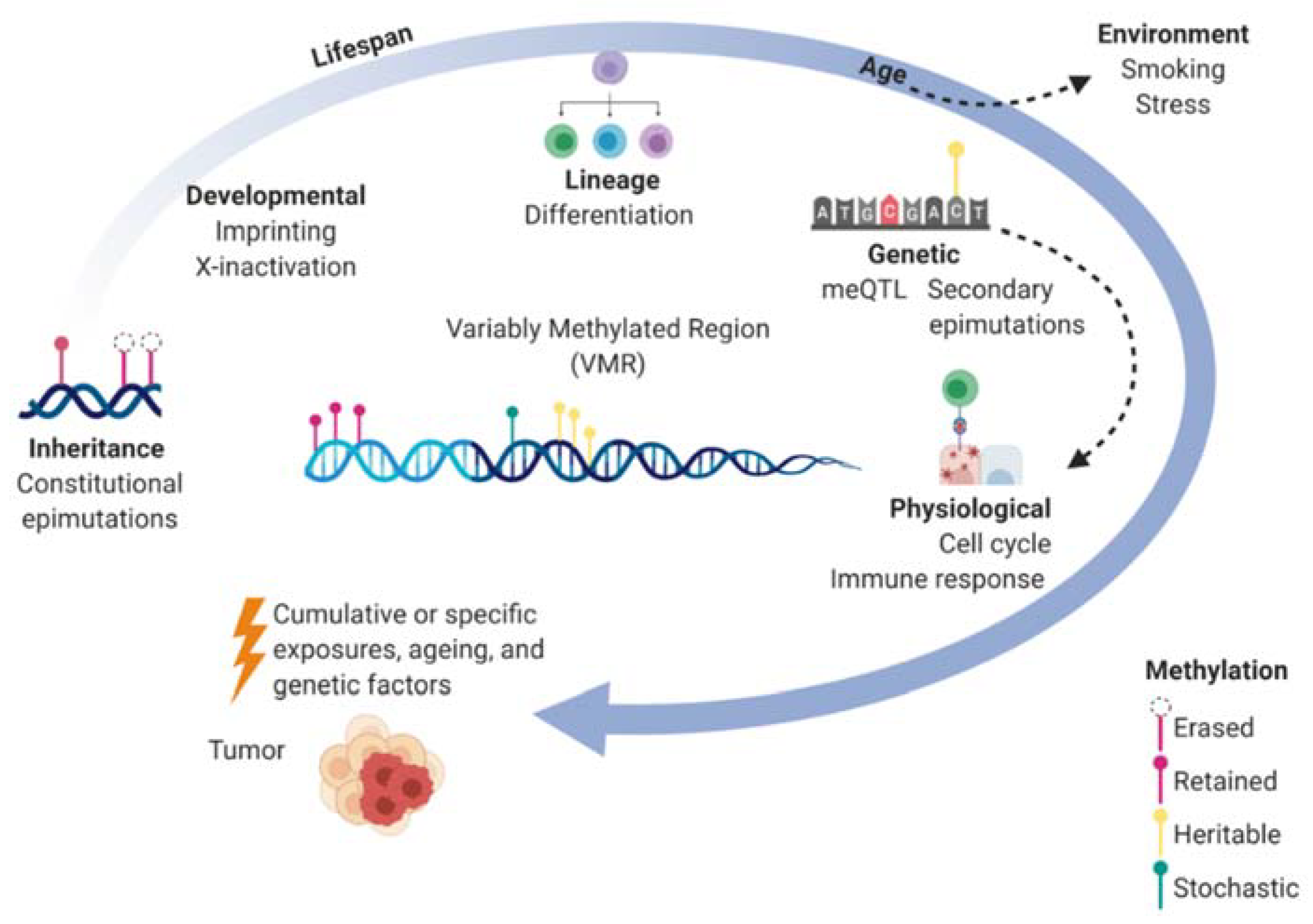
2. DNA Methylation in Epigenetic Control and Heritability

In mammals, DNA modifications predominantly occur through cytosine methylation (CH3) at guanine-cytosine dinucleotides (CpG) which cluster in CpG-dense regions called CpG islands (CGI) [14]. A critical role of CpG DNAm is the regulation of gene expression through transcriptional silencing, an essential mechanism for developmental processes, including parent-of-origin imprinting, X-chromosome inactivation, and transposon silencing, and cellular differentiation [14]. For each cell, DNAm is inherited through successive cell cycles resulting in cell type patterns that extend through a specific lineage [15]. Cellular identity is responsive to numerous spatiotemporal factors including environmental cues (e.g., stimuli, signaling), cell cycle phase, and physical environment (e.g., oxygen availability, anchoring) [16]. Thus, while DNAm are stable epigenetic marks that establish a cellular memory and control of phenotype, they are not static. Recent studies mapping DNAm across cell types and populations has revealed cell-type specific patterns of variable methylated regions (VMRs) with high inter-individual variation [17].
The loss of epigenetic control over cellular identity and physiological homeostasis is an enabling hallmark of cancer. Oncogenic transformation is achieved through the cooperation of genetic and epigenetic reprogramming [18] to alter gene expression resulting in widespread DNAm alterations, often called epimutations [19]. Methylation across a variety of tumors show patterns of global hypomethylation as well as regions of focal hypermethylation at normally unmethylated CGI, often in the promoters of tumor suppressor and DNA mismatch repair genes [20]. EOC presents similar patterns but with histotype specificity [21,22]. High-grade serous ovarian cancers (HGSOCs) exhibit few hypermethylation events, although BRCA1 hypermethylation has been observed in 11.5% [23]. Conversely, endometrioid ovarian cancer (ENOC) is characterized by frequent promoter hypermethylation (CpG island methylation phenotype, CIMP) that is also common in uterine endometrioid tumors and parsimonious with the hypothesized endometriosis origin of ENOC [24]. Clear cell ovarian cancer (CCOC) also distinguishes itself by displaying the highest frequency (>70%) of hypermethylation [25]. It is important to note that differences in tumor DNAm may be associated with response to chemotherapy and prognosis. Several studies have investigated DNAm in association with treatment response and prognosis, and have been reviewed elsewhere [22].
Under normal conditions, DNAm acts as a critical barrier to cellular reprogramming and tumor initiation [26]. In normal tissues, aberrant DNAm can occur as a result of both somatic epimutations that arise and accumulate during the lifespan [27,28] and wide-spread ‘constitutional’ epimutations that originate in the parental germline or during early embryogenesis [29]. While most methylation is removed from the parental germline during embryogenesis, experimental studies have demonstrated a significant level can be retained across generations by resisting erasure [13,30] and through regulation of de novo methylation by non-coding RNA (ncRNA) populations packaged in sperm and oocytes [31].
During the lifespan, DNAm is continuously influenced by environmental exposures and genetic sequence as well as interacting factors such as age and gender [32]. Genetic variants are estimated to account for ~20% of the inter-individual variance in methylation though each CpG site varies in heritability and some (18%) have shown as high as >99% genetic heritability [32]. Constitutional epimutations that occur in association with genetic variants are considered secondary epimutations. Adding further complexity, CpG heritability has been shown to vary over time where environmental factors contribute more to methylation variance with increasing age. Genetic effects on DNAm are also associated with transcriptional response such as activation of immune genes [33] meaning meQTL vary depending on physiological states. Thus, the methylome of an individual at any particular VMR is a function of a multitude of factors that are dynamic over the lifespan and can cumulatively or specifically contribute to cancer (Figure 1). The heritable components of the methylome (both epigenetic and genetic) could contribute to the familial heritability of EOC.
In the following sections, we present a review of the epidemiological studies that have explored DNAm and EOC susceptibility. These have been categorized by study design and include: (1) familial and population-based studies targeting known cancer susceptibility genes (CSGs), (2) epigenome-wide association studies (EWAS), (3) integrative genomic-epigenomic approaches to study DNAm underlying genetic associations with risk, and (4) environmental epigenetic studies evaluating DNA methylation as a mediator of environmental risk.

3. DNA Methylation in Ovarian Cancer Susceptibility Genes

Both primary and secondary constitutional epimutations that contribute to cancer susceptibility have been definitively described in CSGs for Lynch syndrome (MLH1, MSH2) and Wilms Tumor (H19) [29], but other cancers are also emerging such as colorectal cancer (MGMT) and breast cancer (BRCA1) [11]. Four studies have evaluated DNAm to identify constitutional epimutations in known CSGs that contribute to EOC susceptibility: one case-only study [34], two case-control studies [35,36], and one familial segregation study [37] (Table 1). These studies limited cases to women without BRCA1 or BRCA2 sequence mutations to enrich the study population for non-genetic mechanisms. Three studies additionally limited cases to familial and/or early-onset EOC [34,36,37] while the other did not have family history criterion [35]. From peripheral blood leukocytes (PBL), DNAm in gene promoter regions was quantified via bisulfite sequencing technologies. Bisulfite pyrosequencing has been the standard practice for hypermethylation detection in clinical epigenetic studies and is generally followed by clonal or plasmid bisulfite sequencing for validation and confirmation of allele-specific methylation [38].
All four studies evaluated BRCA1 and together suggest BRCA1 promoter hypermethylation could be associated with EOC susceptibility as both constitutional primary and secondary epimutations. In a familial segregation study of families with breast and ovarian cancer, BRCA1 promoter hypermethylation was present in two out of 49 families, including one proband with triple negative breast cancer and one with HGSOC [37]. DNA sequencing identified a 5′ UTR BRCA1 variant that segregated with BRCA1 hypermethylation following a dominant inheritance pattern suggesting a secondary constitutive epimutation. In another study, promoter hypermethylation of BRCA1 was observed in 8% (three in 39) of cases with EOC and 1% (six in 613) of cases with breast cancer but in none of 10 age-matched controls [34]. It was further demonstrated that BRCA1 hypermethylation was confined to a single parental allele, present in all fractions of myeloid cell types (mesoderm) as well as urine (endoderm), and did not correlate with local DNA sequence variants. Together, these findings aligned with the definition of a primary constitutive epimutation although correlated sequence variation may still underlie the hypermethylation observed. This study also detected RAD51C promoter hypermethylation in one case of EOC.
Methylation of BRCA1 has been further implicated in a large-scale, two-stage study totaling 1640 EOC cases and 3682 controls where promoter methylation was detected in 6.4% of EOC cases compared to 4.2% of controls, and was associated with increased risk of EOC (OR = 1.83, 95% CI = 1.27–2.63) [35]. The study also included histotype-specific analysis which has not been reported elsewhere, finding that increased prevalence of in BRCA1 promoter methylation was specific to HGSOC and not significantly higher in LGSOC or non-serous histotypes. Sensitivity analyses showed that methylation frequency did not differ by cancer stage, surgery (before vs. after), chemotherapy, or tissue storage time but did decrease with age which was adjusted for in their comparisons. Additionally, tumor BRCA1 promoter methylation was five-times more frequent in cases with PBL methylation (18 in 29, 62%) than in cases without (seven in 58, 12%). A substantial proportion (54%, 13 in 24) of cases with PBL BRCA1 methylation also displayed methylation amongst other organ tissues whereas none of the patients without PBL methylation showed this feature, suggesting inheritance of the epimutation rather than somatic origin.
In contradiction to these findings, a study of 108 patients with breast and/or ovarian cancer and 60 controls found no subjects with promoter hypermethylation in BRCA1 [36]. The discrepancy in detection may be due to dissimilarities in study design since they did not age-match controls that were significantly younger (median age 61 vs. 41 in cases and controls, respectively) or limit to chemo naïve blood samples. They also employed different methylation quantification technology and analytical methods which can both contribute to differential detection.
In summary, the current literature demonstrates the prevalence of constitutional epimutations in BRCA1 that may well contribute to EOC susceptibility. However additional large-scale, confirmatory studies are needed. Further studies with histotype-specific analysis are particularly warranted given the observed specificity for HGSOC thus far. Finally, it is important to note that it is unknown whether the promoter hypermethylation originates in the early zygote or germline. A recent study evaluated BRCA1 promoter methylation in mother–daughter pairs [39]. However, these data allow no conclusions to be made on maternal transfer [40].

4. Epigenome-Wide Association Studies of DNA Methylation

Genome-wide DNAm has been examined in four case-control studies termed epigenome-wide association studies (EWAS) (Table 2). Blood samples were retrospectively collected and profiled using methylation microarrays with single CpG site probes [41] to identify differentially methylated probes (DMPs). DMPs may be symptomatic—caused by immune or other reactive response to the disease state—or etiologic differences that contributed to disease development. It is not possible to distinguish between the two within the retrospective design, making it difficult to discern the implications for EOC susceptibility. Nevertheless, EWAS have generated a significant compilation of EOC-associated PBL epimutations as well as insight into important dynamics of blood-based DNAm and useful analytical strategies.
The first EWAS in EOC compared 25,642 CpG probes between 113 pre-treatment cases and 148 unaffected controls and detected 2714 DMPs [42]. With just the top 100 DMPs, EOC cases could be accurately predicted (AUC = 0.80, 95% CI = 0.84–0.87). It also performed well for post-treatment cases with active disease (AUC = 0.76, 95% CI = 0.72–0.81) and poorly for those without active disease (AUC = 0.52, 95% CI = 0.48–0.55). Further analysis showed that the DMPs largely overlapped with age-related DMPs and strongly correlated with cell type distributions. Systematic differences in the leukocyte sub-populations were later confirmed as the major contributor of the findings when the ~2700 DMPs were shown to be highly correlated with leukocyte-tagging DMPs (spearman correlation = 0.75) and only 17 DMPs were non-leukocyte DMPs [43,44]. Based on this evidence, subsequent studies of PBL DNAm have used a series of analytical methods to mitigate confounding by cell-type.
Two additional EWAS studies were performed in parallel using the same hospital-based population but employing different analytical methods to mitigate confounding by cell-type. One study analyzed the total available sample size of 336 EOC cases and 398 controls but limited analysis to ~14,000 probes that were not previously associated with leukocyte cell-types (i.e., probe filtering) [45]. The other analyzed a subset of the same population (242 cases/181 controls) that had complete blood count (CBC) measures available for direct adjustment of cell-type distribution within logistic regression models. Though the sample size was reduced, this approach allowed more (~22k) CpG probes to be assessed [46].
While both methods aimed to accomplish the same goal—identification of DMPs that were independent of cell-type differences—the results were strikingly disparate with no overlap between findings. With the probe filtering method [45], 30 DMPs were detected and pathway analysis implicated enrichment for the telomerase signaling pathway (HDAC3, IL2RG, PIK3C2B, PIK3R1, and POT1) and paxillin signaling pathway (ARFIP2, ITGB6, PIK3C2B, PIK3R1, and SRC). Other findings relevant to cancer biology included hypomethylation in the gene body of HHIP (hedgehog-interacting protein) which has been associated with tumor growth and angiogenesis [47], and promoter hypomethylation for antiapoptotic CUL7 that inhibits p53 [48] and Caspase-8 [49]. With the CBC adjustment method [46], 62 DMPs were identified with the most significant local to cancer-associated genes SOCS2 and SEPT9, the latter of which has been previously implicated in ovarian tumorigenesis [50]. Notably, 61 (98%) of the CBC-adjusted DMPs overlapped with previously reported unadjusted DMPs from Teschendorff et al. (2009), suggesting that the replicated probes are either robust to cell-type distribution or that the CBC adjustment method was not able to remove all confounding by cell-type. In the filtering method, only six (20%) DMPs overlapped with prior unadjusted results. However, the complete lack of overlap between filtered and CBC-adjusted probes in the sample clinical population suggests these may be leukocyte-associated DMPs.
The study by Winham et al. [46] also performed novel regional-based testing for a subset (n = 163) of samples profiled using a higher density array. CpG probes were aggregated into 25,607 CpG islands to increase statistical power but this method also has biological relevance since individual CpG methylation is less stable and usually the entire promoter is either methylated or not [51]. Interestingly, highly ranked single DMP sites were not significant at the regional-level based testing and vice versa. Rather, additional regions were identified that corresponded with known CSGs including the CpG island in the promoter of BNC2, a CSG identified through GWAS [52]. Another region identified was in the promoter of XRCC2, a homologous recombination gene that has been indicated in EOC susceptibility and other cancers [53].
Another EWAS study was recently conducted among Chinese women and differed from prior studies by using a two-stage design [54]. The initial discovery stage compared methylation across ~485,000 CpG probes in 24 EOC cases and 24 controls and validated 96 DMPs in an independent set of 205 EOC cases and 205 controls. In total, 40 validated DMPs were identified which where enrichment for immune process genes (e.g., LYST, CADM1, NFATC1). The significant probes did not show correlation with leukocytes. However, they did correlate with platelet count and coagulation factors. Only 16 DMPs were not associated with platelets or coagulation factors which included hypermethylation of CADM1 and CADM2 and hypomethylation of LYST. CADM1 is known to be a tumor suppressor gene for solid tumors [55] including EOC [56] and is frequently inactivated by promoter hypermethylation [57]. Observed expression of CADM1 was lower in blood cells of EOC cases possibly caused by promoter methylation. LYST is a lysosomal trafficking regulator that promotes proliferation and inhibits apoptosis in multiple myeloma [58], and also harbors driver mutations for rare bone tumors [59]. Notably, this is the only EWAS study that has analyzed DNAm by EOC histotypes. In the histotype-specific analysis, most DMPs were associated with serous and endometrioid EOC while 10 were associated with mucinous EOC, in concordance with a distinct etiology for mucinous EOC [3]. DMPs that were only significant in histotype-specific analysis included three serous (LYST, SUN1, C9orf92), two endometrioid (SAMHD1, GLRX2), and one mucinous and endometrioid CpG probe (CD177).
In total, EWAS studies have analyzed a total of 801 EOC cases and 715 controls and identified 2846 DMPs (Table S1). We observed little replication across studies largely owing to confounding by blood-level factors (cell type distribution, coagulation factors) but also study heterogeneity. While most EOC cases were comprised of serous histology they also included a wide range (28–51%) of non-serous ovarian cancers causing phenotypic heterogeneity across studies. Although histotype-specific DMPs were only reported in one study, the findings indicate PBL DNAm may display unique epigenetic alterations that occur during tumorigenesis [21]. EWAS studies also included adjustments for different confounders such as demographic and known EOC risk factors (age, parity, age of first birth, alcohol use, smoking, enrollment year/state) [45,46] and experimental factors (bisulfite conversion efficiency, array batch) [42]. Despite these differences, 82 DMPs were replicated in at least one other study comprising 71 replicated genes (Table S2). It is conceivable that some of the cancer-associated probes could also include predisposition or risk-associated DMPs. As we noted, several known tumor suppressor genes and oncogenes have been identified across EWAS and represent candidate susceptibility genes.

5. Genetic Susceptibility Mediated by DNA Methylation

The functional connection between genetic and epigenetic variation is an integral component of the genetic predisposition to cancer [60]. Numerous studies have mapped genetic variation to CpG methylation levels (methylation quantitative trait loci, meQTL) and shown these associations underlie DNAm variation and the inheritance of complex traits and diseases [33,61]. Three studies have explored methylation-mediated relationships between genetic variation and EOC risk using different integrative approaches.
Shen et al. [62] performed a candidate gene study and comprehensive analysis of tumor DNAm, associated meQTL, and gene expression for HNF1B, a suspected susceptibility gene from tumor methylation patterns. HNF1B was hypermethylated in approximately 50% of HGSOC tumors (n = 608) but not in any of the CCOC tumors profiled (n = 4). Among SNPs within 150kb of HNF1B, nine SNPs were associated with increased risk for HGSOC (rs7405776 OR = 1.13, 95% CI = 1.09−1.17, P = 3.1 × 10⁻¹⁰) but conversely were associated with reduced risk for CCOC (rs11651755 OR = 0.77, 95% CI = 0.70−0.84, P = 1.6 × 10⁻⁸) (Table 3).
Four of the risk SNPs in the 5′ UTR of HNF1B were significantly correlated with promoter CpG methylation in tumors, suggesting the aberrant DNAm observed in tumors could be etiologic. Immunohistochemical (IHC) analysis further showed that the HFN1B protein was expressed in most CCOC tumors where the HNF1B promoter was not methylated whereas the majority of HGSOC did not express the HNF1B protein and had frequent HNF1B promoter methylation. Together, these findings demonstrated that risk-associated variation in HNF1B alters promoter methylation for HGSOC and CCOC in opposing directions, suggesting it may have a tumor suppressor role in HGSOC and a reverse, oncogenic role in CCOC.
Two genome-wide studies have evaluated PBL DNAm and genetic variation in EOC but used different analytical frameworks [63,64]. One was an EWAS-based approach that identified DMPs and then evaluated whether they correlated with risk SNPs [63]. For the EWAS, PBL CpG DNAm was profiled and compared between 214 cases and 214 controls and identified 1993 DMPs that were subsequently filtered to 185 DMPs with meQTL. Twenty-eight of the meQTL were associated with risk and mediation analysis using the causal inference test [65] revealed that 13 DMPs modulated associations of 17 SNPs. Interestingly, prior EWAS studies [42,46] also detected DMPs at AIM2 and STAB1 which could both conceivably affect immune system response and cancer development. AIM2 is a member of innate immune sensors that initiate inflammasomes and trigger secretion of proinflammatory cytokines [66]. Although it has displayed both tumor suppression and promotion roles across cancer types, its upregulation in ovarian cancers suggests it could promote EOC progression [67,68]. STAB1 is a transmembrane receptor expressed on macrophages and lymphatic endothelial cells that acts as an inhibitor of antitumor immunity [69].
It is also notable that all meQTL that linked to the 13 DMPs were trans associations where the SNP was located on a different chromosome than the DMP. This was also true for the entirety of the 427 DMP-meQTL pairs identified. It is surprising that trans-meQTL, rather than cis-meQTL, were strictly observed for DMPs since they are estimated to account for a small portion of heritable PBL CpG (7% vs. 73%, respectively) [70]. Furthermore, VMRs within the genome have shown to be correlated in cis and trans forming co-methylated networks with low genetic heritability and high cell type specificity [17]. Thus, the predominance of trans-meQTL is intriguing and it is provocative to postulate whether it’s a reflection of differential blood cell distributions or some other underlying biology.
The second genome-wide study used a GWAS-based approach that imputed and compared genetically inherited PBL DNAm levels between 22,406 EOC cases and 40,941 controls [64]. In this innovative application, meQTL were used as genetic instruments to estimate CpG methylation levels, effectively removing both symptomatic and confounding differences in methylation that are present in typical EWAS. High density genetic and PBL DNAm data from the Framingham Offspring Study [71] were used to build genetic prediction models and risk associations for 62,938 CpG probes were estimated from 751,031 SNPs using GWAS summary statistics. A resulting 89 differentially methylated CpG were significantly associated with EOC risk and included eight known genomic risk regions where seven were correlated with local gene expression (Table 3). Additionally, one novel risk region was identified where increased risk of EOC was associated with hypermethylation of two CpG sites that were correlated with reduced expression of ADAP1 (7p22.3) a GTPase-activating protein that functions as a scaffold in several signal transduction pathways. Histotype-specific analysis showed that all DMPs were associated with HGSOC and three loci (3q25, 17q21.31, and 19p13.11) had HGSOC-specific DMPs. The 2q31.1 locus was the only region that was also significant for mucinous and endometrioid histotypes.
In comparison to prior EWAS study findings, the integrative GWAS-based approach replicated (p < 0.10) EWAS associations with increased risk of EOC for hypomethylation of cg19399532 at C1orf220 [42,46] and hypomethylation of cg21870884 at GPR25 [46]. C1orf220 is a long non-coding RNA that has exhibited upregulation in lung squamous cell carcinoma [72] and was one of the 71 genes that was replicated across EWAS studies and did not correlate with age. GPR25 is a G-protein coupled receptor that activates signaling cascades as a response to extracellular stress and has been linked to heritable arterial stiffness [73].
Collectively, variation in DNAm has been mapped to nine GWAS risk loci using integrative approaches for tumor and PBL DNAm (Table 3). Future studies mapping meQTL in additional tissue and cell types will be an important undertaking to further elucidate the known genetic susceptibility as well as identify additional risk loci. Currently, meQTL mapping and imputation have been performed with in cis SNPs but it will be intriguing to investigate the relationship of trans-meQTL with EOC risk using this approach.

6. Environmental Risk Mediated by DNA Methylation

In the prior section, the association between DNAm and EOC susceptibility was evaluated within a framework focused on the mediation of genetic effects on phenotypic variance, i.e., genetic (GWAS SNP) → epigenetic (meQTL association) → phenotype (EOC). However, DNAm may also be a mediator of environmental exposures whereby DNAm alterations can occur without DNA sequence alterations and these changes can influence gene expression and hence EOC susceptibility, i.e., environmental exposure (risk factor) → epigenetic (DMP) → phenotype (EOC). Thus far, only one study has evaluated DNAm as a mediator of an environmental risk factor for EOC.
In the study by Wu et al. [74], DNAm was evaluated as a causal mediator of the association between EOC risk and alcohol use which is an exposure known to alter DNAm [75]. Among 196 EOC cases and 202 controls previously analyzed in prior EWAS [45,46,63], DNAm was evaluated in combination with alcohol consumption. Approximately 63% of cases and 85% of controls reported alcohol use at study enrollment which was associated with a significant inverse association with EOC risk (OR = 0.34, p = 0.001). Alcohol use was associated with reduced CpG methylation in both cases and controls for two DMPs on chromosome 11 that were found to be significant mediators of the association between alcohol consumption and reduced EOC risk: cg09358725 at LMO2 and cg11016563 at TRPC6. While this study suggests alcohol use may associate with EOC status by regulating CpG-specific DNAm patterns, effects on DNAm may be temporary and the identified DMPs may not be stable. Though results should be interpreted cautiously, they are encouraging for additional studies to validate these findings.
Studies are needed to assess DNAm as a mediator of other known EOC risk factors. DNAm alterations have been associated with several previously reported EOC risk factors, including sex hormone exposure [76], obesity [77], endometriosis [78], irregular menstruation in women with polycystic ovarian syndrome [79], as well as perceived stress, cortisol output, and inflammation [80]. Integrating epigenetics into epidemiological investigations of these exposures may help to elucidate etiological mechanisms of this disease. Furthermore, future studies should also consider whether DNAm may be a modifier of, or act in combination with (i.e., interaction), both genetic and environmental risk factors for EOC [81].

7. Conclusions

DNAm has been at the forefront of epigenetic research and has provided a paradigm for the epigenetic inheritance of cancer susceptibility. The complexities of not only the inheritance but the dynamics of DNAm across cell types and the lifespan have presented challenges to the investigation and interpretation of epimutations. Nevertheless, a growing collection of epidemiological studies with various designs have begun to elucidate the role of DNAm in EOC susceptibility. While rare hypermethylation occurrences have been observed, phenotypic variance associated with differences in DNAm secondary to genetic variation appears to be a significant component of the heritability of EOC. Across EWAS and integrative study designs, 25% (n = 10) of GWAS risk loci have been correlated with DNAm changes. Notably, a novel genome-wide significant DNAm risk locus has also been detected. As detailed maps of DNAm variation across populations and tissues, and in linkage to methylation (meQTL), expression (eQTL), and transcriptional response (reQTL) continue to be generated, further revelations of not only individual genetic–epigenetic loci but potentially co-methylated networks are expected to follow.
While studies with genetic–epigenetic approaches are increasing, studies that integrate DNAm with environmental exposure data are comparably lacking. More research is needed to elucidate the relationship between epigenetics and environmental exposures and how they contribute to the heritability of EOC. Truly comprehensive evaluations of DNAm will require integration with both genetic and environmental factors. This multi-dimensional data will carry a higher computational burden necessitating larger sample sizes. To-date, EOC EWAS studies have been conducted in a significantly smaller number of samples (<2000 subjects overall) compared to GWAS (>100,000 subjects). Imputation-based methods for DNAm, such as those employed by Yang et al. [64], could be one approach to boost power but will require more complex imputation algorithms that account for environmental exposures and may not be available in reference datasets. Ideally, large-scale DNAm profiling initiatives should be undertaken, preferably with the higher coverage technologies that are now available.
Currently, microarrays offer coverage of over 850,000 CpG sites and targeted bisulfite sequencing is another relatively low-cost option with even higher coverage, capturing up to several million CpGs. Whole-genome bisulfite sequencing (WGBS) has typically been cost-prohibitive for association studies, however, it continues to improve in terms of efficiency and accuracy with lower input amounts and read-depths required, making it an increasingly feasible option [82]. The application of WGBS will be imperative for a more comprehensive assessment of DNAm since the coverage of cell-type-specific VMRs are not well represented with a fixed content design [83] and it captures additional DNAm features such as allele-specific methylation [84]. Overall, as technology continues to advance and costs decrease, future epigenetic studies will be able to evaluate larger numbers of samples with improved feature capture, enabling further discovery that will enhance our understanding of DNAm and its contribution to cancer susceptibility.

Supplementary Materials

The following are available online at https://www.mdpi.com/2072-6694/13/1/108/s1, Table S1: All DMPs identified through EWAS in EOC, Table S2: Genes replicated across EWAS in EOC.

Author Contributions

Conceptualization, B.M.R. and B.L.F.; literature survey, B.M.R.; writing—original draft preparation, B.M.R.; writing—review and editing, B.L.F.; supervision, B.L.F. All authors have read and agreed to the published version of the manuscript.

Funding

This research received no external funding.

Institutional Review Board Statement

Not applicable.

Informed Consent Statement

Not applicable.

Data Availability Statement

No new data were created or analyzed in this study. Data sharing is not applicable to this article.

Conflicts of Interest

The authors declare no conflict of interest.

References

  1. Wray, N.R.; Yang, J.; Goddard, M.E.; Visscher, P.M. The genetic interpretation of area under the ROC curve in genomic profiling. PLoS Genet. 2010, 6, e1000864. [Google Scholar] [CrossRef] [PubMed] [Green Version]
  2. Zheng, G.Q.; Yu, H.Y.; Kanerva, A.; Forsti, A.; Sundquist, K.; Hemminki, K. Familial risks of ovarian cancer by age at diagnosis, proband type and histology. PLoS ONE 2018, 13, e0205000. [Google Scholar] [CrossRef] [Green Version]
  3. Jones, M.R.; Kamara, D.; Karlan, B.Y.; Pharoah, P.D.P.; Gayther, S.A. Genetic epidemiology of ovarian cancer and prospects for polygenic risk prediction. Gynecol. Oncol. 2017, 147, 705–713. [Google Scholar] [CrossRef] [PubMed] [Green Version]
  4. Banta, J.A.; Richards, C.L. Quantitative epigenetics and evolution. Heredity 2018, 121, 210–224. [Google Scholar] [CrossRef] [PubMed]
  5. Waddington, C.H. The Epigenotype. Endeavour 1942, 1, 18–20. [Google Scholar] [CrossRef] [PubMed] [Green Version]
  6. Bernstein, B.E.; Meissner, A.; Lander, E.S. The mammalian epigenome. Cell 2007, 128, 669–681. [Google Scholar] [CrossRef] [PubMed] [Green Version]
  7. Skvortsova, K.; Iovino, N.; Bogdanovic, O. Functions and mechanisms of epigenetic inheritance in animals. Nat. Rev. Mol. Cell Biol. 2018, 19, 774–790. [Google Scholar] [CrossRef] [Green Version]
  8. Frias-Lasserre, D.; Villagra, C.A. The Importance of ncRNAs as Epigenetic Mechanisms in Phenotypic Variation and Organic Evolution. Front. Microbiol. 2017, 8, 2483. [Google Scholar] [CrossRef]
  9. Feinberg, A.P.; Vogelstein, B. Hypomethylation Distinguishes Genes of Some Human Cancers from Their Normal Counterparts. Nature 1983, 301, 89–92. [Google Scholar] [CrossRef]
  10. Hitchins, M.P.; Rapkins, R.W.; Kwok, C.T.; Srivastava, S.; Wong, J.J.L.; Khachigian, L.M.; Polly, P.; Goldblatt, J.; Ward, R.L. Dominantly Inherited Constitutional Epigenetic Silencing of MLH1 in a Cancer-Affected Family Is Linked to a Single Nucleotide Variant within the 5′ UTR. Cancer Cell 2011, 20, 200–213. [Google Scholar] [CrossRef] [Green Version]
  11. Lonning, P.E.; Eikesdal, H.P.; Loes, I.M.; Knappskog, S. Constitutional Mosaic Epimutations-a hidden cause of cancer? Cell Stress 2019, 3, 118–135. [Google Scholar] [CrossRef] [PubMed] [Green Version]
  12. Lacal, I.; Ventura, R. Epigenetic Inheritance: Concepts, Mechanisms and Perspectives. Front. Mol. Neurosci. 2018, 11, 292. [Google Scholar] [CrossRef] [PubMed] [Green Version]
  13. Lesch, B.J.; Tothova, Z.; Morgan, E.A.; Liao, Z.; Bronson, R.T.; Ebert, B.L.; Page, D.C. Intergenerational epigenetic inheritance of cancer susceptibility in mammals. Elife 2019, 8. [Google Scholar] [CrossRef] [PubMed]
  14. Smith, Z.D.; Meissner, A. DNA methylation: Roles in mammalian development. Nat. Rev. Genet. 2013, 14, 204–220. [Google Scholar] [CrossRef]
  15. Kim, M.; Costello, J. DNA methylation: An epigenetic mark of cellular memory. Exp. Mol. Med. 2017, 49, e322. [Google Scholar] [CrossRef] [Green Version]
  16. Wagner, A.; Regev, A.; Yosef, N. Revealing the vectors of cellular identity with single-cell genomics. Nat. Biotechnol. 2016, 34, 1145–1160. [Google Scholar] [CrossRef] [Green Version]
  17. Garg, P.; Joshi, R.S.; Watson, C.; Sharp, A.J. A survey of inter-individual variation in DNA methylation identifies environmentally responsive co-regulated networks of epigenetic variation in the human genome. PLoS Genet. 2018, 14, e1007707. [Google Scholar] [CrossRef]
  18. Shen, H.; Laird, P.W. Interplay between the Cancer Genome and Epigenome. Cell 2013, 153, 38–55. [Google Scholar] [CrossRef] [Green Version]
  19. Holliday, R. The inheritance of epigenetic defects. Science 1987, 238, 163–170. [Google Scholar] [CrossRef]
  20. Esteller, M. Epigenetics in cancer. N. Engl. J. Med. 2008, 358, 1148–1159. [Google Scholar] [CrossRef]
  21. Earp, M.A.; Cunningham, J.M. DNA methylation changes in epithelial ovarian cancer histotypes. Genomics 2015, 106, 311–321. [Google Scholar] [CrossRef] [PubMed]
  22. Natanzon, Y.; Goode, E.L.; Cunningham, J.M. Epigenetics in ovarian cancer. Semin. Cancer Biol. 2018, 51, 160–169. [Google Scholar] [CrossRef] [PubMed]
  23. Cancer Genome Atlas Research, N. Integrated genomic analyses of ovarian carcinoma. Nature 2011, 474, 609–615. [Google Scholar] [CrossRef] [PubMed]
  24. Kolbe, D.L.; DeLoia, J.A.; Porter-Gill, P.; Strange, M.; Petrykowska, H.M.; Guirguis, A.; Krivak, T.C.; Brody, L.C.; Elnitski, L. Differential analysis of ovarian and endometrial cancers identifies a methylator phenotype. PLoS ONE 2012, 7, e32941. [Google Scholar] [CrossRef] [Green Version]
  25. Engqvist, H.; Parris, T.Z.; Biermann, J.; Ronnerman, E.W.; Larsson, P.; Sundfeldt, K.; Kovacs, A.; Karlsson, P.; Helou, K. Integrative genomics approach identifies molecular features associated with early-stage ovarian carcinoma histotypes. Sci. Rep. 2020, 10, 7946. [Google Scholar] [CrossRef]
  26. Mikkelsen, T.S.; Hanna, J.; Zhang, X.L.; Ku, M.C.; Wernig, M.; Schorderet, P.; Bernstein, B.E.; Jaenisch, R.; Lander, E.S.; Meissner, A. Dissecting direct reprogramming through integrative genomic analysis. Nature 2008, 454, 49–55. [Google Scholar] [CrossRef]
  27. Bjornsson, H.T.; Sigurdsson, M.I.; Fallin, M.D.; Irizarry, R.A.; Aspelund, T.; Cui, H.; Yu, W.; Rongione, M.A.; Ekstrom, T.J.; Harris, T.B.; et al. Intra-individual change over time in DNA methylation with familial clustering. JAMA 2008, 299, 2877–2883. [Google Scholar] [CrossRef] [Green Version]
  28. Kwabi-Addo, B.; Chung, W.; Shen, L.; Ittmann, M.; Wheeler, T.; Jelinek, J.; Issa, J.P. Age-related DNA methylation changes in normal human prostate tissues. Clin. Cancer Res. 2007, 13, 3796–3802. [Google Scholar] [CrossRef] [Green Version]
  29. Hitchins, M.P. Constitutional epimutation as a mechanism for cancer causality and heritability? Nat. Rev. Cancer 2015, 15. [Google Scholar] [CrossRef]
  30. Greenberg, M.V.C.; Bourc’his, D. The diverse roles of DNA methylation in mammalian development and disease. Nat. Rev. Mol. Cell Biol. 2019, 20, 590–607. [Google Scholar] [CrossRef]
  31. Rodgers, A.B.; Morgan, C.P.; Leu, N.A.; Bale, T.L. Transgenerational epigenetic programming via sperm microRNA recapitulates effects of paternal stress. Proc. Natl. Acad. Sci. USA 2015, 112, 13699–13704. [Google Scholar] [CrossRef] [PubMed] [Green Version]
  32. Van Dongen, J.; Nivard, M.G.; Willemsen, G.; Hottenga, J.J.; Helmer, Q.; Dolan, C.V.; Ehli, E.A.; Davies, G.E.; Van Iterson, M.; Breeze, C.E.; et al. Genetic and environmental influences interact with age and sex in shaping the human methylome. Nat. Commun. 2016, 7, 11115. [Google Scholar] [CrossRef] [PubMed] [Green Version]
  33. Husquin, L.T.; Rotival, M.; Fagny, M.; Quach, H.; Zidane, N.; McEwen, L.M.; MacIsaac, J.L.; Kobor, M.S.; Aschard, H.; Patin, E.; et al. Exploring the genetic basis of human population differences in DNA methylation and their causal impact on immune gene regulation. Genome. Biol. 2018, 19, 1–17. [Google Scholar] [CrossRef] [Green Version]
  34. Hansmann, T.; Pliushch, G.; Leubner, M.; Kroll, P.; Endt, D.; Gehrig, A.; Preisler-Adams, S.; Wieacker, P.; Haaf, T. Constitutive promoter methylation of BRCA1 and RAD51C in patients with familial ovarian cancer and early-onset sporadic breast cancer. Hum. Mol. Genet. 2012, 21, 4669–4679. [Google Scholar] [CrossRef] [PubMed]
  35. Lonning, P.E.; Berge, E.O.; Bjornslett, M.; Minsaas, L.; Chrisanthar, R.; Hoberg-Vetti, H.; Dulary, C.; Busato, F.; Bjorneklett, S.; Eriksen, C.; et al. White Blood Cell BRCA1 Promoter Methylation Status and Ovarian Cancer Risk. Ann. Intern. Med. 2018, 168, 326–334. [Google Scholar] [CrossRef] [PubMed]
  36. Tabano, S.; Azzollini, J.; Pesenti, C.; Lovati, S.; Costanza, J.; Fontana, L.; Peissel, B.; Miozzo, M.; Manoukian, S. Analysis of BRCA1 and RAD51C Promoter Methylation in Italian Families at High-Risk of Breast and Ovarian Cancer. Cancers 2020, 12, 910. [Google Scholar] [CrossRef] [PubMed] [Green Version]
  37. Evans, D.G.R.; Van Veen, E.M.; Byers, H.J.; Wallace, A.J.; Ellingford, J.M.; Beaman, G.; Santoyo-Lopez, J.; Aitman, T.J.; Eccles, D.M.; Lalloo, F.I.; et al. A Dominantly Inherited 5’ UTR Variant Causing Methylation-Associated Silencing of BRCA1 as a Cause of Breast and Ovarian Cancer. Am. J. Hum. Genet. 2018, 103, 213–220. [Google Scholar] [CrossRef] [Green Version]
  38. Sloane, M.A.; Ward, R.L.; Hesson, L.B. Defining the criteria for identifying constitutional epimutations. Clin. Epigenetics 2016, 8, 1–2. [Google Scholar] [CrossRef] [Green Version]
  39. Al-Moghrabi, N.; Al-Showimi, M.; Al-Yousef, N.; Al-Shahrani, B.; Karakas, B.; Alghofaili, L.; Almubarak, H.; Madkhali, S.; Al Humaidan, H. Methylation of BRCA1 and MGMT genes in white blood cells are transmitted from mothers to daughters. Clin. Epigenetics 2018, 10, 99. [Google Scholar] [CrossRef]
  40. Lonning, P.E.; Knappskog, S. BRCA1 methylation in newborns: Genetic disposition, maternal transfer, environmental influence, or by chance only? Clin. Epigenetics 2018, 10, 128. [Google Scholar] [CrossRef] [Green Version]
  41. Bibikova, M.; Barnes, B.; Tsan, C.; Ho, V.; Klotzle, B.; Le, J.M.; Delano, D.; Zhang, L.; Schroth, G.P.; Gunderson, K.L.; et al. High density DNA methylation array with single CpG site resolution. Genomics 2011, 98, 288–295. [Google Scholar] [CrossRef] [Green Version]
  42. Teschendorff, A.E.; Menon, U.; Gentry-Maharaj, A.; Ramus, S.J.; Gayther, S.A.; Apostolidou, S.; Jones, A.; Lechner, M.; Beck, S.; Jacobs, I.J.; et al. An epigenetic signature in peripheral blood predicts active ovarian cancer. PLoS ONE 2009, 4, e8274. [Google Scholar] [CrossRef] [PubMed] [Green Version]
  43. Koestler, D.C.; Marsit, C.J.; Christensen, B.C.; Accomando, W.; Langevin, S.M.; Houseman, E.A.; Nelson, H.H.; Karagas, M.R.; Wiencke, J.K.; Kelsey, K.T. Peripheral blood immune cell methylation profiles are associated with nonhematopoietic cancers. Cancer Epidemiol. Biomark. Prev. 2012, 21, 1293–1302. [Google Scholar] [CrossRef] [Green Version]
  44. Langevin, S.M.; Houseman, E.A.; Accomando, W.P.; Koestler, D.C.; Christensen, B.C.; Nelson, H.H.; Karagas, M.R.; Marsit, C.J.; Wiencke, J.K.; Kelsey, K.T. Leukocyte-adjusted epigenome-wide association studies of blood from solid tumor patients. Epigenetics 2014, 9, 884–895. [Google Scholar] [CrossRef] [PubMed] [Green Version]
  45. Fridley, B.L.; Armasu, S.M.; Cicek, M.S.; Larson, M.C.; Wang, C.; Winham, S.J.; Kalli, K.R.; Koestler, D.C.; Rider, D.N.; Shridhar, V.; et al. Methylation of leukocyte DNA and ovarian cancer: Relationships with disease status and outcome. BMC Med. Genom. 2014, 7, 21. [Google Scholar] [CrossRef] [PubMed] [Green Version]
  46. Winham, S.J.; Armasu, S.M.; Cicek, M.S.; Larson, M.C.; Cunningham, J.M.; Kalli, K.R.; Fridley, B.L.; Goode, E.L. Genome-wide investigation of regional blood-based DNA methylation adjusted for complete blood counts implicates BNC2 in ovarian cancer. Genet. Epidemiol. 2014, 38, 457–466. [Google Scholar] [CrossRef] [Green Version]
  47. Agrawal, V.; Kim, D.Y.; Kwon, Y.G. Hhip regulates tumor-stroma-mediated upregulation of tumor angiogenesis. Exp. Mol. Med. 2017, 49, e289. [Google Scholar] [CrossRef] [Green Version]
  48. Jung, P.; Verdoodt, B.; Bailey, A.; Yates, J.R., 3rd; Menssen, A.; Hermeking, H. Induction of cullin 7 by DNA damage attenuates p53 function. Proc. Natl. Acad. Sci. USA 2007, 104, 11388–11393. [Google Scholar] [CrossRef] [Green Version]
  49. Kong, Y.J.; Wang, Z.H.; Zhou, Z.M.; Mao, X.Y.; Chen, C.S. CUL7 promotes cancer cell survival through promoting Caspase-8 ubiquitination. Cancer Res. 2019, 79. [Google Scholar] [CrossRef]
  50. Peterson, E.A.; Stanbery, L.; Li, C.; Kocak, H.; Makarova, O.; Petty, E.M. SEPT9_i1 and genomic instability: Mechanistic insights and relevance to tumorigenesis. Genes Chromosomes Cancer 2011, 50, 940–949. [Google Scholar] [CrossRef] [Green Version]
  51. Weber, M.; Hellmann, I.; Stadler, M.B.; Ramos, L.; Paabo, S.; Rebhan, M.; Schubeler, D. Distribution, silencing potential and evolutionary impact of promoter DNA methylation in the human genome. Nat. Genet. 2007, 39, 457–466. [Google Scholar] [CrossRef] [PubMed]
  52. Buckley, M.A.; Woods, N.T.; Tyrer, J.P.; Mendoza-Fandino, G.; Lawrenson, K.; Hazelett, D.J.; Najafabadi, H.S.; Gjyshi, A.; Carvalho, R.S.; Lyra, P.C., Jr.; et al. Functional Analysis and Fine Mapping of the 9p22.2 Ovarian Cancer Susceptibility Locus. Cancer Res. 2019, 79, 467–481. [Google Scholar] [CrossRef] [PubMed] [Green Version]
  53. He, Y.; Zhang, Y.; Jin, C.; Deng, X.; Wei, M.; Wu, Q.; Yang, T.; Zhou, Y.; Wang, Z. Impact of XRCC2 Arg188His polymorphism on cancer susceptibility: A meta-analysis. PLoS ONE 2014, 9, e91202. [Google Scholar] [CrossRef] [PubMed]
  54. Li, L.; Zheng, H.; Huang, Y.; Huang, C.; Zhang, S.; Tian, J.; Li, P.; Sood, A.K.; Zhang, W.; Chen, K. DNA methylation signatures and coagulation factors in the peripheral blood leucocytes of epithelial ovarian cancer. Carcinogenesis 2017, 38, 797–805. [Google Scholar] [CrossRef]
  55. Van Der Weyden, L.; Arends, M.J.; Rust, A.G.; Poulogiannis, G.; McIntyre, R.E.; Adams, D.J. Increased tumorigenesis associated with loss of the tumor suppressor gene Cadm1. Mol. Cancer 2012, 11, 29. [Google Scholar] [CrossRef] [Green Version]
  56. Si, X.; Xu, F.; Xu, F.; Wei, M.; Ge, Y.; Chenge, S. CADM1 inhibits ovarian cancer cell proliferation and migration by potentially regulating the PI3K/Akt/mTOR pathway. Biomed. Pharm. 2020, 123, 109717. [Google Scholar] [CrossRef]
  57. De Strooper, L.M.; Van Zummeren, M.; Steenbergen, R.D.; Bleeker, M.C.; Hesselink, A.T.; Wisman, G.B.; Snijders, P.J.; Heideman, D.A.; Meijer, C.J. CADM1, MAL and miR124-2 methylation analysis in cervical scrapes to detect cervical and endometrial cancer. J. Clin. Pathol. 2014, 67, 1067–1071. [Google Scholar] [CrossRef]
  58. Bong, I.P.; Ng, C.C.; Fakiruddin, S.K.; Lim, M.N.; Zakaria, Z. Small interfering RNA-mediated silencing of nicotinamide phosphoribosyltransferase (NAMPT) and lysosomal trafficking regulator (LYST) induce growth inhibition and apoptosis in human multiple myeloma cells: A preliminary study. Bosn J. Basic Med. Sci. 2016, 16, 268–275. [Google Scholar] [CrossRef] [Green Version]
  59. Tarpey, P.S.; Behjati, S.; Young, M.D.; Martincorena, I.; Alexandrov, L.B.; Farndon, S.J.; Guzzo, C.; Hardy, C.; Latimer, C.; Butler, A.P.; et al. The driver landscape of sporadic chordoma. Nat. Commun. 2017, 8, 890. [Google Scholar] [CrossRef] [Green Version]
  60. Heyn, H.; Sayols, S.; Moutinho, C.; Vidal, E.; Sanchez-Mut, J.V.; Stefansson, O.A.; Nadal, E.; Moran, S.; Eyfjord, J.E.; Gonzalez-Suarez, E.; et al. Linkage of DNA methylation quantitative trait loci to human cancer risk. Cell Rep. 2014, 7, 331–338. [Google Scholar] [CrossRef] [Green Version]
  61. Heyn, H.; Moran, S.; Hernando-Herraez, I.; Sayols, S.; Gomez, A.; Sandoval, J.; Monk, D.; Hata, K.; Marques-Bonet, T.; Wang, L.; et al. DNA methylation contributes to natural human variation. Genome Res. 2013, 23, 1363–1372. [Google Scholar] [CrossRef] [PubMed] [Green Version]
  62. Shen, H.; Fridley, B.L.; Song, H.L.; Lawrenson, K.; Cunningham, J.M.; Ramus, S.J.; Cicek, M.S.; Tyrer, J.; Stram, D.; Larson, M.C.; et al. Epigenetic analysis leads to identification of HNF1B as a subtype-specific susceptibility gene for ovarian cancer. Nat. Commun. 2013, 4, 1–10. [Google Scholar] [CrossRef] [PubMed]
  63. Koestler, D.C.; Chalise, P.; Cicek, M.S.; Cunningham, J.M.; Armasu, S.; Larson, M.C.; Chien, J.; Block, M.; Kalli, K.R.; Sellers, T.A.; et al. Integrative genomic analysis identifies epigenetic marks that mediate genetic risk for epithelial ovarian cancer. BMC Med. Genom. 2014, 7, 8. [Google Scholar] [CrossRef] [PubMed] [Green Version]
  64. Yang, Y.; Wu, L.; Shu, X.; Lu, Y.; Shu, X.O.; Cai, Q.; Beeghly-Fadiel, A.; Li, B.; Ye, F.; Berchuck, A.; et al. Genetic Data from Nearly 63,000 Women of European Descent Predicts DNA Methylation Biomarkers and Epithelial Ovarian Cancer Risk. Cancer Res. 2019, 79, 505–517. [Google Scholar] [CrossRef] [Green Version]
  65. Millstein, J.; Zhang, B.; Zhu, J.; Schadt, E.E. Disentangling molecular relationships with a causal inference test. BMC Genet. 2009, 10, 23. [Google Scholar] [CrossRef] [Green Version]
  66. Lugrin, J.; Martinon, F. The AIM2 inflammasome: Sensor of pathogens and cellular perturbations. Immunol. Rev. 2018, 281, 99–114. [Google Scholar] [CrossRef]
  67. Chang, C.M.; Wang, M.L.; Lu, K.H.; Yang, Y.P.; Juang, C.M.; Wang, P.H.; Hsu, R.J.; Yu, M.H.; Chang, C.C. Integrating the dysregulated inflammasome-based molecular functionome in the malignant transformation of endometriosis-associated ovarian carcinoma. Oncotarget 2018, 9, 3704–3726. [Google Scholar] [CrossRef] [Green Version]
  68. Luborsky, J.; Barua, A.; Edassery, S.; Bahr, J.M.; Edassery, S.L. Inflammasome expression is higher in ovarian tumors than in normal ovary. PLoS ONE 2020, 15, e0227081. [Google Scholar] [CrossRef]
  69. Hollmen, M.; Figueiredo, C.R.; Jalkanen, S. New tools to prevent cancer growth and spread: A ‘Clever’ approach. Brit. J. Cancer 2020, 123, 501–509. [Google Scholar] [CrossRef]
  70. Huan, T.X.; Joehanes, R.; Song, C.; Peng, F.; Guo, Y.C.; Mendelson, M.; Yao, C.; Liu, C.Y.; Ma, J.T.; Richard, M.; et al. Genome-wide identification of DNA methylation QTLs in whole blood highlights pathways for cardiovascular disease. Nat. Commun. 2019, 10, 1–14. [Google Scholar] [CrossRef] [Green Version]
  71. Kannel, W.B.; Feinleib, M.; Mcnamara, P.M.; Garrison, R.J.; Castelli, W.P. An Investigation of Coronary Heart Disease in Families: The Framingham Offspring Study. Am. J. Epidemiol. 2017, 185, 1093–1102. [Google Scholar] [CrossRef] [Green Version]
  72. Shi, Y.F.; Li, Y.P.; Yan, C.; Su, H.; Ying, K.J. Identification of key genes and evaluation of clinical outcomes in lung squamous cell carcinoma using integrated bioinformatics analysis. Oncol. Lett. 2019, 18, 5859–5870. [Google Scholar] [CrossRef] [PubMed] [Green Version]
  73. Sherva, R.; Miller, M.B.; Lynch, A.I.; Devereux, R.B.; Rao, D.C.; Oberman, A.; Hopkins, P.N.; Kitzman, D.W.; Atwood, L.D.; Arnett, D.K. A whole genome scan for pulse pressure/stroke volume ratio in African Americans: The HyperGEN study. Am. J. Hypertens. 2007, 20, 398–402. [Google Scholar] [CrossRef] [PubMed] [Green Version]
  74. Wu, D.; Yang, H.; Winham, S.J.; Natanzon, Y.; Koestler, D.C.; Luo, T.; Fridley, B.L.; Goode, E.L.; Zhang, Y.; Cui, Y. Mediation analysis of alcohol consumption, DNA methylation, and epithelial ovarian cancer. J. Hum. Genet. 2018, 63, 339–348. [Google Scholar] [CrossRef]
  75. Liu, C.; Marioni, R.E.; Hedman, A.K.; Pfeiffer, L.; Tsai, P.C.; Reynolds, L.M.; Just, A.C.; Duan, Q.; Boer, C.G.; Tanaka, T.; et al. A DNA methylation biomarker of alcohol consumption. Mol. Psychiatry 2018, 23, 422–433. [Google Scholar] [CrossRef]
  76. Boyne, D.J.; Friedenreich, C.M.; McIntyre, J.B.; Stanczyk, F.Z.; Courneya, K.S.; King, W.D. Endogenous sex hormone exposure and repetitive element DNA methylation in healthy postmenopausal women. Cancer Causes Control. 2017, 28, 1369–1379. [Google Scholar] [CrossRef]
  77. Crujeiras, A.B.; Casanueva, F.F. Obesity and the reproductive system disorders: Epigenetics as a potential bridge. Hum. Reprod Update 2015, 21, 249–261. [Google Scholar] [CrossRef] [Green Version]
  78. He, J.; Chang, W.; Feng, C.; Cui, M.; Xu, T. Endometriosis Malignant Transformation: Epigenetics as a Probable Mechanism in Ovarian Tumorigenesis. Int. J. Genom. 2018, 2018, 1465348. [Google Scholar] [CrossRef] [Green Version]
  79. Jiao, J.; Sagnelli, M.; Shi, B.; Fang, Y.Y.; Shen, Z.Q.; Tang, T.Y.; Dong, B.Y.; Li, D.; Wang, X.X. Genetic and epigenetic characteristics in ovarian tissues from polycystic ovary syndrome patients with irregular menstruation resemble those of ovarian cancer. BMC Endocr. Disord. 2019, 19, 1–10. [Google Scholar] [CrossRef]
  80. Lam, L.L.; Emberly, E.; Fraser, H.B.; Neumann, S.M.; Chen, E.; Miller, G.E.; Kobor, M.S. Factors underlying variable DNA methylation in a human community cohort. Proc. Natl. Acad. Sci. USA 2012, 109, 17253–17260. [Google Scholar] [CrossRef] [Green Version]
  81. Ladd-Acosta, C.; Fallin, M.D. The role of epigenetics in genetic and environmental epidemiology. Epigenomics 2016, 8, 271–283. [Google Scholar] [CrossRef] [PubMed] [Green Version]
  82. Suzuki, M.; Liao, W.; Wos, F.; Johnston, A.D.; DeGrazia, J.; Ishii, J.; Bloom, T.; Zody, M.C.; Germer, S.; Greally, J.M. Whole-genome bisulfite sequencing with improved accuracy and cost. Genome Res. 2018, 28, 1364–1371. [Google Scholar] [CrossRef] [PubMed] [Green Version]
  83. Won, K.J.; Zhang, X.; Wang, T.; Ding, B.; Raha, D.; Snyder, M.; Ren, B.; Wang, W. Comparative annotation of functional regions in the human genome using epigenomic data. Nucleic Acids Res. 2013, 41, 4423–4432. [Google Scholar] [CrossRef] [PubMed]
  84. Kaplow, I.M.; MacIsaac, J.L.; Mah, S.M.; McEwen, L.M.; Kobor, M.S.; Fraser, H.B. A pooling-based approach to mapping genetic variants associated with DNA methylation. Genome Res. 2015, 25, 907–917. [Google Scholar] [CrossRef] [Green Version]
Figure 1. Dynamics of the methylome over the lifespan and the contribution to cancer. Within a variably methylated region (VMR) for a particular cell, the methylome can be shaped by molecular factors (inside circle) such as inherited methylation marks, developmental processes, cellular differentiation, genetic sequence, and physiological states which are shown to be dependent on genetics as well. Environmental factors also effect methylation where epimutations not only accumulate over time but have larger effects on methylation with ageing. The cumulative effects of genetic, epigenetic, and environmental exposures contribute to the development of cancer.
Cancers 13 00108 g001
Table 1. DNA methylation in known ovarian cancer susceptibility genes.
First Author,
Year, Country
Study DesignSample Size aCase CriterionPlatformMethylation Type, ThresholdsPrevalenceComments
Gene(s)N (%)
Hansmann, 2012, GermanyCase-only641 Cases
(613 BC,
39 EOC)
Familial or early- onseta BC or EOC
Chemotherapy naïve
BRCA1/2 negative
Bisulfite pyrosequencing
(4-7 CpG)
Bisulfite plasmid sequencing
(27 CpG)
Hypermethylation as 75th percentile + 3xIQR,
>6% for all genes
ATM
BRCA1
BRCA2
PTEN
RAD51C
TP53
0
All: 9 (1.4%)
EOC: 3 (8%)
BC: 6 (1%)
0
0
All: 3 (0.5%)
EOC: 1 (2.5%)
BC: 2 (0.3%)
0
10 age-matched controls were tested for BRCA1 and none were hypermethylated. Formal statistical comparison was not included.
Urine, saliva also profiled
Lonning, 2018, NorwayTwo-stage
case-control
Phase I:
934 Cases, 1698 Controls
(332 HGSOC, 298 LGSOC, 295 Nonserous)
Phase II:
607 Cases, 1984 Controls
(286 HGSOC, 151 LGSOC, 170 Nonserous)
Invasive EOC
Chemotherapy naïve
BRCA1/2 negative
qPCR
Bisulfite pyrosequencing (NR)
Manually scored positive/negative methylation status
>Median
BRCA1Controls: 70 (4.2%)
All EOC: 59 (6.4%)
OR=1.8 (1.27-2.63)
HGSOC: 32 (9.6%)
OR=2.9 (1.85-4.56)
LGSOC: 12 (4.0%)
OR=0.98 (0.54-1.79)
Nonserous: 15 (5.1%)
OR=1.23 (0.71-2.13)
932 post- and 784 pre-surgery
333 not tested for BRCA1/2 in phase II
Age-matched controls
Age-adjusted logistic regression
Tumors also profiled
Evans, 2018, Segregation49 probandsFamilial BC or EOC
Manchester score>34
BRCA1/2 negative
Bisulfite pyrosequencing
(10 CpG)
Bisulfite plasmid sequencing
Mean promoter methylation (%)BRCA1Proband: 2 (4%)
~50% methylated
Dominant inheritance; segregates with
c.−107A>T in the BRCA1 5′ UTR
BRCA1 promoter hypermethylation was 71.4% informative
Buccal mucosa, tumor, hair samples also profiled
Tabano, 2020, ItalyCase-control108 Cases, 60 ControlsInvasive or DCIS BC or high-grade non-mucinous EOC
BRCA1/2 negative
MassARRAY® EpiTYPER
(9 CpG BRCA1,
11 CpG RAD51C)
Hypermethylation as >UCL 95% CI b,
>13.6% BRCA1
>12.1% RAD51C
BRCA1
RAD51C
0
Case mean: 4.4%
BC: 4.3%
EOC: 3.9%
Control mean: 4.3%
0
Case mean: 3.7%
BC: 3.7%
EOC: 3.5%
Control mean: 4.3%
NR = not reported; BC = breast cancer; EOC = epithelial ovarian cancer; BC = breast cancer; HGSOC = high-grade serous ovarian cancer; LGSOC = low-grade serous ovarian cancer; PBL = peripheral blood leukocyte; qPCR = quantitative polymerase chain reaction; OR = odds ratio. a Histotype-specific sample size included when reported in study. b Age at diagnosis < 51 years. c 1000 bootstrap samples in controls was performed to derive the one-sided 95% percentile bootstrap confidence interval of the controls’ mean.
Table 2. Epigenome-wide association studies of DNA methylation in ovarian cancer.
First Author,
Year, Region
Study Design, Population, AncestryCases/
Controls
Histology, N (%) aPlatform
(No. Cpg Tested)
ResultsProbe/Gene Replicated
DMPAUC
(95% CI) b
Teschendorff, 2009, UKRetrospective, Population-based, European235/148Serous, 152 (57%)
Endometrioid, 37, (14%)
Mucinous, 30 (11%)
Clear Cell, 28 (11%)
Other, 19 (7%)
HumanMethylation27 (25,642)27140.80
(0.74–0.87) c
0.76
(0.72–0.81) d
73/71
Fridley, 2014, USARetrospective, Hospital-based, European336/338Serous, 243 (72%)
Endometrioid, 63 (19%)
Mucinous, 8 (2%)
Clear Cell, 16 (5%)
Other, 6 (2%)
HumanMethylation27 HumanMethylation450 (13,816)30NE6/6
Winham, 2014, USARetrospective, Hospital-based, European242/181 eSerous, 168 (70%)
Endometrioid, 45 (19%)
Mucinous, 7 (3%)
Clear Cell, 12 (5%)
Other, 10 (4%)
HumanMethylation27 HumanMethylation450 (22,278)62NE61/59
Li, 2017, ChinaRetrospective, Hospital-based, Han Chinese230/229Serous, 109 (49%)
Endometrioid, 59 (26%)
Mucinous, 24 (11%)
Other, 32 (14%)
HumanMethylation450 (450k)Overall:6/5
400.77 (0.73–0.82)
Serous:
320.77 (0.71–0.83)
Endometrioid:
340.80 (0.74–0.86)
Mucinous:
110.73 (0.61–0.84)
TOTAL 801/715Serous, 504 (52%)
Endometrioid, 267 (28%)
Mucinous, 69 (7%)
Clear Cell, 56 (6%)
Other, 67 (7%)
2846 82/71
DMP = Differentially methylated probe; NE = not estimated; AUC = area under the curve; CI = confidence interval. a Histology counts provided for n = 266 enrolled subjects in Teschendorff, 2009 but analysis was limited to n = 235 cases. b AUC was estimated using the top 100 significant CpG probes for Teschendorff et al. and the top 6 CpG probes in Li et al. c AUC for pre-treatment cases with active disease (CA-125 serum > 30). d AUC for post-treatment cases with active disease (CA-125 serum > 30). e Subjects are a subset from Fridley, 2014 and are not counted in total sample size.
Table 3. DNA methylation associated with genome-wide significant genetic risk loci.
StudyLocusGenetic RiskDNA Methylation
SNP a
(Gene)
HistotypeOR
(95% CI)
p-ValueCpG Site bCorrelated ExpressionHistotype cCase Statusp-Value
Shen, 201317q12rs7405776 (HNF1B)Serous1.13 (1.09–1.17)3.1 × 10⁻¹⁰cg14487292HNF1BSerousHyperNE
rs11651755 (HNF1B)Clear cell0.77 (0.70–0.84)1.6 × 10⁻⁸ Clear cellHypoNE
Yang. 20192q31.1rs2072590 (HAGLR)Serous1.20 (1.14–1.25)3.8 × 10⁻¹⁴cg25137403HOXD4SerousHyper9.1 × 10−14
rs711830 (HOXD3)Mucinous1.30 (1.20–1.40)7.5 × 10⁻¹² MucinousHyper9.7 × 10−9
rs2072590 (HAGLR)Endometrioid1.13 (1.04–1.22)2.4 × 10⁻³ EndometrioidHyper5.8 × 10−5
3q25rs7651446 (TIPARP)All1.44 (1.35–1.53)1.5 × 10⁻²⁸cg26405475SSR3HG SerousHypo1.9 × 10−26
7p22.3NA cg03634833ADAP1AllHypo5.8 × 10−7
8q24.21rs10088218 (LINC00824)Serous0.76 (0.70–0.81)8.0 × 10⁻¹⁵cg08478672NAAllHyper3.8 × 10−7
9q34.2rs635634All1.11 (1.07–1.16)4.4 × 10⁻⁹cg14653977GBGT1 ABOAllHyper2.0 × 10−9
10p12rs1243180 (MLLT10)All1.10 (1.06–1.13)1.8 × 10⁻⁸cg04231319MLLT10AllHypo1.1 × 10−8
17q21.31rs2960000 (PLEKHM1)Serous1.16 (1.12–1.20)3.3 × 10⁻¹⁰cg07067577ARHGAP27MAPTAllHypo6.9 × 10−14
HG SerousHypo1.4 × 10−10
17q21.32rs9303542 (SKAP1)All1.12 (1.08–1.16)6.0 × 10⁻¹¹cg19139618SKAP1 HOXB3 HOXB8AllHypo7.1 × 10−7
19p13.11rs2363956 (ANKLE1)Serous1.16 (1.11–1.21)3.8 × 10⁻¹¹cg21956434ABHD8HG SerousHyper5.7 × 10−20
All = All invasive EOC; Serous = High and low grade serous histotypes; Mucinous = Borderline/LMP and invasive mucinous histotypes; HG = High-grade. a First genome-wide significant SNP results reported and referenced. Gene reported is closest gene. b Top CpG site reported when more than one significant CpG was associated with a locus. c Histotype-specific results included for loci with significant differential associations for histotypes (by Cochran test).
Publisher’s Note: MDPI stays neutral with regard to jurisdictional claims in published maps and institutional affiliations.

