Predictive Features of Malignancy in Branch Duct Type Intraductal Papillary Mucinous Neoplasm of the Pancreas: A Meta-Analysis
Simple Summary
Abstract
1. Introduction
2. Results
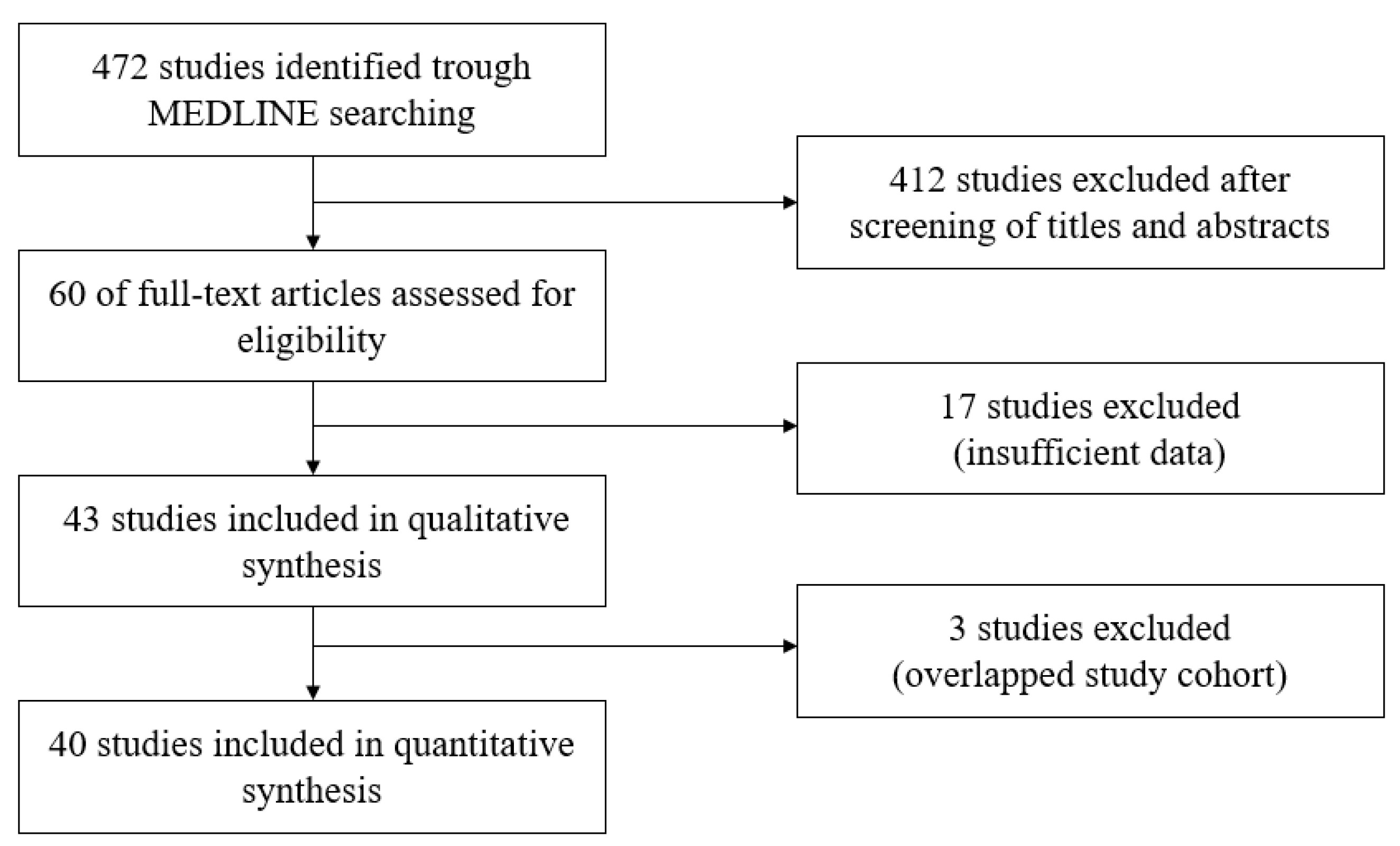
2.1. Search Results
2.2. Characteristics of Included Studies
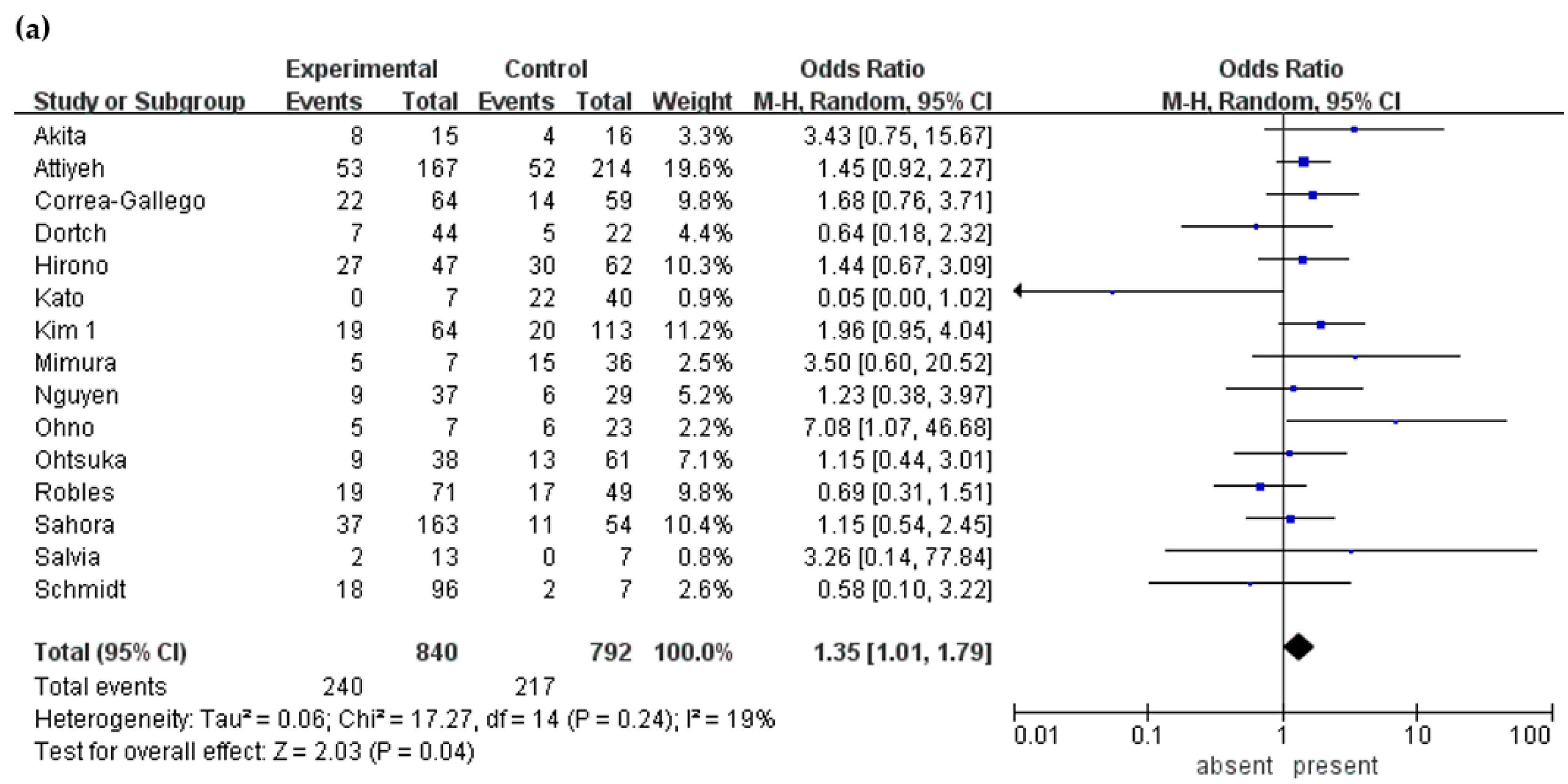
2.3. Clinical Symptoms
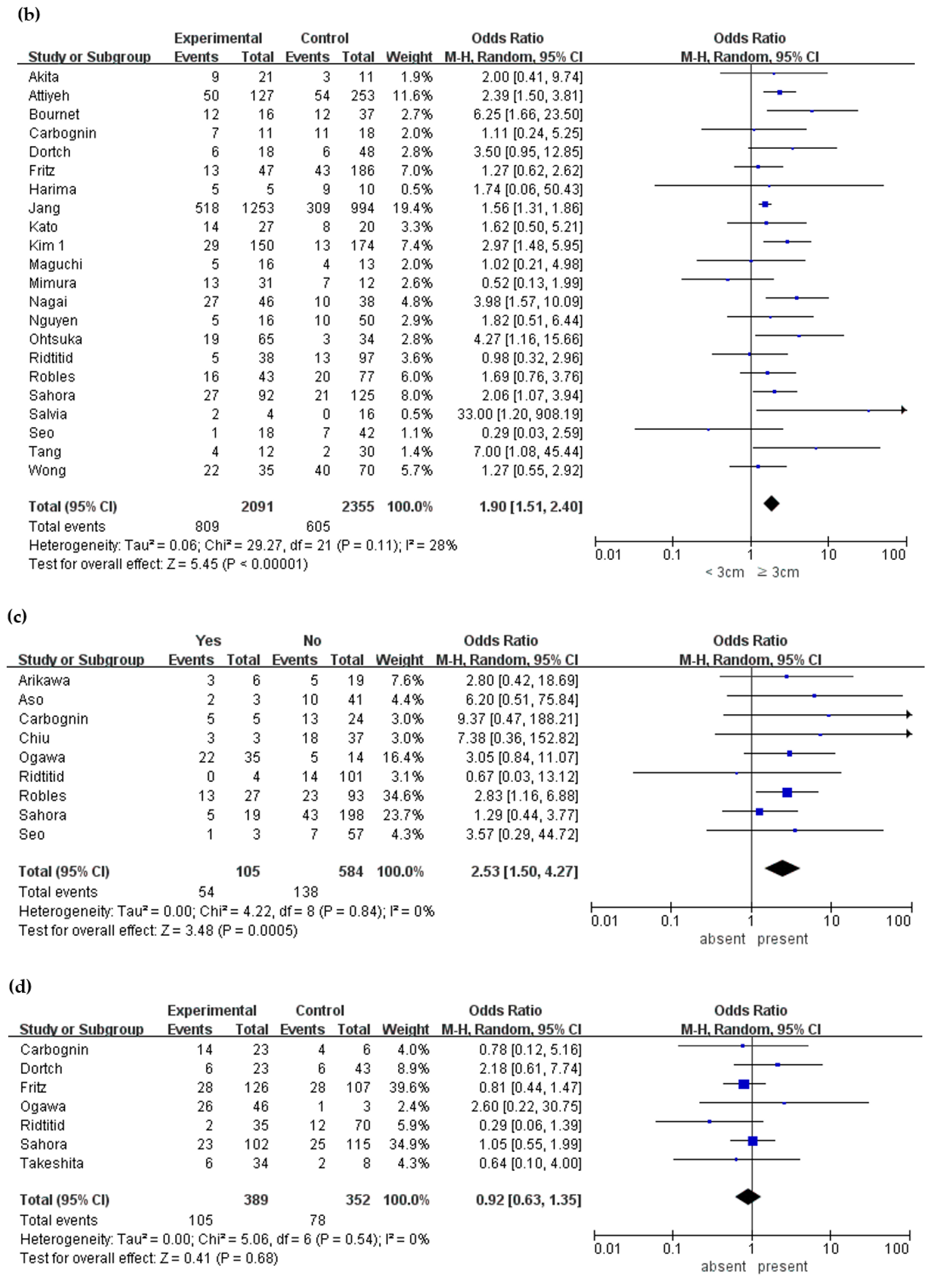
2.4. Characteristics of Cyst
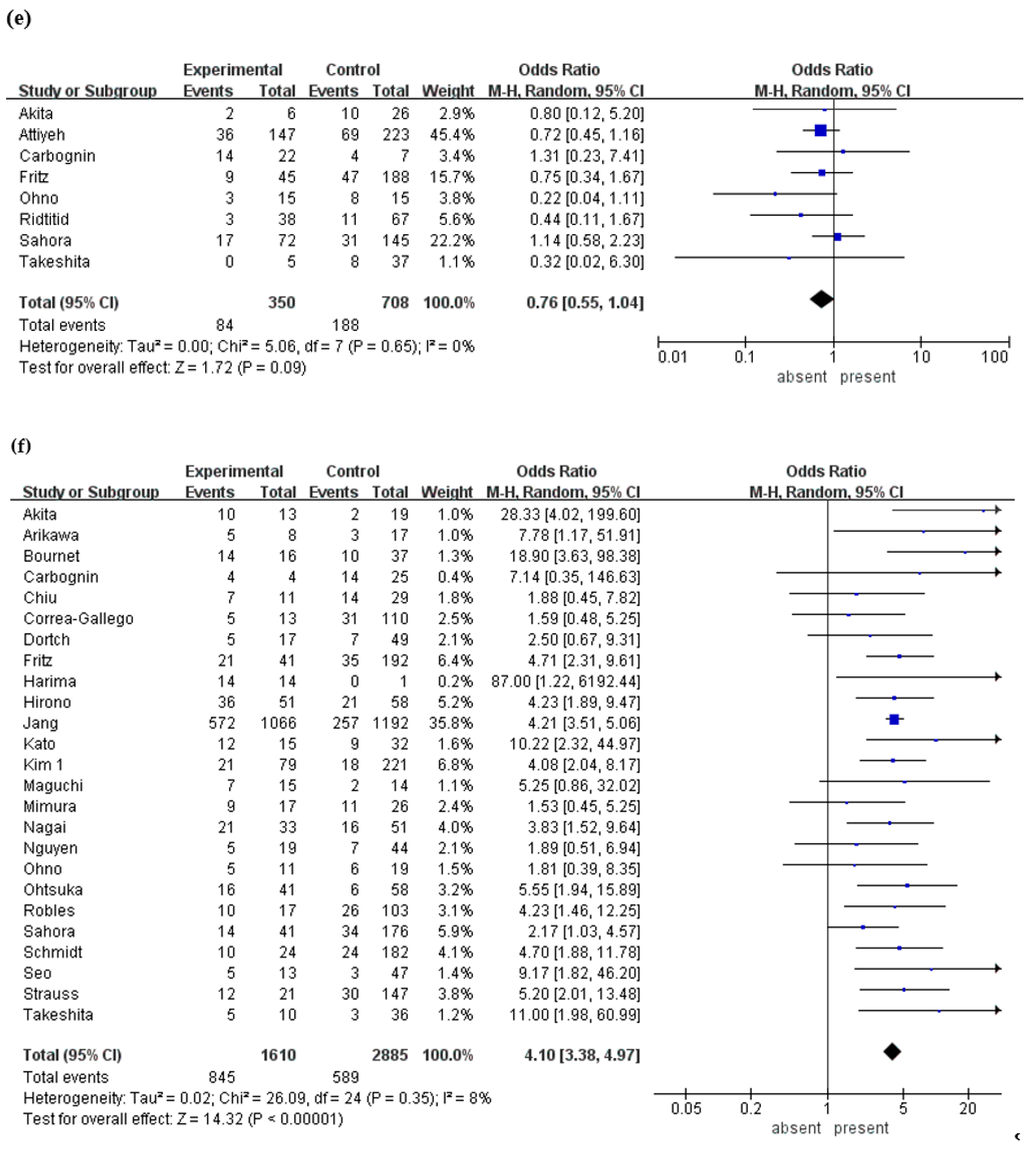
2.5. Mural Nodule
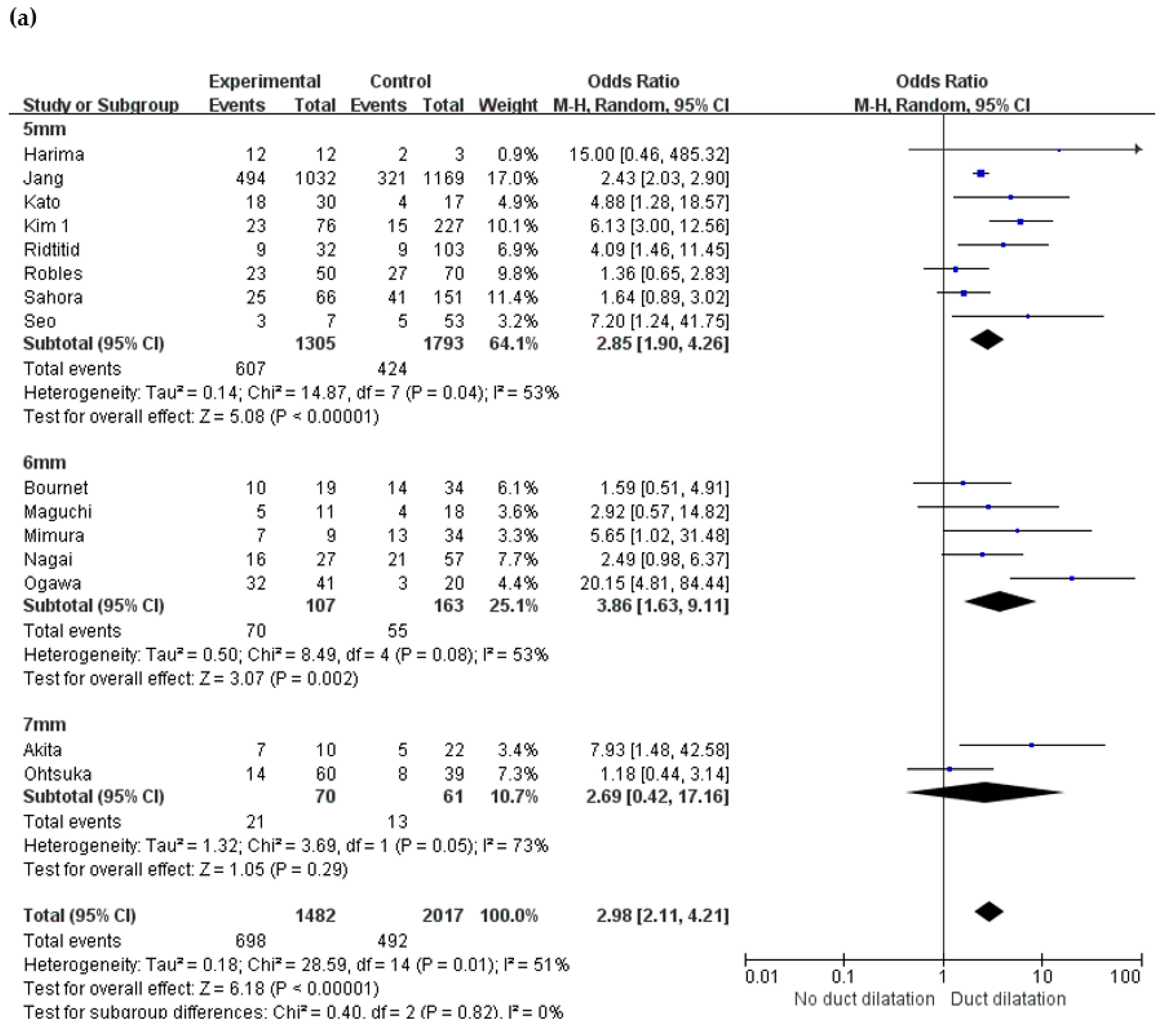
2.6. Changes in Main Pancreatic Duct
2.7. Lymphadenopathy
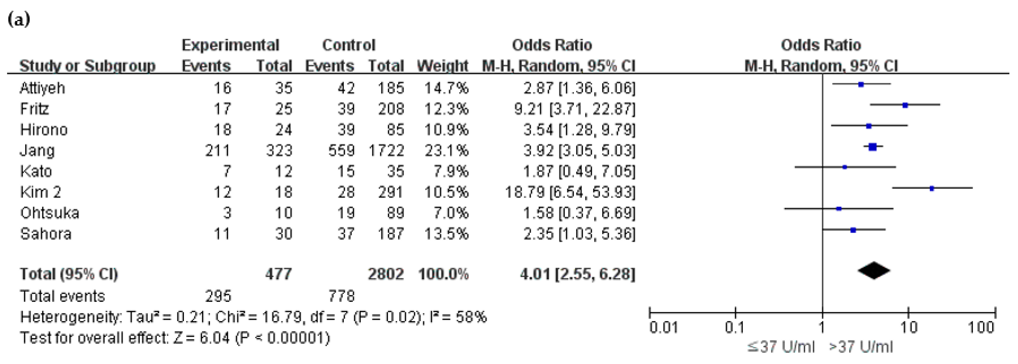
2.8. Biochemical Markers
3. Discussion
4. Materials and Methods
4.1. Literature Search Strategy
4.2. Inclusion/Exclusion Criteria
4.3. Data Extraction and Quality Assessment
4.4. Data Analysis
5. Conclusions
Author Contributions
Funding
Conflicts of Interest
References
- Tanaka, M.; Fernandez-del Castillo, C.; Adsay, V.; Chari, S.; Falconi, M.; Jang, J.Y.; Kimura, W.; Levy, P.; Pitman, M.B.; Schmidt, C.M.; et al. International consensus guidelines 2012 for the management of IPMN and MCN of the pancreas. Pancreatology 2012, 12, 183–197. [Google Scholar] [CrossRef]
- Ohashi, K.; Murakami, Y.; Maruyama, M. Four cases of “mucin-producing” cancer of the pancreas on specific findings of the papilla of Vater. Prog. Dig. Endosc. 1982, 20, 348–352. [Google Scholar]
- Siegel, R.L.; Miller, K.D.; Jemal, A. Cancer statistics, 2019. CA Cancer J. Clin. 2019, 69, 7–34. [Google Scholar] [CrossRef] [PubMed]
- Chang, Y.R.; Park, J.K.; Jang, J.Y.; Kwon, W.; Yoon, J.H.; Kim, S.W. Incidental pancreatic cystic neoplasms in an asymptomatic healthy population of 21,745 individuals: Large-scale, single-center cohort study. Medicine (Baltimore) 2016, 95, e5535. [Google Scholar] [CrossRef] [PubMed]
- Fernandez-del Castillo, C.; Targarona, J.; Thayer, S.P.; Rattner, D.W.; Brugge, W.R.; Warshaw, A.L. Incidental pancreatic cysts: Clinicopathologic characteristics and comparison with symptomatic patients. Arch. Surg. 2003, 138, 423–427. [Google Scholar] [CrossRef]
- Vege, S.S.; Ziring, B.; Jain, R.; Moayyedi, P.; Clinical Guidelines, C.; American Gastroenterology, A. American gastroenterological association institute guideline on the diagnosis and management of asymptomatic neoplastic pancreatic cysts. Gastroenterology 2015, 148, 819–822, quize 812–813. [Google Scholar] [CrossRef]
- Del Chiaro, M.; Verbeke, C.; Salvia, R.; Kloppel, G.; Werner, J.; McKay, C.; Friess, H.; Manfredi, R.; Van Cutsem, E.; Lohr, M.; et al. European experts consensus statement on cystic tumours of the pancreas. Dig. Liver Dis. 2013, 45, 703–711. [Google Scholar] [CrossRef] [PubMed]
- European Study Group on Cystic Tumours of the Pancreas. European evidence-based guidelines on pancreatic cystic neoplasms. Gut 2018, 67, 789–804. [Google Scholar] [CrossRef] [PubMed]
- Tanaka, M.; Fernandez-Del Castillo, C.; Kamisawa, T.; Jang, J.Y.; Levy, P.; Ohtsuka, T.; Salvia, R.; Shimizu, Y.; Tada, M.; Wolfgang, C.L. Revisions of international consensus Fukuoka guidelines for the management of IPMN of the pancreas. Pancreatology 2017, 17, 738–753. [Google Scholar] [CrossRef] [PubMed]
- Tanaka, M.; Chari, S.; Adsay, V.; Fernandez-del Castillo, C.; Falconi, M.; Shimizu, M.; Yamaguchi, K.; Yamao, K.; Matsuno, S. International consensus guidelines for management of intraductal papillary mucinous neoplasms and mucinous cystic neoplasms of the pancreas. Pancreatology 2006, 6, 17–32. [Google Scholar] [CrossRef] [PubMed]
- Akahoshi, K.; Ono, H.; Akasu, M.; Ban, D.; Kudo, A.; Konta, A.; Tanaka, S.; Tanabe, M. Rapid growth speed of cysts can predict malignant intraductal mucinous papillary neoplasms. J. Surg. Res. 2018, 231, 195–200. [Google Scholar] [CrossRef] [PubMed]
- Akita, H.; Takeda, Y.; Hoshino, H.; Wada, H.; Kobayashi, S.; Marubashi, S.; Eguchi, H.; Tanemura, M.; Mori, M.; Doki, Y.; et al. Mural nodule in branch duct-type intraductal papillary mucinous neoplasms of the pancreas is a marker of malignant transformation and indication for surgery. Am. J. Surg. 2011, 202, 214–219. [Google Scholar] [CrossRef] [PubMed]
- Arikawa, S.; Uchida, M.; Uozumi, J.; Sakoda, J.; Kaida, H.; Kunou, Y.; Hirose, Y.; Abe, T.; Hayabuchi, N.; Naito, Y.; et al. Utility of multidetector row CT in diagnosing branch duct IPMNs of the pancreas compared with MR cholangiopancreatography and endoscopic ultrasonography. Kurume Med. J. 2011, 57, 91–100. [Google Scholar] [CrossRef] [PubMed]
- Aso, T.; Ohtsuka, T.; Matsunaga, T.; Kimura, H.; Watanabe, Y.; Tamura, K.; Ideno, N.; Osoegawa, T.; Takahata, S.; Shindo, K.; et al. “High-risk stigmata” of the 2012 international consensus guidelines correlate with the malignant grade of branch duct intraductal papillary mucinous neoplasms of the pancreas. Pancreas 2014, 43, 1239–1243. [Google Scholar] [CrossRef] [PubMed]
- Attiyeh, M.A.; Fernandez-Del Castillo, C.; Al Efishat, M.; Eaton, A.A.; Gonen, M.; Batts, R.; Pergolini, I.; Rezaee, N.; Lillemoe, K.D.; Ferrone, C.R.; et al. Development and Validation of a Multi-institutional Preoperative Nomogram for Predicting Grade of Dysplasia in Intraductal Papillary Mucinous Neoplasms (IPMNs) of the Pancreas: A Report from The Pancreatic Surgery Consortium. Ann. Surg. 2018, 267, 157–163. [Google Scholar] [CrossRef]
- Bournet, B.; Kirzin, S.; Carrere, N.; Portier, G.; Otal, P.; Selves, J.; Musso, C.; Suc, B.; Moreau, J.; Fourtanier, G.; et al. Clinical fate of branch duct and mixed forms of intraductal papillary mucinous neoplasia of the pancreas. J. Gastroenterol. Hepatol. 2009, 24, 1211–1217. [Google Scholar] [CrossRef]
- Carbognin, G.; Zamboni, G.; Pinali, L.; Chiara, E.D.; Girardi, V.; Salvia, R.; Mucelli, R.P. Branch duct IPMTs: Value of cross-sectional imaging in the assessment of biological behavior and follow-up. Abdom. Imaging 2006, 31, 320–325. [Google Scholar] [CrossRef]
- Chiu, S.S.; Lim, J.H.; Lee, W.J.; Chang, K.T.; Oh, D.K.; Lee, K.T.; Lee, J.K.; Choi, S.H. Intraductal papillary mucinous tumour of the pancreas: Differentiation of malignancy and benignancy by CT. Clin. Radiol. 2006, 61, 776–783. [Google Scholar] [CrossRef]
- Correa-Gallego, C.; Do, R.; Lafemina, J.; Gonen, M.; D’Angelica, M.I.; DeMatteo, R.P.; Fong, Y.; Kingham, T.P.; Brennan, M.F.; Jarnagin, W.R.; et al. Predicting dysplasia and invasive carcinoma in intraductal papillary mucinous neoplasms of the pancreas: Development of a preoperative nomogram. Ann. Surg. Oncol. 2013, 20, 4348–4355. [Google Scholar] [CrossRef]
- Dortch, J.D.; Stauffer, J.A.; Asbun, H.J. Pancreatic Resection for Side-Branch Intraductal Papillary Mucinous Neoplasm (SB-IPMN): A Contemporary Single-Institution Experience. J. Gastrointest. Surg. 2015, 19, 1603–1609. [Google Scholar] [CrossRef]
- Fritz, S.; Klauss, M.; Bergmann, F.; Strobel, O.; Schneider, L.; Werner, J.; Hackert, T.; Buchler, M.W. Pancreatic main-duct involvement in branch-duct IPMNs: An underestimated risk. Ann. Surg. 2014, 260, 848–855. [Google Scholar] [CrossRef]
- Harima, H.; Kaino, S.; Shinoda, S.; Kawano, M.; Suenaga, S.; Sakaida, I. Differential diagnosis of benign and malignant branch duct intraductal papillary mucinous neoplasm using contrast-enhanced endoscopic ultrasonography. World J. Gastroenterol. 2015, 21, 6252–6260. [Google Scholar] [CrossRef]
- Hirono, S.; Kawai, M.; Okada, K.I.; Miyazawa, M.; Shimizu, A.; Kitahata, Y.; Ueno, M.; Yanagisawa, A.; Yamaue, H. Factors Associated With Invasive Intraductal Papillary Mucinous Carcinoma of the Pancreas. JAMA Surg. 2017, 152, e165054. [Google Scholar] [CrossRef] [PubMed]
- Jang, J.Y.; Park, T.; Lee, S.; Kim, Y.; Lee, S.Y.; Kim, S.W.; Kim, S.C.; Song, K.B.; Yamamoto, M.; Hatori, T.; et al. Proposed Nomogram Predicting the Individual Risk of Malignancy in the Patients with Branch Duct Type Intraductal Papillary Mucinous Neoplasms of the Pancreas. Ann. Surg. 2017, 266, 1062–1068. [Google Scholar] [CrossRef] [PubMed]
- Kato, Y.; Takahashi, S.; Gotohda, N.; Konishi, M. Risk factors for malignancy in branched-type intraductal papillary mucinous neoplasms of the pancreas during the follow-up period. World J. Surg. 2015, 39, 244–250. [Google Scholar] [CrossRef] [PubMed]
- Kim, Y.I.; Shin, S.H.; Song, K.B.; Hwang, D.W.; Lee, J.H.; Park, K.M.; Lee, Y.J.; Kim, S.C. Branch duct intraductal papillary mucinous neoplasm of the pancreas: Single-center experience with 324 patients who underwent surgical resection. Korean J. Hepatobiliary Pancreat. Surg. 2015, 19, 113–120. [Google Scholar] [CrossRef]
- Kim, T.H.; Song, T.J.; Hwang, J.H.; Yoo, K.S.; Lee, W.J.; Lee, K.H.; Dong, S.H.; Park, C.H.; Park, E.T.; Moon, J.H.; et al. Predictors of malignancy in pure branch duct type intraductal papillary mucinous neoplasm of the pancreas: A nationwide multicenter study. Pancreatology 2015, 15, 405–410. [Google Scholar] [CrossRef]
- Koshita, S.; Noda, Y.; Ito, K.; Kanno, Y.; Ogawa, T.; Masu, K.; Masaki, Y.; Horaguchi, J.; Oikawa, M.; Tsuchiya, T.; et al. Pancreatic juice cytology with immunohistochemistry to detect malignancy and histologic subtypes in patients with branch duct type intraductal papillary mucinous neoplasms of the pancreas. Gastrointest. Endosc. 2017, 85, 1036–1046. [Google Scholar] [CrossRef]
- Lee, K.H.; Lee, S.J.; Lee, J.K.; Ryu, J.K.; Kim, E.Y.; Kim, T.H.; Moon, J.H.; Lee, W.J.; Cho, Y.K.; Kim, J.J. Prediction of malignancy with endoscopic ultrasonography in patients with branch duct-type intraductal papillary mucinous neoplasm. Pancreas 2014, 43, 1306–1311. [Google Scholar] [CrossRef]
- Maguchi, H.; Tanno, S.; Mizuno, N.; Hanada, K.; Kobayashi, G.; Hatori, T.; Sadakari, Y.; Yamaguchi, T.; Tobita, K.; Doi, R.; et al. Natural history of branch duct intraductal papillary mucinous neoplasms of the pancreas: A multicenter study in Japan. Pancreas 2011, 40, 364–370. [Google Scholar] [CrossRef]
- Mimura, T.; Masuda, A.; Matsumoto, I.; Shiomi, H.; Yoshida, S.; Sugimoto, M.; Sanuki, T.; Yoshida, M.; Fujita, T.; Kutsumi, H.; et al. Predictors of malignant intraductal papillary mucinous neoplasm of the pancreas. J. Clin. Gastroenterol. 2010, 44, e224–e229. [Google Scholar] [CrossRef] [PubMed]
- Nagai, K.; Doi, R.; Ito, T.; Kida, A.; Koizumi, M.; Masui, T.; Kawaguchi, Y.; Ogawa, K.; Uemoto, S. Single-institution validation of the international consensus guidelines for treatment of branch duct intraductal papillary mucinous neoplasms of the pancreas. J. Hepatobiliary Pancreat. Surg. 2009, 16, 353–358. [Google Scholar] [CrossRef] [PubMed]
- Nguyen, A.H.; Toste, P.A.; Farrell, J.J.; Clerkin, B.M.; Williams, J.; Muthusamy, V.R.; Watson, R.R.; Tomlinson, J.S.; Hines, O.J.; Reber, H.A.; et al. Current recommendations for surveillance and surgery of intraductal papillary mucinous neoplasms may overlook some patients with cancer. J. Gastrointest. Surg. 2015, 19, 258–265. [Google Scholar] [CrossRef] [PubMed]
- Ogawa, H.; Itoh, S.; Ikeda, M.; Suzuki, K.; Naganawa, S. Intraductal papillary mucinous neoplasm of the pancreas: Assessment of the likelihood of invasiveness with multisection CT. Radiology 2008, 248, 876–886. [Google Scholar] [CrossRef]
- Ohno, E.; Itoh, A.; Kawashima, H.; Ishikawa, T.; Matsubara, H.; Itoh, Y.; Nakamura, Y.; Hiramatsu, T.; Nakamura, M.; Miyahara, R.; et al. Malignant transformation of branch duct-type intraductal papillary mucinous neoplasms of the pancreas based on contrast-enhanced endoscopic ultrasonography morphological changes: Focus on malignant transformation of intraductal papillary mucinous neoplasm itself. Pancreas 2012, 41, 855–862. [Google Scholar] [PubMed]
- Ohtsuka, T.; Kono, H.; Nagayoshi, Y.; Mori, Y.; Tsutsumi, K.; Sadakari, Y.; Takahata, S.; Morimatsu, K.; Aishima, S.; Igarashi, H.; et al. An increase in the number of predictive factors augments the likelihood of malignancy in branch duct intraductal papillary mucinous neoplasm of the pancreas. Surgery 2012, 151, 76–83. [Google Scholar] [CrossRef]
- Ridtitid, W.; DeWitt, J.M.; Schmidt, C.M.; Roch, A.; Stuart, J.S.; Sherman, S.; Al-Haddad, M.A. Management of branch-duct intraductal papillary mucinous neoplasms: A large single-center study to assess predictors of malignancy and long-term outcomes. Gastrointest. Endosc. 2016, 84, 436–445. [Google Scholar] [CrossRef]
- Robles, E.P.; Maire, F.; Cros, J.; Vullierme, M.P.; Rebours, V.; Sauvanet, A.; Aubert, A.; Dokmak, S.; Levy, P.; Ruszniewski, P. Accuracy of 2012 International Consensus Guidelines for the prediction of malignancy of branch-duct intraductal papillary mucinous neoplasms of the pancreas. United Eur. Gastroenterol. J. 2016, 4, 580–586. [Google Scholar] [CrossRef]
- Rodriguez, J.R.; Salvia, R.; Crippa, S.; Warshaw, A.L.; Bassi, C.; Falconi, M.; Thayer, S.P.; Lauwers, G.Y.; Capelli, P.; Mino-Kenudson, M.; et al. Branch-duct intraductal papillary mucinous neoplasms: Observations in 145 patients who underwent resection. Gastroenterology 2007, 133, 72–79, quize 309–310. [Google Scholar] [CrossRef]
- Sahora, K.; Mino-Kenudson, M.; Brugge, W.; Thayer, S.P.; Ferrone, C.R.; Sahani, D.; Pitman, M.B.; Warshaw, A.L.; Lillemoe, K.D.; Fernandez-del Castillo, C.F. Branch duct intraductal papillary mucinous neoplasms: Does cyst size change the tip of the scale? A critical analysis of the revised international consensus guidelines in a large single-institutional series. Ann. Surg. 2013, 258, 466–475. [Google Scholar] [CrossRef]
- Salvia, R.; Crippa, S.; Falconi, M.; Bassi, C.; Guarise, A.; Scarpa, A.; Pederzoli, P. Branch-duct intraductal papillary mucinous neoplasms of the pancreas: To operate or not to operate? Gut 2007, 56, 1086–1090. [Google Scholar] [CrossRef] [PubMed]
- Schmidt, C.M.; White, P.B.; Waters, J.A.; Yiannoutsos, C.T.; Cummings, O.W.; Baker, M.; Howard, T.J.; Zyromski, N.J.; Nakeeb, A.; DeWitt, J.M.; et al. Intraductal papillary mucinous neoplasms: Predictors of malignant and invasive pathology. Ann. Surg. 2007, 246, 644–651. [Google Scholar] [CrossRef] [PubMed]
- Seo, N.; Byun, J.H.; Kim, J.H.; Kim, H.J.; Lee, S.S.; Song, K.B.; Kim, S.C.; Han, D.J.; Hong, S.M.; Lee, M.G. Validation of the 2012 International Consensus Guidelines Using Computed Tomography and Magnetic Resonance Imaging: Branch Duct and Main Duct Intraductal Papillary Mucinous Neoplasms of the Pancreas. Ann. Surg. 2016, 263, 557–564. [Google Scholar] [CrossRef] [PubMed]
- Serikawa, M.; Sasaki, T.; Fujimoto, Y.; Kuwahara, K.; Chayama, K. Management of intraductal papillary-mucinous neoplasm of the pancreas: Treatment strategy based on morphologic classification. J. Clin. Gastroenterol. 2006, 40, 856–862. [Google Scholar] [CrossRef]
- Shimizu, Y.; Hijioka, S.; Hirono, S.; Kin, T.; Ohtsuka, T.; Kanno, A.; Koshita, S.; Hanada, K.; Kitano, M.; Inoue, H.; et al. New Model for Predicting Malignancy in Patients with Intraductal Papillary Mucinous Neoplasm. Ann. Surg. 2020, 272, 155–162. [Google Scholar] [CrossRef]
- Strauss, A.; Birdsey, M.; Fritz, S.; Schwarz-Bundy, B.D.; Bergmann, F.; Hackert, T.; Kauczor, H.U.; Grenacher, L.; Klauss, M. Intraductal papillary mucinous neoplasms of the pancreas: Radiological predictors of malignant transformation and the introduction of bile duct dilation to current guidelines. Br. J. Radiol. 2016, 89, 20150853. [Google Scholar] [CrossRef]
- Takeshita, K.; Kutomi, K.; Takada, K.; Haruyama, T.; Fukushima, J.; Aida, R.; Takada, T.; Furui, S. Differential diagnosis of benign or malignant intraductal papillary mucinous neoplasm of the pancreas by multidetector row helical computed tomography: Evaluation of predictive factors by logistic regression analysis. J. Comput. Assist. Tomogr. 2008, 32, 191–197. [Google Scholar] [CrossRef]
- Tang, R.S.; Weinberg, B.; Dawson, D.W.; Reber, H.; Hines, O.J.; Tomlinson, J.S.; Chaudhari, V.; Raman, S.; Farrell, J.J. Evaluation of the guidelines for management of pancreatic branch-duct intraductal papillary mucinous neoplasm. Clin. Gastroenterol. Hepatol. 2008, 6, 815–819, quiz 719. [Google Scholar] [CrossRef]
- Wong, J.; Weber, J.; Centeno, B.A.; Vignesh, S.; Harris, C.L.; Klapman, J.B.; Hodul, P. High-grade dysplasia and adenocarcinoma are frequent in side-branch intraductal papillary mucinous neoplasm measuring less than 3 cm on endoscopic ultrasound. J. Gastrointest. Surg. 2013, 17, 78–84. [Google Scholar] [CrossRef]
- Woo, S.M.; Ryu, J.K.; Lee, S.H.; Yoon, W.J.; Kim, Y.T.; Yoon, Y.B. Branch duct intraductal papillary mucinous neoplasms in a retrospective series of 190 patients. Br. J. Surg. 2009, 96, 405–411. [Google Scholar] [CrossRef]
- Matsumoto, T.; Aramaki, M.; Yada, K.; Hirano, S.; Himeno, Y.; Shibata, K.; Kawano, K.; Kitano, S. Optimal management of the branch duct type intraductal papillary mucinous neoplasms of the pancreas. J. Clin. Gastroenterol. 2003, 36, 261–265. [Google Scholar] [CrossRef] [PubMed]
- Sugiyama, M.; Izumisato, Y.; Abe, N.; Masaki, T.; Mori, T.; Atomi, Y. Predictive factors for malignancy in intraductal papillary-mucinous tumours of the pancreas. Br. J. Surg. 2003, 90, 1244–1249. [Google Scholar] [CrossRef]
- Walsh, R.M.; Vogt, D.P.; Henderson, J.M.; Hirose, K.; Mason, T.; Bencsath, K.; Hammel, J.; Brown, N. Management of suspected pancreatic cystic neoplasms based on cyst size. Surgery 2008, 144, 677–684. [Google Scholar] [CrossRef] [PubMed]
- Weinberg, B.M.; Spiegel, B.M.; Tomlinson, J.S.; Farrell, J.J. Asymptomatic pancreatic cystic neoplasms: Maximizing survival and quality of life using Markov-based clinical nomograms. Gastroenterology 2010, 138, 531–540. [Google Scholar] [CrossRef] [PubMed]
- Jang, J.Y.; Kim, S.W.; Lee, S.E.; Yang, S.H.; Lee, K.U.; Lee, Y.J.; Kim, S.C.; Han, D.J.; Choi, D.W.; Choi, S.H.; et al. Treatment guidelines for branch duct type intraductal papillary mucinous neoplasms of the pancreas: When can we operate or observe? Ann. Surg. Oncol. 2008, 15, 199–205. [Google Scholar] [CrossRef]
- Tanaka, M. Controversies in the management of pancreatic IPMN. Nat. Rev. Gastroenterol. Hepatol. 2011, 8, 56–60. [Google Scholar] [CrossRef] [PubMed]
- Scheiman, J.M.; Hwang, J.H.; Moayyedi, P. American gastroenterological association technical review on the diagnosis and management of asymptomatic neoplastic pancreatic cysts. Gastroenterology 2015, 148, 824–848, e822. [Google Scholar] [CrossRef]
- Uehara, H.; Ishikawa, O.; Katayama, K.; Kawada, N.; Ikezawa, K.; Fukutake, N.; Takakura, R.; Takano, Y.; Tanaka, S.; Takenaka, A. Size of mural nodule as an indicator of surgery for branch duct intraductal papillary mucinous neoplasm of the pancreas during follow-up. J. Gastroenterol. 2011, 46, 657–663. [Google Scholar] [CrossRef]
- Marchegiani, G.; Andrianello, S.; Borin, A.; Dal Borgo, C.; Perri, G.; Pollini, T.; Romano, G.; D’Onofrio, M.; Gabbrielli, A.; Scarpa, A.; et al. Systematic review, meta-analysis, and a high-volume center experience supporting the new role of mural nodules proposed by the updated 2017 international guidelines on IPMN of the pancreas. Surgery 2018, 163, 1272–1279. [Google Scholar] [CrossRef]
- Ohno, E.; Hirooka, Y.; Itoh, A.; Ishigami, M.; Katano, Y.; Ohmiya, N.; Niwa, Y.; Goto, H. Intraductal papillary mucinous neoplasms of the pancreas: Differentiation of malignant and benign tumors by endoscopic ultrasound findings of mural nodules. Ann. Surg. 2009, 249, 628–634. [Google Scholar] [CrossRef]
- Kawada, N.; Uehara, H.; Nagata, S.; Tsuchishima, M.; Tsutsumi, M.; Tomita, Y. Mural nodule of 10 mm or larger as predictor of malignancy for intraductal papillary mucinous neoplasm of the pancreas: Pathological and radiological evaluations. Pancreatology 2016, 16, 441–448. [Google Scholar] [CrossRef]
- Moher, D.; Liberati, A.; Tetzlaff, J.; Altman, D.G.; Group, P. Preferred reporting items for systematic reviews and meta-analyses: The PRISMA statement. BMJ 2009, 339, b2535. [Google Scholar] [CrossRef]
- Higgins, J.P.T.; Thomas, J.; Chandler, J.; Cumpston, M.; Li, T.; Page, M.J.; Welch, V.A. Cochrane Handbook for Systematic Reviews of Interventions, version 6.0 (updated July 2019); Cochrane: London, UK, 2019; Available online: www.training.cochrane.org/handbook (accessed on 20 July 2020).
- Stroup, D.F.; Berlin, J.A.; Morton, S.C.; Olkin, I.; Williamson, G.D.; Rennie, D.; Moher, D.; Becker, B.J.; Sipe, T.A.; Thacker, S.B. Meta-analysis of observational studies in epidemiology: A proposal for reporting. Meta-analysis Of Observational Studies in Epidemiology (MOOSE) group. JAMA 2000, 283, 2008–2012. [Google Scholar] [CrossRef]



| Study | Year | Study Period | No. of Patients | Mean Age (Years) | Male/Female | Type of IPMN | Benign | Malignant | Malignancy Proportion (%) | Diagnostic Modality |
|---|---|---|---|---|---|---|---|---|---|---|
| Akahoshi et al. [11] | 2018 | 2006–2017 | 50 | 68 | 33/17 | BD | 33 | 17 | 34.0% | CT, MRI |
| Akita et al. [12] | 2011 | 1992–2007 | 32 | 62.6 | 19/13 | BD | 20 | 12 | 37.5% | CT, MRI, MRCP |
| Arikawa et al. [13] | 2011 | 2003–2008 | 25 | 65.2 | 20/5 | BD | 17 | 8 | 32.0% | CT, MRCP, EUS |
| Aso et al. [14] | 2014 | 2006–2013 | 70 | N/A | N/A | BD | 42 | 28 | 40.0% | CT, MRCP, EUS |
| Attiyeh et al. [15] | 2018 | 2005–2015 | 381 | 67 | 160/221 | BD | 276 | 105 | 27.6% | Not stated |
| Bournet et al. [16] | 2009 | 1988–2005 | 53 | 63.9 | 52/47 | BD, mixed | 29 | 24 | 24.2% | CT, MRCP, EUS, ERCP |
| Carbognin et al. [17] | 2006 | 1995–2005 | 29 | Benign 64.7 Malignant 62.2 | 17/12 | BD, mixed | 11 | 18 | 62.1% | CT, MRI, MRCP |
| Chiu et al. [18] | 2006 | 1995–2005 | 40 | N/A | N/A | BD, MD, mixed | 30 | 10 | 25.0% | CT |
| Correa-Gallego et al. [19] | 2013 | 1994–2010 | 123 | 68 | 50/73 | BD | 87 | 36 | 29.3% | Not stated |
| Dortch et al. [20] | 2015 | 2002–2013 | 66 | 68 | 26/42 | BD | 54 | 12 | 18.2% | CT, MRCP, EUS, FNA |
| Fritz et al. [21] | 2014 | 2004–2012 | 233 | N/A | 95/138 | BD | 177 | 56 | 24.0% | CT, MRI |
| Harima et al. [22] | 2015 | 2009–2014 | 15 | N/A | N/A | BD | 1 | 14 | 93.3% | CT, EUS |
| Hirono et al. [23] | 2017 | 1999–2015 | 109 | N/A | 46/63 | BD | 52 | 57 | 52.3% | CT, EUS |
| Jang et al. [24] | 2017 | 1992–2012 | 2258 | 65.0 | 1408/850 | BD | 1429 | 829 | 36.7% | CT, EUS |
| Kato et al. [25] | 2015 | 1994–2012 | 47 | 66.2 | 30/17 | BD | 25 | 22 | 46.8% | Not stated |
| Kim YI et al. [26] | 2015 | 1997–2013 | 324 | 62 | 179/145 | BD | 282 | 42 | 13.0% | CT, MRCP, EUS, ERCP |
| Kim TH et al. [27] | 2015 | 2004–2012 | 177 | 63 | 108/69 | BD | 138 | 39 | 22.0% | CT, EUS |
| Koshita et al. [28] | 2017 | 2005–2014 | 28 | 62.2 | 17/11 | BD | 14 | 14 | 50.0% | CT, MRCP, EUS, ERCP |
| Lee et al. [29] | 2014 | 2002–2011 | 84 | 64.7 | 55/29 | BD | 68 | 16 | 19.0% | EUS |
| Maguchi et al. [30] | 2011 | N/A | 29 | N/A | N/A | BD | 20 | 9 | 31.0% | CT, EUS |
| Mimura et al. [31] | 2010 | 1998–2009 | 43 | Benign 66.0 Malignant 66.7 | 29/14 | BD, mixed | 23 | 20 | 46.5% | CT, EUS |
| Nagai et al. [32] | 2009 | 1984–2007 | 84 | 63 | 48/36 | BD | 47 | 37 | 44.0% | CT, ERCP, MRI, EUS |
| Nguyen et al. [33] | 2015 | 1996–2012 | 66 | 69 | 26/42 | BD | 51 | 15 | 22.7% | CT, MRI, EUS |
| Ogawa et al. [34] | 2008 | 2000–2006 | 49 | 64.9 | 39/20 | BD | 22 | 27 | 55.1% | CT |
| Ohno et al. [35] | 2012 | 2001–2009 | 30 | 65.1 | 15/15 | BD | 19 | 11 | 63.3% | CT, ERCP, CE-EUS |
| Ohtsuka et al. [36] | 2012 | 1990–2009 | 99 | NA | 60/39 | BD | 77 | 22 | 22.2% | CT, MRCP, US, EUS |
| Ridtitid et al. [37] | 2016 | 2001–2013 | 135 | 65.2 | 71/64 | BD | 117 | 18 | 13.3% | CT, MRI, EUS |
| Robles et al. [38] | 2016 | 2006–2014 | 120 | 57.9 | 65/55 | BD | 84 | 36 | 30.0% | CT, MRI, EUS |
| Rodriguez et al. [39] | 2007 | 1990–2005 | 145 | 67* | 62/83 | BD | 113 | 32 | 22.1% | CEUS, CT, MRI |
| Sahora et al. [40] | 2013 | 1995–2012 | 217 | N/A | 82/135 | BD, mixed | 169 | 48 | 22.1% | CT, MRI, MRCP, EUS |
| Salvia et al. [41] | 2007 | 2000–2003 | 20 | 58 | 10/10 | BD | 18 | 2 | 10.0% | US, MRI, MRCP, CEUS, EUS, ERCP |
| Schmidt et al. [42] | 2007 | 1991–2006 | 103 | 63 | 50/53 | BD | 83 | 20 | 19.4% | CT, MRI, ERCP, EUS |
| Seo et al. [43] | 2016 | 2011–2013 | 60 | 64.3 | 35/25 | BD | 52 | 8 | 13.3% | CT, MRI |
| Serikawa et al. [44] | 2006 | 1992–2005 | 56 | 65.8 | 42/14 | BD | 49 | 7 | 10.3% | US, EUS, CT, ERCP, MRCP |
| Shimizu et al. [45] | 2020 | 1996–2014 | 466 | 67.9 | 274/192 | BD, MD, mixed | 208 | 258 | 55.4% | CT, EUS, MRCP |
| Strauss et al. [46] | 2016 | 2004–2012 | 168 | N/A | N/A | BD | 126 | 42 | 25.0% | CT, MRI, MRCP |
| Takeshita et al. [47] | 2008 | 2002–2006 | 46 | 65 | 28/25 | BD | 38 | 8 | 17.4% | CT |
| Tang et al. [48] | 2008 | 1995–2006 | 31 | 66.5 | 10/21 | BD | 26 | 5 | 16.1% | CT, MRI, MRCP, ERCP, EUS |
| Wong et al. [49] | 2013 | 2000–2010 | 105 | 68 | 47/58 | BD | 43 | 62 | 59.0% | CT, MRI, EUS |
| Woo et al. [50] | 2009 | 1998–2005 | 85 | 63 | 50/35 | BD | 71 | 14 | 16.5% | CT, EUS, ERCP, MR |
| Parameters | No. Studies | No. of Patient | No. of Positive Feature (%) | No. of Malignancy (%) | No. of Malignancy among Positive Features (%) | No. of Malignancy among Negative Features (%) | OR | 95% CI | p-Value |
|---|---|---|---|---|---|---|---|---|---|
| Symptoms (+) | 16 | 2844 | 1089 (38.3) | 966 (34.0) | 369 (33.9) | 597 (34.0) | 1.35 | 1.01, 1.79 | 0.040 |
| Cyst size (≥3 cm) | 22 | 4446 | 2091 (47.0) | 1414 (31.8) | 814 (38.9) | 605 (25.7) | 1.90 | 1.51, 2.40 | <0.001 |
| Wall thickening | 9 | 689 | 105 (15.2) | 192 (27.9) | 54 (51.4) | 138 (23.6) | 2.53 | 1.50, 4.27 | <0.001 |
| Multilocular | 7 | 741 | 389 (52.5) | 183 (24.7) | 105 (27.0) | 78 (22.2) | 0.92 | 0.63, 1.35 | 0.68 |
| Multiplicity | 8 | 1058 | 350 (33.1) | 272 (25.7) | 84 (24.0) | 188 (26.6) | 0.76 | 0.55, 1.04 | 0.09 |
| Mural nodule | 25 | 4495 | 1610 (35.8) | 1434 (31.9) | 845 (52.5) | 589 (20.4) | 4.10 | 3.38, 4.97 | <0.001 |
| MPD dilatation | 15 | 3499 | 1482 (42.4) | 1190 (34.0) | 698 (47.1) | 492 (24.4) | 2.98 | 2.11, 4.21 | <0.001 |
| > 5 mm | 8 | 3098 | 1305 (42.1) | 1031 (33.3) | 607 (46.5) | 424 (23.6) | 2.85 | 1.90, 4.26 | <0.001 |
| > 6 mm | 5 | 270 | 107 (39.6) | 125 (46.3) | 70 (65.4) | 55 (33.7) | 3.86 | 1.63, 9.11 | 0.002 |
| > 7 mm | 2 | 131 | 70 (53.4) | 72 (55.0) | 21 (30.0) | 13 (21.3) | 2.69 | 0.42, 17.16 | 0.29 |
| Abrupt caliber change | 4 | 467 | 34 (7.3) | 74 (15.8) | 18 (52.9) | 56 (12.9) | 7.41 | 2.49, 22.06 | <0.001 |
| Lymphadenopathy | 4 | 390 | 70 (17.9) | 70 (17.9) | 14 (20.0) | 56 (15.3) | 8.55 | 3.25, 22.51 | <0.001 |
| CA 19-9 (> 37 U/mL) | 8 | 3279 | 477 (14.5) | 1073 (32.7) | 295 (61.8) | 778 (27.8) | 4.01 | 2.55, 6.28 | <0.001 |
| CEA (> 5 ng/mL) | 4 | 2405 | 301 (12.5) | 912 (37.9) | 161 (53.5) | 751 (35.7) | 2.04 | 1.60, 2.61 | <0.001 |
© 2020 by the authors. Licensee MDPI, Basel, Switzerland. This article is an open access article distributed under the terms and conditions of the Creative Commons Attribution (CC BY) license (http://creativecommons.org/licenses/by/4.0/).
Share and Cite
Kwon, W.; Han, Y.; Byun, Y.; Kang, J.S.; Choi, Y.J.; Kim, H.; Jang, J.-Y. Predictive Features of Malignancy in Branch Duct Type Intraductal Papillary Mucinous Neoplasm of the Pancreas: A Meta-Analysis. Cancers 2020, 12, 2618. https://doi.org/10.3390/cancers12092618
Kwon W, Han Y, Byun Y, Kang JS, Choi YJ, Kim H, Jang J-Y. Predictive Features of Malignancy in Branch Duct Type Intraductal Papillary Mucinous Neoplasm of the Pancreas: A Meta-Analysis. Cancers. 2020; 12(9):2618. https://doi.org/10.3390/cancers12092618
Chicago/Turabian StyleKwon, Wooil, Youngmin Han, Yoonhyeong Byun, Jae Seung Kang, Yoo Jin Choi, Hongbeom Kim, and Jin-Young Jang. 2020. "Predictive Features of Malignancy in Branch Duct Type Intraductal Papillary Mucinous Neoplasm of the Pancreas: A Meta-Analysis" Cancers 12, no. 9: 2618. https://doi.org/10.3390/cancers12092618
APA StyleKwon, W., Han, Y., Byun, Y., Kang, J. S., Choi, Y. J., Kim, H., & Jang, J.-Y. (2020). Predictive Features of Malignancy in Branch Duct Type Intraductal Papillary Mucinous Neoplasm of the Pancreas: A Meta-Analysis. Cancers, 12(9), 2618. https://doi.org/10.3390/cancers12092618


