Central Pathology Review in SENTIX, a Prospective Observational International Study on Sentinel Lymph Node Biopsy in Patients with Early-Stage Cervical Cancer (ENGOT-CX2)

 ,
,  , , , ,
  ,
,  , , ,
, , ,  , , , add
Show full author list
, , , add
Show full author list
Abstract
1. Introduction
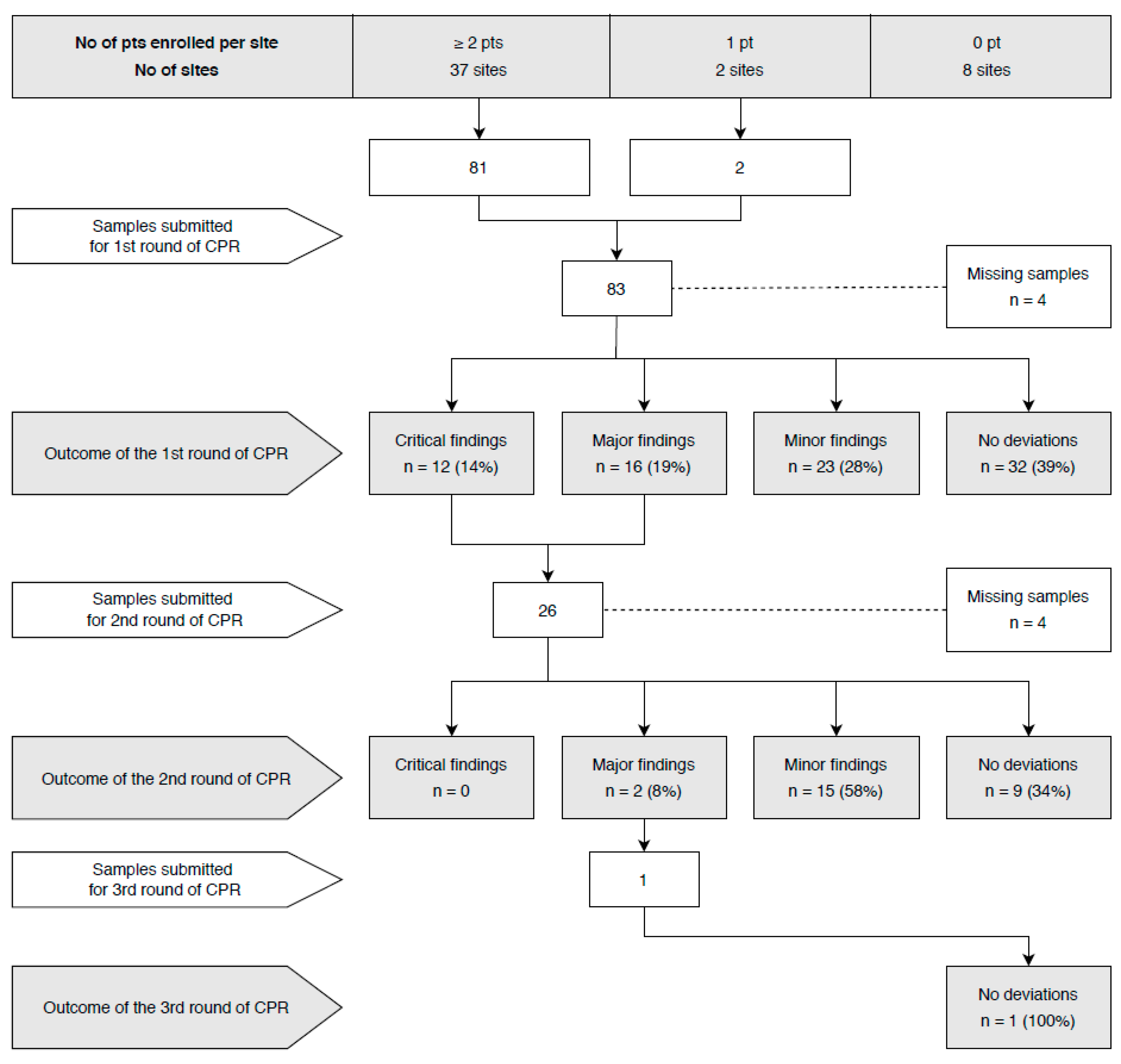
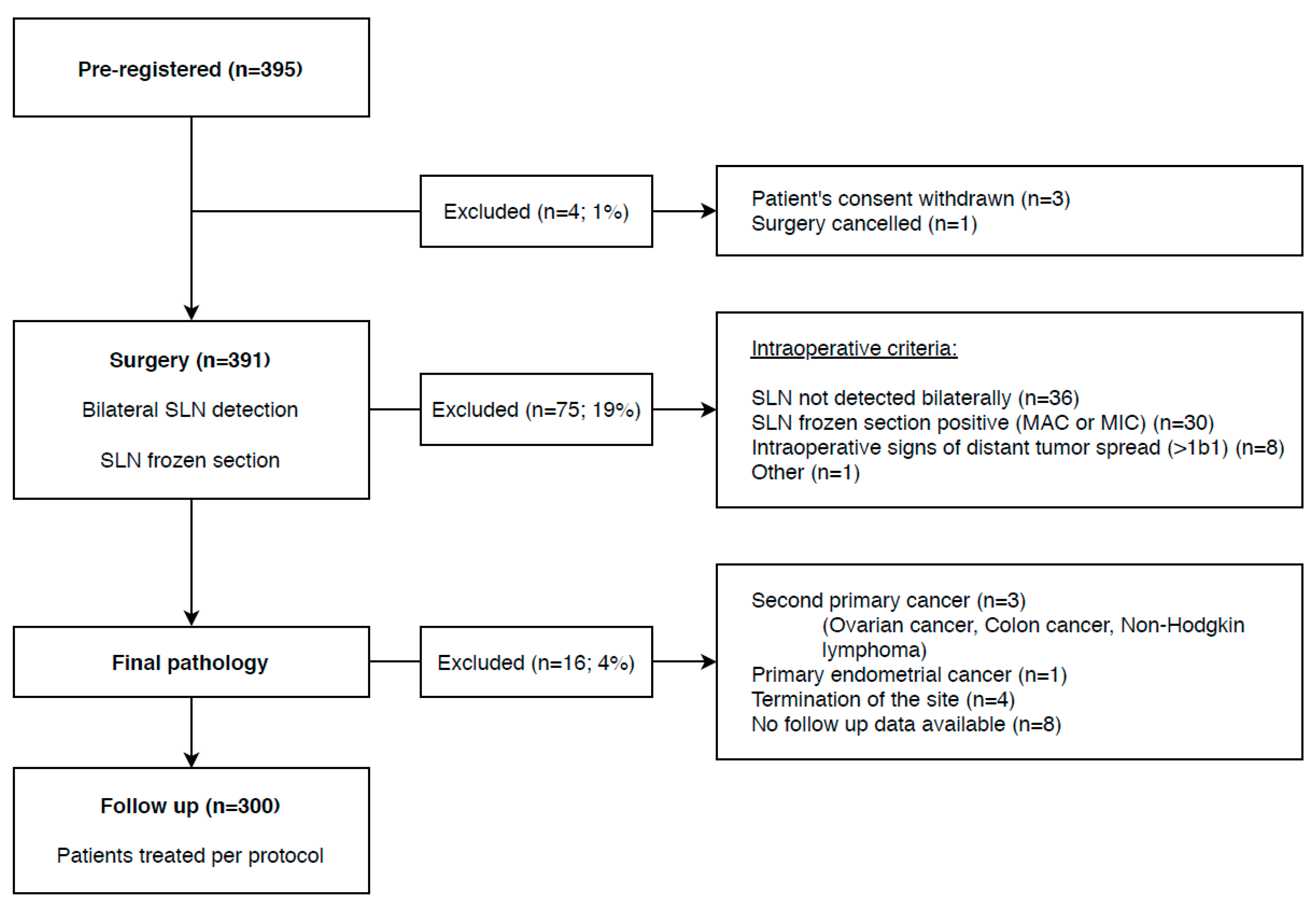
2. Results
3. Discussion
4. Materials and Methods
4.1. Ethics
4.2. Study Sites
4.3. Patients
4.4. SLN Detection
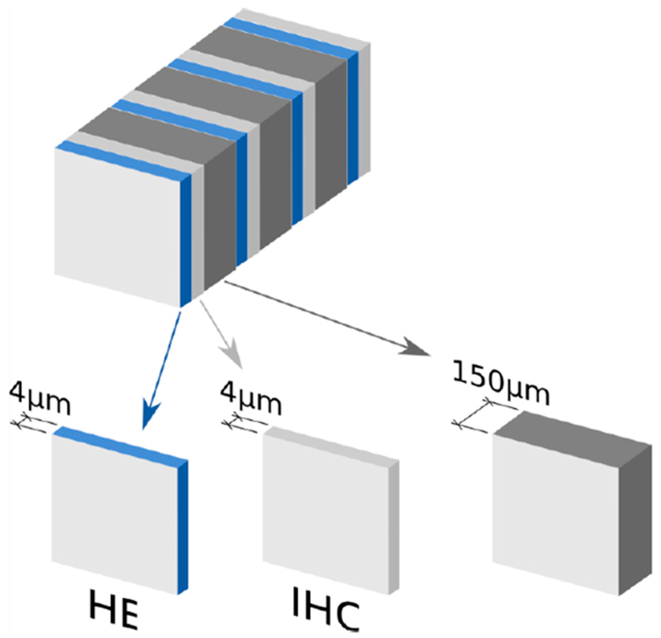
4.5. SLN Ultrastaging Protocol
4.6. Central Pathology Review
5. Conclusions
Author Contributions
Funding
Acknowledgments
Conflicts of Interest
References
- Cibula, D.; Dusek, J.; Jarkovsky, J.; Dundr, P.; Querleu, D.; van der Zee, A.; Kucukmetin, A.; Kocian, R. A prospective multicenter trial on sentinel lymph node biopsy in patients with early-stage cervical cancer (SENTIX). Int. J. Gynecol. Cancer 2019, 29, 212–215. [Google Scholar] [CrossRef] [PubMed]
- Delgado, G.; Bundy, B.; Zaino, R.; Sevin, B.U.; Creasman, W.T.; Major, F. Prospective surgical-pathological study of disease-free interval in patients with stage IB squamous cell carcinoma of the cervix: A Gynecologic Oncology Group study. Gynecol. Oncol. 1990, 38, 352–357. [Google Scholar] [CrossRef]
- Derks, M.; van der Velden, J.; de Kroon, C.D.; Nijman, H.W.; van Lonkhuijzen, L.; van der Zee, A.G.J.; Zwinderman, A.H.; Kenter, G.G. Surgical Treatment of Early-Stage Cervical Cancer: A Multi-Institution Experience in 2124 Cases in The Netherlands Over a 30-Year Period. Int. J. Gynecol. Cancer 2018, 28, 757–763. [Google Scholar] [CrossRef] [PubMed]
- Ho, C.M.; Chien, T.Y.; Huang, S.H.; Wu, C.J.; Shih, B.Y.; Chang, S.C. Multivariate analysis of the prognostic factors and outcomes in early cervical cancer patients undergoing radical hysterectomy. Gynecol. Oncol. 2004, 93, 458–464. [Google Scholar] [CrossRef]
- Chen, Y.; Zhang, L.; Tian, J.; Fu, X.; Ren, X.; Hao, Q. Significance of the absolute number and ratio of metastatic lymph nodes in predicting postoperative survival for the International Federation of Gynecology and Obstetrics stage IA2 to IIA cervical cancer. Int. J. Gynecol. Cancer 2013, 23, 157–163. [Google Scholar] [CrossRef]
- Lee, Y.J.; Kim, D.Y.; Lee, S.W.; Park, J.Y.; Suh, D.S.; Kim, J.H.; Kim, Y.M.; Kim, Y.T.; Nam, J.H. A postoperative scoring system for distant recurrence in node-positive cervical cancer patients after radical hysterectomy and pelvic lymph node dissection with para-aortic lymph node sampling or dissection. Gynecol. Oncol. 2017, 144, 536–540. [Google Scholar] [CrossRef]
- Wang, C.; Yang, C.; Wang, W.; Xia, B.; Li, K.; Sun, F.; Hou, Y. A Prognostic Nomogram for Cervical Cancer after Surgery from SEER Database. J. Cancer 2018, 9, 3923–3928. [Google Scholar] [CrossRef]
- Cibula, D.; McCluggage, W.G. Sentinel lymph node (SLN) concept in cervical cancer: Current limitations and unanswered questions. Gynecol. Oncol. 2019, 152, 202–207. [Google Scholar] [CrossRef]
- Cibula, D.; Potter, R.; Planchamp, F.; Avall-Lundqvist, E.; Fischerova, D.; Haie Meder, C.; Kohler, C.; Landoni, F.; Lax, S.; Lindegaard, J.C.; et al. The European Society of Gynaecological Oncology/European Society for Radiotherapy and Oncology/European Society of Pathology Guidelines for the Management of Patients With Cervical Cancer. Int. J. Gynecol. Cancer 2018, 28, 641–655. [Google Scholar] [CrossRef]
- Darai, E.; Rouzier, R.; Ballester, M.; Barranger, E.; Coutant, C. Sentinel lymph node biopsy in gynaecological cancers: The importance of micrometastases in cervical cancer. Surg. Oncol. 2008, 17, 227–235. [Google Scholar] [CrossRef]
- Ferrandina, G.; Pedone Anchora, L.; Gallotta, V.; Fagotti, A.; Vizza, E.; Chiantera, V.; De Iaco, P.; Ercoli, A.; Corrado, G.; Bottoni, C.; et al. Can We Define the Risk of Lymph Node Metastasis in Early-Stage Cervical Cancer Patients? A Large-Scale, Retrospective Study. Ann. Surg. Oncol. 2017, 24, 2311–2318. [Google Scholar] [CrossRef] [PubMed]
- Cibula, D.; Abu-Rustum, N.R.; Dusek, L.; Slama, J.; Zikan, M.; Zaal, A.; Sevcik, L.; Kenter, G.; Querleu, D.; Jach, R.; et al. Bilateral ultrastaging of sentinel lymph node in cervical cancer: Lowering the false-negative rate and improving the detection of micrometastasis. Gynecol. Oncol. 2012, 127, 462–466. [Google Scholar] [CrossRef] [PubMed]
- Kadkhodayan, S.; Hasanzadeh, M.; Treglia, G.; Azad, A.; Yousefi, Z.; Zarifmahmoudi, L.; Sadeghi, R. Sentinel node biopsy for lymph nodal staging of uterine cervix cancer: A systematic review and meta-analysis of the pertinent literature. Eur. J. Surg. Oncol. 2015, 41, 1–20. [Google Scholar] [CrossRef] [PubMed]
- Lecuru, F.; Mathevet, P.; Querleu, D.; Leblanc, E.; Morice, P.; Darai, E.; Marret, H.; Magaud, L.; Gillaizeau, F.; Chatellier, G.; et al. Bilateral negative sentinel nodes accurately predict absence of lymph node metastasis in early cervical cancer: Results of the SENTICOL study. J. Clin. Oncol. 2011, 29, 1686–1691. [Google Scholar] [CrossRef] [PubMed]
- Tax, C.; Rovers, M.M.; de Graaf, C.; Zusterzeel, P.L.; Bekkers, R.L. The sentinel node procedure in early stage cervical cancer, taking the next step; a diagnostic review. Gynecol. Oncol. 2015, 139, 559–567. [Google Scholar] [CrossRef] [PubMed]
- Wang, X.J.; Fang, F.; Li, Y.F. Sentinel-lymph-node procedures in early stage cervical cancer: A systematic review and meta-analysis. Med. Oncol. 2015, 32, 385. [Google Scholar] [CrossRef]
- Biglia, N.; Librino, A.; Ottino, M.C.; Panuccio, E.; Daniele, A.; Chahin, A. Lower limb lymphedema and neurological complications after lymphadenectomy for gynecological cancer. Int. J. Gynecol. Cancer 2015, 25, 521–525. [Google Scholar] [CrossRef]
- Hareyama, H.; Hada, K.; Goto, K.; Watanabe, S.; Hakoyama, M.; Oku, K.; Hayakashi, Y.; Hirayama, E.; Okuyama, K. Prevalence, classification, and risk factors for postoperative lower extremity lymphedema in women with gynecologic malignancies: A retrospective study. Int. J. Gynecol. Cancer 2015, 25, 751–757. [Google Scholar] [CrossRef]
- Weinberger, V.; Cibula, D.; Zikan, M. Lymphocele: Prevalence and management in gynecological malignancies. Expert Rev. Anticancer Ther. 2014, 14, 307–317. [Google Scholar] [CrossRef]
- Zikan, M.; Fischerova, D.; Pinkavova, I.; Slama, J.; Weinberger, V.; Dusek, L.; Cibula, D. A prospective study examining the incidence of asymptomatic and symptomatic lymphoceles following lymphadenectomy in patients with gynecological cancer. Gynecol. Oncol. 2015, 137, 291–298. [Google Scholar] [CrossRef]
- Bats, A.S.; Clement, D.; Larousserie, F.; Lefrere-Belda, M.A.; Faraggi, M.; Froissart, M.; Lecuru, F. Sentinel lymph node biopsy improves staging in early cervical cancer. Gynecol. Oncol. 2007, 105, 189–193. [Google Scholar] [CrossRef] [PubMed]
- Popa, I.; Plante, M.; Renaud, M.C.; Roy, M.; Tetu, B. Negative sentinel lymph node accurately predicts negative status of pelvic lymph nodes in uterine cervix carcinoma. Gynecol. Oncol. 2006, 103, 649–653. [Google Scholar] [CrossRef] [PubMed]
- Salvo, G.; Ramirez, P.T.; Levenback, C.F.; Munsell, M.F.; Euscher, E.D.; Soliman, P.T.; Frumovitz, M. Sensitivity and negative predictive value for sentinel lymph node biopsy in women with early-stage cervical cancer. Gynecol. Oncol. 2017, 145, 96–101. [Google Scholar] [CrossRef] [PubMed]
- Cibula, D.; Abu-Rustum, N.R.; Dusek, L.; Zikan, M.; Zaal, A.; Sevcik, L.; Kenter, G.G.; Querleu, D.; Jach, R.; Bats, A.S.; et al. Prognostic significance of low volume sentinel lymph node disease in early-stage cervical cancer. Gynecol. Oncol. 2012, 124, 496–501. [Google Scholar] [CrossRef] [PubMed]
- Dundr, P.; Cibula, D.; Nemejcova, K.; Ticha, I.; Bartu, M.; Jaksa, R. Pathologic Protocols for Sentinel Lymph Nodes Ultrastaging in Cervical Cancer. Arch. Pathol. Lab. Med. 2019. [Google Scholar] [CrossRef] [PubMed]
- Delomenie, M.; Bonsang-Kitzis, H.; Bats, A.S.; Ngo, C.; Balaya, V.; Xuan, H.T.N.; Koual, M.; Mathevet, P.; Lecuru, F. The clinical implication of lymph nodes micrometastases and isolated tumor cells in patients with cervical cancer: A systematic review. Eur. J. Obstet. Gynecol. Reprod. Biol. 2019, 241, 71–76. [Google Scholar] [CrossRef]
- La Rosa, V.L.; Shah, M.; Kahramanoglu, I.; Cerentini, T.M.; Ciebiera, M.; Lin, L.T.; Dirnfeld, M.; Minona, P.; Tesarik, J. Quality of life and fertility preservation counseling for women with gynecological cancer: An integrated psychological and clinical perspective. J. Psychosom. Obstet. Gynaecol. 2019, 1–7. [Google Scholar] [CrossRef]
- Vitale, S.G.; La Rosa, V.L.; Rapisarda, A.M.C.; Lagana, A.S. The Importance of Fertility Preservation Counseling in Patients with Gynecologic Cancer. J. Reprod. Infertil. 2017, 18, 261–263. [Google Scholar]
- Nezhat, C.; Roman, R.A.; Rambhatla, A.; Nezhat, F. Reproductive and oncologic outcomes after fertility-sparing surgery for early stage cervical cancer: A systematic review. Fertil. Steril. 2020, 113, 685–703. [Google Scholar] [CrossRef]
- Vergote, I.; Pujade-Lauraine, E.; Pignata, S.; Kristensen, G.B.; Ledermann, J.; Casado, A.; Sehouli, J.; Mirza, M.; Fossati, R.; Marth, C.; et al. European Network of Gynaecological Oncological Trial Groups’ requirements for trials between academic groups and pharmaceutical companies. Int. J. Gynecol. Cancer 2010, 20, 476–478. [Google Scholar] [CrossRef]
- FIGO Committee on Gynecologic Oncology. FIGO staging for carcinoma of the vulva, cervix, and corpus uteri. Int. J. Gynaecol. Obstet. 2014, 125, 97–98. [Google Scholar] [CrossRef]
- Buda, A.; Papadia, A.; Zapardiel, I.; Vizza, E.; Ghezzi, F.; De Ponti, E.; Lissoni, A.A.; Imboden, S.; Diestro, M.D.; Verri, D.; et al. From Conventional Radiotracer Tc-99(m) with Blue Dye to Indocyanine Green Fluorescence: A Comparison of Methods Towards Optimization of Sentinel Lymph Node Mapping in Early Stage Cervical Cancer for a Laparoscopic Approach. Ann. Surg. Oncol. 2016, 23, 2959–2965. [Google Scholar] [CrossRef] [PubMed]
- Jewell, E.L.; Huang, J.J.; Abu-Rustum, N.R.; Gardner, G.J.; Brown, C.L.; Sonoda, Y.; Barakat, R.R.; Levine, D.A.; Leitao, M.M., Jr. Detection of sentinel lymph nodes in minimally invasive surgery using indocyanine green and near-infrared fluorescence imaging for uterine and cervical malignancies. Gynecol. Oncol. 2014, 133, 274–277. [Google Scholar] [CrossRef] [PubMed]
- Luhrs, O.; Ekdahl, L.; Lonnerfors, C.; Geppert, B.; Persson, J. Combining Indocyanine Green and Tc(99)-nanocolloid does not increase the detection rate of sentinel lymph nodes in early stage cervical cancer compared to Indocyanine Green alone. Gynecol. Oncol. 2019. [Google Scholar] [CrossRef]
- Sobin, L.H.; Gospodarowicz, M.K.; Wittekind, C.H. International Union against Cancer (UICC). TNM Classification of Malignant Tumours, 7th ed.; Wiley: New York, NY, USA, 2009. [Google Scholar]


| Characteristics | Values | N (%) |
|---|---|---|
| Site category according to number of enrolled patients | ≤10 | 150 (50%) |
| 11–20 | 39 (13%) | |
| >20 | 111 (37%) | |
| Age (continuous) | 41 (29; 65) | |
| Age category | ≤40 | 129 (43%) |
| 41–60 | 139 (46.3%) | |
| >60 | 32 (10.7%) | |
| BMI | ≤25 | 172 (57.3%) |
| 26–30 | 68 (22.7%) | |
| >30 | 59 (19.7%) | |
| Missing | 1 (0.3%) | |
| ECOG performance status | 0 | 287 (95.7%) |
| 1 | 12 (4.0%) | |
| Missing | 1 (0.3%) | |
| No. of prior pregnancies | 0 | 64 (21.3%) |
| 1 | 53 (17.7%) | |
| 2 | 99 (33%) | |
| >2 | 83 (27.7%) | |
| Missing | 1 (0.3%) | |
| No. of prior deliveries | 0 | 77 (25.7%) |
| 1 | 74 (24.7%) | |
| 2 | 102 (34%) | |
| >2 | 46 (15.3%) | |
| Missing | 1 (0.3%) | |
| Diagnostic procedure Biopsy | 118 (39.3%) | |
| Conization | 185 (61.7%) | |
| Stage (preoperative) | T1a1 + LVSI | 16 (5.3%) |
| T1a2 | 24 (8.0%) | |
| T1b1 | 259 (86.3%) | |
| Missing | 1 (0.3%) | |
| Grade | G1 | 72 (24.0%) |
| G2 | 160 (53.3%) | |
| G3 | 64 (21.3%) | |
| Missing | 4 (1.3%) | |
| Tumor type | Squamous cell carcinoma | 211 (70.3%) |
| Adenocarcinoma usual type | 84 (28.0%) | |
| Adenosquamous carcinoma | 4 (1.3%) | |
| Missing | 1 (0.3%) | |
| Tumor size | ≤2 cm | 209 (69.7%) |
| >2 cm | 90 (30.0%) | |
| Missing | 1 (0.3%) | |
| LVSI | Yes | 86 (28.7%) |
| No | 210 (70.0%) | |
| Missing | 4 (1.0%) | |
| Number of SLN | 2 | 127 (42.3%) |
| 3 | 86 (28.7%) | |
| 4 | 45 (15.0%) | |
| >4 | 42 (13.9%) | |
| Fertility-sparing surgery (FSS) | All FSS Conization Simple trachelectomy Radical trachelectomy | 52 (17.3%) 66675 (1.7%) 666719 (6.3%) 666728 (9.3%) |
© 2020 by the authors. Licensee MDPI, Basel, Switzerland. This article is an open access article distributed under the terms and conditions of the Creative Commons Attribution (CC BY) license (http://creativecommons.org/licenses/by/4.0/).
Share and Cite
Nemejcova, K.; Kocian, R.; Kohler, C.; Jarkovsky, J.; Klat, J.; Berjon, A.; Pilka, R.; Sehnal, B.; Gil-Ibanez, B.; Lupo, E.; et al. Central Pathology Review in SENTIX, a Prospective Observational International Study on Sentinel Lymph Node Biopsy in Patients with Early-Stage Cervical Cancer (ENGOT-CX2). Cancers 2020, 12, 1115. https://doi.org/10.3390/cancers12051115
Nemejcova K, Kocian R, Kohler C, Jarkovsky J, Klat J, Berjon A, Pilka R, Sehnal B, Gil-Ibanez B, Lupo E, et al. Central Pathology Review in SENTIX, a Prospective Observational International Study on Sentinel Lymph Node Biopsy in Patients with Early-Stage Cervical Cancer (ENGOT-CX2). Cancers. 2020; 12(5):1115. https://doi.org/10.3390/cancers12051115
Chicago/Turabian StyleNemejcova, Kristyna, Roman Kocian, Christhardt Kohler, Jiri Jarkovsky, Jaroslav Klat, Alberto Berjon, Radovan Pilka, Borek Sehnal, Blanca Gil-Ibanez, Ezequiel Lupo, and et al. 2020. "Central Pathology Review in SENTIX, a Prospective Observational International Study on Sentinel Lymph Node Biopsy in Patients with Early-Stage Cervical Cancer (ENGOT-CX2)" Cancers 12, no. 5: 1115. https://doi.org/10.3390/cancers12051115
APA StyleNemejcova, K., Kocian, R., Kohler, C., Jarkovsky, J., Klat, J., Berjon, A., Pilka, R., Sehnal, B., Gil-Ibanez, B., Lupo, E., Petiz, A., Arencibia Sanchez, O., Kascak, P., Martinelli, F., Buda, A., Presl, J., Barahona, M., van Lonkhuijzen, L., Szatkowski, W., ... Cibula, D. (2020). Central Pathology Review in SENTIX, a Prospective Observational International Study on Sentinel Lymph Node Biopsy in Patients with Early-Stage Cervical Cancer (ENGOT-CX2). Cancers, 12(5), 1115. https://doi.org/10.3390/cancers12051115