Article

Cognitive Functions in Repeated Glioma Surgery

1
Department of Neurosurgery, Azienda Ospedaliero-Universitaria Santa Maria della Misericordia, 33100 Udine, Italy
2
Scientific Institute, IRCCS E. Medea, San Vito al Tagliamento, 33078 Pordenone, Italy
3
Department of Medical and Biological Sciences, Section of Statistics, University of Udine, 33100 Udine, Italy
*
Authors to whom correspondence should be addressed.
Cancers 2020, 12(5), 1077; https://doi.org/10.3390/cancers12051077
Submission received: 13 April 2020 / Accepted: 21 April 2020 / Published: 26 April 2020
(This article belongs to the Special Issue Recurrent Glioblastoma)

Abstract

:
Low-grade gliomas (LGG) are slow-growing brain tumors infiltrating the central nervous system which tend to recur, often with malignant degeneration after primary treatment. Re-operations are not always recommended due to an assumed higher risk of neurological and cognitive deficits. However, this assumption is relatively ungrounded due to a lack of extensive neuropsychological testing. We retrospectively examined a series of 40 patients with recurrent glioma in eloquent areas of the left hemisphere, who all completed comprehensive pre- (T3) and post-surgical (T4) neuropsychological assessments after a second surgery (4-month follow up). The lesions were most frequent in the left insular cortex and the inferior frontal gyrus. Among this series, in 17 patients the cognitive outcomes were compared before the first surgery (T1), 4 months after the first surgery (T2), and at T3 and T4. There was no significant difference either in the number of patients scoring within the normal range between T3 and T4, or in their level of performance. Further addressing the T1–T4 evolution, there was no significant difference in the number of patients scoring within the normal range. As to their level of performance, the only significant change was in phonological fluency. This longitudinal follow-up study showed that repeated glioma surgery is possible without major damage to cognitive functions in the short-term period (4 months) after surgery.

1. Introduction

Low-grade gliomas (LGG) are a heterogeneous group of primary brain tumors that often arise in young, otherwise healthy, patients, often located close to or actually infiltrating eloquent brain areas. In the case of these tumors, survival is associated with maximal extent of resection (EOR) [1,2,3,4,5,6,7,8,9], though this may affect neurological and cognitive functions, and consequently, patient quality of life (QoL). Management is then based on histology, molecular profile, tumor location, and patient characteristics. To combine maximal resection with a reduced risk of clinical and neuropsychological impairment, mapping with awake craniotomies and neuropsychological testing has become an almost mandatory stage in therapeutic management in supratentorial locations. We know that LGG tend to recur, both migrating along subcortical pathways and growing into the previous cavity, evolving towards a higher grade of malignancy in a high percentage of cases [9,10,11,12,13,14,15]. Only a few recent surgical studies [3,16,17,18] have provided evidence that repeated surgery improves the chances of overall survival (OS) and progression-free survival (PFS). Several clinicians are reluctant to re-operate on LGG patients, preferring new cycles of chemotherapy and/or radiotherapy, their major concern related to making further, serious damage to cognitive function in exchange for poor prognostic benefit. However, cortical reorganization of functional brain areas and the remodeling of neural circuitry may allow the re-operation of previously un-removable lesions, and help to preserve cognitive function in the presence of an evolving structural lesion [19,20,21].
Despite the amount of available data on this topic, the role and impact of re-resection in the management of recurrent glioma has not been clearly defined [3]. In this study, we sought to elucidate the impact of re-operation on the neurocognitive skills of patients affected by glioma recurrence.

Aim of the Study

The aim of our study was to assess the neuropsychological performance in patients who undergo a recurrent surgery. In particular, we measured whether the impact of a second surgery on neuropsychological performance was significantly higher than the impact a first surgery might have. The literature on this topic is currently scarce.
We assessed the cognitive functioning of a consecutive series of 40 patients (mean age was 37 years, range 15−66 y) with recurrent glioma in eloquent areas [22,23,24] of the left hemisphere, treated by the senior author (M.S.) between 2010 and 2017, before (T3) and after (T4) second surgery. All patients were evaluated by two physicians (an attending neurosurgeon and a neuropsychologist) at least three times during the perioperative period of re-operation: one week before surgery, one week after surgery, and 4 months later. A subset of patients of this series (n = 17; mean age was 35 years, range 20–56 y), the control group, were assessed also before (T1) and after (T2) their first surgery. Patients had no comorbidities or other symptoms, with the exception of epilepsy in some cases. They underwent surgery the first time, then a repeat surgery on the recurrence of the disease years later. For all the patients, surgery was performed at our institution. All the 40 patients had T3–T4 neuropsychological data. Only 17 had complete T1–T4 neuropsychological data because the remaining underwent first surgery years before the introduction of extensive pre- and post-surgery neuropsychological testing.
All tumors were left-hemispheric, with histological confirmation of an infiltrative glioma at the time of initial surgery and showed progression in their MRI (magnetic resonance imaging) sequence, often with symptomatic worsening (seizures).

2. Results

The final study population was comprised of 40 patients. Mean (±SD) age was 37 (±11) years, mean (±SD) educational level or years of schooling was 12.7 ± 4 years, with 68% male and 32% female patients who were right-handed (92.1 ± 20.2 mean handedness). The subgroup of patients (N = 17) that were tested at four time points (T1–T4) had a mean age of 35 ± 8.78 and a mean years of schooling of 13.5 ± 3.6 years and were right-handed (94.4 ± 12.7 mean handedness). The baseline clinical, radiological, and histopathological characteristics of the patients are summarized in Table 1. All the tumors were located in the left hemisphere, and in all the cases the recurrence was local (see Figure 1). However, the time between the first and recurrent surgery was variable (mean months 49.2 ± 38.6). Previous treatments were antiepileptic treatments and MRI controls at 6/8 months post-surgery. fMRI data were routinely acquired, and results determined that the left hemisphere was the dominant one.
Eight gliomas were involved in the fronto-parietal lobe (somatosensory–motor cortex), 23 were in the paralimbic system (9 frontoinsular, 1 temporoinsular, 13 frontotemporoinsular), 5 involved the temporal lobe (operculum), 2 were in the frontal operculum, and 2 were in the temporo-parietal lobe. Figure 1 shows the lesion overlaps. Recurrent lesions were often located both along the walls of and within the previous-tumor cavity (see Figure 2 for a representative case).
According to the 2016 WHO classification of the brain tumors, based on morphology and molecular alterations, 15 tumors remained astrocytoma, and 2 were oligodendroglioma, while 16 patients had progressed to anaplastic status, and 7 had progressed to grade IV status at recurrence.

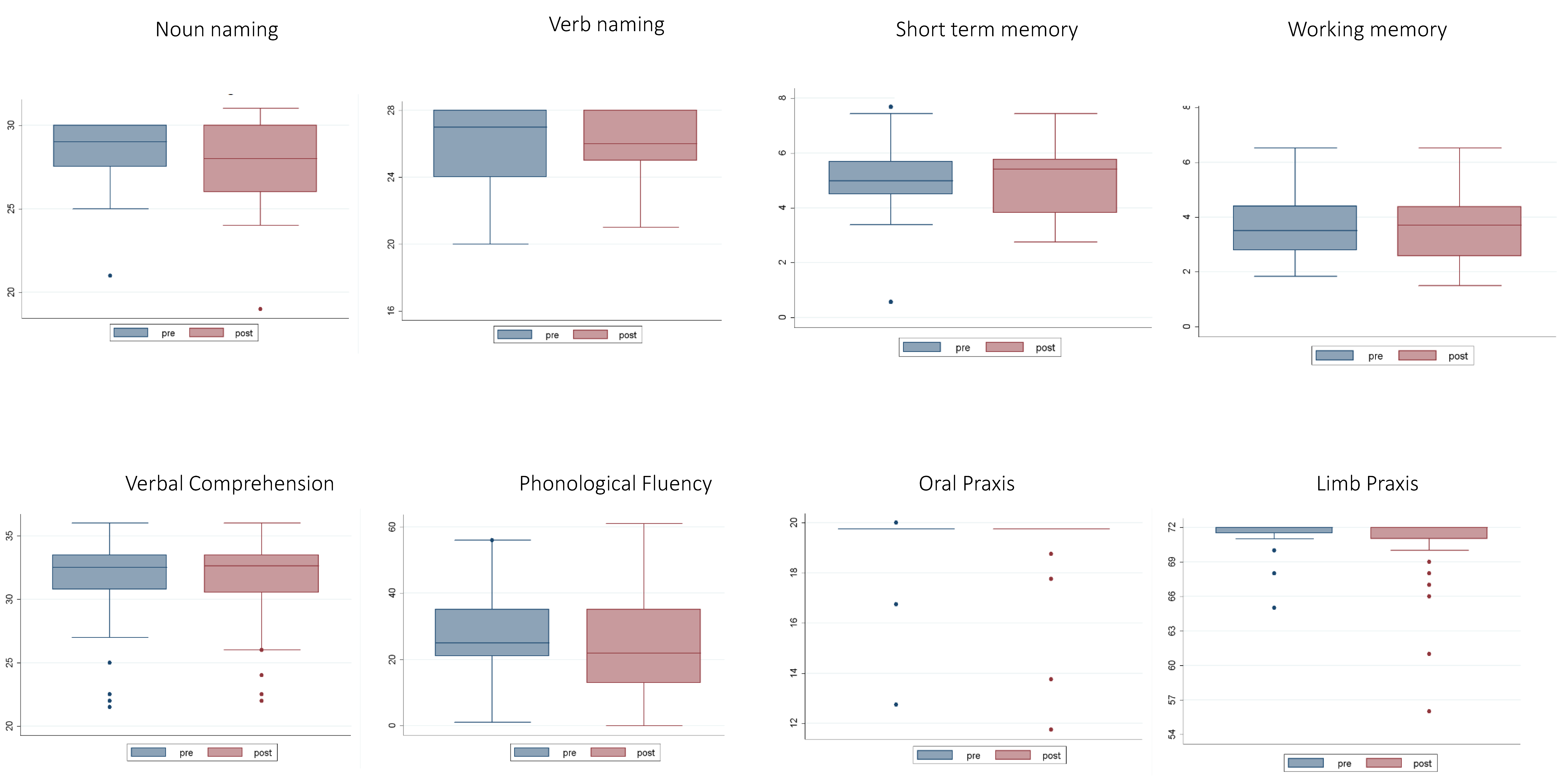
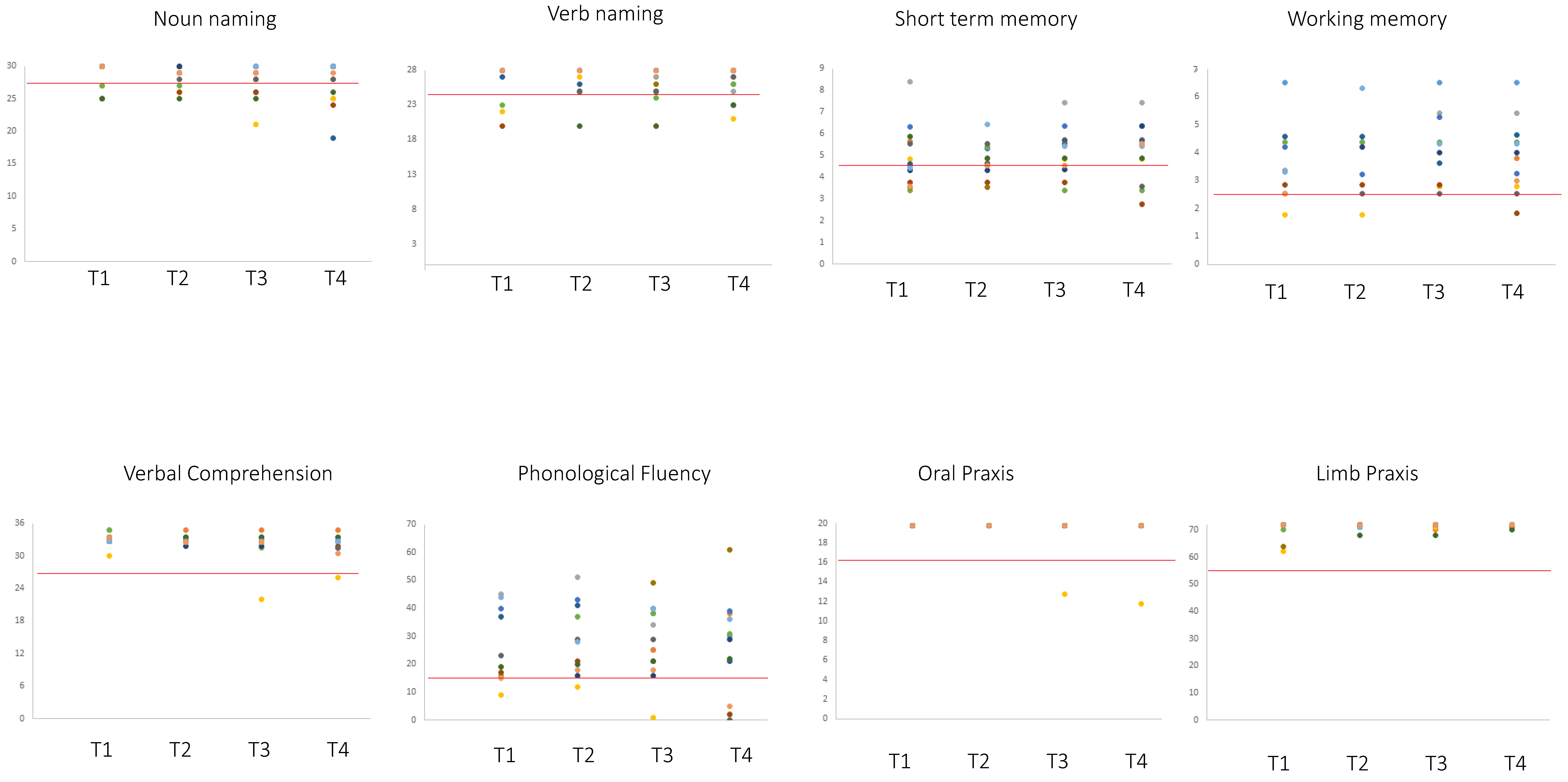
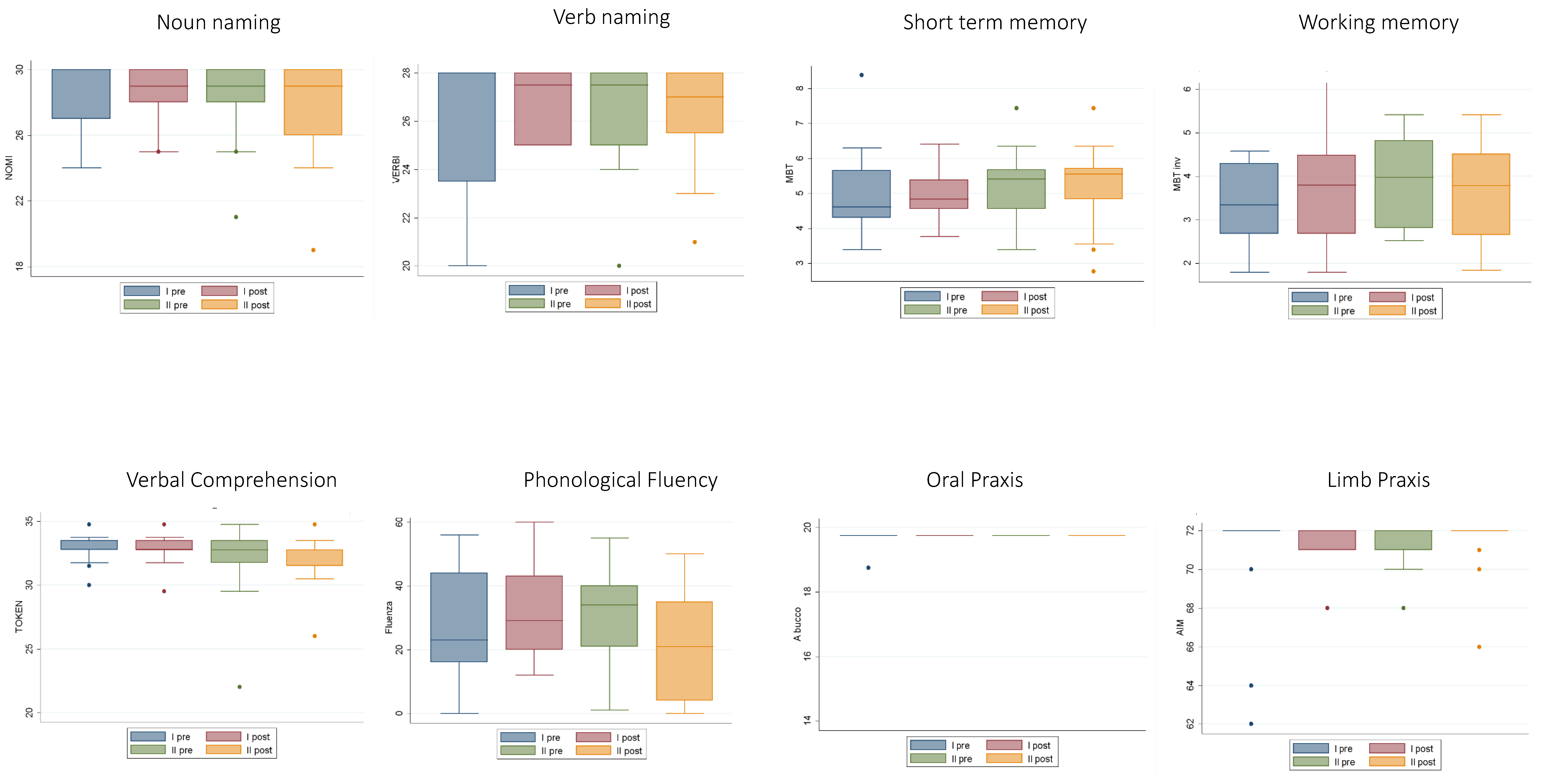
2.1. Neuropsychological Data

Table 2 shows the pre- and post-surgery number of patients within the normal range of performance for all the analyzed cognitive tasks. Some patients improved at T4, some worsened at T4, and the others remained unchanged, namely some patients were below the normal range at T3 and remained so at T4.
The analyses of the number of patients exhibiting normal performance at T4 vs. T3 did not provide evidence of significant differences. The analyses of the level of performance (working memory and phonological fluency data were normally distributed; whereas comprehension, oral and ideomotor apraxia, short-term memory, noun and verb naming data were not normally distributed (see Table 3)), showed that except for the oral praxis, even remaining within the normal range, in which a minimal difference between a pre-surgery level of 19.5 ± 1.19 and a post-surgery level of 19.3 ± 1.5 was evidenced, there was no significant difference between T3 vs. T4 performance for any of the other tasks (Table 4 and Figure 3 and Figure 4).
We further addressed the neuropsychological pattern for the subgroup (N = 17) of patients who were tested pre- and post- first and second surgery (T1–T4). The analyses of the number of patients exhibiting normal performance in the T1–T4 measures did not evidence significant differences (Table 5). The analyses of the level of performance, repeated measures ANOVA (Table 6) showed that among all the tests, only phonological fluency showed a significant change in results between T4 and T1, between T4 and T2, and between T4 and T3 (see Figure 5 and Figure 6).

2.2. Radiotherapy

No effect was observed as a result of conventional radiotherapy performed after the recurrent surgery (see Table 7).

2.3. MRI Structural Data Analysis

Figure 1 shows the overlap of the patients’ lesion masks (lesion masks include both the surgical cavity of the first surgery and the lesion regrowth). This overlay of the patients’ lesion masks shows that the lesions involved eloquent areas. The maximum overlap of the pre-surgical lesion masks, occurring in at least 25% of the patients with recurrence surgery (N = 40), involved the inferior frontal gyrus (pars opercularis). We performed a Voxel Lesion Symptom Mapping (VLSM) analysis trying to correlate lesion location and neuropsychological performance. No significant voxels survived the multiple comparisons corrected threshold.

2.4. Intraoperative Findings in Recurrence Surgery

In first surgery, all the patients were operated under the awake condition. The same approach was used in recurrent surgery, since the indications and justifications were the same as for the first surgery, given that the same area was involved in the recurrent resection.
At recurrent surgery, from a technical/surgical point of view there was the appearance of the scar left by the first surgery, while resection was actually facilitated by the presence of recurring pathological tissue in the surgical cavity, which was well defined and easily distinguishable from the healthy tissue. As far as the infiltrative component was concerned, the methodological testing remained the same.
All lesions were on the dominant left-hand side, where language-related areas were the most frequent regions exposed by the craniotomy. Therefore, the intra-surgery changes most often involved language-related functions: seven patients exhibited a moderate decrease in language performance, with a reduction in naming and fluency. Three patients showed transitory fluctuations in language performance, two patients were subject to generalized seizures, and one exhibited extreme drowsiness at the end of the procedure.

2.5. Extent of Resection

The EOR was computed in difference MRI sequences, according to histological data. Specifically, the EOR was computed on T2-weighted MRI images for Low grade glioma (LGG) and on post-contrast T1-weighted MRI images for High grade glioma (HGG). The median EOR was 94% (range 65%−100%) and 96% (range 50%−100%) in patients who underwent surgery for LGG and HGG, respectively.

2.6. Overall Survival

Overall in the study population the 5-year estimated Overall Survival (Oss) was 92%.

2.7. Correlation Analysis

We performed a correlation analysis and we found significant negative correlations between both the extent of resection and verbal comprehension (Token test) and between the extent of resection and phonological fluency (r = −371, p = 0.02 and r = −36, p = −0.027, respectively). In addition, we found significant positive correlations between both histology (LGG; HGG) and working memory and between histology and naming verbs (r = −36, p = 0.02 and r = −36, p = −0.028, respectively). Sex did not significantly correlate with any measure. Previous oncological treatment was significantly correlated with histology, (r = −0.51, p = 0.001).

3. Discussion

Despite a modern, multidisciplinary approach in the management of LGG, early relapse and progression will often occur, especially in lesions with a particular molecular pattern [32,33]. In this scenario and given the variability of heterogeneous postoperative treatment for LGG, the role of second surgery outcomes has been poorly investigated [3,16,17,18,25,26,28,29,30,31,34]. The few studies performed on recurrent surgery focus on the neuro-oncological aspect, without considering the neuropsychological point of view, which was the aim of this paper.
Several clinicians are reluctant to re-operate on LGG patients, preferring new cycles of chemotherapy and/or radiotherapy, and avoiding increased surgical risk [27]. Their major concern is related to making further, serious damage to cognitive function in exchange for poor prognostic benefit. More recently, other authors have suggested that surgical resection improves quality of life by reducing the mass effect, incidence of seizures, risk of malignant transformation, and improving overall survival [3,16,17,18,25,26,29,30,31,34]. Fluorescence-guided surgery has also been shown to improve the extent of resection in recurrent gliomas [35]. Despite all these achievements, the authors considered the role of surgery as undefined and that decisions should be made case by case. Their assumption is that there is insufficient evidence to make any specific recommendations [3,16,17,18,25,26,29,30,31,34,35].
Considering the principle that the larger the resection, the longer the survival, which is confirmed by recent literature [1,2,3,4,5,6,7,8,9], this study focuses on the potential change in neuropsychological status of patients who underwent aggressive re-operation. Forty patients have been subject to consecutive homogeneous neuropsychological evaluation, before and after second surgery.
In addition, we analyzed the evolution of neuropsychological changes that might occur after first and second surgery in 17 cases from the same cohort of 40 cases. We followed the entire neurocognitive “path” of these patients, from the first brain surgery until and after the second operation, which has never before been presented. The functional preoperative studies and surgical technique with intraoperative functional mapping used for the first procedure remained the same in the case of the second procedure, considering that the tumor was close to an eloquent area, which was the reason that a tumoral residuum was originally left (see also [36,37]).
Several new studies have shown that the plastic potential of the area may continue with a new functional reorganization that takes place during relapse [19,20,38,39]. This possible behavior of functional areas has allowed the principle of “two-step surgery” in the case of giant lesions.
Moreover, in the cases examined in this study, the main part of the recurrent neoplastic tissue was located either in the previous-tumor cavity or along its walls. This part of the tumor was usually soft, well defined in regard to the parenchyma, and relatively easy to remove. The surgical-functional problem was related to just the infiltrated parenchyma where mapping had been performed (for example, see Figure 2). The only delicate surgical step was the dissection of the scar tissue, which in some cases involved the cortex.
By analyzing the neuropsychological data from 40 patients after second surgery, we considered two different measures: the number of patients scoring within the normal range, namely we tested for changes in the number of patients scoring normally after the second surgery, with respect to the cut-offs, and, as a second measure of the patients’ levels of performance, we tested for changes in their accuracy (number of correct responses).
The analyses were performed by comparing pre- and follow up (4 months after surgery) performances. The patients sample at 4 months included also the 19 patients who had radiotherapy post the second surgery due to an oncological worsening.
As expected, after a short follow up of 4 months, there was no significant difference between patients who underwent radiotherapy and those who did not, but these data just provided us with their status after surgery [40]. These results are reliable since after 4 months the impact of radiotherapy is not likely to be observed.
The neuropsychological tests covered several cognitive domains. Detailed neuropsychological testing was not conducted at terms longer than 4 months because our objective was to analyze the impact of surgery. No significant differences were found for cognitive tests such as naming nouns, comprehension, ideomotor apraxia, short-term memory, working memory, and oral praxis. We found no significant changes either in the number of patients scoring normally between pre- and post-second surgery, or in their accuracy, i.e., their level of performance.
To further analyze the neuropsychological evolution, we considered also the pre- and post- first surgery and post-second surgery data of a smaller sample of 17 patients. We obtained the same results as for the 40 patients used in this study. Only the phonological fluency, considering the level of performance, showed a significant difference in the results between T4 and T1, T2 and T3. The relatively small sample size and the overall good performance limited the results of this correlation analysis between clinical details and neuropsychological data to the results reported just above. The same limitation applies to lesion location. Despite being a small sample size, the set of 17 patients are considered to provide a supplementary study, since the main analyses here involve all 40 patients in the main patient group. Though the sample size used here is relatively small, this study helps to elucidate neuropsychological status in recurrent surgeries, as studies in the literature on this topic are rare.
The neuropsychological status of a patient after a second surgery on a LGG is almost an unknown argument, with only a few studies addressing this aspect. Other studies [21] have reported on the intraoperative mapping of the first and second resection, but there are no previous studies that provide an extensive neuropsychological examination of LGG patients with recurrent surgery. Although our experience and findings are clearly based on a limited number of cases, these preliminary data indicate positive outcomes. These results indicate that second surgery could be considered in cases where surgery with a new extensive resection still remains the better solution for the patient. Further experience and further data are required to validate these findings and draw more generalized conclusions.

4. Materials and Methods

4.1. Neuropsychological Testing

All participants underwent neuropsychological testing with the same neuropsychologist. The neuropsychological battery was selected to comprehensively cover several cognitive domains, to provide age and years of schooling-corrected normative data, and to provide a short administration time to minimize patient fatigue. For the purpose of the study and statistical analysis we selected specific domains: memory, language (naming nouns, naming verbs, and verbal comprehension) executive abilities (phonological fluency and working memory), and praxis.
The battery of neuropsychological tests involved the assessment of spatio-temporal orientation, handedness (Edinburgh Handedness Inventory [41]), executive functions (working memory [42,43]; verbal fluency [44]), verbal short-term memory (forward [42,43]), praxis (ideomotor limb apraxia [45] and oral apraxia [46]), together with language processing tasks, such as object naming, action naming (battery for the analysis of language disorders [47]), and language comprehension (Token test [48]). The pre-op and post-op assessments in the case of recurrence were indicated T3 and T4, respectively. A subset group of patients (N = 17) were tested at four points in time i.e., before the first surgery (T1), 4 months following the first surgery (T2), one week before recurrence surgery (T3), and 4 months following recurrence surgery (T4).
The local institution ethics committee on human research approved this study, and the patients all signed a consent form.

4.2. MRI Structural Data

We retrospectively used T1 and T2 structural imaging data that was routinely acquired during pre-surgery investigations with a 3-T Philips Achieva whole-body scanner.
Structural MRI data processing and statistical analyses were performed using MATLAB r2007b (Mathworks Inc., Natick, MA, USA) and SPM8 (Statistical Parametric Mapping software; Wellcome Department of Imaging Neuroscience, London, UK; http://www.fil.ion.ucl.ac.uk/spm/software/spm8/). Volumes of interest (VOIs) for the patients’ lesions (lesion masks include both the surgical cavity of the first surgery and the lesion regrowth) were drawn on their T2-weighted MRI scans using the MRIcron software (http://www.mccauslandcenter.sc.edu/mricro/mricron/index.html) and were normalized to the Montreal Neurological Institute (MNI) space using the “Clinical Toolbox” (http://www.mccauslandcenter.sc.edu/CRNL/clinical-toolbox) for SPM8.
The MRIcron procedure (http://www.mccauslandcenter.sc.edu/mricro/mricron/index.html) was employed to overlap the lesion templates. An image was generated, selecting the overlap of the lesion masks (VOIs) for the 40 recurring (T3) patient lesions. The number of overlapping lesions is illustrated by different colors that correspond to increasing frequencies (as indicated in the bar code).

4.3. Awake Surgery

The decision to re-operate was based primarily on unequivocal evidence of tumor regrowth provided by MRI. The goal of the repeat surgeries remained the maximum resection of the area in the T2-weighted image that was visible on the MRI sequences, including the contrast-enhanced area in the lesions that exhibited anaplastic transformation.
Surgical procedures for first and repeat surgeries were planned using functional MRI and DTI data and conducted under an “awake-online protocol” based on the methodology of Berger and Ojemann [49,50,51] implemented with real-time neuropsychological testing (RTNT), continuous neuropsychological testing during tumor resection enriching the feedback process, as previously described [52].
Patients were fixed to a Mayfield frame and were slightly sedated during exposure and closure of the surgical field [1,53]. Motor evoked potentials (MEPs) and somatosensory evoked potentials (SSEPs) were recorded during surgery to continuously monitor the integrity of motor-sensory pathways (64-channel Eclipse Neurovascular Workstation, Axon Systems Inc.; 32-channel video polygraphic station, Brain Quick SystemPlus, MicroMed). Continuous electrocorticography was performed to detect discharge phenomena and rule out the possibility that the patient’s decrement in performance or oscillatory performance was caused by the concomitant presence of short focal seizures. The EOR was evaluated on post-contrast T1-weighted MRI sequences and on T2-weighted MRI sequences acquired 4 months after surgery for HGG and LGG, respectively [1,54]. For all the tumors in this study, the histological and molecular analysis was conducted and revised according to the World Health Organization (WHO) classification for tumors of the central nervous system [55].

4.4. Radiotherapy

Among the patients who exhibited malignant transformation at recurrence, 19 underwent radiation therapy. We considered this variable in the interpretation of the final neuropsychological tests, to determine whether there was any significant difference between patients who had and had not been subject to this treatment.

4.5. Statistical Analysis of Neuropsychological Data

The number of patients who scored below the normal range in the consecutive series of 40 patients with recurrent glioma and the subset of 17 patients (i.e., the control group) were analyzed using a McNemar’s test, comparing T3 and T4 in the case of the first series. For the second series we used Cochran’s Q test comparing T1–T4. The neuropsychological scores of the series of 40 patients with recurrent glioma were analyzed using a parametric statistical test (paired t-test) or a non-parametric statistical test (Wilcoxon sign rank-test), depending on whether the variables had a Gaussian distribution. The normal distribution of the data were tested using the Shapiro–Wilk test for normal data. The neuropsychological scores of the subset of 17 patients, who were assessed pre- and post-first and recurrent surgery were analyzed using parametric statistical tests (repeated measures ANOVA). All analyses were conducted using Stata/SE 12.0 for Microsoft Windows. All two-tailed statistical significance levels were set to p < 0.05.

5. Conclusions

The aim of this study was the evaluation of cognitive aspects in cases of second surgery for tumor resection. Repeat surgery correlates with the oncological assessment that the minor the tumor mass, the better chance of survival follows, although this purely oncological aspect has not been considered here. Our longitudinal follow-up study indicates that glioma surgery is possible without major damage to cognitive functions in the short-term period (4 months) after surgery. Re-operation for recurrent glioma in an eloquent area with accurate neuropsychological assessment and RTNT intraoperative control is an efficacious and safe therapeutic strategy. It is possible to maximize tumor resection while retaining a reduced risk of permanent neurological deficiency, resulting in preservation or even improvement in patient quality of life. This is however a preliminary study with a small number of patients, and further studies with longer follow-up times are necessary to elucidate the conclusive and definitive effects of this surgical approach. The low morbidity associated with re-operation nevertheless suggests that it is preferable to “over indicate” an early re-intervention than to perform a late surgery.

Author Contributions

Conceptualization, M.S. and B.T.; methodology, B.T.; M.S.; G.C.; T.I.; formal analysis, M.I.; C.B.; investigation, I.G.; B.T.; G.C.; T.I.; writing—original draft preparation, G.C.; B.T.; M.S.; writing—review and editing, G.C.; B.T.; T.I; M.S.; supervision, B.T.; M.S.; All authors have read and agreed to the published version of the manuscript.

Funding

This research received no external funding.

Conflicts of Interest

The authors declare no conflict of interest.

References

  1. Ius, T.; Isola, M.; Budai, R.; Pauletto, G.; Tomasino, B.; Fadiga, L.; Skrap, M. Low-grade glioma surgery in eloquent areas: Volumetric analysis of extent of resection and its impact on overall survival. A single-institution experience in 190 patients—Clinical article. J. Neurosurg. 2012, 117, 1039–1052. [Google Scholar] [CrossRef] [PubMed]
  2. Dugdale, E.; Gerrard, G. Low-grade gliomas in adults. Cancer Med. 2006, 3, 40–45. [Google Scholar]
  3. Rezvan, A.; Christine, D.; Christian, H.; Olga, Z.; Lutz, E.; Marius, H.; Stephanie, C.; Christel, H.M.; Rainer, W.C.; Andreas, U. Long-term outcome and survival of surgically treated supratentorial low-grade glioma in adult patients. Acta Neurochir. (Wien.) 2009, 151, 1359–1365. [Google Scholar] [CrossRef] [PubMed]
  4. Duffau, H.; Taillandier, L. New concepts in the management of diffuse low-grade glioma: Proposal of a multistage and individualized therapeutic approach. Neuro Oncol. 2015, 17, 332–342. [Google Scholar] [CrossRef] [PubMed] [Green Version]
  5. Myrmel, K.S. Comparison of a Strategy Favoring Early Surgical Resection vs a Strategy Favoring Watchful Waiting in Low-Grade Gliomas. JAMA 2012, 308, 1881–1888. [Google Scholar]
  6. McGirt, M.J.; Chaichana, K.L.; Attenello, F.J.; Weingart, J.D.; Than, K.; Burger, P.C.; Olivi, A.; Brem, H.Q.-H.A. Extent of surgical resection is independently associated with survival in patients with hemispheric infiltrating low-grade gliomas. Neurosurgery 2008, 63, 700–708. [Google Scholar] [CrossRef] [Green Version]
  7. Schomas, D.A.; Laack, N.N.I.; Rao, R.D.; Meyer, F.B.; Shaw, E.G.; Neill, B.P.O.; Giannini, C.; Brown, P.D. Intracranial low-grade gliomas in adults: 30-year experience with long-term follow-up at Mayo Clinic. Neuro Oncol. 2009, 11, 437–445. [Google Scholar] [CrossRef]
  8. Smith, J.S.; Chang, E.F.; Lamborn, K.R.; Chang, S.M.; Prados, M.D.; Cha, S.; Tihan, T.; Vandenberg, S.; Mcdermott, M.W.; Berger, M.S. Role of Extent of Resection in the Long-Term Outcome of Low-Grade Hemispheric Gliomas. J. Clin. Oncol. 2015, 26, 1338–1345. [Google Scholar] [CrossRef]
  9. Duffau, H. Long-term outcomes after supratotal resection of diffuse low-grade gliomas: A consecutive series with 11-year follow-up. Acta Neurochir. (Wien.) 2016, 158, 51–58. [Google Scholar] [CrossRef] [PubMed]
  10. Baumert, B.G.; Stupp, R. Low-grade glioma: A challenge in therapeutic options: The role of radiotherapy. Ann. Oncol. 2008, 19, 217–223. [Google Scholar] [CrossRef]
  11. Ius, T.; Pauletto, G.; Cesselli, D.; Isola, M.; Turella, L.; Budai, R.; Demaglio, G.; Eleopra, R.; Fadiga, L.; Lettieri, C.; et al. Second Surgery in Insular Low-Grade Gliomas. Biomed. Res. Int. 2015, 2015, 497610. [Google Scholar] [CrossRef] [PubMed] [Green Version]
  12. Murphy, E.S.; Leyrer, C.M.; Parsons, M.; Suh, J.H.; Chao, S.T.; Yu, J.S.; Kotecha, R.; Jia, X.; Peereboom, D.M.; Prayson, R.A.; et al. Risk Factors for Malignant Transformation of Low-Grade Glioma. Int. J. Radiat. Oncol. 2018, 100, 965–971. [Google Scholar] [CrossRef]
  13. Rotta, J.M.; de Oliveira, M.F.; Reis, R.C.; Botelho, R.V. Malignant transformation of low-grade gliomas in patients undergoing adjuvant therapy. Acta Neurol. Belg. 2017, 117, 235–239. [Google Scholar] [CrossRef] [PubMed]
  14. Bogdańska, M.U.; Bodnar, M.; Piotrowska, M.J.; Murek, M.; Schucht, P.; Beck, J.; Martínez-González, A.; Pérez-García, V.M. A mathematical model describes the malignant transformation of low grade gliomas: Prognostic implications. PLoS ONE 2017, 12, e0179999. [Google Scholar] [CrossRef] [PubMed] [Green Version]
  15. Zhang, S.; Chiang, G.C.-Y.; Magge, R.S.; Fine, H.A.; Ramakrishna, R.; Chang, E.W.; Pulisetty, T.; Wang, Y.; Zhu, W.; Kovanlikaya, I. Texture analysis on conventional MRI images accurately predicts early malignant transformation of low-grade gliomas. Eur. Radiol. 2019, 29, 2751–2759. [Google Scholar] [CrossRef]
  16. Ramakrishna, R.; Hebb, A.; Barber, J.; Rostomily, R.; Silbergeld, D. Outcomes in Reoperated Low-Grade Gliomas. Neurosurgery 2015, 77, 175–184. [Google Scholar] [CrossRef] [Green Version]
  17. Schmidt, M.H.; Berger, M.S.; Lamborn, K.R.; Aldape, K.; McDermott, M.W.; Prados, M.D.; Chang, S.M. Repeated operations for infiltrative low-grade gliomas without intervening therapy. J. Neurosurg. 2003, 98, 1165–1169. [Google Scholar] [CrossRef] [Green Version]
  18. Hamisch, C.; Ruge, M.; Kellermann, S.; Kohl, A.C.; Duval, I.; Goldbrunner, R.; Grau, S.J. Impact of treatment on survival of patients with secondary glioblastoma. J. Neurooncol. 2017, 133, 309–313. [Google Scholar] [CrossRef]
  19. Duffau, H. Brain plasticity and tumors. In Advances and Technical Standards in Neurosurgery; Pickard, J.D., Akalan, N., Di Rocco, C., Dolenc, V.V., Antunes, J.L., Mooij, J.J.A., Schramm, J., Sindou, M., Eds.; Springer: Vienna, Austria, 2008; pp. 3–33. ISBN 978-3-211-72283-1. [Google Scholar]
  20. Duffau, H. Brain plasticity: From pathophysiological mechanisms to therapeutic applications. J. Clin. Neurosci. 2006, 13, 885–897. [Google Scholar] [CrossRef]
  21. Southwell, D.G.; Hervey-Jumper, S.L.; Perry, D.W.; Berger, M.S. Intraoperative mapping during repeat awake craniotomy reveals the functional plasticity of adult cortex. J. Neurosurg. 2016, 124, 1460–1469. [Google Scholar] [CrossRef] [Green Version]
  22. Duffau, H.; Capelle, L. Preferential brain locations of low-grade gliomas: Comparison with glioblastomas and review of hypothesis. Cancer 2004, 100, 2622–2626. [Google Scholar] [CrossRef] [PubMed]
  23. Duffau, H.; Capelle, L.; Denvil, D.; Sichez, N.; Gatignol, P.; Lopes, M.; Mitchell, M.C.; Sichez, J.P.; Van Effenterre, R. Functional recovery after surgical resection of low grade gliomas in eloquent brain: Hypothesis of brain compensation. J. Neurol. Neurosurg. Psychiatry 2003, 74, 901–907. [Google Scholar] [CrossRef] [PubMed] [Green Version]
  24. Duffau, H.; Lopes, M.; Arthuis, F.; Bitar, A.; Sichez, J.P.; Van Effenterre, R.; Capelle, L. Contribution of intraoperative electrical stimulations in surgery of low grade gliomas: A comparative study between two series without (1985-96) and with (1996–2003) functional mapping in the same institution. J. Neurol. Neurosurg. Psychiatry 2005, 76, 845–851. [Google Scholar] [CrossRef] [PubMed] [Green Version]
  25. Juratli, T.A.; Kirsch, M.; Geiger, K.; Klink, B.; Leipnitz, E.; Pinzer, T.; Soucek, S.; Schrok, E.; Schackert, G.; Krex, D. The prognostic value of IDH mutations and MGMT promoter status in secondary high-grade gliomas. J. Neurooncol. 2012, 110, 325–333. [Google Scholar] [CrossRef] [PubMed]
  26. Thon, N.; Eigenbrod, S.; Kreth, S.; Lutz, J.; Tonn, J.C.; Kretzschmar, H.; Peraud, A.; Kreth, F.W. IDH1 mutations in grade II astrocytomas are associated with unfavorable progression-free survival and prolonged postrecurrence survival. Cancer 2012, 118, 452–460. [Google Scholar] [CrossRef] [PubMed]
  27. Berger, A.; Tzarfati, G.; Costa, M.; Serafimova, M.; Korn, A.; Vendrov, I.; Alfasi, T.; Krill, D.; Aviram, D.; Ben Moshe, S.; et al. Incidence and impact of stroke following surgery for low-grade gliomas. J. Neurosurg. JNS 2019, 1–9. [Google Scholar] [CrossRef]
  28. Uppstrom, T.J.; Singh, R.; Hadjigeorgiou, G.F.; Magge, R.; Ramakrishna, R. Repeat surgery for recurrent low-grade gliomas should be standard of care. Clin. Neurol. Neurosurg. 2016, 151, 18–23. [Google Scholar] [CrossRef] [PubMed]
  29. Blonski, M.; Taillandier, L.; Herbet, G.; Maldonado, I.L.; Beauchesne, P.; Fabbro, M.; Campello, C.; Gozé, C.; Rigau, V.; Moritz-Gasser, S.; et al. Combination of neoadjuvant chemotherapy followed by surgical resection as a new strategy for WHO grade II gliomas: A study of cognitive status and quality of life. J. Neurooncol. 2012, 106, 353–366. [Google Scholar] [CrossRef]
  30. Martino, J.; Taillandier, L.; Moritz-gasser, S.; Gatignol, P.; Duffau, H. Re-operation is a safe and effective therapeutic strategy in recurrent WHO grade II gliomas within eloquent areas. Acta Neurochir. (Wien.) 2009, 151, 427–436. [Google Scholar] [CrossRef]
  31. Jaeckle, K.A.; Decker, P.A.; Ballman, K.V.; Flynn, P.J.; Giannini, C.; Scheithauer, B.W.; Jenkins, R.B.; Buckner, J.C. Transformation of low grade glioma and correlation with outcome: An NCCTG database analysis. J. Neurooncol. 2011, 104, 253–259. [Google Scholar] [CrossRef] [Green Version]
  32. Fukuya, Y.; Ikuta, S.; Maruyama, T.; Nitta, M.; Saito, T.; Tsuzuki, S.; Chernov, M.; Kawamata, T.; Muragaki, Y. Tumor recurrence patterns after surgical resection of intracranial low-grade gliomas. J. Neurooncol. 2019, 144, 519–528. [Google Scholar] [CrossRef] [PubMed]
  33. Patil, C.G.; Walker, D.G.; Miller, D.M.; Butte, P.; Morrison, B.; Kittle, D.S.; Hansen, S.J.; Nufer, K.L.; Byrnes-Blake, K.A.; Yamada, M.; et al. Phase 1 Safety, Pharmacokinetics, and Fluorescence Imaging Study of Tozuleristide (BLZ-100) in Adults With Newly Diagnosed or Recurrent Gliomas. Neurosurgery 2019, 85, E641–E649. [Google Scholar] [CrossRef] [PubMed]
  34. Blonski, M.; Pallud, J.; Gozé, C.; Mandonnet, E.; Rigau, V.; Bauchet, L.; Fabbro, M.; Beauchesne, P.; Baron, M.H.; Fontaine, D.; et al. Neoadjuvant chemotherapy may optimize the extent of resection of World Health Organization grade II gliomas: A case series of 17 patients. J. Neurooncol. 2013, 113, 267–275. [Google Scholar] [CrossRef] [PubMed]
  35. Li, J.; Niu, X.; Gan, Y.; Yang, Y.; Wang, T.; Zhang, H.; Liu, Y.; Mao, Q. Clinical and Pathologic Features and Prognostic Factors for Recurrent Gliomas. World Neurosurg. 2019, 128, e21–e30. [Google Scholar] [CrossRef]
  36. Dirks, P.; Bernstein, M.; Muller, P.J.; Tucker, W.S. The value of reoperation for recurrent glioblastoma. Can. J. Surg. 1993, 36, 271–275. [Google Scholar]
  37. Harsh, G.R.; Levin, V.A.; Gutin, P.H.; Seager, M.; Silver, P.; Wilson, C.B. Reoperation for Recurrent Glioblastoma and Anaplastic Astrocytoma. Neurosurgery 1987, 21, 615–621. [Google Scholar] [CrossRef]
  38. Desmurget, M.; Bonnetblanc, F.; Duffau, H. Contrasting acute and slow-growing lesions: A new door to brain plasticity. Brain 2007, 130, 898–914. [Google Scholar] [CrossRef] [Green Version]
  39. Duffau, H. Lessons from brain mapping in surgery for low-grade glioma: Insights into associations between tumour and brain plasticity. Lancet Neurol. 2005, 4, 476–486. [Google Scholar] [CrossRef]
  40. Campanella, F.; Palese, A.; Del Missier, F.; Moreale, R.; Ius, T.; Shallice, T.; Fabbro, F.; Skrap, M. Long-Term Cognitive Functioning and Psychological Well-Being in Surgically Treated Patients with Low-Grade Glioma. World Neurosurg. 2017, 103, 799–808. [Google Scholar] [CrossRef]
  41. Oldfield, R.C. The assessment and analysis of handedness: The Edinburgh inventory. Neuropsychologia 1971, 9, 97–113. [Google Scholar] [CrossRef]
  42. Monaco, M.; Costa, A.; Caltagirone, C.; Carlesimo, G.A. Forward and backward span for verbal and visuo-spatial data: Standardization and normative data from an Italian adult population. Neurol. Sci. 2013, 34, 749–754. [Google Scholar] [CrossRef]
  43. Monaco, M.; Costa, A.; Caltagirone, C.; Carlesimo, G.A. Erratum to: Forward and backward span for verbal and visuo-spatial data: Standardization and normative data from an Italian adult population. Neurol. Sci. 2015, 36, 345–347. [Google Scholar] [CrossRef] [PubMed] [Green Version]
  44. Novelli, G.; Papagno, C.; Capitani, E.; Laiacona, M.; Vallar, G.; Cappa, S.F. Tre test clinici di ricerca e produzione lessicale. Taratura su sogetti normali. Arch. Psicol. Neurol. Psichiatr. 1986, 47, 477–506. [Google Scholar]
  45. De Renzi, E.; Motti, F.; Nichelli, P. Imitating Gestures: A Quantitative Approach to Ideomotor Apraxia. Arch. Neurol. 1980, 37, 6–10. [Google Scholar] [CrossRef] [PubMed]
  46. Spinnler, M.; Tognoni, G. Standardizzazione e validazione di test neuropsicologici. Ital. J. Neurol. Sci. 1987, 8, 1–120. [Google Scholar]
  47. Miceli, G.; Laudanna, A.; Burani, C. Batteria per l’analisi dei Deficit Afasici. B. A. D. A. (B. A. D. A.: A Battery for the Assessment of Aphasic Disorders). Roma Cepsag 1994. Available online: https://www.iris.unisa.it/handle/11386/3828878?mode=full.19 (accessed on 6 April 2020).
  48. De Renzi, E.; Faglioni, P. Normative Data and Screening Power of a Shortened Version of the Token Test. Cortex 1978, 14, 41–49. [Google Scholar] [CrossRef]
  49. Berger, M.S. Lesions in functional (“eloquent”) cortex and subcortical white matter. Clin. Neurosurg. 1994, 41, 444–463. [Google Scholar]
  50. Berger, M.S.; Ojemann, G.A. Intraoperative brain mapping techniques in neuro-oncology. Stereotact. Funct. Neurosurg. 1992, 58, 153–161. [Google Scholar] [CrossRef]
  51. Ojemann, G.; Ojemann, J.; Lettich, E.; Berger, M. Cortical language localization in left, dominant hemisphere. J. Neurosurg. 1989, 71, 316–326. [Google Scholar] [CrossRef]
  52. Skrap, M.; Marin, D.; Ius, T.; Fabbro, F.; Tomasino, B. Brain mapping: A novel intraoperative neuropsychological approach. J. Neurosurg. 2016, 125, 877–887. [Google Scholar] [CrossRef] [Green Version]
  53. Skrap, M.; Mondani, M.; Tomasino, B.; Weis, L.; Budai, R.; Pauletto, G.; Eleopra, R.; Fadiga, L.; Ius, T. Surgery of insular nonenhancing gliomas: Volumetric analysis of tumoral resection, clinical outcome, and survival in a consecutive series of 66 cases. Neurosurgery 2012, 70, 1081–1093. [Google Scholar] [CrossRef] [PubMed] [Green Version]
  54. Ius, T.; Pignotti, F.; Della Pepa, G.M.; Bagatto, D.; Isola, M.; Battistella, C.; Gaudino, S.; Pegolo, E.; Chiesa, S.; Arcicasa, M.; et al. Glioblastoma: From volumetric analysis to molecular predictors. J. Neurosurg. Sci. 2020. [Google Scholar] [CrossRef]
  55. Louis, D.N.; Perry, A.; Reifenberger, G.; von Deimling, A.; Figarella-Branger, D.; Cavenee, W.K.; Ohgaki, H.; Wiestler, O.D.; Kleihues, P.; Ellison, D.W. The 2016 World Health Organization Classification of Tumors of the Central Nervous System: A summary. Acta Neuropathol. 2016, 131, 803–820. [Google Scholar] [CrossRef] [PubMed] [Green Version]
Figure 1. Overlap of all the 40 pre-recurrence (T3) patients’ lesions masks. Lesion masks include both the surgical cavity of the first surgery and the lesion regrowth. The number of overlapping lesions is illustrated by different colors coding increasing frequencies (as indicated in the bar code). As shown by the image, the epicenter of the overlap is located at the level of the frontotemporoinsular region. MR images are displayed in radiological convention.
Cancers 12 01077 g001
Figure 2. Axial view T1-weighted MRI. Upper image shows frontotemporoinsular glioma before first surgery. (a) Lower images show recurrence before second surgery. (b) Recurrent neoplastic tissue was located both along the walls of and within the previous-tumor cavity.
Cancers 12 01077 g002
Figure 3. Performance of the 40 patients with recurrent glioma at T3 and T4 (pre- and post-recurrence surgery, respectively). Red lines indicate the cut-off, below which the patient results were pathological: verbal short-term memory (<4.26 is equal to an equivalent score of 0), working memory (<2.65 is equal to an equivalent score of 0), ideomotor limb apraxia (<53) [25] and oral apraxia (<16) [26], language comprehension (<26.25 is equal to an equivalent score of 0) [27], object naming and action naming (<28 and <26 respectively) [28], and verbal fluency (<16 is equal to an equivalent score of 0) [29]. Patients showing identical scores are represented by overlapping dots.
Cancers 12 01077 g003
Figure 4. Mean performance of the 40 patients with recurrent glioma T3 and T4 (pre- and post-recurrence surgery, respectively). Bars represent the standard deviations.
Cancers 12 01077 g004
Figure 5. Performance of the 17 patients with recurrent glioma at T1 and T2 (pre- and post-first surgery, respectively) and at T3 and T4 (pre- and post-recurrence surgery, respectively). Red lines indicate the cut-off, below which the patient results are pathological (<4.26 is equal to an equivalent score of 0) [30,31], working memory (<2.65 is equal to an equivalent score of 0) [30], ideomotor limb apraxia (<53) [25] and oral apraxia (<16) [26], language comprehension (<26.25 is equal to an equivalent score of 0) [27], object naming and action naming (<28 and <26 respectively) [28] and verbal fluency (<16 is equal to an equivalent score of 0) [29]. Patients showing identical scores are represented by overlapping dots.
Cancers 12 01077 g005
Figure 6. Mean performance of the 17 patients with recurrent glioma T1 and T2 (pre- and post-first surgery, respectively) and T3 and T4 (pre- and post-recurrence surgery, respectively). Bars represent the standard deviations.
Cancers 12 01077 g006
Table 1. Patients’ demographic and clinical data (H = hemisphere; P# = case number; M = male; F = female; S = years of schooling; Hist= histology; HGG =high grade glioma; LGG= low grade glioma, EOR = Extent of resection (%), RT = Radiotherapy post-2nd surgery.
P#GenderAgeSSite SurgeryHHistEORRT Post 2nd Surgery
1F3313F-T-ILHLGG66V
2M328F-PLHHGG100V
3M3513T-ILHHGG100V
4F5213F-PLHLGG85V
5M2313FLHLGG100X
6M4013TLHHGG100V
7F5613F-ILHHGG100X
8M2413T-PLHHGG100X
9M368F-T-ILHHGG100X
10M3018FLHLGG90X
11F3718F-T-ILHHGG85V
12M41-F-T-ILHHGG85V
13M2011T-PLHLGG65X
14M4917F-T-ILHHGG90V
15M4518FLHLGG90X
16M4413TLHHGG100V
17M358F-PLHHGG100V
18F348F-PLHHGG100X
19F3818F-PLHLGG85X
20M488F-T-ILHHGG100V
21M3713F-T-ILHLGG90X
22F1710FLHLGG85X
23M398FLHHGG100V
24F665F-T-ILHHGG100V
25M4518F-T-ILHLGG70X
26M4718F-T-ILHHGG100V
27M5718TLHHGG X
28M1510F-ILHLGG100X
29M2918F-ILHLGG100X
30M325FLHHGG84X
31M365FLHHGG100V
32F4813F-T-ILHHGG50V
33M338F-ILHLGG80V
34M368F-ILHHGG85X
35M3418F-T-ILHLGG85X
36F2813F-T-ILHHGG100V
37M1812TLHLGG100X
38F3413F-ILHLGG92X
39F3513F-ILHLGG90X
40F478TLHHGG100V
Table 2. Number of patients with recurrent glioma whose results were within the normal range at T3 and T4 (pre and post-recurrence surgery, respectively), and the results of the McNemar test (T3 vs. T4). We stratified patients who worsened (w, those within the normal range at T3 who were below the normal range at T4), and improved (i, those below the normal range at T3 who were within the normal range at T4). The mean and standard deviation of patients who scored below the normal range (and the cut-off) are shown in grey. To date, the level of performance of those patients was only slightly below the cut-offs.
Number of Patients within the Normal RangeLevel of Performance of Patients Below the Normal Range
TestT3T4T3 vs. T4 McNemarT3T4Cut-offs
STM31/38 28/38 (4/38 w; 1/38 i)p = 0.375, n.s.3.05 ± 1.43.39 ± 0.414.26
o_PRAXIS 40/40 40/40----
IMA38/38 38/38----
Compr35/39 35/39 (1/39 w, 1/39 i)p = 1, n.s.22.75 ± 1.5523.63 ± 1.8026.26
N_nam30/40 27/40 (4/40 w, 1/40 i)p = 0.37, n.s.25.60 ± 1.7825.23 ± 2.0528
v_nam24/39 25/39 (4/39 w, 5/39 i)p = 1, n.s.23.13 ± 1.8523.57 ± 1.4526
WM25/31 23/31 (4/35w, 2/31 i)p = 0.68, n.s.2.32 ± 0.312.26 ± 0.392.65
Ph_Fl32/38 26/38 (7/38 w, 1/38 i)p = 0.07, n.s.11 ± 5.198 ± 5.3316
STM = short-term memory, o_PRAXIS = oral praxis, IMA = ideomotor apraxia, Compr = verbal comprehension, N_nam = noun naming, v_nam = verb naming, WM = working memory, Ph_Fl = phonological fluency.
Table 3. Shapiro–Wilk test, using a = 0.05, on the normality distribution of the patients’ level of performance.
TaskPre-SurgeryPost-Surgery
WMp = 0.28p = 0.74
Ph_Flp = 0.19p = 0.51
Comprp < 0.001p < 0.001
o_PRAXISp < 0.001p < 0.001
STMp = 0.01p = 0.2
IMAp < 0.001p < 0.001
N_namp < 0.001p < 0.001
v_namp < 0.001p < 0.01
STM = short-term memory, o_PRAXIS = oral praxis, IMA = ideomotor apraxia, Compr = verbal comprehension, N_nam = noun naming, v_nam = verb naming, WM = working memory, Ph_Fl = phonological fluency.
Table 4. Results of the student t-test or Wilcoxon test comparing pre-surgery (T3) to post-surgery (T4) patients’ level of performance.
TaskT3T4T3 vs. T4
STM5.04 ± 1.235 ± 1.18Z = −0.5, p = 0.59
WM3.63 ± 1.133.63 ± 1.14t (30) = −0.18 p = 0.5
IMA71.38 ± 1.4470.55 ± 3.28Z = 0.84, p = 0.39
N_nam28.23 ± 1.927.95 ± 2.35Z = 0.416, p = 0.67
v_nam25.79 ± 1.925.92 ± 2.08Z = −0.506, p = 0.612
Compr31.37 ± 3.531.49 ± 3.22Z = −0.379, p = 0.750
Ph_Fl27.38 ± 12.2925.26 ± 15.07t (37) = 0.907, p = 0.18
o_PRAXIS19.5 ± 1.1919.30 ± 1.58Z = −2.44, p = 0.014
STM = short-term memory, o_PRAXIS = oral praxis, IMA = ideomotor apraxia, Compr = verbal comprehension, N_nam = noun naming, v_nam = verb naming, WM = working memory, Ph_Fl = phonological fluency.
Table 5. Number of the 17 patients with recurrent glioma whose results were within the normal range at time points T1 and T2 (pre- and post-first surgery, respectively) and at T3 and T4 (pre- and post-recurrence surgery, respectively), with Cochran’s Q test of equality of proportions between the different times points. We stratified patients who decreased (w, normal range at T3 and below the normal range at T4), and improved (i, below the normal range at T3 but within the normal range at T4). The mean and s.d. below the normal patient range for patient performance (and the cut-off) are shown in grey. To date, the level of performance in those patients was only slightly below the cut-offs (reported in the last column).
TaskNumber of Patients within the Normal RangeLevel of Performance of Patients Below the Normal Range
T1T2T3T4T1–T4
Cochran’s Q test
T1T2T3T4Cut-offs
STM10/1312/13 (0 w,2 i)11/1310/13 (1w, 0i)p = 0.299, n.s.3.56 ± 0.15 3.65 ± 0.843.58 ± 0.263.38 ± 0.454.26
o_PRAXIS16/1616/1616/1616/16-----16
IMA17/1717/1717/1717/17-----52
Compr17/1717/1717/1717/17p = 0.391, n.s.--222626.26
N_nam12/1714/17 (0w, 2 i)13/1712/17 (1w, 0i)p = 0.468, n.s.25.67 ± 1.3425.67 ± 0.5824.50 ± 2.3823.80 ± 2.7728
v_nam8/128/12 (2w, 2 i)7/129/12 (1w, 3 i)p = 0.801, n.s22.25 ± 1.7124 ± 2.523.43 ± 2.3723 ± 1.6326
WM6/86/8 (0w, o i)7/86/8 (1w, 0 i)p = 0.732, n.s.2.28 ± 0.992.16 ± 0.992.28 ± 1.701.70 ± 0.372.65
Ph_Fl9/1312/13 (o w, 3 i)11/138/13 (3w, 0i)p = 0.057, n.s.10 ± 7.3514 ± 2.8310 ± 7.942.60 ± 1.9516
Table 6. Repeated measures ANOVA results for patients’ level of performance in various tasks pre- and post-first surgery (T1 and T2, respectively) and pre- and post-recurrence surgery (T3 and T4, respectively).
TaskCut-offsT1T2T3T4Repeated Measures ANOVAPost-Hoc Analyses (Tukey-Corrections)
Ph_Fl1628.92 ± 18.1631.9 ± 15.7530.24 ± 14.6925.06 ± 17.94F(3.36) = 8.18, p = 0.0003T4 vs. T1. t(12) = −3.09, p = 0.019
T4 vs. T2 t(12) = −4.66, p < 0.001
T4 vs. T3 t(12) = −3.75, p = 0.003
N_nam2828.71 ± 2.1728.59 ± 1.6628.06 ± 2.4127.76 ± 3.07F(3.48) = 1.13, p = 0.33-
v_nam2626.00 ± 2.9226.36 ± 2.2725.88 ± 2.6226.35 ± 2.15F(3.33) = 0.42, p = 0.67-
Compr26.2633.01 ± 1.1532.84 ± 1.0932.00 ± 2.8131.94 ± 1.94F(3.48) = 3.06, p = 0.07-
IMA5270.82 ± 3.0071.59 ± 1.0071.53 ± 1.0771.40 ± 1.59F(3.42) = 0.67, p = 0.48-
STM4.264.91 ± 1.364.81 ± 0.725.14 ± 0.995.08 ± 1.23F(3.36) = 0.34, p = 0.78-
WM2.653.61 ± 1.373.80 ± 1.363.75 ± 1.383.75 ± 1.22F(3.21) = 0.69, p = 0.568-
o_PRAXIS2619.69 ± 0.2519.75 ± 0.0019.34 ± 1.7019.28 ± 1.94F(3.45) = 1, p = 0.33-
STM = short-term memory, o_PRAXIS = oral praxis, IMA = ideomotor apraxia, Compr = verbal comprehension, N_nam = noun naming, v_nam = verb naming, WM = working memory, Ph_Fl = phonological fluency.
Table 7. Effect of performing radiotherapy post-recurrent surgery: comparison of performance between patients who did and did not undergo radiotherapy.
Task Level of Performance
TestT-TestRTNo RT
STMt(37) = 0.32, p = 0.74, n.s.5.06 ± 1.104.93 ± 1.28
o_PRAXISt(38) = 0.116, p = 0.908, n.s.19.32 ± 1.4319.27 ± 1.74
IMAt(36) = −0.98, p = 0.33, n.s.70 ± 3.99
Comprt(38) = −0.53, p = 0.59, n.s.31.20 ± 3.4231.75 ± 3.11
N_namt(38) = −0.67, p = 0.504, n.s.27.68 ± 2,7528.19 ± 1.97
v_namt(37) = −1.02, p = 0.314, n.s.25.56 ± 2,0926.24 ± 2.07
WMt(33) = 0.704, p = 0.48, n.s.3.78 ± 1.283.504 ± 1.04
Ph_Flt(37) = 0.05, p = 0.96, n.s.25.39 ± 13.4125.14 ± 16.70
STM = short-term memory, o_PRAXIS = oral praxis, IMA = ideomotor apraxia, Compr = verbal comprehension, N_nam = noun naming, v_nam = verb naming, WM = working memory, Ph_Fl = phonological fluency.

