Leveraging the Role of the Metastatic Associated Protein Anterior Gradient Homologue 2 in Unfolded Protein Degradation: A Novel Therapeutic Biomarker for Cancer
Abstract
1. Introduction
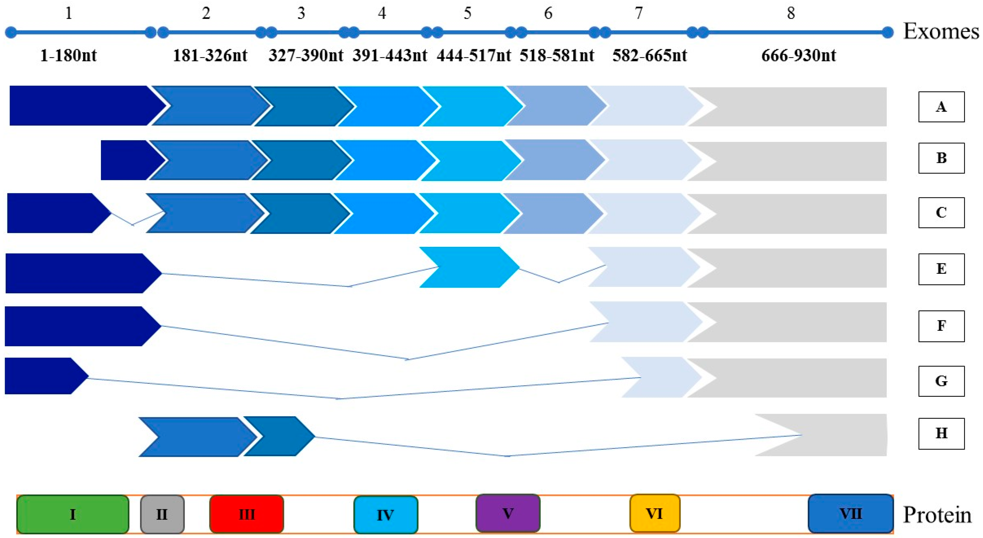
2. Molecular Structure
3. Expression and Survival
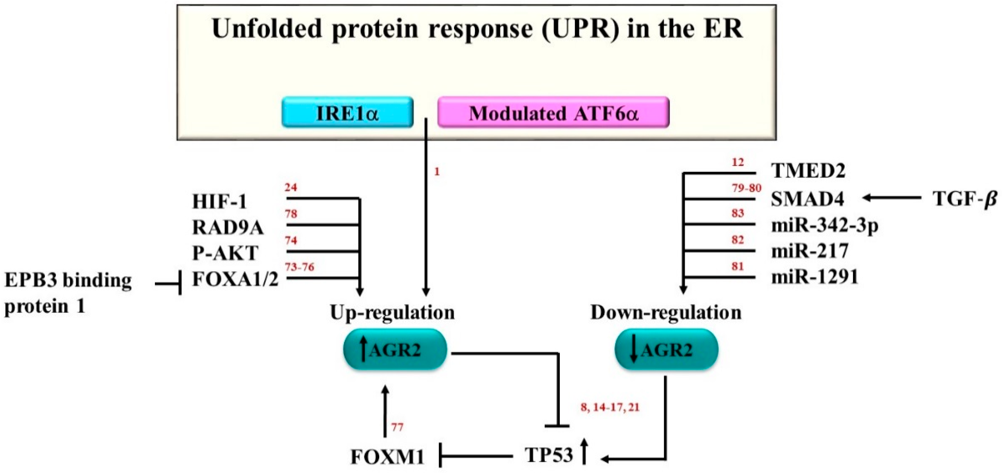
4. AGR2 Regulation
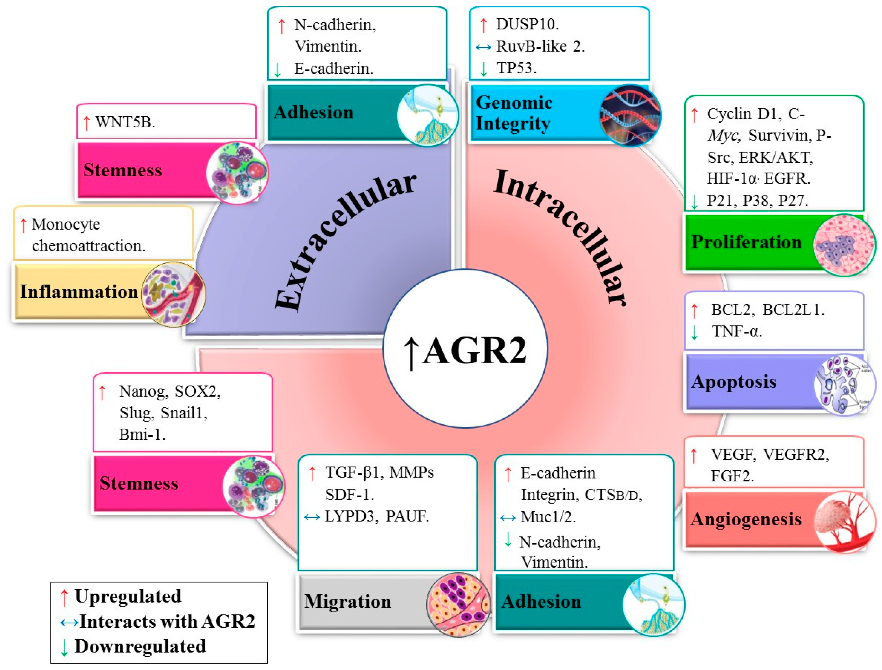
5. Role of AGR2 in the Cell Signaling Network
5.1. Genomic Integrity
5.2. Proliferation and Apoptosis
5.3. Angiogenesis
5.4. Adhesion and Migration
5.5. Stemness
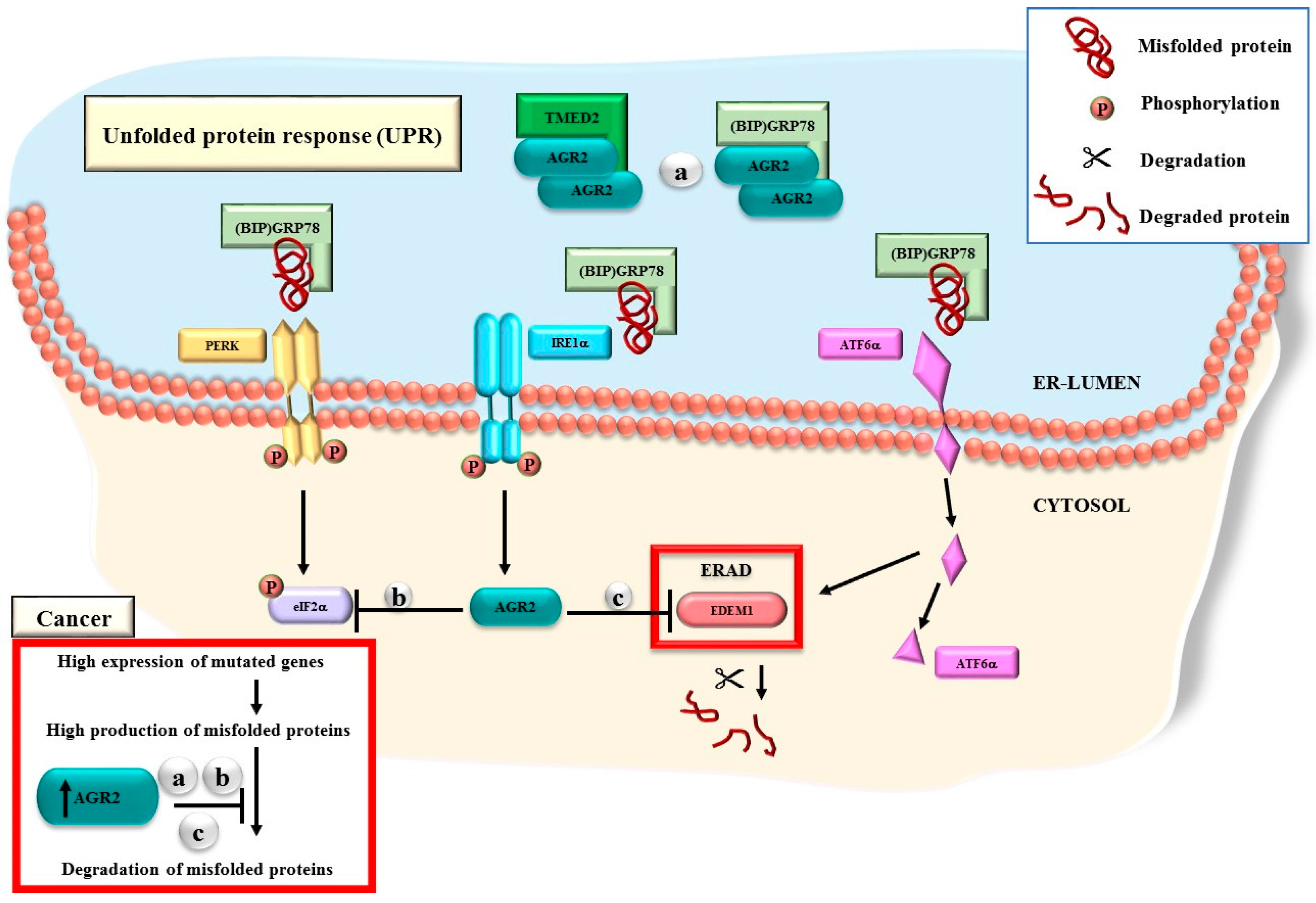
6. AGR2 Functions through the UPR Machinery
7. Drug Resistance
8. Therapeutic Potential
9. AGR2 Gene Expression Meta-Analysis in CNS Tumors
10. Conclusions and Way Forward
Supplementary Materials
Author Contributions
Funding
Conflicts of Interest
Abbreviations
| AdAGR2 | Encode AGR2 |
| AdCs | Adenocarcinoma |
| AGR2 | Anterior Gradient 2 Homolog |
| AhR | Aryl hydrocarbon receptor |
| AKT1 | RAC-alpha serine/threonine-protein kinase1 |
| ALDH1 | Aldehyde Dehydrogenase 1 |
| ATF6α | Activating Transcription Factor 6 |
| AuNPs | Nanoparticles |
| CALB1 | Calbindin 1 |
| CNS | Central Nervous System tumors |
| CCA | Cholangiocarcinoma |
| CCA | Cholangiocarcinoma |
| CD133 | Prominin 1 |
| CDH2 | N-Cadherin |
| CDH1 | E-Cadherin |
| CML | Chronic Myelogenous Leukemia |
| CRISPR | Clustered Regularly Interspaced Short Palindromic Repeats |
| CSCs | Cancer Stem Cells |
| CTLs | Cytotoxic T-lymphocyte |
| CTSB | Cathepsins B |
| CTSD | Cathepsins D |
| DAG1 | α dystroglycan1 |
| DCs | Dendritic Cells |
| DFS | Disease free survival |
| DUSP10 | Dual Specificity Phosphatase 10 |
| eAGR2 | extracellular AGR2 |
| EBP1 | ErbB3 Binding Protein 1 |
| EDEM1 | Alpha-mannosidase like protein1 |
| EGFR | Epidermal Growth Factor Receptor |
| eIF2α | Translation Initiation Factor |
| ELISA | Enzyme-Linked Immunosorbent Assay |
| EMT | Epithelial-mesenchymal transition |
| ER | Endoplasmic Reticulum |
| ERAD | Endoplasmic Reticulum-Associated Degradation |
| FGF2 | Fibroblast Growth Factor 2 |
| FOXA1 | Forkhead Box A1 |
| FOXA2 | Forkhead Box A3 |
| FOXM1 | Forkhead Box M1 |
| GBM | Glioblastoma Multiforme |
| GEO | Gene Expression Omnibus |
| GEP | Gene Expression Profiling |
| GRp78 | Endoplasmic reticulum luminal Calcium Binding Protein |
| H&E | Hematoxylin and eosin stain |
| HBEC | Human Bronchial Epithelial Cells |
| HCCs | Hepatocellular carcinoma |
| HepG2 | Human Hepatocarcinoma cell line |
| HER2 | Human Epidermal Growth Factor Receptor2 |
| HIF-1α | Hypoxia-inducible factor-1α |
| HNSCC | Head and Neck Squamous Cell Carcinoma |
| IBD | Inflammatory Bowel Disease |
| IDCs | Invasive Ductal Carcinomas |
| IPA | Ingenuity Pathway Analysis Software |
| IRE1α | Inositol-Requiring Enzyme1 |
| K562DR | Dasatinib-resistant K562 |
| LYPD3 | Ly6/PLAUR domain-containing protein 3 |
| MAPK | Mitogen Activated Kinase-Like Protein |
| miR-342-3p | MicroRNA-342-3p |
| MMPs | Matrix metalloproteinases |
| mRNA | Messenger RNA |
| MUC1 | Mucin1 |
| miR-1291 | MicroRNA-1291 |
| NSCLC | Non-small cell lung cancer |
| Oct4 | POU class 5 homeobox 1 |
| OS | Overall Survival |
| PA | Pituitary Adenoma |
| PanINs | Pancreatic Intraepithelial Neoplasia |
| PCa | Pancreatic cancer cell line |
| PC-3 | Prostate cancer cell line |
| PDAC | Pancreatic Ductal Adenocarcinomas |
| PDI | Protein Disulfide Isomerase |
| PDPK1 | 3-phosphoinositide-dependent protein kinase 1 |
| PIMA | Pulmonary Invasive Mucinous Adenocarcinoma |
| PR | Progesterone Receptor |
| RAD9A | RAD9 checkpoint clamp component A |
| rhAGR2 | Recombinant AGR2 |
| RNA-seq | Transcript Profiling |
| RT-PCR | Reverse transcription |
| RT-qPCR | Reverse transcription-Quantitative reverse transcription |
| RuvB-like2 | Reptin |
| SACC | Salivary Adenoid Cystic Carcinoma cell line |
| SCCs | Squamous Cell Carcinoma |
| SDF-1 | Stromal-Derived Factor-1 |
| shRNA | Short Hairpin RNA |
| siRNA | Small interferon RNA |
| Slug | Snail family transcriptional repressor 2 |
| Snail1 | Snail family transcriptional repressor 1 |
| Sox2 | SRY-box transcription factor 2 |
| SP | Secretory Pathway |
| SRCC | Gastric Signet-Ring Cell Carcinoma |
| TC | Tubular Complexes |
| TCDD | 2,3,7,8-Tetrachlorodibenzo-p-dioxin |
| TMED2 | Transmembrane p24 trafficking protein 2 |
| TGF-β | Transforming Growth Factor Beta |
| TTP | Time to Tumor Progression |
| TKI | Tyrosine Kinase Inhibitor |
| TNBC | Triple-negative breast cancers |
| TNF-α | Tumor Necrosis Factor |
| TNM | Tumor node metastasis |
| UPR | Unfolded Protein Response |
| VEGF | Vascular Endothelial Growth Factor |
| VEGFR2 | Vascular Endothelial Growth Factor Receptor |
| Wnt5b | Wnt family member 5B |
References
- Higa, A.; Mulot, A.; Delom, F.; Bouchecareilh, M.; Nguyen, D.T.; Boismenu, D.; Wise, M.J.; Chevet, E. Role of pro-oncogenic protein disulfide isomerase (pdi) family member anterior gradient 2 (agr2) in the control of endoplasmic reticulum homeostasis. J. Biol. Chem. 2011, 286, 44855–44868. [Google Scholar] [CrossRef] [PubMed]
- Thompson, D.A.; Weigel, R.J. Hag-2, the human homologue of the xenopus laevis cement gland gene xag-2, is coexpressed with estrogen receptor in breast cancer cell lines. Biochem. Biophys. Res. Commun. 1998, 251, 111–116. [Google Scholar] [CrossRef] [PubMed]
- Fletcher, G.C.; Patel, S.; Tyson, K.; Adam, P.J.; Schenker, M.; Loader, J.A.; Daviet, L.; Legrain, P.; Parekh, R.; Harris, A.L.; et al. Hag-2 and hag-3, human homologues of genes involved in differentiation, are associated with oestrogen receptor-positive breast tumours and interact with metastasis gene c4.4a and dystroglycan. Br. J. Cancer 2003, 88, 579–585. [Google Scholar] [CrossRef] [PubMed]
- Liu, D.; Rudland, P.S.; Sibson, D.R.; Platt-Higgins, A.; Barraclough, R. Human homologue of cement gland protein, a novel metastasis inducer associated with breast carcinomas. Cancer Res. 2005, 65, 3796–3805. [Google Scholar] [CrossRef] [PubMed]
- Patel, P.; Clarke, C.; Barraclough, D.L.; Jowitt, T.A.; Rudland, P.S.; Barraclough, R.; Lian, L.Y. Metastasis-promoting anterior gradient 2 protein has a dimeric thioredoxin fold structure and a role in cell adhesion. J. Mol. Biol. 2013, 425, 929–943. [Google Scholar] [CrossRef] [PubMed]
- Kumar, A.; Godwin, J.W.; Gates, P.B.; Garza-Garcia, A.A.; Brockes, J.P. Molecular basis for the nerve dependence of limb regeneration in an adult vertebrate. Science 2007, 318, 772–777. [Google Scholar] [CrossRef] [PubMed]
- Gourevitch, D.L.; Clark, L.; Bedelbaeva, K.; Leferovich, J.; Heber-Katz, E. Dynamic changes after murine digit amputation: The mrl mouse digit shows waves of tissue remodeling, growth, and apoptosis. Wound Repair Regen. 2009, 17, 447–455. [Google Scholar] [CrossRef] [PubMed]
- Pohler, E.; Craig, A.L.; Cotton, J.; Lawrie, L.; Dillon, J.F.; Ross, P.; Kernohan, N.; Hupp, T.R. The barrett’s antigen anterior gradient-2 silences the p53 transcriptional response to DNA damage. Mol. Cell. Proteom. 2004, 3, 534–547. [Google Scholar] [CrossRef]
- Gray, T.A.; Alsamman, K.; Murray, E.; Sims, A.H.; Hupp, T.R. Engineering a synthetic cell panel to identify signalling components reprogrammed by the cell growth regulator anterior gradient-2. Mol. Biosyst. 2014, 10, 1409–1425. [Google Scholar] [CrossRef]
- Niederreiter, L.; Kaser, A. Endoplasmic reticulum stress and inflammatory bowel disease. Acta Gastro-Enterol. Belg. 2011, 74, 330–333. [Google Scholar]
- Zhu, Q.; Mangukiya, H.B.; Mashausi, D.S.; Guo, H.; Negi, H.; Merugu, S.B.; Wu, Z.; Li, D. Anterior gradient 2 is induced in cutaneous wound and promotes wound healing through its adhesion domain. FEBS J. 2017, 284, 2856–2869. [Google Scholar] [CrossRef] [PubMed]
- Maurel, M.; Obacz, J.; Avril, T.; Ding, Y.P.; Papadodima, O.; Treton, X.; Daniel, F.; Pilalis, E.; Horberg, J.; Hou, W.; et al. Control of anterior gradient 2 (agr2) dimerization links endoplasmic reticulum proteostasis to inflammation. EMBO Mol. Med. 2019, 11, e10120. [Google Scholar] [CrossRef] [PubMed]
- Zweitzig, D.R.; Smirnov, D.A.; Connelly, M.C.; Terstappen, L.W.; O’Hara, S.M.; Moran, E. Physiological stress induces the metastasis marker agr2 in breast cancer cells. Mol. Cell. Biochem. 2007, 306, 255–260. [Google Scholar] [CrossRef] [PubMed]
- Ambolet-Camoit, A.; Bui, L.C.; Pierre, S.; Chevallier, A.; Marchand, A.; Coumoul, X.; Garlatti, M.; Andreau, K.; Barouki, R.; Aggerbeck, M. 2,3,7,8-tetrachlorodibenzo-p-dioxin counteracts the p53 response to a genotoxicant by upregulating expression of the metastasis marker agr2 in the hepatocarcinoma cell line hepg2. Toxicol. Sci. 2010, 115, 501–512. [Google Scholar] [CrossRef] [PubMed]
- Hu, Z.; Gu, Y.; Han, B.; Zhang, J.; Li, Z.; Tian, K.; Young, C.Y.; Yuan, H. Knockdown of agr2 induces cellular senescence in prostate cancer cells. Carcinogenesis 2012, 33, 1178–1186. [Google Scholar] [CrossRef]
- Li, Z.; Zhu, Q.; Chen, H.; Hu, L.; Negi, H.; Zheng, Y.; Ahmed, Y.; Wu, Z.; Li, D. Binding of anterior gradient 2 and estrogen receptor-alpha: Dual critical roles in enhancing fulvestrant resistance and igf-1-induced tumorigenesis of breast cancer. Cancer Lett. 2016, 377, 32–43. [Google Scholar] [CrossRef] [PubMed]
- Hrstka, R.; Bouchalova, P.; Michalova, E.; Matoulkova, E.; Muller, P.; Coates, P.J.; Vojtesek, B. Agr2 oncoprotein inhibits p38 mapk and p53 activation through a dusp10-mediated regulatory pathway. Mol. Oncol. 2016, 10, 652–662. [Google Scholar] [CrossRef]
- Brychtova, V.; Mohtar, A.; Vojtesek, B.; Hupp, T.R. Mechanisms of anterior gradient-2 regulation and function in cancer. Semin. Cancer Biol. 2015, 33, 16–24. [Google Scholar] [CrossRef]
- Chevet, E.; Fessart, D.; Delom, F.; Mulot, A.; Vojtesek, B.; Hrstka, R.; Murray, E.; Gray, T.; Hupp, T. Emerging roles for the pro-oncogenic anterior gradient-2 in cancer development. Oncogene 2013, 32, 2499–2509. [Google Scholar] [CrossRef]
- Ryu, J.; Park, S.G.; Lee, P.Y.; Cho, S.; Lee, D.H.; Kim, G.H.; Kim, J.H.; Park, B.C. Dimerization of pro-oncogenic protein anterior gradient 2 is required for the interaction with bip/grp78. Biochem. Biophys. Res. Commun. 2013, 430, 610–615. [Google Scholar] [CrossRef]
- Clarke, D.J.; Murray, E.; Faktor, J.; Mohtar, A.; Vojtesek, B.; MacKay, C.L.; Smith, P.L.; Hupp, T.R. Mass spectrometry analysis of the oxidation states of the pro-oncogenic protein anterior gradient-2 reveals covalent dimerization via an intermolecular disulphide bond. Biochim. Biophys. Acta 2016, 1864, 551–561. [Google Scholar] [CrossRef] [PubMed]
- Tian, S.B.; Tao, K.X.; Hu, J.; Liu, Z.B.; Ding, X.L.; Chu, Y.N.; Cui, J.Y.; Shuai, X.M.; Gao, J.B.; Cai, K.L.; et al. The prognostic value of agr2 expression in solid tumours: A systematic review and meta-analysis. Sci. Rep. 2017, 7, 15500–15510. [Google Scholar] [CrossRef] [PubMed]
- Khan, I.; Baeesa, S.; Bangash, M.; Schulten, H.J.; Alghamdi, F.; Qashqari, H.; Madkhali, N.; Carracedo, A.; Saka, M.; Jamal, A.; et al. Pleomorphism and drug resistant cancer stem cells are characteristic of aggressive primary meningioma cell lines. Cancer Cell Int. 2017, 17, 72–86. [Google Scholar] [CrossRef] [PubMed]
- Hong, X.Y.; Wang, J.; Li, Z. Agr2 expression is regulated by hif-1 and contributes to growth and angiogenesis of glioblastoma. Cell Biochem. Biophys. 2013, 67, 1487–1495. [Google Scholar] [CrossRef] [PubMed]
- Xu, C.; Liu, Y.; Xiao, L.; Guo, C.; Deng, S.; Zheng, S.; Zeng, E. The involvement of anterior gradient 2 in the stromal cell-derived factor 1-induced epithelial-mesenchymal transition of glioblastoma. Tumor Biol. 2016, 37, 6091–6097. [Google Scholar] [CrossRef] [PubMed]
- Obacz, J.; Brychtova, V.; Podhorec, J.; Fabian, P.; Dobes, P.; Vojtesek, B.; Hrstka, R. Anterior gradient protein 3 is associated with less aggressive tumors and better outcome of breast cancer patients. Oncotargets Ther. 2015, 8, 1523–1532. [Google Scholar]
- Gray, T.A.; MacLaine, N.J.; Michie, C.O.; Bouchalova, P.; Murray, E.; Howie, J.; Hrstka, R.; Maslon, M.M.; Nenutil, R.; Vojtesek, B.; et al. Anterior gradient-3: A novel biomarker for ovarian cancer that mediates cisplatin resistance in xenograft models. J. Immunol. Methods 2012, 378, 20–32. [Google Scholar] [CrossRef] [PubMed]
- Obacz, J.; Takacova, M.; Brychtova, V.; Dobes, P.; Pastorekova, S.; Vojtesek, B.; Hrstka, R. The role of agr2 and agr3 in cancer: Similar but not identical. Eur. J. Cell Biol. 2015, 94, 139–147. [Google Scholar] [CrossRef]
- Petek, E.; Windpassinger, C.; Egger, H.; Kroisel, P.M.; Wagner, K. Localization of the human anterior gradient-2 gene (agr2) to chromosome band 7p21.3 by radiation hybrid mapping and fluorescencein situ hybridisation. Cytogenet. Cell Genet. 2000, 89, 141–142. [Google Scholar] [CrossRef]
- Kovalev, L.I.; Shishkin, S.S.; Khasigov, P.Z.; Dzeranov, N.K.; Kazachenko, A.V.; Toropygin, I.; Mamykina, S.V. Identification of agr2 protein, a novel potential cancer marker, using proteomics technologies. Prikladnaia Biokhimiia I Mikrobiologiia 2006, 42, 480–484. [Google Scholar] [CrossRef]
- Ahmad, R.; Nicora, C.D.; Shukla, A.K.; Smith, R.D.; Qian, W.J.; Liu, A.Y. An efficient method for native protein purification in the selected range from prostate cancer tissue digests. Chin. Clin. Oncol. 2016, 5, 78–90. [Google Scholar] [CrossRef] [PubMed]
- Neeb, A.; Hefele, S.; Bormann, S.; Parson, W.; Adams, F.; Wolf, P.; Miernik, A.; Schoenthaler, M.; Kroenig, M.; Wilhelm, K.; et al. Splice variant transcripts of the anterior gradient 2 gene as a marker of prostate cancer. Oncotarget 2014, 5, 8681–8689. [Google Scholar] [CrossRef] [PubMed]
- Gupta, A.; Wodziak, D.; Tun, M.; Bouley, D.M.; Lowe, A.W. Loss of anterior gradient 2 (agr2) expression results in hyperplasia and defective lineage maturation in the murine stomach. J. Biol. Chem. 2013, 288, 4321–4333. [Google Scholar] [CrossRef] [PubMed]
- Shishkin, S.S.; Eremina, L.S.; Kovalev, L.I.; Kovaleva, M.A. Agr2, erp57/grp58, and some other human protein disulfide isomerases. Biochemistry 2013, 78, 1415–1430. [Google Scholar] [CrossRef] [PubMed]
- Gray, T.A.; Murray, E.; Nowicki, M.W.; Remnant, L.; Scherl, A.; Muller, P.; Vojtesek, B.; Hupp, T.R. Development of a fluorescent monoclonal antibody-based assay to measure the allosteric effects of synthetic peptides on self-oligomerization of agr2 protein. Protein Sci. 2013, 22, 1266–1278. [Google Scholar] [CrossRef]
- Clarke, C.; Rudland, P.; Barraclough, R. The metastasis-inducing protein agr2 is o-glycosylated upon secretion from mammary epithelial cells. Mol. Cell. Biochem. 2015, 408, 245–252. [Google Scholar] [CrossRef] [PubMed]
- Fagerberg, L.; Hallstrom, B.M.; Oksvold, P.; Kampf, C.; Djureinovic, D.; Odeberg, J.; Habuka, M.; Tahmasebpoor, S.; Danielsson, A.; Edlund, K.; et al. Analysis of the human tissue-specific expression by genome-wide integration of transcriptomics and antibody-based proteomics. Mol. Cell. Proteom. 2014, 13, 397–406. [Google Scholar] [CrossRef] [PubMed]
- Riener, M.O.; Thiesler, T.; Hellerbrand, C.; Amann, T.; Cathomas, G.; Fritzsche, F.R.; Dahl, E.; Bahra, M.; Weichert, W.; Terracciano, L.; et al. Loss of anterior gradient-2 expression is an independent prognostic factor in colorectal carcinomas. Eur. J. Cancer 2014, 50, 1722–1730. [Google Scholar] [CrossRef]
- Di Maro, G.; Salerno, P.; Unger, K.; Orlandella, F.M.; Monaco, M.; Chiappetta, G.; Thomas, G.; Oczko-Wojciechowska, M.; Masullo, M.; Jarzab, B.; et al. Anterior gradient protein 2 promotes survival, migration and invasion of papillary thyroid carcinoma cells. Mol. Cancer 2014, 13, 160–171. [Google Scholar] [CrossRef]
- Uhlen, M.; Fagerberg, L.; Hallstrom, B.M.; Lindskog, C.; Oksvold, P.; Mardinoglu, A.; Sivertsson, A.; Kampf, C.; Sjostedt, E.; Asplund, A.; et al. Proteomics. Tissue-based map of the human proteome. Science 2015, 347, 1260419. [Google Scholar] [CrossRef]
- Thul, P.J.; Akesson, L.; Wiking, M.; Mahdessian, D.; Geladaki, A.; Ait Blal, H.; Alm, T.; Asplund, A.; Bjork, L.; Breckels, L.M.; et al. A subcellular map of the human proteome. Science 2017, 356, 1126–1130. [Google Scholar] [CrossRef] [PubMed]
- Uhlen, M.; Zhang, C.; Lee, S.; Sjostedt, E.; Fagerberg, L.; Bidkhori, G.; Benfeitas, R.; Arif, M.; Liu, Z.; Edfors, F.; et al. A pathology atlas of the human cancer transcriptome. Science 2017, 357, 18–28. [Google Scholar] [CrossRef] [PubMed]
- Innes, H.E.; Liu, D.; Barraclough, R.; Davies, M.P.; O’Neill, P.A.; Platt-Higgins, A.; de Silva Rudland, S.; Sibson, D.R.; Rudland, P.S. Significance of the metastasis-inducing protein agr2 for outcome in hormonally treated breast cancer patients. Br. J. Cancer 2006, 94, 1057–1065. [Google Scholar] [CrossRef] [PubMed]
- Ondrouskova, E.; Sommerova, L.; Nenutil, R.; Coufal, O.; Bouchal, P.; Vojtesek, B.; Hrstka, R. Agr2 associates with her2 expression predicting poor outcome in subset of estrogen receptor negative breast cancer patients. Exp. Mol. Pathol. 2017, 102, 280–283. [Google Scholar] [CrossRef] [PubMed]
- Guo, J.; Gong, G.; Zhang, B. Identification and prognostic value of anterior gradient protein 2 expression in breast cancer based on tissue microarray. Tumour Biol. 2017, 39, 117–1186. [Google Scholar] [CrossRef] [PubMed]
- Ramachandran, V.; Arumugam, T.; Wang, H.; Logsdon, C.D. Anterior gradient 2 is expressed and secreted during the development of pancreatic cancer and promotes cancer cell survival. Cancer Res. 2008, 68, 7811–7818. [Google Scholar] [CrossRef] [PubMed]
- Riener, M.O.; Pilarsky, C.; Gerhardt, J.; Grutzmann, R.; Fritzsche, F.R.; Bahra, M.; Weichert, W.; Kristiansen, G. Prognostic significance of agr2 in pancreatic ductal adenocarcinoma. Histol. Histopathol. 2009, 24, 1121–1128. [Google Scholar] [PubMed]
- Mizuuchi, Y.; Aishima, S.; Ohuchida, K.; Shindo, K.; Fujino, M.; Hattori, M.; Miyazaki, T.; Mizumoto, K.; Tanaka, M.; Oda, Y. Anterior gradient 2 downregulation in a subset of pancreatic ductal adenocarcinoma is a prognostic factor indicative of epithelial-mesenchymal transition. Lab. Investig. 2015, 95, 193–206. [Google Scholar] [CrossRef] [PubMed]
- Uthaisar, K.; Vaeteewoottacharn, K.; Seubwai, W.; Talabnin, C.; Sawanyawisuth, K.; Obchoei, S.; Kraiklang, R.; Okada, S.; Wongkham, S. Establishment and characterization of a novel human cholangiocarcinoma cell line with high metastatic activity. Oncol. Rep. 2016, 36, 1435–1446. [Google Scholar] [CrossRef] [PubMed]
- Vivekanandan, P.; Micchelli, S.T.; Torbenson, M. Anterior gradient-2 is overexpressed by fibrolamellar carcinomas. Hum. Pathol. 2009, 40, 293–299. [Google Scholar] [CrossRef] [PubMed]
- Lepreux, S.; Bioulac-Sage, P.; Chevet, E. Differential expression of the anterior gradient protein-2 is a conserved feature during morphogenesis and carcinogenesis of the biliary tree. Liver Int. 2011, 31, 322–328. [Google Scholar] [CrossRef] [PubMed]
- Maresh, E.L.; Mah, V.; Alavi, M.; Horvath, S.; Bagryanova, L.; Liebeskind, E.S.; Knutzen, L.A.; Zhou, Y.; Chia, D.; Liu, A.Y.; et al. Differential expression of anterior gradient gene agr2 in prostate cancer. BMC Cancer 2010, 10, 680–688. [Google Scholar] [CrossRef] [PubMed]
- Wayner, E.A.; Quek, S.I.; Ahmad, R.; Ho, M.E.; Loprieno, M.A.; Zhou, Y.; Ellis, W.J.; True, L.D.; Liu, A.Y. Development of an elisa to detect the secreted prostate cancer biomarker agr2 in voided urine. Prostate 2012, 72, 1023–1034. [Google Scholar] [CrossRef] [PubMed]
- Shi, T.; Gao, Y.; Quek, S.I.; Fillmore, T.L.; Nicora, C.D.; Su, D.; Zhao, R.; Kagan, J.; Srivastava, S.; Rodland, K.D.; et al. A highly sensitive targeted mass spectrometric assay for quantification of agr2 protein in human urine and serum. J. Proteome Res. 2014, 13, 875–882. [Google Scholar] [CrossRef] [PubMed]
- Rice, G.E.; Edgell, T.A.; Autelitano, D.J. Evaluation of midkine and anterior gradient 2 in a multimarker panel for the detection of ovarian cancer. J. Exp. Clin. Cancer Res. 2010, 29, 62–69. [Google Scholar] [CrossRef] [PubMed]
- Kamal, A.; Valentijn, A.; Barraclough, R.; Rudland, P.; Rahmatalla, N.; Martin-Hirsch, P.; Stringfellow, H.; Decruze, S.B.; Hapangama, D.K. High agr2 protein is a feature of low grade endometrial cancer cells. Oncotarget 2018, 9, 31459–31472. [Google Scholar] [CrossRef] [PubMed]
- Fritzsche, F.R.; Dahl, E.; Dankof, A.; Burkhardt, M.; Pahl, S.; Petersen, I.; Dietel, M.; Kristiansen, G. Expression of agr2 in non small cell lung cancer. Histol. Histopathol. 2007, 22, 703–708. [Google Scholar] [PubMed]
- Pizzi, M.; Fassan, M.; Balistreri, M.; Galligioni, A.; Rea, F.; Rugge, M. Anterior gradient 2 overexpression in lung adenocarcinoma. Appl. Immunohistochem. Mol. Morphol. 2012, 20, 31–36. [Google Scholar] [CrossRef] [PubMed]
- Chung, K.; Nishiyama, N.; Wanibuchi, H.; Yamano, S.; Hanada, S.; Wei, M.; Suehiro, S.; Kakehashi, A. Agr2 as a potential biomarker of human lung adenocarcinoma. Osaka City Med. J. 2012, 58, 13–24. [Google Scholar]
- Narumi, S.; Miki, Y.; Hata, S.; Ebina, M.; Saito, M.; Mori, K.; Kobayashi, M.; Suzuki, T.; Iwabuchi, E.; Sato, I.; et al. Anterior gradient 2 is correlated with egfr mutation in lung adenocarcinoma tissues. Int. J. Biol. Markers 2015, 30, e234–e242. [Google Scholar] [CrossRef]
- Tomoshige, K.; Guo, M.; Tsuchiya, T.; Fukazawa, T.; Fink-Baldauf, I.M.; Stuart, W.D.; Naomoto, Y.; Nagayasu, T.; Maeda, Y. An egfr ligand promotes egfr-mutant but not kras-mutant lung cancer in vivo. Oncogene 2018, 37, 3894–3908. [Google Scholar] [CrossRef] [PubMed]
- Alavi, M.; Mah, V.; Maresh, E.L.; Bagryanova, L.; Horvath, S.; Chia, D.; Goodglick, L.; Liu, A.Y. High expression of agr2 in lung cancer is predictive of poor survival. BMC Cancer 2015, 15, 655–664. [Google Scholar] [CrossRef] [PubMed]
- Li, Y.; Wang, W.; Liu, Z.; Jiang, Y.; Lu, J.; Xie, H.; Tang, F. Agr2 diagnostic value in nasopharyngeal carcinoma prognosis. Clin. Chim. Acta 2018, 484, 323–327. [Google Scholar] [CrossRef] [PubMed]
- Pizzi, M.; Fassan, M.; Realdon, S.; Balistreri, M.; Battaglia, G.; Giacometti, C.; Zaninotto, G.; Zagonel, V.; De Boni, M.; Rugge, M. Anterior gradient 2 profiling in barrett columnar epithelia and adenocarcinoma. Hum. Pathol. 2012, 43, 1839–1844. [Google Scholar] [CrossRef] [PubMed]
- Zhang, J.; Jin, Y.; Xu, S.; Zheng, J.; Zhang, Q.I.; Wang, Y.; Chen, J.; Huang, Y.; He, X.; Zhao, Z. Agr2 is associated with gastric cancer progression and poor survival. Oncol. Lett. 2016, 11, 2075–2083. [Google Scholar] [CrossRef] [PubMed]
- Valladares-Ayerbes, M.; Blanco-Calvo, M.; Reboredo, M.; Lorenzo-Patino, M.J.; Iglesias-Diaz, P.; Haz, M.; Diaz-Prado, S.; Medina, V.; Santamarina, I.; Pertega, S.; et al. Evaluation of the adenocarcinoma-associated gene agr2 and the intestinal stem cell marker lgr5 as biomarkers in colorectal cancer. Int. J. Mol. Sci. 2012, 13, 4367–4387. [Google Scholar] [CrossRef]
- Kim, S.J.; Jun, S.; Cho, H.Y.; Lee, D.C.; Yeom, Y.I.; Kim, J.H.; Kang, D. Knockdown of anterior gradient 2 expression extenuates tumor-associated phenotypes of snu-478 ampulla of vater cancer cells. BMC Cancer 2014, 14, 804–814. [Google Scholar] [CrossRef]
- Kim, S.J.; Kim, D.H.; Kang, D.; Kim, J.H. Expression of anterior gradient 2 is decreased with the progression of human biliary tract cancer. Tohoku J. Exp. Med. 2014, 234, 83–88. [Google Scholar] [CrossRef]
- Ho, M.E.; Quek, S.I.; True, L.D.; Seiler, R.; Fleischmann, A.; Bagryanova, L.; Kim, S.R.; Chia, D.; Goodglick, L.; Shimizu, Y.; et al. Bladder cancer cells secrete while normal bladder cells express but do not secrete agr2. Oncotarget 2016, 7, 15747–15756. [Google Scholar] [CrossRef]
- Ma, S.R.; Wang, W.M.; Huang, C.F.; Zhang, W.F.; Sun, Z.J. Anterior gradient protein 2 expression in high grade head and neck squamous cell carcinoma correlated with cancer stem cell and epithelial mesenchymal transition. Oncotarget 2015, 6, 8807–8821. [Google Scholar] [CrossRef]
- Tohti, M.; Li, J.; Ma, C.; Li, W.; Lu, Z.; Hu, Y. Expression of agr2 in pituitary adenomas and its association with tumor aggressiveness. Oncol. Lett. 2015, 10, 2878–2882. [Google Scholar] [CrossRef] [PubMed]
- Tohti, M.; Li, J.; Tang, C.; Wen, G.; Abdujilil, A.; Yizim, P.; Ma, C. Serum agr2 as a useful biomarker for pituitary adenomas. Clin. Neurol. Neurosurg. 2017, 154, 19–22. [Google Scholar] [CrossRef] [PubMed]
- Vitello, E.A.; Quek, S.I.; Kincaid, H.; Fuchs, T.; Crichton, D.J.; Troisch, P.; Liu, A.Y. Cancer-secreted agr2 induces programmed cell death in normal cells. Oncotarget 2016, 7, 49425–49434. [Google Scholar] [CrossRef] [PubMed]
- Hrstka, R.; Murray, E.; Brychtova, V.; Fabian, P.; Hupp, T.R.; Vojtesek, B. Identification of an akt-dependent signalling pathway that mediates tamoxifen-dependent induction of the pro-metastatic protein anterior gradient-2. Cancer Lett. 2013, 333, 187–193. [Google Scholar] [CrossRef] [PubMed]
- Zhang, Y.; Ali, T.Z.; Zhou, H.; D’Souza, D.R.; Lu, Y.; Jaffe, J.; Liu, Z.; Passaniti, A.; Hamburger, A.W. Erbb3 binding protein 1 represses metastasis-promoting gene anterior gradient protein 2 in prostate cancer. Cancer Res. 2010, 70, 240–248. [Google Scholar] [CrossRef] [PubMed]
- Zheng, W.; Rosenstiel, P.; Huse, K.; Sina, C.; Valentonyte, R.; Mah, N.; Zeitlmann, L.; Grosse, J.; Ruf, N.; Nurnberg, P.; et al. Evaluation of agr2 and agr3 as candidate genes for inflammatory bowel disease. Genes Immun. 2006, 7, 11–18. [Google Scholar] [CrossRef] [PubMed]
- Milewski, D.; Balli, D.; Ustiyan, V.; Le, T.; Dienemann, H.; Warth, A.; Breuhahn, K.; Whitsett, J.A.; Kalinichenko, V.V.; Kalin, T.V. Foxm1 activates agr2 and causes progression of lung adenomas into invasive mucinous adenocarcinomas. PLoS Genet. 2017, 13, e1007097. [Google Scholar] [CrossRef] [PubMed]
- Broustas, C.G.; Hopkins, K.M.; Panigrahi, S.K.; Wang, L.; Virk, R.K.; Lieberman, H.B. Rad9a promotes metastatic phenotypes through transcriptional regulation of anterior gradient 2 (agr2). Carcinogenesis 2019, 40, 164–172. [Google Scholar] [CrossRef] [PubMed]
- Sommerova, L.; Ondrouskova, E.; Vojtesek, B.; Hrstka, R. Suppression of agr2 in a tgf-beta-induced smad regulatory pathway mediates epithelial-mesenchymal transition. BMC Cancer 2017, 17, 546–557. [Google Scholar] [CrossRef]
- Norris, A.M.; Gore, A.; Balboni, A.; Young, A.; Longnecker, D.S.; Korc, M. Agr2 is a smad4-suppressible gene that modulates muc1 levels and promotes the initiation and progression of pancreatic intraepithelial neoplasia. Oncogene 2013, 32, 3867–3876. [Google Scholar] [CrossRef]
- Tu, M.J.; Pan, Y.Z.; Qiu, J.X.; Kim, E.J.; Yu, A.M. Microrna-1291 targets the foxa2-agr2 pathway to suppress pancreatic cancer cell proliferation and tumorigenesis. Oncotarget 2016, 7, 45547–45561. [Google Scholar] [CrossRef] [PubMed]
- Pan, B.; Yang, J.; Wang, X.; Xu, K.; Ikezoe, T. Mir-217 sensitizes chronic myelogenous leukemia cells to tyrosine kinase inhibitors by targeting pro-oncogenic anterior gradient 2. Exp. Hematol. 2018, 68, 80–88. [Google Scholar] [CrossRef] [PubMed]
- Xue, X.; Fei, X.; Hou, W.; Zhang, Y.; Liu, L.; Hu, R. Mir-342-3p suppresses cell proliferation and migration by targeting agr2 in non-small cell lung cancer. Cancer Lett. 2018, 412, 170–178. [Google Scholar] [CrossRef] [PubMed]
- Maslon, M.M.; Hrstka, R.; Vojtesek, B.; Hupp, T.R. A divergent substrate-binding loop within the pro-oncogenic protein anterior gradient-2 forms a docking site for reptin. J. Mol. Biol. 2010, 404, 418–438. [Google Scholar] [CrossRef] [PubMed]
- Park, K.; Chung, Y.J.; So, H.; Kim, K.; Park, J.; Oh, M.; Jo, M.; Choi, K.; Lee, E.J.; Choi, Y.L.; et al. Agr2, a mucinous ovarian cancer marker, promotes cell proliferation and migration. Exp. Mol. Med. 2011, 43, 91–100. [Google Scholar] [CrossRef] [PubMed]
- Dumartin, L.; Alrawashdeh, W.; Trabulo, S.M.; Radon, T.P.; Steiger, K.; Feakins, R.M.; di Magliano, M.P.; Heeschen, C.; Esposito, I.; Lemoine, N.R.; et al. Er stress protein agr2 precedes and is involved in the regulation of pancreatic cancer initiation. Oncogene 2017, 36, 3094–3103. [Google Scholar] [CrossRef] [PubMed]
- Gupta, A.; Dong, A.; Lowe, A.W. Agr2 gene function requires a unique endoplasmic reticulum localization motif. J. Biol. Chem. 2012, 287, 4773–4782. [Google Scholar] [CrossRef] [PubMed]
- Vanderlaag, K.E.; Hudak, S.; Bald, L.; Fayadat-Dilman, L.; Sathe, M.; Grein, J.; Janatpour, M.J. Anterior gradient-2 plays a critical role in breast cancer cell growth and survival by modulating cyclin d1, estrogen receptor-alpha and survivin. Breast Cancer Res. 2010, 12, 32–47. [Google Scholar] [CrossRef] [PubMed]
- Guo, H.; Zhu, Q.; Yu, X.; Merugu, S.B.; Mangukiya, H.B.; Smith, N.; Li, Z.; Zhang, B.; Negi, H.; Rong, R.; et al. Tumor-secreted anterior gradient-2 binds to vegf and fgf2 and enhances their activities by promoting their homodimerization. Oncogene 2017, 36, 5098–5109. [Google Scholar] [CrossRef] [PubMed]
- Jia, M.; Guo, Y.; Zhu, D.; Zhang, N.; Li, L.; Jiang, J.; Dong, Y.; Xu, Q.; Zhang, X.; Wang, M.; et al. Pro-metastatic activity of agr2 interrupts angiogenesis target bevacizumab efficiency via direct interaction with vegfa and activation of nf-kappab pathway. Biochim. Biophys. Acta Mol. Basis Dis. 2018, 1864, 1622–1633. [Google Scholar] [CrossRef] [PubMed]
- Sung, H.Y.; Choi, E.N.; Lyu, D.; Park, A.K.; Ju, W.; Ahn, J.H. Aberrant hypomethylation-mediated agr2 overexpression induces an aggressive phenotype in ovarian cancer cells. Oncol. Rep. 2014, 32, 815–820. [Google Scholar] [CrossRef] [PubMed]
- Dumartin, L.; Whiteman, H.J.; Weeks, M.E.; Hariharan, D.; Dmitrovic, B.; Iacobuzio-Donahue, C.A.; Brentnall, T.A.; Bronner, M.P.; Feakins, R.M.; Timms, J.F.; et al. Agr2 is a novel surface antigen that promotes the dissemination of pancreatic cancer cells through regulation of cathepsins b and d. Cancer Res. 2011, 71, 7091–7102. [Google Scholar] [CrossRef] [PubMed]
- Chanda, D.; Lee, J.H.; Sawant, A.; Hensel, J.A.; Isayeva, T.; Reilly, S.D.; Siegal, G.P.; Smith, C.; Grizzle, W.; Singh, R.; et al. Anterior gradient protein-2 is a regulator of cellular adhesion in prostate cancer. PLoS ONE 2014, 9, e89940. [Google Scholar] [CrossRef] [PubMed]
- Park, S.W.; Zhen, G.; Verhaeghe, C.; Nakagami, Y.; Nguyenvu, L.T.; Barczak, A.J.; Killeen, N.; Erle, D.J. The protein disulfide isomerase agr2 is essential for production of intestinal mucus. Proc. Natl. Acad. Sci. USA 2009, 106, 6950–6955. [Google Scholar] [CrossRef] [PubMed]
- Bhatia, R.; Gautam, S.K.; Cannon, A.; Thompson, C.; Hall, B.R.; Aithal, A.; Banerjee, K.; Jain, M.; Solheim, J.C.; Kumar, S.; et al. Cancer-associated mucins: Role in immune modulation and metastasis. Cancer Metastasis Rev. 2019. [Google Scholar] [CrossRef] [PubMed]
- Reynolds, I.S.; Fichtner, M.; McNamara, D.A.; Kay, E.W.; Prehn, J.H.M.; Burke, J.P. Mucin glycoproteins block apoptosis; promote invasion, proliferation, and migration; and cause chemoresistance through diverse pathways in epithelial cancers. Cancer Metastasis Rev. 2019. [Google Scholar] [CrossRef] [PubMed]
- Tsuji, T.; Satoyoshi, R.; Aiba, N.; Kubo, T.; Yanagihara, K.; Maeda, D.; Goto, A.; Ishikawa, K.; Yashiro, M.; Tanaka, M. Agr2 mediates paracrine effects on stromal fibroblasts that promote invasion by gastric signet-ring carcinoma cells. Cancer Res. 2015, 75, 356–366. [Google Scholar] [CrossRef] [PubMed]
- Yosudjai, J.; Inpad, C.; Chomwong, S.; Dana, P.; Sawanyawisuth, K.; Phimsen, S.; Wongkham, S.; Jirawatnotai, S.; Kaewkong, W. An aberrantly spliced isoform of anterior gradient-2, agr2vh promotes migration and invasion of cholangiocarcinoma cell. Biomed. Pharmacother. 2018, 107, 109–116. [Google Scholar] [CrossRef] [PubMed]
- Fessart, D.; Domblides, C.; Avril, T.; Eriksson, L.A.; Begueret, H.; Pineau, R.; Malrieux, C.; Dugot-Senant, N.; Lucchesi, C.; Chevet, E.; et al. Secretion of protein disulphide isomerase agr2 confers tumorigenic properties. eLife 2016, 5, e13887. [Google Scholar] [CrossRef]
- Kim, J.; Chung, J.Y.; Kim, T.J.; Lee, J.W.; Kim, B.G.; Bae, D.S.; Choi, C.H.; Hewitt, S.M. Genomic network-based analysis reveals pancreatic adenocarcinoma up-regulating factor-related prognostic markers in cervical carcinoma. Front. Oncol. 2018, 8, 465. [Google Scholar] [CrossRef]
- Delom, F.; Nazaraliyev, A.; Fessart, D. The role of protein disulphide isomerase agr2 in the tumour niche. Biol. Cell 2018, 110, 271–282. [Google Scholar] [CrossRef] [PubMed]
- Hrstka, R.; Brychtova, V.; Fabian, P.; Vojtesek, B.; Svoboda, M. Agr2 predicts tamoxifen resistance in postmenopausal breast cancer patients. Dis. Markers 2013, 35, 207–212. [Google Scholar] [CrossRef] [PubMed]
- Hrstka, R.; Podhorec, J.; Nenutil, R.; Sommerova, L.; Obacz, J.; Durech, M.; Faktor, J.; Bouchal, P.; Skoupilova, H.; Vojtesek, B. Tamoxifen-dependent induction of agr2 is associated with increased aggressiveness of endometrial cancer cells. Cancer Investig. 2017, 35, 313–324. [Google Scholar] [CrossRef] [PubMed]
- Li, Z.; Zhu, Q.; Hu, L.; Chen, H.; Wu, Z.; Li, D. Anterior gradient 2 is a binding stabilizer of hypoxia inducible factor-1alpha that enhances cocl2 -induced doxorubicin resistance in breast cancer cells. Cancer Sci. 2015, 106, 1041–1049. [Google Scholar] [CrossRef] [PubMed]
- Wu, J.; Wang, C.; Li, X.; Song, Y.; Wang, W.; Li, C.; Hu, J.; Zhu, Z.; Li, J.; Zhang, W.; et al. Identification, characterization and application of a g-quadruplex structured DNA aptamer against cancer biomarker protein anterior gradient homolog 2. PLoS ONE 2012, 7, e46393. [Google Scholar] [CrossRef] [PubMed]
- Wu, Z.H.; Zhu, Q.; Gao, G.W.; Zhou, C.C.; Li, D.W. Preparation, characterization and potential application of monoclonal antibody 18a4 against agr2. Chin. J. Cell. Mol. Immunol. 2010, 26, 49–51. [Google Scholar]
- Lee, H.J.; Hong, C.Y.; Kim, M.H.; Lee, Y.K.; Nguyen-Pham, T.N.; Park, B.C.; Yang, D.H.; Chung, I.J.; Kim, H.J.; Lee, J.J. In vitro induction of anterior gradient-2-specific cytotoxic t lymphocytes by dendritic cells transduced with recombinant adenoviruses as a potential therapy for colorectal cancer. Exp. Mol. Med. 2012, 44, 60–67. [Google Scholar] [CrossRef]
- Lee, H.J.; Hong, C.Y.; Jin, C.J.; Kim, M.H.; Lee, Y.K.; Nguyen-Pham, T.N.; Lee, H.; Park, B.C.; Chung, I.J.; Kim, H.J.; et al. Identification of novel hla-a*0201-restricted epitopes from anterior gradient-2 as a tumor-associated antigen against colorectal cancer. Cell. Mol. Immunol. 2012, 9, 175–183. [Google Scholar] [CrossRef]
- Garri, C.; Howell, S.; Tiemann, K.; Tiffany, A.; Jalali-Yazdi, F.; Alba, M.M.; Katz, J.E.; Takahashi, T.T.; Landgraf, R.; Gross, M.E.; et al. Identification, characterization and application of a new peptide against anterior gradient homolog 2 (agr2). Oncotarget 2018, 9, 27363–27379. [Google Scholar] [CrossRef]
- Arumugam, T.; Deng, D.; Bover, L.; Wang, H.; Logsdon, C.D.; Ramachandran, V. New blocking antibodies against novel agr2-c4.4a pathway reduce growth and metastasis of pancreatic tumors and increase survival in mice. Mol. Cancer Ther. 2015, 14, 941–951. [Google Scholar] [CrossRef]
- Guo, H.; Chen, H.; Zhu, Q.; Yu, X.; Rong, R.; Merugu, S.B.; Mangukiya, H.B.; Li, D. A humanized monoclonal antibody targeting secreted anterior gradient 2 effectively inhibits the xenograft tumor growth. Biochem. Biophys. Res. Commun. 2016, 475, 57–63. [Google Scholar] [CrossRef] [PubMed]
- Pan, F.; Li, W.; Yang, W.; Yang, X.Y.; Liu, S.; Li, X.; Zhao, X.; Ding, H.; Qin, L.; Pan, Y. Anterior gradient 2 as a supervisory marker for tumor vessel normalization induced by anti-angiogenic treatment. Oncol. Lett. 2018, 16, 3083–3091. [Google Scholar] [CrossRef] [PubMed]
- Barrett, T.; Wilhite, S.E.; Ledoux, P.; Evangelista, C.; Kim, I.F.; Tomashevsky, M.; Marshall, K.A.; Phillippy, K.H.; Sherman, P.M.; Holko, M.; et al. Ncbi geo: Archive for functional genomics data sets–update. Nucleic Acids Res. 2013, 41, D991–D995. [Google Scholar] [CrossRef] [PubMed]
- Kolesnikov, N.; Hastings, E.; Keays, M.; Melnichuk, O.; Tang, Y.A.; Williams, E.; Dylag, M.; Kurbatova, N.; Brandizi, M.; Burdett, T.; et al. Arrayexpress update—Simplifying data submissions. Nucleic Acids Res. 2015, 43, 1113–1116. [Google Scholar] [CrossRef] [PubMed]
- Ritchie, M.E.; Phipson, B.; Wu, D.; Hu, Y.; Law, C.W.; Shi, W.; Smyth, G.K. Limma powers differential expression analyses for RNA-sequencing and microarray studies. Nucleic Acids Res. 2015, 43, 47–59. [Google Scholar] [CrossRef] [PubMed]
- Holsken, A.; Sill, M.; Merkle, J.; Schweizer, L.; Buchfelder, M.; Flitsch, J.; Fahlbusch, R.; Metzler, M.; Kool, M.; Pfister, S.M.; et al. Adamantinomatous and papillary craniopharyngiomas are characterized by distinct epigenomic as well as mutational and transcriptomic profiles. Acta Neuropathol. Commun. 2016, 4, 20–32. [Google Scholar] [CrossRef] [PubMed]
- Lee, Y.; Liu, J.; Patel, S.; Cloughesy, T.; Lai, A.; Farooqi, H.; Seligson, D.; Dong, J.; Liau, L.; Becker, D.; et al. Genomic landscape of meningiomas. Brain Pathol. 2010, 20, 751–762. [Google Scholar] [CrossRef]
- Gump, J.M.; Donson, A.M.; Birks, D.K.; Amani, V.M.; Rao, K.K.; Griesinger, A.M.; Kleinschmidt-DeMasters, B.K.; Johnston, J.M.; Anderson, R.C.; Rosenfeld, A.; et al. Identification of targets for rational pharmacological therapy in childhood craniopharyngioma. Acta Neuropathol. Commun. 2015, 3, 30–41. [Google Scholar] [CrossRef]
- Michaelis, K.A.; Knox, A.J.; Xu, M.; Kiseljak-Vassiliades, K.; Edwards, M.G.; Geraci, M.; Kleinschmidt-DeMasters, B.K.; Lillehei, K.O.; Wierman, M.E. Identification of growth arrest and DNA-damage-inducible gene beta (gadd45beta) as a novel tumor suppressor in pituitary gonadotrope tumors. Endocrinology 2011, 152, 3603–3613. [Google Scholar] [CrossRef] [PubMed]
- Zhou, G.; Soufan, O.; Ewald, J.; Hancock, R.E.W.; Basu, N.; Xia, J. Networkanalyst 3.0: A visual analytics platform for comprehensive gene expression profiling and meta-analysis. Nucleic Acids Res. 2019. [Google Scholar] [CrossRef]
- Xia, J.; Gill, E.E.; Hancock, R.E. Networkanalyst for statistical, visual and network-based meta-analysis of gene expression data. Nat. Protoc. 2015, 10, 823–844. [Google Scholar] [CrossRef] [PubMed]
- Vazquez-Sanchez, A.Y.; Hinojosa, L.M.; Parraguirre-Martinez, S.; Gonzalez, A.; Morales, F.; Montalvo, G.; Vera, E.; Hernandez-Gallegos, E.; Camacho, J. Expression of katp channels in human cervical cancer: Potential tools for diagnosis and therapy. Oncol. Lett. 2018, 15, 6302–6308. [Google Scholar] [PubMed]
- Chang, Y.C.; Chan, Y.C.; Chang, W.M.; Lin, Y.F.; Yang, C.J.; Su, C.Y.; Huang, M.S.; Wu, A.T.H.; Hsiao, M. Feedback regulation of aldoa activates the hif-1alpha/mmp9 axis to promote lung cancer progression. Cancer Lett. 2017, 403, 28–36. [Google Scholar] [CrossRef] [PubMed]
- Musgrove, E.A.; Caldon, C.E.; Barraclough, J.; Stone, A.; Sutherland, R.L. Cyclin d as a therapeutic target in cancer. Nat. Rev. Cancer 2011, 11, 558–572. [Google Scholar] [CrossRef] [PubMed]
- Inoue, K.; Fry, E.A. Aberrant expression of p16(ink4a) in human cancers—A new biomarker? Cancer Rep. Rev. 2018, 2. [Google Scholar] [CrossRef]
- Olar, A.; Goodman, L.D.; Wani, K.M.; Boehling, N.S.; Sharma, D.S.; Mody, R.R.; Gumin, J.; Claus, E.B.; Lang, F.F.; Cloughesy, T.F.; et al. A gene expression signature predicts recurrence-free survival in meningioma. Oncotarget 2018, 9, 16087–16098. [Google Scholar] [CrossRef] [PubMed]
- Qian, C.; Xia, Y.; Ren, Y.; Yin, Y.; Deng, A. Identification and validation of psat1 as a potential prognostic factor for predicting clinical outcomes in patients with colorectal carcinoma. Oncol. Lett. 2017, 14, 8014–8020. [Google Scholar] [CrossRef] [PubMed]
- Jiang, J.; Zhang, L.; Chen, H.; Lei, Y.; Zhang, T.; Wang, Y.; Jin, P.; Lan, J.; Zhou, L.; Huang, Z.; et al. Regorafenib induces lethal autophagy arrest by stabilizing psat1 in glioblastoma. Autophagy 2019, 1–17. [Google Scholar] [CrossRef]
- Tabernero, M.D.; Maillo, A.; Gil-Bellosta, C.J.; Castrillo, A.; Sousa, P.; Merino, M.; Orfao, A. Gene expression profiles of meningiomas are associated with tumor cytogenetics and patient outcome. Brain Pathol. 2009, 19, 409–420. [Google Scholar] [CrossRef] [PubMed]
- Jandial, R.; Choy, C.; Levy, D.M.; Chen, M.Y.; Ansari, K.I. Astrocyte-induced reelin expression drives proliferation of her2(+) breast cancer metastases. Clin. Exp. Metastasis 2017, 34, 185–196. [Google Scholar] [CrossRef]
- Lin, L.; Yan, F.; Zhao, D.; Lv, M.; Liang, X.; Dai, H.; Qin, X.; Zhang, Y.; Hao, J.; Sun, X.; et al. Reelin promotes the adhesion and drug resistance of multiple myeloma cells via integrin beta1 signaling and stat3. Oncotarget 2016, 7, 9844–9858. [Google Scholar] [PubMed]
- Wu, X.; Giobbie-Hurder, A.; Liao, X.; Connelly, C.; Connolly, E.M.; Li, J.; Manos, M.P.; Lawrence, D.; McDermott, D.; Severgnini, M.; et al. Angiopoietin-2 as a biomarker and target for immune checkpoint therapy. Cancer Immunol. Res. 2017, 5, 17–28. [Google Scholar] [CrossRef] [PubMed]
- Li, Y.H.; Yu, C.Y.; Li, X.X.; Zhang, P.; Tang, J.; Yang, Q.; Fu, T.; Zhang, X.; Cui, X.; Tu, G.; et al. Therapeutic target database update 2018: Enriched resource for facilitating bench-to-clinic research of targeted therapeutics. Nucleic Acids Res. 2018, 46, 1121–1127. [Google Scholar]
- Schulten, H.J.; Hussein, D.; Al-Adwani, F.; Karim, S.; Al-Maghrabi, J.; Al-Sharif, M.; Jamal, A.; Al-Ghamdi, F.; Baeesa, S.S.; Bangash, M.; et al. Microarray expression data identify dcc as a candidate gene for early meningioma progression. PLoS ONE 2016, 11, e0153681. [Google Scholar] [CrossRef] [PubMed]
- Vassilopoulos, A.; Xiao, C.; Chisholm, C.; Chen, W.; Xu, X.; Lahusen, T.J.; Bewley, C.; Deng, C.X. Synergistic therapeutic effect of cisplatin and phosphatidylinositol 3-kinase (pi3k) inhibitors in cancer growth and metastasis of brca1 mutant tumors. J. Biol. Chem. 2014, 289, 24202–24214. [Google Scholar] [CrossRef] [PubMed]
- Schulten, H.J.; Hussein, D. Array expression meta-analysis of cancer stem cell genes identifies upregulation of podxl especially in dcc low expression meningiomas. PLoS ONE 2019, 14, e0215452. [Google Scholar] [CrossRef] [PubMed]
- Xu, C.; Sun, L.; Jiang, C.; Zhou, H.; Gu, L.; Liu, Y.; Xu, Q. Spp1, analyzed by bioinformatics methods, promotes the metastasis in colorectal cancer by activating emt pathway. Biomed. Pharmacother. 2017, 91, 1167–1177. [Google Scholar] [CrossRef] [PubMed]
- Seo, K.J.; Kim, M.; Kim, J. Prognostic implications of adhesion molecule expression in colorectal cancer. Int. J. Clin. Exp. Pathol. 2015, 8, 4148–4157. [Google Scholar] [PubMed]
- Hulleman, E.; Quarto, M.; Vernell, R.; Masserdotti, G.; Colli, E.; Kros, J.M.; Levi, D.; Gaetani, P.; Tunici, P.; Finocchiaro, G.; et al. A role for the transcription factor hey1 in glioblastoma. J. Cell. Mol. Med. 2009, 13, 136–146. [Google Scholar] [CrossRef] [PubMed]
- Salvi, S.; Bonafe, M.; Bravaccini, S. Androgen receptor in breast cancer: A wolf in sheep’s clothing? A lesson from prostate cancer. Semin. Cancer Biol. 2019. [Google Scholar] [CrossRef] [PubMed]
- Thike, A.A.; Yong-Zheng Chong, L.; Cheok, P.Y.; Li, H.H.; Wai-Cheong Yip, G.; Huat Bay, B.; Tse, G.M.; Iqbal, J.; Tan, P.H. Loss of androgen receptor expression predicts early recurrence in triple-negative and basal-like breast cancer. Mod. Pathol. 2014, 27, 352–360. [Google Scholar] [CrossRef] [PubMed]
- Schroeder, A.; Herrmann, A.; Cherryholmes, G.; Kowolik, C.; Buettner, R.; Pal, S.; Yu, H.; Muller-Newen, G.; Jove, R. Loss of androgen receptor expression promotes a stem-like cell phenotype in prostate cancer through stat3 signaling. Cancer Res. 2014, 74, 1227–1237. [Google Scholar] [CrossRef] [PubMed]
- Valkenburg, K.C.; De Marzo, A.M.; Williams, B.O. Deletion of tumor suppressors adenomatous polyposis coli and smad4 in murine luminal epithelial cells causes invasive prostate cancer and loss of androgen receptor expression. Oncotarget 2017, 8, 80265–80277. [Google Scholar] [CrossRef] [PubMed]
- Breier, G.; Grosser, M.; Rezaei, M. Endothelial cadherins in cancer. Cell Tissue Res. 2014, 355, 523–527. [Google Scholar] [CrossRef] [PubMed]
- Zanetta, L.; Corada, M.; Grazia Lampugnani, M.; Zanetti, A.; Breviario, F.; Moons, L.; Carmeliet, P.; Pepper, M.S.; Dejana, E. Downregulation of vascular endothelial-cadherin expression is associated with an increase in vascular tumor growth and hemorrhagic complications. Thromb. Haemost. 2005, 93, 1041–1046. [Google Scholar] [CrossRef] [PubMed]
- Rezaei, M.; Friedrich, K.; Wielockx, B.; Kuzmanov, A.; Kettelhake, A.; Labelle, M.; Schnittler, H.; Baretton, G.; Breier, G. Interplay between neural-cadherin and vascular endothelial-cadherin in breast cancer progression. Breast Cancer Res. 2012, 14, R154. [Google Scholar] [CrossRef] [PubMed]
- Evans, D.R.; Venkitachalam, S.; Revoredo, L.; Dohey, A.T.; Clarke, E.; Pennell, J.J.; Powell, A.E.; Quinn, E.; Ravi, L.; Gerken, T.A.; et al. Evidence for galnt12 as a moderate penetrance gene for colorectal cancer. Hum. Mutat. 2018, 39, 1092–1101. [Google Scholar] [CrossRef]
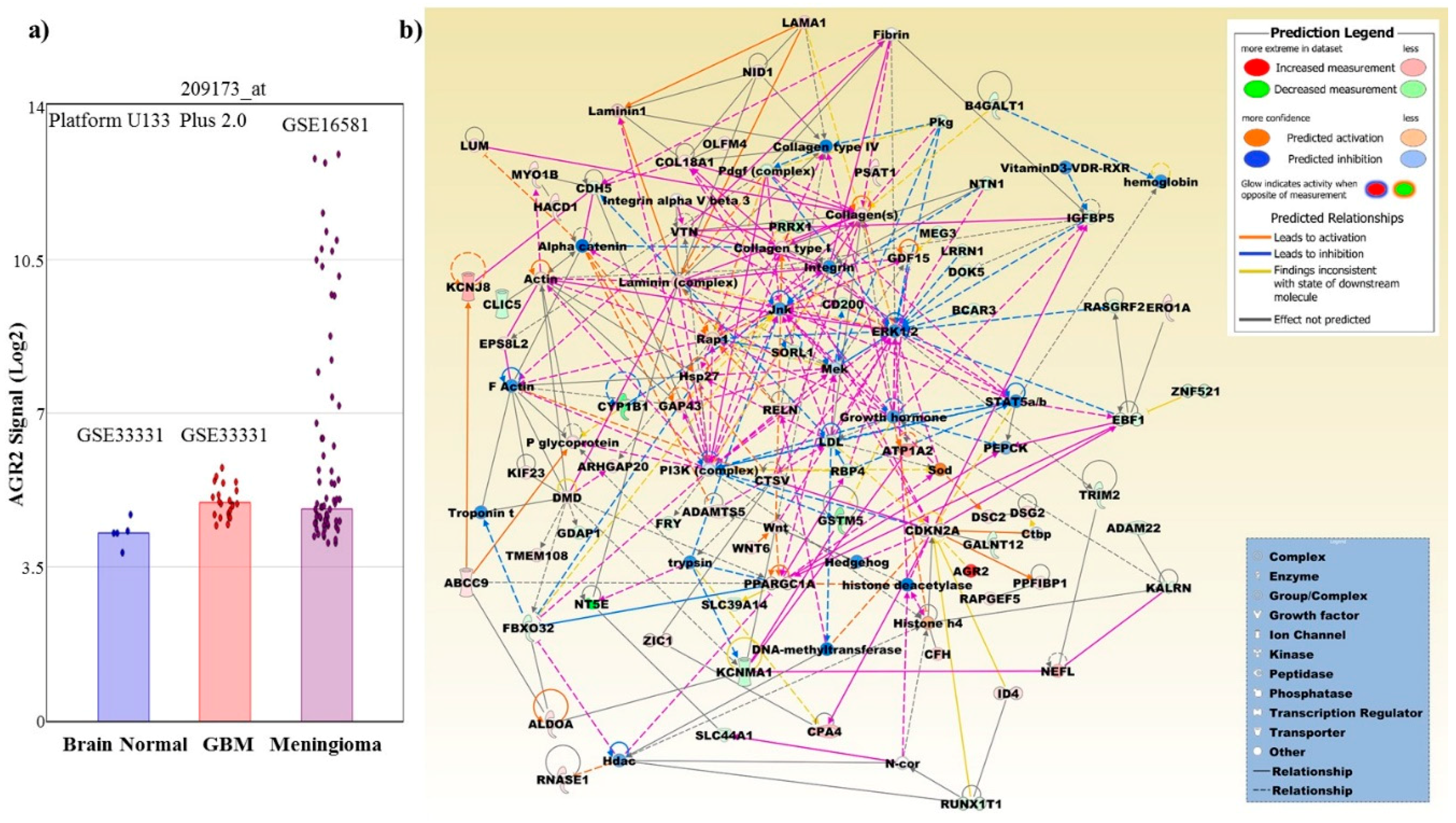
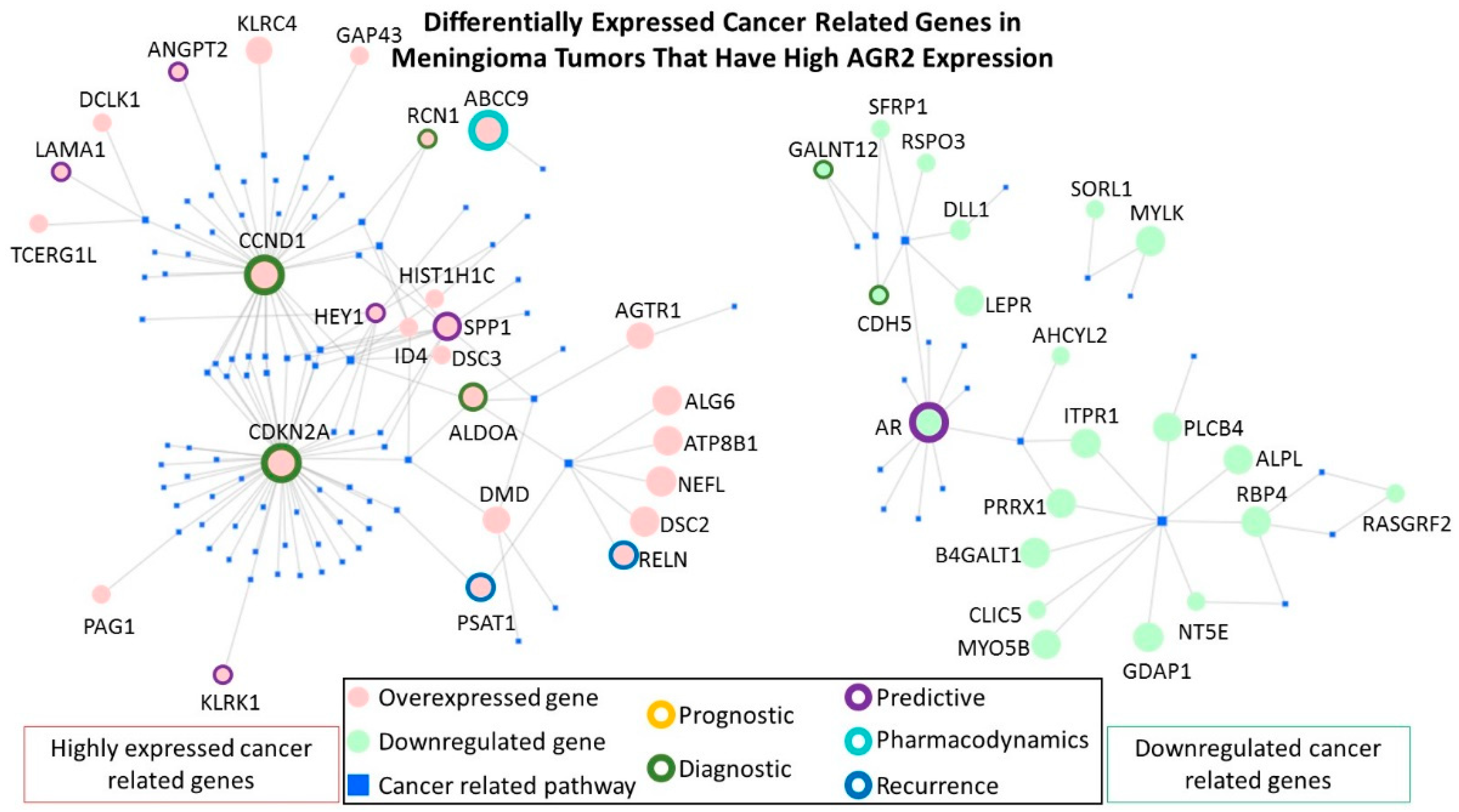
| GEO/Array Express Access Number | Array Type | Number of Samples | Tumor Type | |
|---|---|---|---|---|
| AGR2 High | AGR2 Low | |||
| GSE54934 | HuGene 1.0 ST | 3 | 19 | MN |
| GSE77259, GSE100534 | HuGene 1.0 ST | 6 | 16 | MN |
| GSE88720 | HuGene 2.1 ST | 2 | 12 | MN |
| GSE16581 | U133 Plus 2.0 | 16 | 52 | MN |
| GSE68015 | U133 Plus 2.0 | 3 | 6 | MN |
| E-MTAB-1852 | U133 Plus 2.0 | 5 | 10 | MN |
| E-GEOD-9438 | U133 Plus 2.0 | 3 | 16 | MN |
| E-MEXP-3586 | U133 Plus 2.0 | 5 | 11 | MN |
| GSE68015 | U133 Plus 2.0 | 5 | 10 | adCP 1 |
| GSE26966 | U133 Plus 2.0 | 13 | 9 | PGT 2 |
| Top Canonical Pathways | p-Value | Overlap |
| Apelin Cardiac Fibroblast Signaling Pathway | 7.55E−05 | 18.2% (4/22) |
| Axonal Guidance Signaling | 2.76E−04 | 3.0% (15/498) |
| Osteoarthritis Pathway | 4.17E−04 | 4.3% (9/211) |
| Hepatic Fibrosis/Hepatic Stellate Cell Activation | 8.22E−04 | 4.3% (8/186) |
| Glioblastoma Multiforme Signaling | 2.21E−03 | 4.1% (7/170) |
| Top Upstream Regulator | p-Value of Overlap | |
| progesterone | 1.31E−08 | |
| CTNNB1 | 1.68E−08 | |
| ERBB2 | 4.47E−08 | |
| forskolin | 5.07E−08 | |
| AHR | 5.61E−08 | |
| Diseases and Disorders | p-Value, # Molecules | |
| Neurological Disease | 2.24E−04–1.51E−15, 112 | |
| Cancer | 2.47E−04–2.22E−15, 228 | |
| Gastrointestinal Disease | 2.47E−04–2.22E−15, 216 | |
| Organismal Injury and Abnormalities | 2.47E−04–2.22E−15, 229 | |
| Cardiovascular Disease | 1.74E−04–3.10E−15, 85 | |
| Molecular and Cellular Functions | p-Value, # Molecules | |
| Cellular Movement | 2.36E−04–9.50E−14, 97 | |
| Cell Death and Survival | 2.05E−04–1.74E−10, 97 | |
| Cell-To-Cell Signaling and Interaction | 1.95E−04–2.26E−10, 49 | |
| Cellular Assembly and Organization | 2.51E−04–2.26E−10, 70 | |
| Cell Morphology | 2.31E−04–1.15E−09, 75 | |
| Physiological System Development and Function | p-Value, # Molecules | |
| Organismal Development | 1.87E−04–1.46E−15, 121 | |
| Nervous System Development and Function | 2.36E−04–1.51E−15, 90 | |
| Cardiovascular System Development and Function | 1.87E−04–1.60E−15, 74 | |
| Skeletal and Muscular System Development and Function | 2.31E−04–4.41E−11, 70 | |
| Tissue Morphology | 2.36E−04–9.89E−11, 97 | |
| Assays: Clinical Chemistry and Hematology | p-Value, # Molecules | |
| Increased Levels of Alkaline Phosphatase | 6.39E−03–1.48E−03, 5 | |
| Increased Levels of Hematocrit | 1.94E−02–1.94E−02, 4 | |
| Increased Levels of Red Blood Cells | 2.43E−02–2.43E−02, 4 | |
| Decreased Levels of Albumin | 6.16E−02–6.16E−02, 1 | |
| Increased Levels of ALT | 8.12E−02–8.12E−02, 1 | |
| Cardiotoxicity | p-Value, # Molecules | |
| Cardiac Dysfunction | 2.65E−01–2.23E−10, 19 | |
| Cardiac Arteriopathy | 1.56E−01–3.78E−08, 19 | |
| Cardiac Arrythmia | 3.92E−01–3.97E−07, 13 | |
| Cardiac Enlargement | 1.19E−01–3.81E−06, 23 | |
| Cardiac Dilation | 2.49E−01–1.25E−05, 15 | |
| Hepatotoxicity | p-Value, # Molecules | |
| Liver Hyperplasia/Hyperproliferation | 5.24E−01–2.28E−07, 118 | |
| Liver Steatosis | 1.91E−01–1.14E−04, 14 | |
| Liver Cirrhosis | 6.16E−02–2.42E−04, 12 | |
| Hepatocellular Carcinoma | 3.86E−01–3.32E−04, 27 | |
| Liver Necrosis/Cell Death | 2.94E−02–1.23E−03, 9 | |
| Nephrotoxicity | p-Value, # Molecules | |
| Renal Damage | 3.92E−01–4.38E−05, 12 | |
| Renal Dysfunction | 3.29E−04–3.29E−04, 2 | |
| Renal Necrosis/Cell Death | 2.65E−01–1.69E−03, 13 | |
| Glomerular Injury | 3.07E−01–9.78E−03, 8 | |
| Renal Inflammation | 3.78E−01–1.25E−02, 7 | |
| Top Networks | Score | |
| Skeletal and Muscular System Development and Function, Cancer, Endocrine System Disorders | 43 | |
| Skeletal and Muscular System Development and Function, Embryonic Development, Nervous System Development and Function | 38 | |
| Cellular Function and Maintenance, Skeletal and Muscular System, Development and Function, Tissue Development | 38 | |
| Cardiovascular Disease, Cardiovascular System. Development andFunction, Organismal Injury and Abnormalities | 31 | |
| Organismal Development, Organismal Functions, Cardiac Dysfunction | 29 | |
| Top Tox Lists | p-Value | Overlap |
| Hepatic Fibrosis | 1.67E−05 | 7.5% (8/106) |
| Cardiac Necrosis/Cell Death | 1.73E−05 | 4.4% (13/297) |
| Cardiac Fibrosis | 2.61E−05 | 4.9% (11/223) |
| Recovery from Ischemic Acute Renal Failure (Rat) | 3.85E−04 | 21.4% (3/14) |
| Cardiac Hypertrophy | 4.86E−04 | 3.3% (12/362) |
| Fold Change Up-Regulated Top Molecules, Exp. Value | Fold Change Down-Regulated Top Molecules, Exp. Value | |
| AGR2, 90.210 | TCEAL2, −48.790 | |
| CALB1, 49.780 | COL8A1, −36.360 | |
| KCNT2, 34.470 | MFAP5, −35.660 | |
| DSC3, 33.800 | NT5E, −31.300 | |
| HOPX, 27.670 | SYNPO2, −29.750 | |
| GPR83, 22.020 | SLPI, −20.350 | |
| CA3, 21.830 | CYP1B1, −18.420 | |
| ATP1A2, 21.780 | RSPO3, −17.040 | |
| KCNJ8, 19.690 | SFRP1, −16.210 | |
| GRB14, 17.690 | UCHL1, −15.030 | |
| Gene | Clinical Relevance |
|---|---|
| Upregulated | |
| ABCC9 (also known as SUR2) | The ATP Binding Cassette Subfamily C Member 9 gene encodes a protein which is a member of the superfamily of ATP-binding cassette (ABC) transporters and is known to be involved in multi-drug resistance, particularly in human cervical cancer [122]. |
| ALDOA | The aldolase, fructose-bisphosphate A gene encodes a proteins, which belongs to the class I fructose-bisphosphate aldolase protein family. This protein is thought to catalyze the reversible conversion of fructose-1,6-bisphosphate to glyceraldehyde 3-phosphate and dihydroxyacetone phosphate, a process which is vital for developmental pathways. The elevated protein levels were shown to highly correlate with a poor prognosis in patients with non-small cell lung cancer (NSCLC) [123]. |
| CCND1 | Cyclin D1 protein encoded by the CCND1 gene belongs to the cyclin family that consists of proteins that functions as regulators of CDK kinases. The CCND1 cyclin was shown to be required for the cell cycle G1/S transition, and associated gene overexpression has been observed frequently in a variety of tumors [124]. Previous studies confirmed the downregulation of cyclin D1 upon silencing of AGR2, as mentioned in Figure 3 [88]. |
| CDKN2A | The Cyclin Dependent Kinase Inhibitor 2A gene encodes the two cell cycle regulators p14ARF and p16INK4a. Overexpression of p16INK4a has been demonstrated in benign tumors and is seemingly a negative response mechanism to oncogenic signaling that otherwise would enhance cell proliferation [125]. In contrast, in cancers with retinoblastoma (RB) inactivation, overexpression of p16INK4a is associated with worse prognosis and therefore, using p16INK4a as a biomarker should be carefully considered in the specific context. A meta-analysis on microarray expression data in meningiomas identified a data set of 18 genes including CDKN2 whose deregulated expression was a predictive marker for recurrence [126]. This gene is often included in diagnostic onco-gene panels. |
| PSAT1 | Phosphoserine Aminotransferase 1 encodes an enzymatic component of the serine synthesis pathway. Overexpression of PSAT1 in different cancer types is associated with negative prognosis [127]. In glioblastoma cells, the multi-kinase inhibitor regorafenib effected a lethal autophagy arrest by stabilizing PSAT1 [128]. |
| RCN1 | A class prediction analysis demonstrated that Reticulocalbin 1, a calcium-binding protein, is overexpressed in meningiomas with cytogenetically complex karyotypes that are commonly associated with unfavorable prognosis [129]. AGR2 has been shown to regulate the expression of this ER chaperone in the PDAC tumor cells [92]. |
| RELN | The Reelin gene encodes an extracellular matrix glycoprotein protein which is involved in cell-cell interactions and is critical for cell positioning and neuronal migration during brain development. The expression of the RELN gene has been shown to be increased in Her2+ breast cancer brain metastases [130], and to be negatively correlated with the overall survival in multiple myeloma patients [131]. |
| ANGPT2 | A meta-analysis on microarray expression data in meningiomas identified a data set of 18 genes including Angiopoietin 2 whose deregulated expression was a predictive marker for recurrence [126]. Serum ANGPT2 is currently assessed for its application as a predictive and prognostic marker for immune checkpoint therapy in cancer [132]. |
| KLRK1 | Killer Cell Lectin Like Receptor K1 encodes the transmembrane protein NKG2D, which is part of the CD94/NKG2 family of C-type lectin-like transmembrane proteins. This receptor constitutes a possible therapeutic target for immune diseases and cancer [133]. In meningiomas, a microarray expression analysis detected higher expression of the read-through transcript KLRK1-KLRC4 in female cases compared male cases [134]. |
| LAMA1 | Laminin Subunit Alpha 1 encodes for a protein, which is a component of the extracellular matrix and was found to be comparably higher expressed in breast cancer stem cells [135]. A meta-analysis on microarray expression data on cancer stem cells in meningiomas identified recurrently upregulation of LAMA1 in GII + GIII compared to GI meningiomas [136]. |
| SPP1 | Secreted phosphoprotein 1 (SPP1) gene encodes a protein which is involved in the attachment of osteoclasts to the mineralized bone matrix, and in the upregulated expression of interferon-gamma and interleukin-12. The protein overexpression has been observed in various malignant neoplasms including medullary thyroid carcinoma, lung cancer, gastric cancer, breast cancer and colorectal cancer [137]. In addition, the upregulation of SPP1 protein has been shown to be significantly associated with adherence and invasion [138]. |
| HEY1 | The hes related family bHLH transcription factor with YRPW motif 1 (HEY1) gene encodes a nuclear protein that belongs to the hairy and enhancer of split-related (HESR) family of transcriptional repressors. Notch and c-Jun signal transduction pathways has been shown to induce the expression of this gene. HEY1 expression has been shown to increase with increasing astrocytoma tumor grade and to correlate with decreased overall survival and disease-free survival [139]. However, it is important to note that the protein levels of HEY1 have been reported to be high in the normal brain [37]. |
| Downregulated | |
| AR | The Androgen Receptor gene encodes a protein that functions as a steroid-hormone activated transcription factor. Many studies have shown tumor associated overexpression of this gene [140]. However, in meningioma, our data shows a significant downregulation of AR expression. Other studies have linked low expression with poorer prognosis. The loss of the AR expression in triple-negative breast cancers was associated with a worse prognosis [141]. Loss of AR expression was also shown to promote a stem-like cell phenotype and progression in prostate cancer cells through the STAT3 signaling pathway [142]. In addition, a double conditional knockout of adenomatous polyposis coli and Smad4 caused invasive prostate cancers that had lost the expression of the AR [143]. |
| CDH5 | The cadherin 5 gene encodes for a glycoprotein important for cell-cell adhesion. The protein is thought to contribute to endothelial cell biology by organizing intracellular junctions. The high expression of CDH5 protein, also known as vascular endothelial (VE-) cadherin, has been strongly associated with aggressiveness in many tumors, including breast cancer, melanoma, and small cell lung cancer [144]. However, deficiency of VE-cadherin was also observed in tumors, such as angiosarcomas [145]. Furthermore, absence of VE-cadherin expression has been associated with epithelial mesenchymal transition (EMT) [146]. Perhaps lower expression of VE-cadherin in meningiomas with high AGR2 promotes a smoother EMT. |
| GALNT12 | The polypeptide N-acetylgalactosaminyltransferase 12 gene encodes an enzyme which catalyzes the modulation of N-acetylgalactosamine (GalNAc) on a polypeptide acceptor as part of O-linked protein glycosylation. Gene loss and protein defects have been strongly associated with susceptibility to Colorectal Cancer 1 and Familial Colorectal Cancer Type X [147]. Perhaps the downregulation of GALNT12 is indicative of deregulated glycosylation pathways in meningioma. |
© 2019 by the authors. Licensee MDPI, Basel, Switzerland. This article is an open access article distributed under the terms and conditions of the Creative Commons Attribution (CC BY) license (http://creativecommons.org/licenses/by/4.0/).
Share and Cite
Alsereihi, R.; Schulten, H.-J.; Bakhashab, S.; Saini, K.; Al-Hejin, A.M.; Hussein, D. Leveraging the Role of the Metastatic Associated Protein Anterior Gradient Homologue 2 in Unfolded Protein Degradation: A Novel Therapeutic Biomarker for Cancer. Cancers 2019, 11, 890. https://doi.org/10.3390/cancers11070890
Alsereihi R, Schulten H-J, Bakhashab S, Saini K, Al-Hejin AM, Hussein D. Leveraging the Role of the Metastatic Associated Protein Anterior Gradient Homologue 2 in Unfolded Protein Degradation: A Novel Therapeutic Biomarker for Cancer. Cancers. 2019; 11(7):890. https://doi.org/10.3390/cancers11070890
Chicago/Turabian StyleAlsereihi, Reem, Hans-Juergen Schulten, Sherin Bakhashab, Kulvinder Saini, Ahmed M. Al-Hejin, and Deema Hussein. 2019. "Leveraging the Role of the Metastatic Associated Protein Anterior Gradient Homologue 2 in Unfolded Protein Degradation: A Novel Therapeutic Biomarker for Cancer" Cancers 11, no. 7: 890. https://doi.org/10.3390/cancers11070890
APA StyleAlsereihi, R., Schulten, H.-J., Bakhashab, S., Saini, K., Al-Hejin, A. M., & Hussein, D. (2019). Leveraging the Role of the Metastatic Associated Protein Anterior Gradient Homologue 2 in Unfolded Protein Degradation: A Novel Therapeutic Biomarker for Cancer. Cancers, 11(7), 890. https://doi.org/10.3390/cancers11070890


