Value of Three-Dimensional Imaging Systems for Image-Guided Carbon Ion Radiotherapy


Abstract
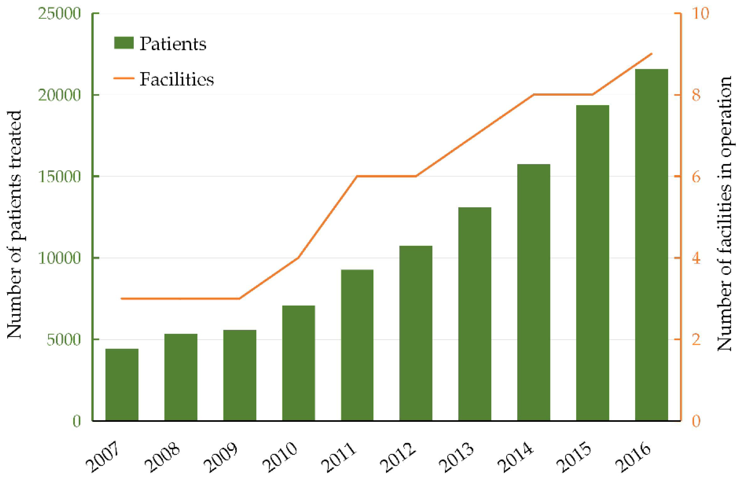
1. Introduction
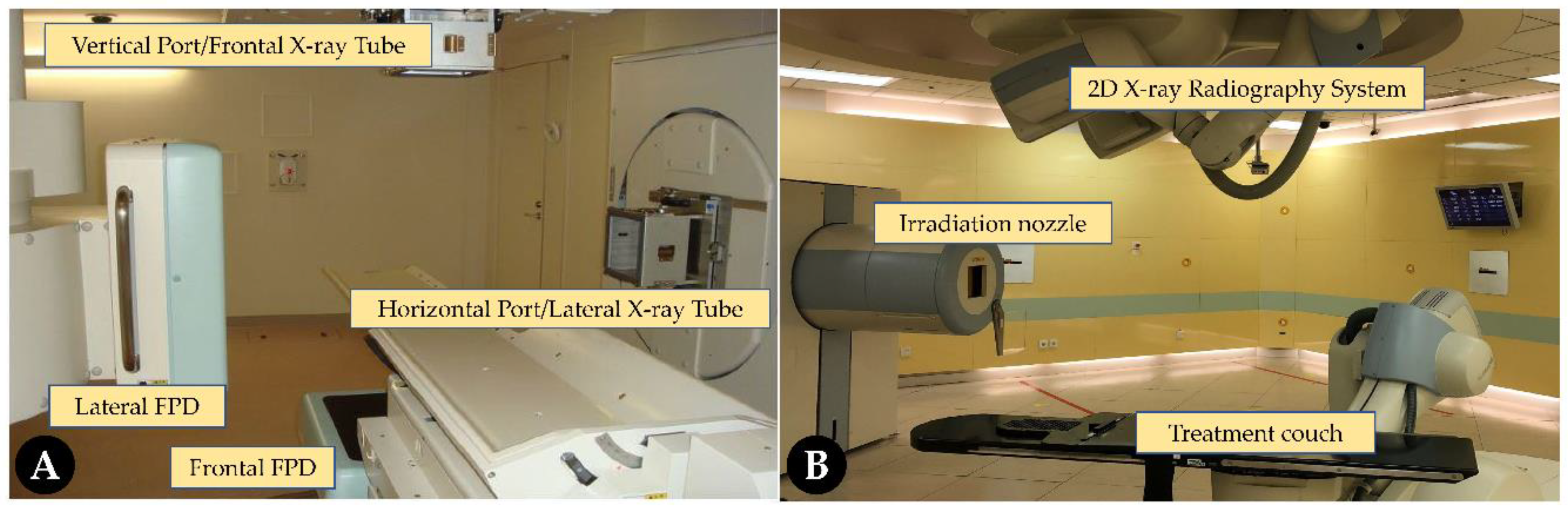
2. Present Verification System in Carbon Ion Radiotherapy
3. 3D Imaging System
4. Precise Positioning
5. Precise Radiation Delivery
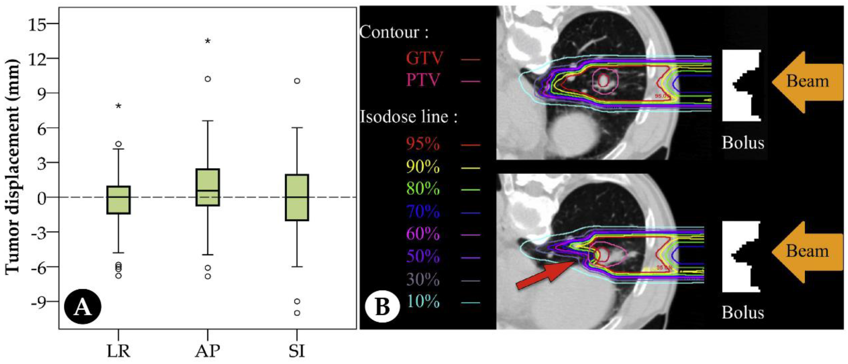
6. Planning Target Volume Margin Reduction
7. Hypofractionated C-Ion Radiotherapy and Motion Management
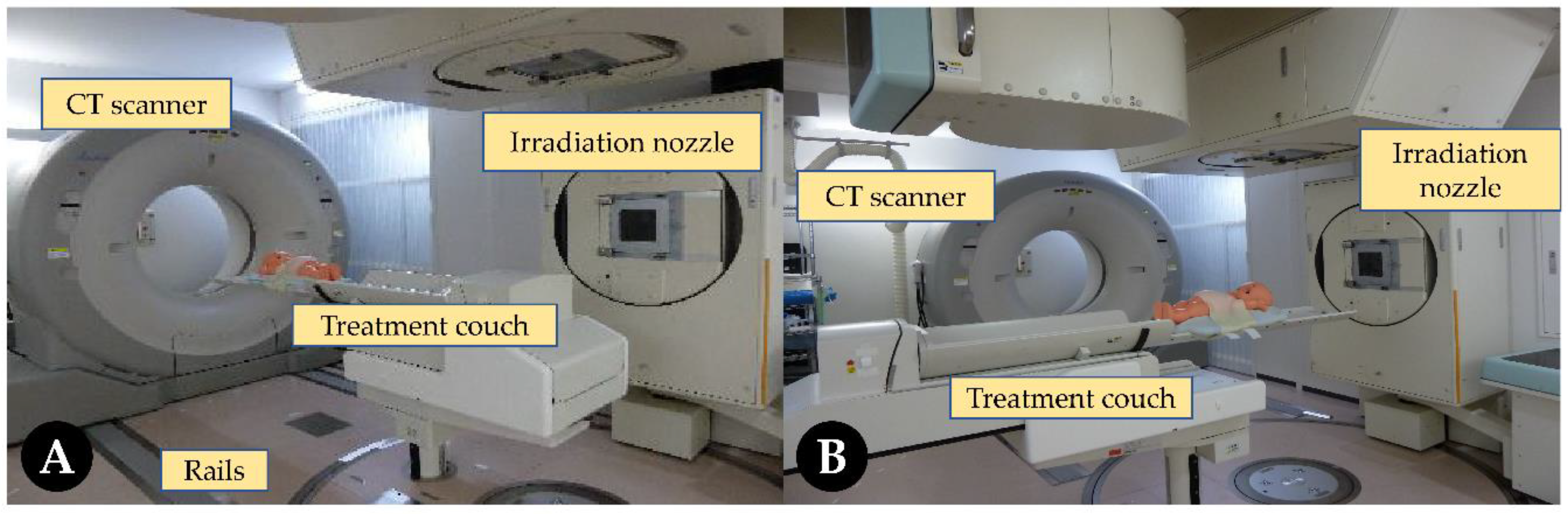
8. In-Room Computed Tomography
9. Image-Guided Adaptive C-Ion Radiotherapy
10. Conclusions
Author Contributions
Funding
Acknowledgments
Conflicts of Interest
References
- Radiation Therapy, Management and Dosimetry Workplace Survey 2010. Available online: http://www.webcitation.org/75AgMAgI2 (accessed on 4 January 2019).
- Simpson, D.R.; Lawson, J.D.; Nath, S.K.; Rose, B.S.; Mundt, A.J.; Mell, L.K.J.C. A survey of image-guided radiation therapy use in the United States. Cancer 2010, 116, 3953–3960. [Google Scholar] [CrossRef] [PubMed]
- Ohno, T.; Kanai, T.; Yamada, S.; Yusa, K.; Tashiro, M.; Shimada, H.; Torikai, K.; Yushida, Y.; Kitada, Y.; Katoh, H. Carbon Ion Radiotherapy at the Gunma University Heavy Ion Medical Center: New Facility Set-up. Cancers 2011, 3, 4046–4060. [Google Scholar] [CrossRef] [PubMed]
- Kamada, T.; Tsujii, H.; Blakely, E.A.; Debus, J.; Neve, W.D.; Durante, M.; Jäkel, O.; Mayer, R.; Orecchia, R.; Pötter, R. Carbon ion radiotherapy in Japan: An assessment of 20 years of clinical experience. Lancet Oncol. 2015, 16, e93–e100. [Google Scholar] [CrossRef]
- Tsujii, H.; Kamada, T. A review of update clinical results of carbon ion radiotherapy. Jpn. J. Clin. Oncol. 2012, 42, 670–685. [Google Scholar] [CrossRef] [PubMed]
- Mori, S.; Shibayama, K.; Tanimoto, K.; Kumagai, M.; Matsuzaki, Y.; Furukawa, T.; Inaniwa, T.; Shirai, T.; Noda, K.; Tsuji, H. First clinical experience in carbon ion scanning beam therapy: Retrospective analysis of patient positional accuracy. J. Radiat. Res. 2012, 53, 760–768. [Google Scholar] [CrossRef] [PubMed]
- Shiba, S.; Saitoh, J.I.; Irie, D.; Shirai, K.; Abe, T.; Kubota, Y.; Sakai, M.; Okada, R.; Ohno, T.; Nakano, T. Potential Pitfalls of a Fiducial Marker-matching Technique in Carbon-ion Radiotherapy for Lung Cancer. Anticancer Res. 2017, 37, 5673–5680. [Google Scholar] [PubMed]
- Irie, D.; Saitoh, J.I.; Shirai, K.; Abe, T.; Kubota, Y.; Sakai, M.; Noda, S.E.; Ohno, T.; Nakano, T. Verification of dose distribution in carbon ion radiotherapy for stage I lung cancer. Int. J. Radiat. Oncol. Biol. Phys. 2016, 96, 1117–1123. [Google Scholar] [CrossRef] [PubMed]
- Abe, S.; Kubota, Y.; Shibuya, K.; Koyama, Y.; Abe, T.; Ohno, T.; Nakano, T. Fiducial marker matching versus vertebral body matching: Dosimetric impact of patient positioning in carbon ion radiotherapy for primary hepatic cancer. Phys. Medica 2017, 33, 114–120. [Google Scholar] [CrossRef] [PubMed]
- Sakai, M.; Kubota, Y.; Saitoh, J.I.; Irie, D.; Shirai, K.; Okada, R.; Torikoshi, M.; Ohno, T.; Nakano, T. Robustness of patient positioning for interfractional error in carbon ion radiotherapy for stage I lung cancer: Bone matching versus tumor matching. Radiother. Oncol. 2017, 129, 95–100. [Google Scholar] [CrossRef] [PubMed]
- Kubota, Y.; Tashiro, M.; Shinohara, A.; Abe, S.; Souda, S.; Okada, R.; Ishii, T.; Kanai, T.; Ohno, T.; Nakano, T. Development of an automatic evaluation method for patient positioning error. J. Appl. Clin. Med. Phys. 2015, 16, 100–111. [Google Scholar] [CrossRef] [PubMed]
- Gurjar, O.P.; Mishra, S.P.; Bhandari, V.; Pathak, P.; Pant, S.; Patel, P. A study on the necessity of kV-CBCT imaging compared to kV-orthogonal portal imaging based on setup errors: Considering other socioeconomical factors. J. Canc. Res. Ther. 2014, 10, 583–586. [Google Scholar]
- Dzierma, Y.; Beyhs, M.; Palm, J.; Niewald, M.; Bell, K.; Nuesken, F.; Licht, N.; Rübe, C. Set-up errors and planning margins in planar and CBCT image-guided radiotherapy using three different imaging systems: A clinical study for prostate and head-and-neck cancer. Phys. Medica 2015, 31, 1055–1059. [Google Scholar] [CrossRef] [PubMed]
- Mayyas, E.; Chetty, I.J.; Chetvertkov, M.; Wen, N.; Neicu, T.; Nurushev, T.; Ren, L.; Lu, M.; Stricker, H.; Pradhan, D. Evaluation of multiple image-based modalities for image-guided radiation therapy (IGRT) of prostate carcinoma: A prospective study. Med. Phys. 2013, 40, 041707. [Google Scholar] [CrossRef] [PubMed]
- Topolnjak, R.; Sonke, J.J.; Nijkamp, J.; Rasch, C.; Minkema, D.; Remeijer, P.; Vliet-Vroegindeweij, C.V. Breast Patient Setup Error Assessment: Comparison of Electronic Portal Image Devices and Cone-Beam Computed Tomography Matching Results. Int. J. Radiat. Oncol. Biol. Phys. 2010, 78, 1235–1243. [Google Scholar] [CrossRef] [PubMed]
- Borst, G.R.; Sonke, J.J.; Betgen, A.; Remeijer, P.; Herk, M.V.; Lebesque, J.V. Kilo-voltage cone-beam computed tomography setup measurements for lung cancer patients; first clinical results and comparison with electronic portal-imaging device. Int. J. Radiat. Oncol. Biol. Phys. 2007, 68, 555–561. [Google Scholar] [CrossRef] [PubMed]
- Martins, L.; Couto, J.G.; Barbosa, B. Use of planar kV vs. CBCT in evaluation of setup errors in oesophagus carcinoma radiotherapy. Rep. Pract. Oncol. Radiother. 2016, 21, 57–62. [Google Scholar] [CrossRef] [PubMed]
- Kubota, Y.; Hayashi, H.; Abe, S.; Souda, S.; Okada, R.; Ishii, T.; Tashiro, M.; Torikoshi, M.; Kanai, T.; Ohno, T. Evaluation of the accuracy and clinical practicality of a calculation system for patient positional displacement in carbon ion radiotherapy at five sites. J. Appl. Clin. Med. Phys. 2018, 19, 144–153. [Google Scholar] [CrossRef] [PubMed]
- Shirato, H.; Suzuki, K.; Sharp, G.C.; Fujita, K.; Onimaru, R.; Fujino, M.; Kato, N.; Osaka, Y.; Kinoshita, R.; Taguchi, H. Speed and amplitude of lung tumor motion precisely detected in four-dimensional setup and in real-time tumor-tracking radiotherapy. Int. J. Radiat. Oncol. Biol. Phys. 2006, 64, 1229–1236. [Google Scholar] [CrossRef] [PubMed]
- Habermehl, D.; Henkner, K.; Ecker, S.; Jäkel, O.; Debus, J.; Combs, S.E. Evaluation of different fiducial markers for image-guided radiotherapy and particle therapy. J. Radiat. Res. 2013, 54 (Suppl. 1), i61–i68. [Google Scholar] [CrossRef]
- Herrmann, R.; Carl, J.; Jäkel, O.; Bassler, N.; Petersen, J.B. Investigation of the dosimetric impact of a Ni-Ti fiducial marker in carbon ion and proton beams. Acta Oncol. 2010, 49, 1160–1164. [Google Scholar] [CrossRef] [PubMed]
- Cheung, J.; Kudchadker, R.J.; Zhu, X.R.; Lee, A.K.; Newhauser, W.D. Dose perturbations and image artifacts caused by carbon-coated ceramic and stainless steel fiducials used in proton therapy for prostate cancer. Phys. Med. Biol. 2010, 55, 7135–7147. [Google Scholar] [CrossRef] [PubMed]
- Maeda, Y.; Sato, Y.; Minami, H.; Yasukawa, Y.; Yamamoto, K.; Tamamura, H.; Shibata, S.; Bou, S.; Sasaki, M.; Tameshige, Y. Positioning accuracy and daily dose assessment for prostate cancer treatment using in-room CT-image guidance at a proton therapy facility. Med. Phys. 2018, 45, 1832–1843. [Google Scholar] [CrossRef] [PubMed]
- Wang, Y.; Efstathiou, J.A.; Sharp, G.C.; Lu, H.M.; Ciernik, I.F.; Trofimov, A.V. Evaluation of the dosimetric impact of interfractional anatomical variations on prostate proton therapy using daily in-room CT images. Med. Phys. 2011, 38, 4623–4633. [Google Scholar] [CrossRef] [PubMed]
- Liu, F.; Ahunbay, E.; Lawton, C.; Li, XA. Assessment and management of interfractional variations in daily diagnostic-quality-CT guided prostate-bed irradiation after prostatectomy. Med. Phys. 2014, 41, 031710. [Google Scholar] [CrossRef] [PubMed]
- Houweling, A.C.; Fukata, K.; Kubota, Y.; Shimada, H.; Rasch, C.R.; Ohno, T.; Bel, A.; Horst, A.V.D. The impact of interfractional anatomical changes on the accumulated dose in carbon ion therapy of pancreatic cancer patients. Radiother. Oncol. 2016, 119, 319–325. [Google Scholar] [CrossRef] [PubMed]
- Miki, K.; Mori, S.; Shiomi, M.; Yamada, S. Gated carbon-ion scanning treatment for pancreatic tumour with field specific target volume and organs at risk. Phys. Medica 2016, 32, 1521–1528. [Google Scholar] [CrossRef] [PubMed]
- Den, R.B.; Doemer, A.G.; Bednarz, G.; Galvin, J.M.; Keane, W.M.; Xiao, Y.; Machtay, M. Daily image guidance with cone-beam computed tomography for head-and-neck cancer intensity-modulated radiotherapy: A prospective study. Int. J. Radiat. Oncol. Biol. Phys. 2010, 76, 1353–1359. [Google Scholar] [CrossRef] [PubMed]
- Ariyaratne, H.; Chesham, H.; Pettingell, J.; Alonzi, R. Image-guided radiotherapy for prostate cancer with cone beam CT: Dosimetric effects of imaging frequency and PTV margin. Radiother. Oncol. 2016, 121, 103–108. [Google Scholar] [CrossRef] [PubMed]
- Knopf, A.C.; Boye, D.; Lomax, A.; Mori, S. Adequate margin definition for scanned particle therapy in the incidence of intrafractional motion. Phys. Med. Biol. 2013, 58, 6079–6094. [Google Scholar] [CrossRef] [PubMed]
- Huijskens, S.C.; van Dijk, I.W.E.M.; Visser, J.; Balgobind, B.V.; Rasch, C.R.N.; Alderliesten, T.; Bel, A. Predictive value of pediatric respiratory-induced diaphragm motion quantified using pre-treatment 4DCT and CBCTs. Radiat. Oncol. 2018, 13, 198. [Google Scholar] [CrossRef] [PubMed]
- Mohamad, O.; Makishima, H.; Kamada, T. Evolution of Carbon Ion Radiotherapy at the National Institute of Radiological Sciences in Japan. Cancers 2018, 10, 66. [Google Scholar] [CrossRef] [PubMed]
- Paz, A.E.; Yamamoto, N.; Sakama, M.; Matsufuji, N.; Kanai, T. Tumor Control Probability Analysis for Single-Fraction Carbon-Ion Radiation Therapy of Early-Stage Non-small Cell Lung Cancer. Int. J. Radiat. Oncol. Biol. Phys. 2018, 102, 1551–1559. [Google Scholar] [CrossRef] [PubMed]
- Kasuya, G.; Kato, H.; Yasuda, S.; Tsuji, H.; Yamada, S.; Haruyama, Y.; Kobashi, G.; Ebner, D.K.; Okada, N.N.; Makishima, H. Progressive hypofractionated carbon-ion radiotherapy for hepatocellular carcinoma: Combined analyses of 2 prospective trials. Cancer 2017, 123, 3955–3965. [Google Scholar] [CrossRef] [PubMed]
- Tashiro, M.; Ishii, T.; Koya, J.; Okada, R.; Kurosawa, Y.; Arai, K.; Abe, S.; Ohashi, Y.; Shimada, H.; Yusa, K. Technical approach to individualized respiratory-gated carbon-ion therapy for mobile organs. Radiol. Phys. Technol. 2013, 6, 356–366. [Google Scholar] [CrossRef] [PubMed]
- Gierga, D.P.; Brewer, J.; Sharp, G.C.; Betke, M.; Willett, C.G.; Chen, G.T. The correlation between internal and external markers for abdominal tumors: Implications for respiratory gating. Int. J. Radiat. Oncol. Biol. Phys. 2005, 61, 1551–1558. [Google Scholar] [CrossRef] [PubMed]
- Zhang, X.; Homma, N.; Ichiji, K.; Abe, M.; Sugita, N.; Takai, Y.; Narita, Y.; Yoshizawa, M. A kernel-based method for markerless tumor tracking in kV fluoroscopic images. Phys. Med. Biol. 2014, 59, 4897–4911. [Google Scholar] [CrossRef] [PubMed]
- Hirai, R.; Sakata, Y.; Taguchi, Y.; Mori, S. Regression model of tumor and diaphragm position for marker-less tumor tracking in carbon ion scanning therapy for hepatocellular carcinoma. Int. J. Radiat. Oncol. 2016, 96, E639–E640. [Google Scholar] [CrossRef]
- Cui, Y.; Dy, JG.; Sharp, G.C.; Alexander, B.; Jiang, S.B. Multiple template based fluoroscopic tracking of lung tumor mass without implanted fiducial markers. Phys. Med. Biol. 2007, 52, 6229–6242. [Google Scholar] [CrossRef] [PubMed]
- Mori, S.; Karube, M.; Shirai, T.; Tajiri, M.; Takekoshi, T.; Miki, K.; Shiraishi, Y.; Tanimoto, K.; Shibayama, K.; Yasuda, S. Carbon-ion pencil beam scanning treatment with gated markerless tumor tracking: An analysis of posintional accuracy. Int. J. Radiat. Oncol. Biol. Phys. 2016, 96, E623–E624. [Google Scholar] [CrossRef]
- Ebner, D.K.; Tsuji, H.; Yasuda, S.; Yamamoto, N.; Mori, S.; Kamada, T. Respiration-gated fast-rescanning carbon-ion radiotherapy. Jpn. J. Clin. Oncol. 2017, 47, 80–83. [Google Scholar] [CrossRef] [PubMed]
- Mori, S.; Furukawa, T. Rapid phase-correlated rescanning irradiation improves treatment time in carbon-ion scanning beam treatment under irregular breathing. Phys. Med. Biol. 2016, 61, 3857–3866. [Google Scholar] [CrossRef] [PubMed]
- Jaffray, D.A. Image-guided radiotherapy: From current concept to future perspectives. Nat. Rev. Clin. Oncol. 2012, 9, 688–699. [Google Scholar] [CrossRef] [PubMed]
- Batumalai, V.; Holloway, L.C.; Kumar, S.; Dundas, K.; Jameson, M.G.; Vinod, S.K.; Delaney, G.P. Survey of image-guided radiotherapy use in Australia. J. Med. Imaging Radiat. Oncol. 2016, 61, 394–401. [Google Scholar] [CrossRef] [PubMed]
- Seco, J.; Spadea, M.F. Imaging in particle therapy: State of the art and future perspective. Acta Oncol. 2015, 54, 1254–1258. [Google Scholar] [CrossRef] [PubMed]
- Iwata, Y.; Noda, K.; Murakami, T.; Shirai, T.; Furukawa, T.; Fujita, T.; Mori, S.; Mizushima, K.; Shouda, K.; Fujimoto, T. Development of a compact superconducting rotating-gantry for heavy-ion therapy. J. Radiat. Res. 2013, 317, 793–797. [Google Scholar] [CrossRef]
- Wong, J.R.; Grimm, L.; Uematsu, M.; Oren, R.; Cheng, C.W.; Merrick, S.; Schiff, P. Image-guided radiotherapy for prostate cancer by CT–linear accelerator combination: Prostate movements and dosimetric considerations. Int. J. Radiat. Oncol. Biol. Phys. 2005, 61, 561–569. [Google Scholar] [CrossRef] [PubMed]
- Aoyama, H.; Azuma, Y.; Inamura, K. Comparison of daily prostate positions during conformal radiation therapy of prostate cancer using an integrated CT-linear accelerator system: In-room CT image versus digitally reconstructed radiograph. J. Radiat. Res. 2011, 52, 220–228. [Google Scholar] [CrossRef] [PubMed]
- Maeda, Y.; Sato, Y.; Shibata, S.; Bou, S.; Yamamoto, K.; Tamamura, H.; Fuwa, N.; Takamatsu, S.; Sasaki, M.; Tameshige, Y. Effects of organ motion on proton prostate treatments, as determined from analysis of daily CT imaging for patient positioning. Med. Phys. 2018, 1844–1856. [Google Scholar] [CrossRef] [PubMed]
- Lim-Reinders, S.; Keller, B.M.; Al-Ward, S.; Sahgal, A.; Kim, A. Online Adaptive Radiation Therapy. Int. J. Radiat. Oncol. Biol. Phys. 2017, 99, 994–1003. [Google Scholar] [CrossRef] [PubMed]
- Kubota, Y.; Sakai, M.; Tashiro, M.; Saitoh, J.; Abe, T.; Ohno, T.; Nakano, T. Technical Note: Predicting dose distribution with replacing stopping power ratio for inter-fractional motion and intra-fractional motion during carbon ion radiotherapy with passive irradiation method for stage I lung cancer. Med. Phys. 2018, 45, 3435–3441. [Google Scholar] [CrossRef] [PubMed]
- Park, Y.K.; Sharp, G.C.; Phillips, J.; Winey, B.A. Proton dose calculation on scatter-corrected CBCT image: Feasibility study for adaptive proton therapy. Med. Phys. 2015, 42, 4449–4459. [Google Scholar] [CrossRef] [PubMed]
- Arai, K.; Kadoya, N.; Kato, T.; Endo, H.; Komori, S.; Abe, Y.; Nakamura, T.; Wada, H.; Kikuchi, Y.; Takai, Y. Feasibility of CBCT-based proton dose calculation using a histogram-matching algorithm in proton beam therapy. Phys. Medica 2017, 33, 68–76. [Google Scholar] [CrossRef] [PubMed]
- Wu, H.; Chen, X.; Yang, X.; Tao, Y.; Xia, Y.; Deng, X.; Zheng, C.; Robbins, J.; Schultz, C.; Li, X.A. Early Prediction of Acute Xerostomia During Radiation Therapy for Head and Neck Cancer Based on Texture Analysis of Daily CT. Int. J. Radiat. Oncol. Biol. Phys. 2018, 102, 1308–1318. [Google Scholar] [CrossRef] [PubMed]
- Schulze, R.; Heil, U.; Gross, D.; Bruellmann, D.; Dranischnikow, E.; Schwanecke, U.; Schoemer, E. Artefacts in CBCT: A review. Dentomaxillofac. Radiol. 2011, 40, 265–273. [Google Scholar] [CrossRef] [PubMed]
- Ueda, U.; Hu, W.; Pouliot, J.; Yom, S.; Quivey, J.; Aubin, M.; Chen, J. SU-DD-A3-02: The Impact of Cone-Beam Computed Tomography (CBCT) Artifacts on Deformable Image Registration Algorithms. Med. Phys. 2010, 37, 3091. [Google Scholar] [CrossRef]
- Disher, B.; Hajdok, G.; Wang, A.; Craig, J.; Gaede, S.; Battista, J.J. Correction for ‘artificial’ electron disequilibrium due to cone-beam CT density errors: Implications for on-line adaptive stereotactic body radiation therapy of lung. Phys. Med. Biol. 2013, 58, 4157–4174. [Google Scholar] [CrossRef] [PubMed]
- Veiga, C.; Janssens, G.; Teng, C.L.; Baudier, T.; Hotoiu, L.; McClelland, J.R.; Royle, G.; Lin, L.; Yin, L.; Metz, J.; et al. First Clinical Investigation of Cone Beam Computed Tomography and Deformable Registration for Adaptive Proton Therapy for Lung Cancer. Int. J. Radiat. Oncol. Biol. Phys. 2016, 95, 549–559. [Google Scholar] [CrossRef] [PubMed]
- Nagano, A.; Minohara, S.; Kato, S.; Kiyohara, H.; Ando, K. Adaptive radiotherapy based on the daily regression of a tumor in carbon-ion beam irradiation. Phys. Med. Biol. 2012, 57, 8343–8356. [Google Scholar] [CrossRef] [PubMed]
- Chen, W.; Gemmel, A.; Rietzel, E. A patient-specific planning target volume used in ‘plan of the day’ adaptation for interfractional motion mitigation. J. Radiat. Res. 2013, 54 (Suppl. 1), i82–i90. [Google Scholar] [CrossRef]
- Hild, S.; Graeff, C.; Rucinski, A.; Zink, K.; Habl, G.; Durante, M.; Herfarth, K.; Bert, C. Scanned ion beam therapy for prostate carcinoma: Comparison of single plan treatment and daily plan-adapted treatment. Strahlenther. Onkol. 2016, 192, 118–126. [Google Scholar] [CrossRef] [PubMed]
- Minohara, S.; Fukuda, S.; Kanematsu, N.; Takei, Y.; Furukawa, T.; Inaniwa, T.; Matsufuji, N.; Mori, S.; Noda, K. Recent innovations in carbon-ion radiotherapy. J. Radiat. Res. 2010, 51, 385–392. [Google Scholar] [CrossRef] [PubMed]
- Koay, E.J.; Lege, D.; Mohan, R.; Komaki, R.; Cox, J.D.; Chang, J.Y. Adaptive/Nonadaptive Proton Radiation Planning and Outcomes in a Phase II Trial for Locally Advanced Non-small Cell Lung Cancer. Int. J. Radiat. Oncol. Biol. Phys. 2012, 84, 1093–1100. [Google Scholar] [CrossRef] [PubMed]
- Mori, S.; Takei, Y.; Shirai, T.; Hara, Y.; Furukawa, T.; Inaniwa, T.; Tanimoto, K.; Tajiri, M.; Kuroiwa, D.; Kimura, T. Scanned carbon-ion beam therapy throughput over the first 7 years at National Institute of Radiological Sciences. Phys. Medica 2018, 52, 18–26. [Google Scholar] [CrossRef] [PubMed]
- Hild, S.; Graeff, C.; Trautmann, J.; Kraemer, M.; Zink, K.; Durante, M.; Bert, C. Fast optimization and dose calculation in scanned ion beam therapy. Med. Phys. 2014, 41, 071703. [Google Scholar] [CrossRef] [PubMed]
- Ghilezan, M.; Yan, D.; Martinez, A. Adaptive Radiation Therapy for Prostate Cancer. Semin. Radiat. Oncol. 2010, 20, 130–137. [Google Scholar] [CrossRef] [PubMed]
- McPartlin, A.J.; Li, X.A.; Kershaw, L.E.; Heide, U.; Kerkmeijer, L.; Lawton, C.; Mahmood, U.; Pos, F.; As, N.V.; Herk, M.V. MRI-guided prostate adaptive radiotherapy—A systematic review. Radiother. Oncol. 2016, 119, 371–380. [Google Scholar] [CrossRef] [PubMed]
- Lagendijk, J.J.; Raaymakers, B.W.; Vulpen, M.V. The Magnetic Resonance Imaging-Linac System. Semin. Radiat. Oncol. 2014, 24, 207–209. [Google Scholar] [CrossRef] [PubMed]
- Pollard, J.M.; Wen, Z.; Sadagopan, R.; Wang, J.; Ibbott, G.S. The Future of Image-Guided Radiotherapy will be MR-Guided. Br. J. Radiol. 2017, 90, 20160667. [Google Scholar] [CrossRef] [PubMed]
- Ohno, T. Particle radiotherapy with carbon ion beams. EPMA J. 2013, 4, 1–7. [Google Scholar] [CrossRef] [PubMed]
- Oborn, B.M.; Dowdell, S.; Metcalfe, P.E.; Crozier, S.; Mohan, R.; Keall, P.J. Future of Medical Physics: Real-time MRI guided Proton Therapy. Med. Phys. 2017, 44, e77–e90. [Google Scholar] [CrossRef] [PubMed]
- Shao, Y.P.; Sun, X.S.; Lou, K.; Zhu, X.R.; Mirkovic, D.; Poenisch, F.; Grosshans, D. In-beam PET imaging for on-line adaptive proton therapy: An initial phantom study. Phys. Med. Biol. 2014, 59, 3373–3388. [Google Scholar] [CrossRef] [PubMed]


| Authors | Year | Tumor Type | Patient Number | BM vs. TM/MM | p-Value |
|---|---|---|---|---|---|
| Abe S. [9] | 2017 | Liver | 20 | 57.9 Gy vs. 59.8 Gy (Median D98) | 0.001 |
| Sakai M. [10] | 2017 | Lung | 30 | 98.9% vs. 100% (Median V95%) | <0.001 |
| Maeda Y. [23] | 2018 | Prostate | 30 | 90.4% vs. 98.7% (Ratio of V95% > 95%) | <0.001 |
© 2019 by the authors. Licensee MDPI, Basel, Switzerland. This article is an open access article distributed under the terms and conditions of the Creative Commons Attribution (CC BY) license (http://creativecommons.org/licenses/by/4.0/).
Share and Cite
Li, Y.; Kubota, Y.; Tashiro, M.; Ohno, T. Value of Three-Dimensional Imaging Systems for Image-Guided Carbon Ion Radiotherapy. Cancers 2019, 11, 297. https://doi.org/10.3390/cancers11030297
Li Y, Kubota Y, Tashiro M, Ohno T. Value of Three-Dimensional Imaging Systems for Image-Guided Carbon Ion Radiotherapy. Cancers. 2019; 11(3):297. https://doi.org/10.3390/cancers11030297
Chicago/Turabian StyleLi, Yang, Yoshiki Kubota, Mutsumi Tashiro, and Tatsuya Ohno. 2019. "Value of Three-Dimensional Imaging Systems for Image-Guided Carbon Ion Radiotherapy" Cancers 11, no. 3: 297. https://doi.org/10.3390/cancers11030297
APA StyleLi, Y., Kubota, Y., Tashiro, M., & Ohno, T. (2019). Value of Three-Dimensional Imaging Systems for Image-Guided Carbon Ion Radiotherapy. Cancers, 11(3), 297. https://doi.org/10.3390/cancers11030297