Article

Modular Flow Synthesis of Citric Acid-Coated Superparamagnetic Iron Oxide Nanoparticles: Preliminary Results

Faculty of Chemistry and Chemical Engineering, University of Maribor, Smetanova 17, SI-2000 Maribor, Slovenia
*
Author to whom correspondence should be addressed.
Micromachines 2025, 16(11), 1228; https://doi.org/10.3390/mi16111228
Submission received: 13 October 2025 / Revised: 27 October 2025 / Accepted: 28 October 2025 / Published: 29 October 2025
(This article belongs to the Section D:Materials and Processing)

Abstract

Superparamagnetic iron oxide nanoparticles (SPIONs) with sizes below 10 nm are biocompatible and non-toxic, making them promising for biomedical applications. To prevent their agglomeration and enhance their functionality, the nanoparticles were coated with citric acid (CA), which modifies the surface charge, improves dispersion stability, and facilitates biomedical use. In this work, a modular flow-through microreactor system was employed to synthesize and coat the nanoparticles in a single, continuous two-step process. The system enables precise control over temperature and mixing, ensuring uniform reaction conditions and minimizing hot spots. The synthesized Fe3O4 nanoparticles exhibited an average crystallite size of ~5 nm (XRD) and particle sizes of 4–6 nm (TEM). FTIR analysis confirmed the successful surface functionalization with CA, while TGA indicated a coating mass fraction of approximately 4–20 wt%, increasing with higher CA concentration. Zeta potential measurements revealed strong colloidal stability, with values around −35 mV at pH 6.5. Among the tested CA concentrations, the sample with a molar ratio of Fe3O4:CA = 1:0.25 exhibited the most favorable properties, including narrow size distribution and improved dispersion stability. These findings demonstrate that the continuous modular flow approach enables the reproducible synthesis of highly stable, sub-10 nm CA-coated SPIONs, offering promising potential for biomedical applications, particularly as magnetic resonance imaging (MRI) contrast agents.

1. Introduction

Superparamagnetic iron oxide nanoparticles (SPIONs) are typically defined as iron oxide nanoparticles with a core diameter below 10 nm, at which size thermal fluctuations are sufficient to randomize a magnetic moment, resulting in superparamagnetic behavior [1,2]. Intensive research over the past decades has demonstrated the broad applicability of SPIONs across diverse fields [3,4,5]. Studies on biocompatibility and toxicity have shown their suitability for biomedical application, particularly as contrast agents for magnetic resonance imaging (MRI) [6,7,8,9]. Furthermore, functionalization strategies enable the extension of their utilization to image-directed drug delivery. Their distinctive properties, such as high surface-area-to-volume ratio, magnetic properties that differ from their bulk counterparts, very high relaxivity, and extensive possibilities for surface functionalization with targeting molecules, have further advance their use in magnetic hyperthermia (MH) [3,10,11], magnetic separation [4,12], tissue engineering [3,13,14], and targeted delivery of drugs/therapeutic agents [9,15,16].
MRI remains one of the most reliable, non-invasive diagnostic techniques, capable of producing highly detailed anatomical images. In this technique, contrast agents are used to enhance the contrast between the target site and surrounding tissue [6,7]. Ferrite nanoparticles have proven to be effective contrast agents, as they modulate the nuclear spin relaxation process, thereby improving sensitivity and detection accuracy. Importantly, the performance of magnetic nanoparticles (MNPs) is strongly linked to their composition, size, surface properties, and degree of agglomeration in the biological environment [4,8]. Therefore, understanding the relationship between the intrinsic parameters and the relaxivities of nuclei influenced by MNPs can provide crucial insights for accurately predicting the properties of engineered MNPs and improving their performance in MRI-based theranostic applications [17].
The initial step in developing SPIONs is the synthesis of the magnetic core. Numerous methods have been proposed to achieve precise control over the size, shape, and magnetic properties. A highly reproducible synthesis process is crucial, as this is one of the essential requirements for the potential clinical translation of these nanomaterials [4]. Generally, nanoparticles smaller than 15 nm are preferred. At these sizes, ferri- and ferromagnetic nanoparticles achieve a single magnetic domain, characterized by a large magnetic moment. Such particles exhibit superparamagnetic properties, including high magnetic moments and rapid response to external magnetic fields, while showing negligible remanent magnetization and coercivity when the external magnetic field is removed [18,19]. This ensures that SPIONs lose their magnetization immediately after the magnetic field is removed, preventing unwanted nanoparticle aggregation and ensuring safe removal from the body. However, due to their small size and high surface-to-volume ratio, SPIONs tend to aggregate as a result of their high surface energy [6,19,20]. Uncoated iron oxide particles are unstable and susceptible to oxidation in the air, which reduces magnetic properties. To maintain and enhance their long-term stability in physiological environments, MNPs must be appropriately surface-modified or coated. The coating material also determines the final surface charge of the MNPs [3,6]. Various synthesis methods have been developed to produce SPIONs with the desired size, shape, saturation magnetization, and other key properties. These methods can be either one-step or two-step, particularly when additional functionalization is required, such as protective coating or surface functionalization aimed at specific applications [6,19,21,22].
Carboxylates greatly influence the development and magnetic properties of SPIONs. For example, surface modification with heavy-chain fatty acids or thiols enhances stability of aqueous SPION suspensions [23]. Coating nanoparticles with citric acid (CA) results in a thermodynamically stable colloidal solution. CA is a widely used organic coating material because it can modify surface charge and hydrophobicity while leaving free carboxyl groups available on the nanoparticle surface. In addition to its stabilizing role, CA exhibits bactericidal and bacteriostatic properties, and solutions containing CA are commonly used as stabilizing agents and plant growth stimulators. Importantly, nanosystems composed of water-dispersed SPIONs coated with CA demonstrate high biocompatibility and low toxicity. This makes them particularly valuable in biomedical applications, where CA acts as a linker for anticancer drugs and enhances the use of SPIONs as drug delivery systems and MRI contrast agents [23,24,25].
In this work, a modular flow-through microreactor system was used to synthesize and coat nanoparticles within a single, continuous process setup using two different microreactors. This change from batch production to semi-continuous flow chemistry offers several key advantages [26,27]. First, the precise control of reaction parameters minimizes temperature and concentration gradients, thereby reducing the risk of hot spots. In batch systems, mixing typically occurs by diffusion over several seconds, which often results in broad particle size distributions (standard deviation > 20%) and increased aggregation. In contrast, microreactors provide characteristic mixing times below 100 ms due to their high surface-to-volume ratios and efficient mass and heat transfer, leading to narrower particle size distributions (standard deviation < 5–10%) and enhanced control over nucleation and growth dynamics [28,29]. Second, the inherently enhanced heat and mass transfer in microreactors significantly accelerates reaction kinetics compared to batch processes. Third, microreactors offer excellent scalability and reproducibility; scale-up can be achieved through the parallelization of identical units without altering the optimized reaction environment, ensuring consistent product quality across production scales. These quantitative advantages are especially beneficial for achieving homogeneous citric acid coating and stable colloidal dispersions of magnetic nanoparticles, as rapid and uniform mixing minimizes local concentration and pH gradients that could otherwise compromise coating efficiency and long-term stability [30]. Moreover, the modular design of the microreactor system employed in this study aligns conceptually with recently reported “modular structure” strategies in nanomaterial synthesis, which emphasize design flexibility, process control, and adaptability to diverse reaction schemes [31].
In this work, we present a modular flow-through microreactor system for the continuous synthesis and in situ CA coating of SPIONs within a single, integrated process. This approach allows precise control over reaction parameters, enabling the production of nanoparticles with uniform size and morphology. Among various biomedical applications of SPIONs, MRI is particularly sensitive to nanoparticle size and magnetic properties. Particles smaller than 10 nm exhibit strong superparamagnetic behavior, high transverse relaxivity, and excellent colloidal stability, making them ideal T2 contrast agents. The SPIONs synthesized in this study, with an average size of approximately 5 nm, therefore possess characteristics that are especially favorable for MRI-based applications.
The novelty of this work lies in the integration of continuous flow synthesis and in situ surface functionalization with CA within a modular microreactor platform. This combined, sequential, and continuous approach offers enhanced control over particle size, dispersion, and surface stability compared to conventional batch or multi-step coating methods, representing an important step toward scalable and reproducible production of CA-coated SPIONs for biomedical use.

2. Materials and Methods

2.1. Materials

Iron(II) chloride, tetrahydrate, 98%,
Iron(III) chloride,
Sodium hydroxide,
Hydochloric acid, 37%,
CA,
Purified water,
Absolute ethanol (CH3CH2OH).
All the chemicals needed were purchased from Sigma Aldrich. Purified water was used for preparing solutions, and purified water and absolute ethanol were used for washing MNPs.
Three solutions were prepared for this synthesis.
(i)
3 M NaOH solution,
(ii)
Iron salt precursor solution (ISPS) that contained 366 mg FeCl2·4H2O and 566 mg FeCl3 in 36 mL 1 M HCl,
(iii)
1 M CA.

2.2. Modular Flow Microreactor System Setup

The synthesis and coating of nanoparticles were performed using a modular Asia Syrris flow microreactor system, where the nanoparticle synthesis follows a demonstration case of Asia Syrris, while the upgrading process coats them with citric acid, consisting of modules as follows:
Asia pump,
Asia Automated RIM,
Chip reactor 250 µL and 1 mL,
Back pressure controller,
Automated collector.
Purified water served as the carrier liquid in the system. The first (Buffer 1 in Figure 1) and second channel (Buffer 2 in Figure 1) of the first pump were connected to the first channel (Automated RIM 1 in Figure 1) and second channel (Automated RIM 2 in Figure 1) of the automated reagent injection system, enabling automated dosage reagents. The 3 M NaOH solution and ISPS were introduced into 250 µL (Chip (CC) 1 in Figure 1) at 90 °C with residence time 0.25 min (15 s), where they reacted to form magnetite/maghemite nanoparticles according to Equation (1).
2 Fe3+(aq) + Fe2+(aq) + 8 Cl(aq) + 8 Na+(aq) + 8 OH(aq) → Fe3O4(s) + 8 NaCl(aq) + 4 H2O(l),
The nanoparticle suspension was transferred into a second 1 mL (Chip CC(2) in Figure 1), where it was mixed with 1 M CA, supplied by the manual reagent injection system (Reagent Injector 1 in Figure 1) using the first channel of the second pump (Buffer 3 in Figure 1) to provide continuous flow. For all three reagent injection systems the 5 mL loop was used, which then presented the limiting factor in the quantity of product that can be produced in one run. In the second microreactor, the following reactions take place. CA dissociates in aqueous solution (Equation (2)) and the citrate ions coordinate with Fe3O4 nanoparticles via their carboxylate groups (-COO) as simplistically presented in Equation (3). By these reactions, a formation of stable Fe3O4@CA coating is obtained. The residence time in the second reactor was 1 min at a temperature of 80 °C.
C6H8O7(aq) → C6H5O73−(aq) + 3 H+(aq),
Fe3O4(s, nanoparticles) + C6H5O73−(aq) → Fe3O4@CA(coated nanoparticles),
Direct pH monitoring within the microreactor was not feasible, as conventional pH probes require larger sample volumes and exhibit response times longer than the residence times in the flow channels. However, due to the precise and reproducible control of flow and mixing provided by the Asia Syrris microreactor system, and based on validation experiments, it can be assumed that the local pH conditions remained consistent for all synthesis runs.
The overall net reaction together in both reactions is presented in Equation (4).
FeCl2·4H2O(aq) + 2 FeCl3(aq)+ 8 NaOH(aq)+ C6H8O7(aq)→ Fe3O4@CA(s)+ 8 NaCl(aq) + 13 H2O(l),
NaCl and water do not participate in the coating process, but they can influence the ionic strength and pH of the solution that have an impact on the coating efficiency and stability. The common pH range for the coating process is between 4 and 7. The citrate coating provides a negative charge to the surface of MNPs, obtaining electrostatic repulsion between particles and preventing aggregation of coated nanoparticles. It should be noted that the amount of added CA can greatly affect the products, as instead of coating the nanoparticles the iron oxide can be dissolved when pH is below 4.
The process was maintained at 1 bar with the backpressure controller (Pressure Controller 1 in Figure 1) at 1 bar of pressure, and the coated nanoparticles in suspension were collected with the automated collector (Automated Collector 1 in Figure 1).

2.3. Experimental Setup

Different molar ratios of CA to nanoparticles were tested to optimize coating efficiency. The samples are denoted as MNP@CAx, where x represents the molar ratio of citric acid to Fe3O4 (Table 1).

2.4. Analysis/Characterization of Fe3O4@CA

To confirm the successful coating of magnetic MNP with CA, the average hydrodynamic particle size, zeta potential, and isoelectric point (IEP) were determined by dynamic light scattering (DLS, Zetasizer Nano ZS, Malvern, Worcestershire, UK) using purified MNP@CA water dispersions. Measurements were conducted with purified water, and the pH was adjusted from the current pH value to 2.0 with 0.1 M hydrochloric acid (HCl) using an auto titrator.
The functional groups of MNP@CA were analyzed using Fourier transform infrared spectroscopy (FTIR, IRAffinity-1S, Shimadzu, Tokyo, Japan). The FTIR spectrum was measured in the range of 4000–400 cm−1. The size and morphology of the nanoparticles were determined using transmission electron microscopy (TEM, JEM-2010F, JEOL, Tokyo, Japan).
The empirical size distribution of the MNPs was estimated by measuring the area of the MNPs on the TEM image. The average particle size is given as a number-weighted average equivalent diameter, representing the diameter of a circle with the same area as the imaged particle.
Phase identification and core particle characterization were performed by X-ray powder diffraction (XRD, high-resolution X-ray powder diffractometer PANalytical X’Pert PRO MPD with Cu–Kα1 radiation (λ = 1.5406 Å)).
The weight loss of Fe3O4@CA was characterized by thermogravimetric analysis (TGA, TGA2, Mettler Toledo, Greifensee, Switzerland). The measurement was performed under nitrogen (100 mL/min) from 25 °C to 600 °C at a heating rate of 10 K/min.

3. Results and Discussion

3.1. X-Ray Powder Diffraction

Based on the diffraction data of the obtained samples, the diffraction peaks at 2Θ values were assigned to the crystal planes (220), (311), (400), (511), and (440), respectively (Figure 2). For Fe3O4, peaks were observed at 30.2°, 35.6°, 43.2°, 53.4°, 57.2°, and 62.7° at 2Θ degrees, indicating the cubic spinel structure of magnetite [32]. All peaks correspond well with the characteristic peaks of magnetite (JCPDS file no. 00-019-0629). The average crystallite diameter, dx = 5 nm, was calculated using the Scherrer equation. The average value of the lattice parameter was determined to be a = 0.8358 nm. Similar peaks were found for Fe3O4@CA, revealing that the CA coating does not cause a phase change in the bare Fe3O4 [33]. The average crystallite size, estimated using the Scherrer equation, was approximately 5 nm. This value serves as an approximate indicator of particle size, as effects such as lattice strain or instrumental broadening were not separately analyzed. The obtained size is consistent with TEM observations (4–6 nm), confirming the nanoscale dimensions of the synthesized Fe3O4 particles.

3.2. Fourier Transform Infrared Spectroscopy

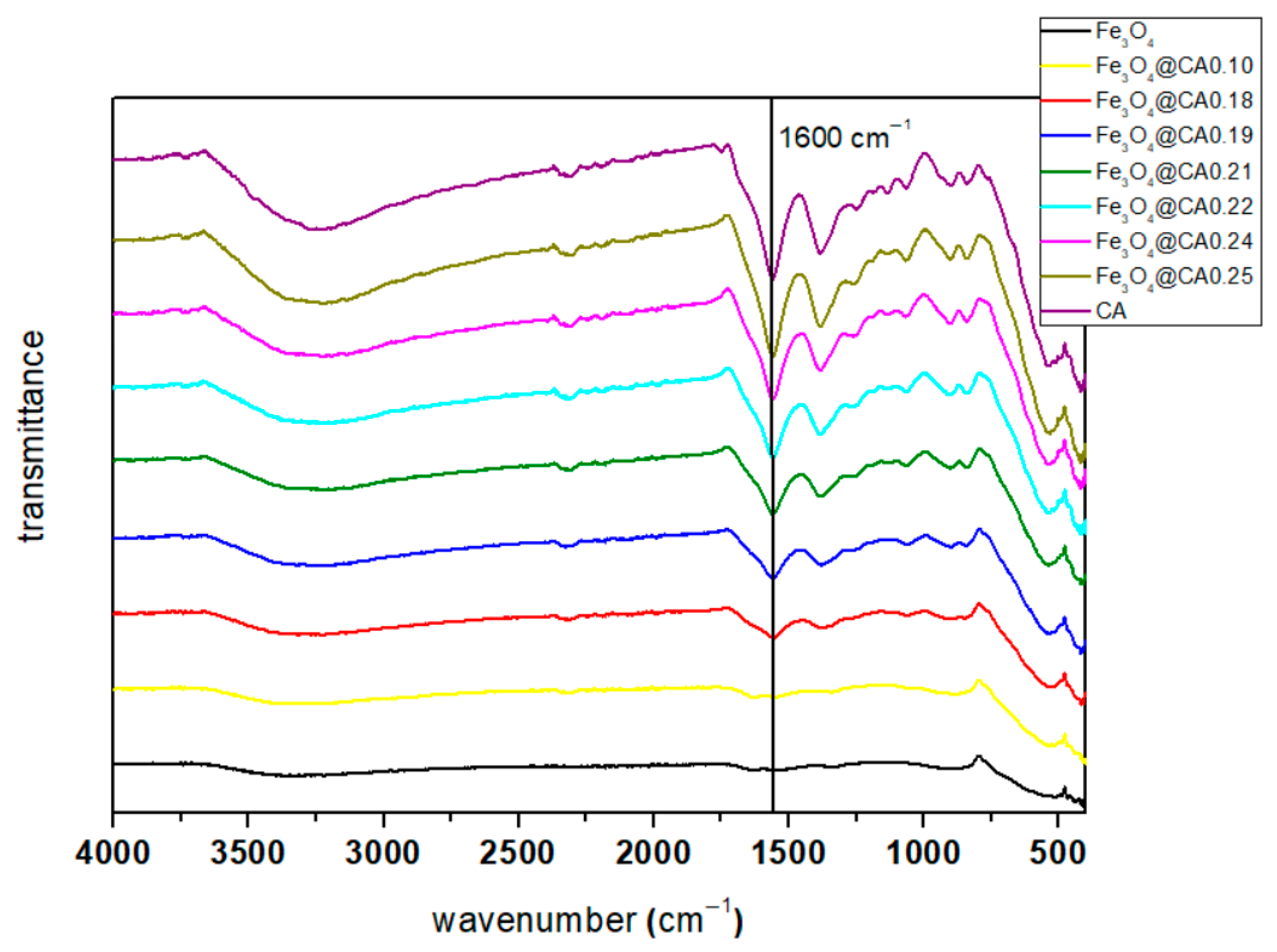
The FTIR spectrum range (4000–400 cm−1) of Fe3O4@CA corresponding to the iron citrate spectrum is presented. A large and intense band at 3450 cm−1 can be attributed to the structural OH groups as well as traces of molecular water and CA. The peak at 1700 cm−1, assignable to the C=O vibration (symmetric stretching) from the COOH group of CA, shifts to an intense band at about 1600 cm−1 for Fe3O4 coated with CA (Fe3O4@CA), indicating the binding of a CA radical to the magnetite surface, as shown in Figure 3. The band at 1400 cm−1 can be assigned to the asymmetric stretching of CO from the COOH group. The low-intensity bands between 400 and 600 cm−1 can be associated with the stretching and torsional vibration modes of the magnetite. These assignments are consistent with a previous study [34] that described two broad bands at 580 and 400 cm−1 associated with magnetite (Fe3O4). As can be seen from Figure 3, coating with CA was successful at some rates for most of the mass ratio of MNP and CA. The depth of the transmittance suggests that a higher amount of CA leads to more coated SPIONs. Only for sample Fe3O4@CA0.10 can it be concluded that coating with CA was not successful.

3.3. Thermogravimetric Analysis

A TGA was conducted to measure the organic content of Fe3O4@CA. The TGA results are presented in Figure 4, which illustrates the mass loss of Fe3O4@CA with different molar ratios between Fe3O4 and CA. A correction of 3% due to the oxidation of magnetite to hematite was taken into account, and the mass loss of CA was accordingly adjusted. The mass loss curve observed between 100 and 600 °C is attributed to the evaporation of water and the decomposition of the bound citrate molecules on the Fe3O4 MNP surfaces. The initial mass loss below 160 °C was due to the evaporation of physically adsorbed water, and the rest of the mass loss was determined as organic content of the coated Fe3O4@CA. This represents the mass percentage of all surface-coated organic groups on the MNPs, corresponding to the desorption of CA molecules from the magnetite particles. The mass percentage loss of CA of each sample is presented in Table 2. The formation of a CA coating layer on the particle surface is attributed to the chemical bonding between the carboxyl groups of CA and the Fe-OH sites on the iron oxide nanoparticles [35].
The amount of organic shell on the surface of nanoparticles was evaluated with thermogravimetry. Based on the graph (Figure 4) and Table 2, it can be concluded that CA constitutes between 4% and 20% of the sample mass [25].

3.4. Transmission Electron Microscopy

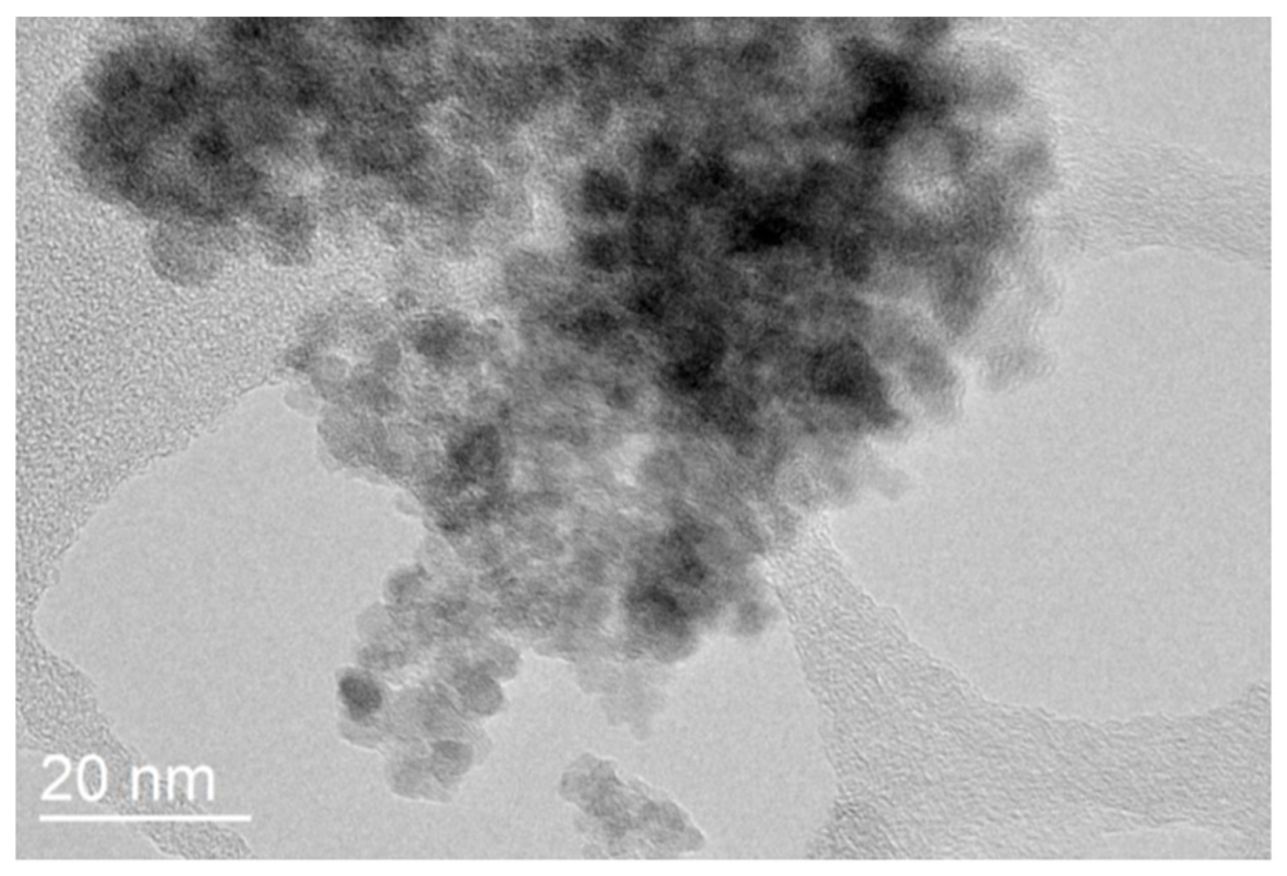
Figure 5 shows that the monodispersed rough surface of Fe3O4 was confirmed through TEM analysis, with particle sizes in the range of 4–6 nm. These values are consistent with the XRD results, where the crystallite size was estimated to be around 5 nm. The crystallite size calculated from XRD is in good agreement with the particle size observed in TEM images. Minor deviations are expected, as XRD provides the size of coherent crystalline domains, while TEM measures the overall particle size, which may include surface coatings or slight aggregation. In comparison with the literature, our findings demonstrate that the microreactor-based flow synthesis yields exceptionally small MNPs relative to those obtained by conventional synthesis methods [36]. For example, Boosz et al. [37] reported a Z-average size of approximately 10 nm for SPIONs synthesized via batch processes, which is notably larger than the particles produced in our study. Additionally, batch synthesis can lead to less uniform coating and stability issues, which are mitigated in our continuous flow approach, highlighting the advantages of modular microreactor-based synthesis for producing ultrasmall, well-dispersed nanoparticles.

3.5. Zeta Potential Measurements

An effective way to enhance the water stability of nanoparticles is by altering their isoelectric point (IEP) through surface modification. In this study, CA was used to coat the Fe3O4 nanoparticles. Citrate, with its three carboxyl groups, served as a potent ligand for stabilizing Fe3O4 particles [38]. The surface charges of the bare magnetite nanoparticles and Fe3O4@CA were characterized by measuring their zeta potential as a function of pH ranging from 7.0 to 2.0, with each measurement conducted in triplicate. Dispersion stability is related to the zeta potential value (mV): a range of 0 to ±5 can lead to rapid agglomeration and precipitation of nanoparticle suspensions; ±10 to ±30 marks the threshold for delicate dispersion; ±30 to ±40 indicates moderate stability of colloidal nanoparticles; and ±40 to ±60 signifies excellent stability of nanoparticle suspensions, indicating a high charge on their surface [23].
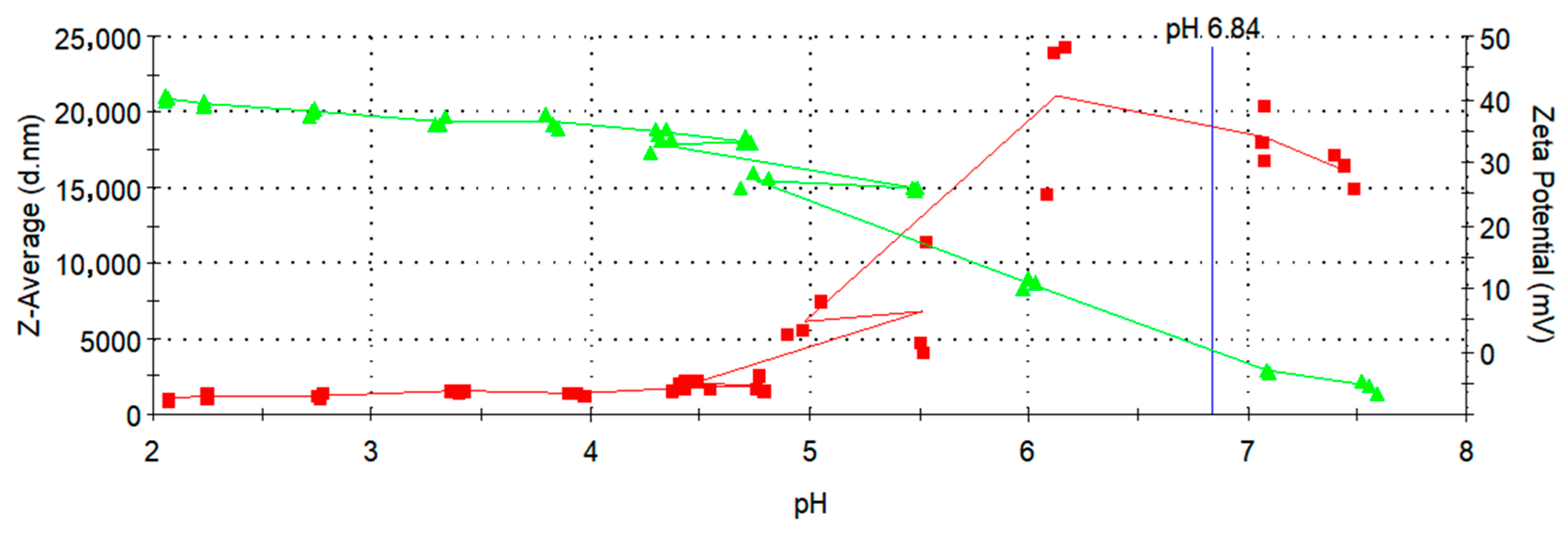
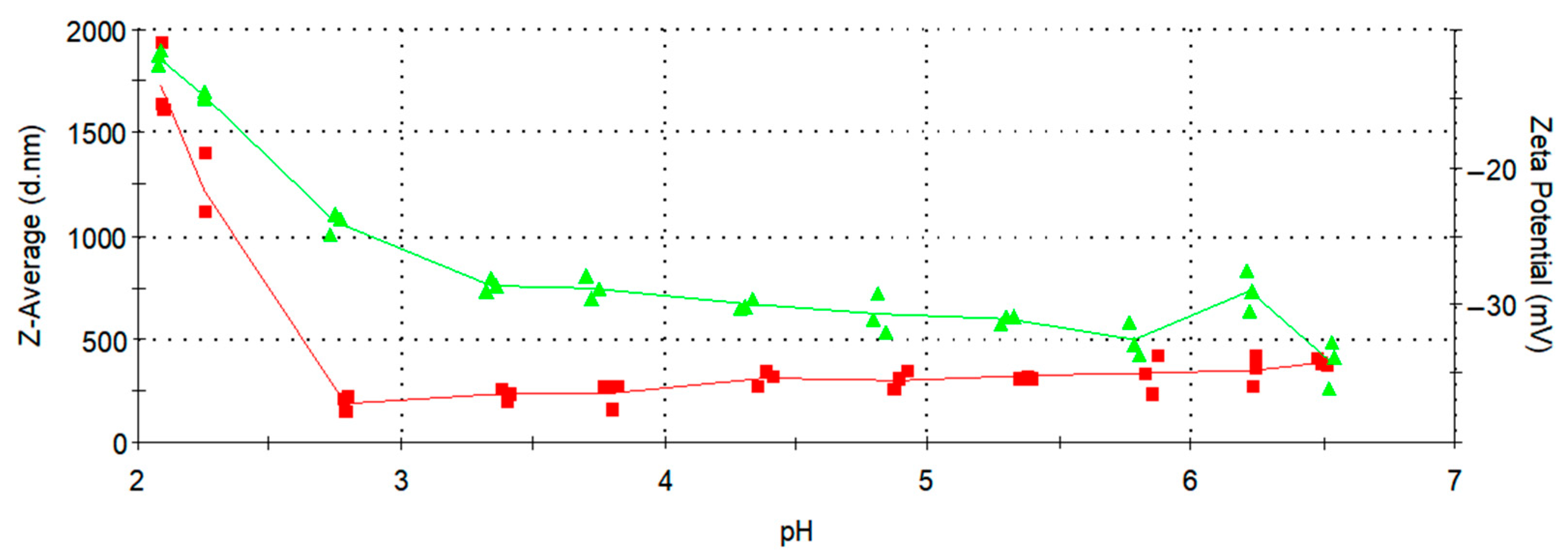
In Figure 6 and Figure 7, the zeta potential values are indicated by green triangles and a green line. As shown in Figure 6, the bare MNPs exhibits an IEP at pH 6.84, consistent with literature reports for Fe3O4 [36], whereas the Fe3O4@CA displays a negative zeta potential of −35 mV at pH 6.5 (Figure 7), respectively. This observed behavior is likely due to the adsorption of citrate onto the bare MNPs, altering the surface charges because of the presence of surface-bound carboxylate groups. The zeta potential becomes more negative with increasing pH due to the rise in OH ions in the solution and the deprotonation of the carboxyl groups of CA, given that the pKa values of citrate are 3.1, 4.8, and 6.4, respectively [36].
This confirms the presence of negatively charged carboxylate groups on the surface of the MNPs. The resulting electrostatic repulsion ensures their colloidal stability in aqueous suspension, as some of the carboxylate groups from CA are adsorbed or coordinated on the MNPs’ surface, while uncoordinated species extend into the water medium [39]. Consequently, the effective charge at pH 6,5 provides stabilization of Fe3O4@CA MNPs. The effective hydrodynamic diameter of Fe3O4@CA MNPs, measured by DLS at 25 °C, varies from 188.7 to 1730 nm with changing pH. The effective particle size started to increase at low pH values below 3, as in this range (pH 3 to 2) the zeta potential markedly decreased and approached zero. As shown in Figure 7, the IEP was not reached within the measured interval, since its value lies below pH 2, while our measurements were conducted in the range from pH 7 to 2.
Based on the results shown in Figure 6 and Figure 7, the IEP differs substantially between bare Fe3O4 nanoparticles, with an IEP of 6.84, and Fe3O4@CA0.25, where the IEP is below 2. This indicates that the nanoparticles were successfully coated with CA, as a surface modification has clearly occurred. Our study reports an average particle size of approximately 5 nm and a zeta potential of around −35 mV for the CA-coated SPIONs synthesized using a modular flow microreactor system. These values are consistent with those reported in previous studies employing similar synthesis or surface modification strategies. For instance, Hussein et al. [40] observed comparable zeta potential values and enhanced colloidal stability for CA-coated SPIONs prepared via chemical co-precipitation, confirming the effectiveness of citric acid in providing electrostatic stabilization. This agreement further validates that the microreactor-based synthesis approach yields nanoparticles with physicochemical properties suitable for biomedical applications.
The modular flow microreactor system offers several advantages over traditional batch methods, including precise control over reaction parameters such as temperature, mixing, and reagent flow, which results in more uniform reaction conditions and consistent particle sizes. This precise control minimizes temperature and concentration gradients, thereby improving reproducibility and colloidal stability of the synthesized nanoparticles. Moreover, the continuous nature of the process allows for facile scalability through parallelization of identical units without altering the optimized conditions.
These benefits are consistent with recent studies that emphasize the advantages of continuous flow synthesis in achieving improved size uniformity, stability, and production efficiency of nanomaterials. Estévez et al. [41] similarly demonstrated that continuous flow microreactor systems enhance nanoparticle quality and process reproducibility compared to conventional batch methods, underscoring the potential of this approach for scalable and controlled nanoparticle production.

4. Conclusions

In this study, SPIONs were successfully synthesized and simultaneously coated with CA using a modular flow microreactor system. This integrated approach enabled continuous, well-controlled synthesis, combining nucleation, growth, and surface functionalization in a single process. XRD analysis confirmed the formation of Fe3O4 nanoparticles with an average crystallite size of approximately 5 nm, which was consistent with the TEM observations showing nearly spherical particles with sizes in the range of 4–6 nm. FTIR spectra verified the effective surface coating with CA, while TGA analysis demonstrated increased mass loss with higher CA concentrations, confirming successful coating. Zeta potential measurements around −35 mV at pH 6.5 further indicated strong colloidal stability of the coated particles.
Compared to conventional batch synthesis methods reported in the literature, the microreactor-based approach provides enhanced control over reaction parameters and reproducible particle characteristics within the sub-10 nm range. The combination of small particle size, narrow size distribution, and good dispersion confirms the potential of this continuous flow process for scalable nanoparticle production.
Overall, this study presents preliminary results demonstrating the feasibility and promise of modular flow synthesis for producing CA-coated SPIONs. Future work will focus on quantitative scalability assessment, process reproducibility, and optimization of coating uniformity to further advance the application of this approach for biomedical purposes, particularly as MRI contrast agents and drug delivery platforms.
Future work will focus on improving experimental reproducibility through replicate synthesis and statistical evaluation, as well as on detailed characterization of coating uniformity using advanced techniques. Since direct pH monitoring within the microreactor was not feasible in the present setup, the development of integrated micro-scale pH sensing or feedback control will be pursued to further enhance coating precision and process consistency. In addition, the scalability of the modular microreactor system will be examined through extended operation and parallelization studies to ensure stable and uniform nanoparticle production.

Author Contributions

S.V.: research, formal analysis, conceptualization, methodology, investigation. A.N.: methodology, investigation, research, conceptualization, writing—review and editing, supervision. J.S.: methodology, investigation, writing—original draft, validation, methodology, research, conceptualization. All authors have read and agreed to the published version of the manuscript.

Funding

The authors gratefully acknowledge the financial support of Slovenian Research and Innovation Agency (ARIS) throughout the research, and infrastructure programmes P2-0006 and P2-0414 and the research project J2-60044.

Data Availability Statement

The data presented in this study are available on request from the corresponding author.

Acknowledgments

We extend our gratitude to Sašo Gyergyek for his collaboration in conducting TEM and magnetic measurements.

Conflicts of Interest

The authors declare no conflicts of interest.

Abbreviations

The following abbreviations are used in this manuscript:
CACitric acid
DLSDynamic light scattering
FTIRFourier transform infrared spectroscopy
IEPIsoeletric point
ISPSIron salt precursor solution
MHMagnetic hyperthermia
MNPsMagnetic nanoparticles
MRIMagnetic resonance imaging
SPIONSuperparamagnetic iron oxide nanoparticles
TEMTransmission electron microscopy
TGAThermogravimetric analysis
XRDX-ray powder diffraction

References

  1. Dias, A.M.M.; Courteau, A.; Bellaye, P.-S.; Kohli, E.; Oudot, A.; Doulain, P.-E.; Petitot, C.; Walker, P.-M.; Decréau, R.; Collin, B. Superparamagnetic Iron Oxide Nanoparticles for Immunotherapy of Cancers Through Macrophages and Magnetic Hyperthermia. Pharmaceutics 2022, 14, 2388. [Google Scholar] [CrossRef] [PubMed]
  2. Wei, H.; Hu, Y.; Wang, J.; Gao, X.; Qian, X.; Tang, M. Superparamagnetic Iron Oxide Nanoparticles: Cytotoxicity, Metabolism, and Cellular Behavior in Biomedicine Applications. Int. J. Nanomed. 2021, 16, 6097–6113. [Google Scholar] [CrossRef]
  3. Martins, P.M.; Lima, A.C.; Ribeiro, S.; Lanceros-Mendez, S.; Martins, P. Magnetic Nanoparticles for Biomedical Applications: From the Soul of the Earth to the Deep History of Ourselves. ACS Appl. Bio Mater. 2021, 4, 5839–5870. [Google Scholar] [CrossRef]
  4. Mittal, A.; Roy, I.; Gandhi, S. Magnetic Nanoparticles: An Overview for Biomedical Applications. Magnetochemistry 2022, 8, 107. [Google Scholar] [CrossRef]
  5. Rümenapp, C.; Gleich, B.; Haase, A. Magnetic Nanoparticles in Magnetic Resonance Imaging and Diagnostics. Pharm. Res. 2012, 29, 1165–1179. [Google Scholar] [CrossRef]
  6. Avasthi, A.; Caro, C.; Pozo-Torres, E.; Leal, M.P.; García-Martín, M.L. Magnetic Nanoparticles as MRI Contrast Agents. Top. Curr. Chem. 2020, 378, 40. [Google Scholar] [CrossRef] [PubMed]
  7. Caspani, S.; Magalhães, R.; Araújo, J.P.; Sousa, C.T. Magnetic Nanomaterials as Contrast Agents for MRI. Materials 2020, 13, 2586. [Google Scholar] [CrossRef]
  8. Chow, J.C.L. 9-Magnetic nanoparticles as contrast agents in magnetic resonance imaging and radiosensitizers in radiotherapy. In Fundamentals and Industrial Applications of Magnetic Nanoparticles; Hussain, C.M., Patankar, K.K., Eds.; Woodhead Publishing: Cambridge, UK, 2022; pp. 291–316. [Google Scholar]
  9. Neuberger, T.; Schöpf, B.; Hofmann, H.; Hofmann, M.; von Rechenberg, B. Superparamagnetic nanoparticles for biomedical applications: Possibilities and limitations of a new drug delivery system. J. Magn. Magn. Mater. 2005, 293, 483–496. [Google Scholar] [CrossRef]
  10. Maity, D.; Kandasamy, G. Superparamagnetic Nanoparticles for Cancer Hyperthermia Treatment. In Nanotechnology Characterization Tools for Tissue Engineering and Medical Therapy; Kumar, C.S.S.R., Ed.; Springer: Berlin/Heidelberg, Germany, 2019; pp. 299–332. [Google Scholar]
  11. Fatima, H.; Charinpanitkul, T.; Kim, K.-S. Fundamentals to Apply Magnetic Nanoparticles for Hyperthermia Therapy. Nanomaterials 2021, 11, 1203. [Google Scholar] [CrossRef]
  12. Yildiz, I. Applications of magnetic nanoparticles in biomedical separation and purification. Nanotechnol. Rev. 2016, 5, 331–340. [Google Scholar] [CrossRef]
  13. Dasari, A.; Xue, J.; Deb, S. Magnetic Nanoparticles in Bone Tissue Engineering. Nanomaterials 2022, 12, 757. [Google Scholar] [CrossRef]
  14. Fan, D.; Wang, Q.; Zhu, T.; Wang, H.; Liu, B.; Wang, Y.; Liu, Z.; Liu, X.; Fan, D.; Wang, X. Recent Advances of Magnetic Nanomaterials in Bone Tissue Repair. Front. Chem. 2020, 8, 745. [Google Scholar] [CrossRef] [PubMed]
  15. kianfar, E. Magnetic Nanoparticles in Targeted Drug Delivery: A Review. J. Supercond. Nov. Magn. 2021, 34, 1709–1735. [Google Scholar] [CrossRef]
  16. Liu, J.F.; Jang, B.; Issadore, D.; Tsourkas, A. Use of magnetic fields and nanoparticles to trigger drug release and improve tumor targeting. WIREs Nanomed. Nanobiotechnol. 2019, 11, e1571. [Google Scholar] [CrossRef]
  17. Huang, J.; Zhong, X.; Wang, L.; Yang, L.; Mao, H. Improving the magnetic resonance imaging contrast and detection methods with engineered magnetic nanoparticles. Theranostics 2012, 2, 86–102. [Google Scholar] [CrossRef]
  18. Dulińska-Litewka, J.; Łazarczyk, A.; Hałubiec, P.; Szafrański, O.; Karnas, K.; Karewicz, A. Superparamagnetic Iron Oxide Nanoparticles—Current and Prospective Medical Applications. Materials 2019, 12, 617. [Google Scholar] [CrossRef]
  19. Kritika; Roy, I. Therapeutic applications of magnetic nanoparticles: Recent advances. Mater. Adv. 2022, 3, 7425–7444. [Google Scholar] [CrossRef]
  20. Si, G.; Hapuarachchige, S.; Artemov, D. Ultrasmall Superparamagnetic Iron Oxide Nanoparticles as Nanocarriers for Magnetic Resonance Imaging: Development and In Vivo Characterization. ACS Appl. Nano Mater. 2022, 5, 9625–9632. [Google Scholar] [CrossRef] [PubMed]
  21. Ali, A.; Shah, T.; Ullah, R.; Zhou, P.; Guo, M.; Ovais, M.; Tan, Z.; Rui, Y. Review on Recent Progress in Magnetic Nanoparticles: Synthesis, Characterization, and Diverse Applications. Front. Chem. 2021, 9, 629054. [Google Scholar] [CrossRef] [PubMed]
  22. Stiufiuc, G.F.; Stiufiuc, R.I. Magnetic Nanoparticles: Synthesis, Characterization, and Their Use in Biomedical Field. Appl. Sci. 2024, 14, 1623. [Google Scholar] [CrossRef]
  23. Dheyab, M.A.; Aziz, A.A.; Jameel, M.S.; Abu Noqta, O.; Khaniabadi, P.M.; Mehrdel, B. Simple rapid stabilization method through citric acid modification for magnetite nanoparticles. Sci. Rep. 2020, 10, 10793. [Google Scholar] [CrossRef] [PubMed]
  24. Mikelashvili, V.; Kekutia, S.; Markhulia, J.; Saneblidze, L.; Maisuradze, N.; Kriechbaum, M.; Almásy, L. Synthesis and Characterization of Citric Acid-Modified Iron Oxide Nanoparticles Prepared with Electrohydraulic Discharge Treatment. Materials 2023, 16, 746. [Google Scholar] [CrossRef]
  25. Nieciecka, D.; Rękorajska, A.; Cichy, D.; Końska, P.; Żuk, M.; Krysiński, P. Synthesis and Characterization of Magnetic Drug Carriers Modified with Tb(3+) Ions. Nanomaterials 2022, 12, 795. [Google Scholar] [CrossRef] [PubMed]
  26. Peng, T.; Liang, J.; Zhang, X.; Chen, L.; Chen, Y.; Qiang, J.; Yuan, S. Investigation of geometric modifications in T-type micromixers for enhanced microfluidic mixing and ZIF-8 synthesis. Results Eng. 2025, 26, 105233. [Google Scholar] [CrossRef]
  27. Tan, Z.; Shi, H.; Zheng, Y.; Cao, Y. A 3D homogeneous microreactor with high mixing intensity at wide Re range for MOF preparation and POCT application. Chem. Eng. J. 2023, 476, 146481. [Google Scholar] [CrossRef]
  28. Watanabe, S.; Miyahara, M.T. Synthesis of Functional Nanoparticles Using a Microreactor. KONA Powder Part J. 2025, 42, 156–169. [Google Scholar] [CrossRef]
  29. Gutierrez, L.; Gomez, L.; Irusta, S.; Arruebo, M.; Santamaria, J. Comparative study of the synthesis of silica nanoparticles in micromixer–microreactor and batch reactor systems. Chem. Eng. J. 2011, 171, 674–683. [Google Scholar] [CrossRef]
  30. Lu, J.Y.; Bu, Z.Q.; Lei, Y.Q.; Wang, D.; He, B.; Wang, J.; Huang, W.T. Facile microwave-assisted synthesis of Sb2O3-CuO nanocomposites for catalytic degradation of p-nitrophenol. J. Mol. Liq. 2024, 409, 125503. [Google Scholar] [CrossRef]
  31. Li, T.; Xiao, L.; Ling, H.; Yang, Y.; Zhong, S. Preparation of artificial substrate binding sites of nanozyme with “modular structure” strategy used for the construction of visual sensing analysis platform for levodopa. Microchem. J. 2025, 212, 113292. [Google Scholar] [CrossRef]
  32. Liu, J.; Dai, C.; Hu, Y. Aqueous aggregation behavior of citric acid coated magnetite nanoparticles: Effects of pH, cations, anions, and humic acid. Environ. Res. 2018, 161, 49–60. [Google Scholar] [CrossRef]
  33. Alzoubi, F.; Jabaly, A.; Noqta, A.; Al-Khateeb, H.M.; Alqadi, M.K.; Bououdina, M. Citric Acid Coated Iron Oxide Nanoparticles as Contrast Agent for Magnetic Resonance Imaging. Res. Sq. 2022. [Google Scholar] [CrossRef]
  34. Singh, D.; Gautam, R.K.; Kumar, R.; Shukla, B.K.; Shankar, V.; Krishna, V. Citric acid coated magnetic nanoparticles: Synthesis, characterization and application in removal of Cd(II) ions from aqueous solution. J. Water Process Eng. 2014, 4, 233–241. [Google Scholar] [CrossRef]
  35. Hajdú, A.; Tombácz, E.; Illés, E.; Bica, D.; Vékás, L. Magnetite nanoparticles stabilized under physiological conditions for biomedical application. In Colloids for Nano-and Biotechnology; Springer: Berlin/Heidelberg, Germany, 2008. [Google Scholar]
  36. Campelj, S.; Makovec, D.; Drofenik, M. Preparation and properties of water-based magnetic fluids. J. Phys. Condens. Matter 2008, 20, 204101. [Google Scholar] [CrossRef]
  37. Boosz, P.; Pfister, F.; Stein, R.; Friedrich, B.; Fester, L.; Band, J.; Mühlberger, M.; Schreiber, E.; Lyer, S.; Dudziak, D.; et al. Citrate-Coated Superparamagnetic Iron Oxide Nanoparticles Enable a Stable Non-Spilling Loading of T Cells and Their Magnetic Accumulation. Cancers 2021, 13, 4143. [Google Scholar] [CrossRef] [PubMed]
  38. Lattuada, M.; Hatton, T.A. Functionalization of monodisperse magnetic nanoparticles. Langmuir 2007, 23, 2158–2168. [Google Scholar] [CrossRef] [PubMed]
  39. Petrinic, I.; Stergar, J.; Bukšek, H.; Drofenik, M.; Gyergyek, S.; Hélix-Nielsen, C.; Ban, I. Superparamagnetic Fe3O4@CA nanoparticles and their potential as draw solution agents in forward osmosis. Nanomaterials 2021, 11, 2965. [Google Scholar] [CrossRef]
  40. Che Mohamed Hussein, S.N.; Amir, Z.; Jan, B.M.; Khalil, M.; Azizi, A. Colloidal Stability of CA, SDS and PVA Coated Iron Oxide Nanoparticles (IONPs): Effect of Molar Ratio and Salinity. Polymers 2022, 14, 4787. [Google Scholar] [CrossRef]
  41. Estévez, M.; Cicuéndez, M.; Crespo, J.; Serrano-López, J.; Colilla, M.; Fernández-Acevedo, C.; Oroz-Mateo, T.; Rada-Leza, A.; González, B.; Izquierdo-Barba, I.; et al. Large-scale production of superparamagnetic iron oxide nanoparticles by flame spray pyrolysis: In vitro biological evaluation for biomedical applications. J. Colloid Interface Sci. 2023, 650, 560–572. [Google Scholar] [CrossRef] [PubMed]
Figure 1. Schematic setup of the simultaneous production of nanoparticles and coating with CA.
Micromachines 16 01228 g001
Figure 2. XRD spectra of Fe3O4.
Micromachines 16 01228 g002
Figure 3. FTIR spectra of Fe3O4, Fe3O4@CA.
Micromachines 16 01228 g003
Figure 4. TGA of the Fe3O4 and Fe3O4@CA.
Micromachines 16 01228 g004
Figure 5. TEM image of bare Fe3O4 nanoparticles.
Micromachines 16 01228 g005
Figure 6. Change in zeta potential and Z-average for Fe3O4 with pH values.
Micromachines 16 01228 g006
Figure 7. Change in zeta potential and Z-average for Fe3O4@CA0.25 with pH values.
Micromachines 16 01228 g007
Table 1. Sample names and corresponding molar values of MNPs and CA used in this study.
Name of SampleAmount of MNPs [mol]Amount of CA [mol]
Fe3O4@CA0.1010.10
Fe3O4@CA0.1810.18
Fe3O4@CA0.1910.19
Fe3O4@CA0.2110.21
Fe3O4@CA0.2210.22
Fe3O4@CA0.2410.24
Fe3O4@CA0.2510.25
Table 2. Mass loss of CA for different molar ratios of Fe3O4@CA.
SampleMass Loss of CA (%)
Fe3O4@CA0.184.4
Fe3O4@CA0.194.9
Fe3O4@CA0.215.0
Fe3O4@CA0.225.3
Fe3O4@CA0.2415.0
Fe3O4@CA0.2519.6
Disclaimer/Publisher’s Note: The statements, opinions and data contained in all publications are solely those of the individual author(s) and contributor(s) and not of MDPI and/or the editor(s). MDPI and/or the editor(s) disclaim responsibility for any injury to people or property resulting from any ideas, methods, instructions or products referred to in the content.