


An Assessment of Carotid Flow Time Using a Portable Handheld Ultrasound Device: The Ideal Tool for Guiding Intraoperative Fluid Management?


Abstract
1. Introduction
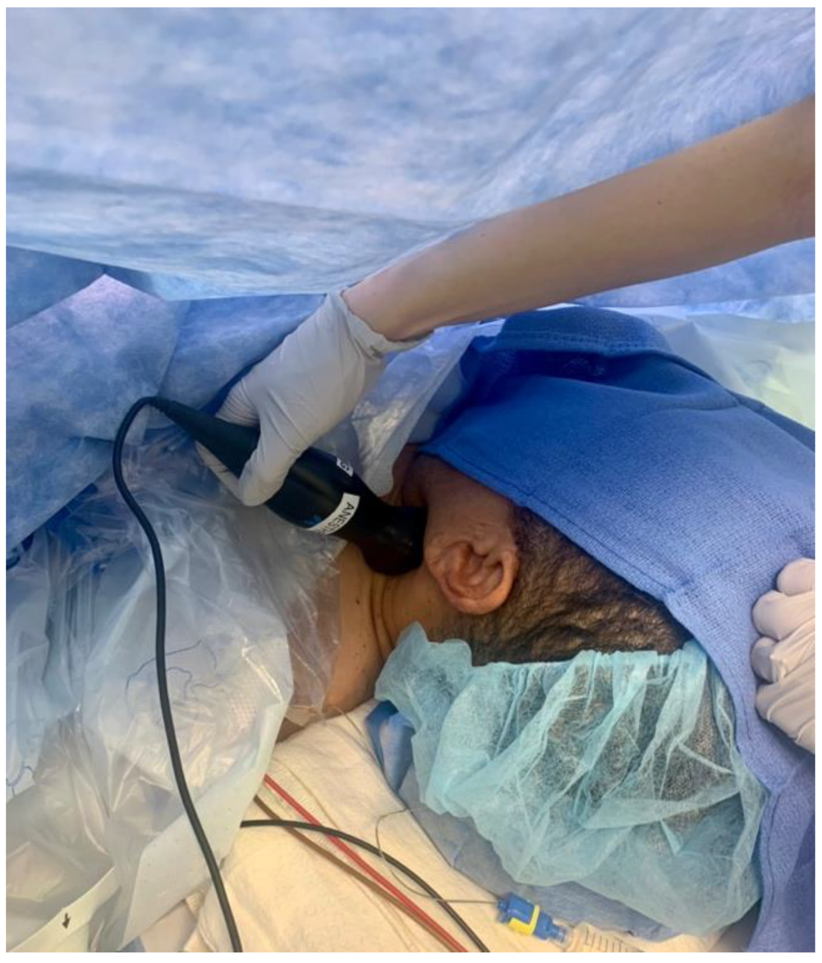
2. Case Report
3. Results
4. Conclusions
Author Contributions
Funding
Institutional Review Board Statement
Data Availability Statement
Conflicts of Interest
References
- Shin, C.H.; Long, D.R.; McLean, D.; Grabitz, S.D.; Ladha, K.; Timm, F.P.; Thevathasan, T.; Pieretti, A.; Ferrone, C.; Hoeft, A.; et al. Effects of intraoperative fluid management on postoperative outcomes: A hospital registry study. Ann. Surg. 2018, 267, 1084–1092. [Google Scholar] [CrossRef]
- Waddle, J.P.; Evers, A.S.; Piccirillo, J.F. Postanesthesia care unit length of stay: Quantifying and assessing dependent factors. Anesth. Analg. 1998, 87, 628–633. [Google Scholar] [PubMed]
- Blehar, D.J.; Glazier, S.; Gaspari, R.J. Correlation of corrected flow time in the carotid artery with changes in intravascular volume status. J. Crit. Care 2014, 29, 486–488. [Google Scholar] [CrossRef] [PubMed]
- Kim, D.H.; Shin, S.; Kim, N.; Choi, T.; Choi, S.H.; Choi, Y.S. Carotid ultrasound measurements for assessing fluid responsiveness in spontaneously breathing patients: Corrected flow time and respirophasic variation in blood flow peak velocity. Br. J. Anaesth. 2018, 121, 541–549. [Google Scholar] [CrossRef] [PubMed]
- Wodey, E.; Carre, F.; Beneux, X.; Schaffuser, A.; Ecoffey, C. Limits of corrected flow time to monitor hemodynamic status in children. J. Clin. Monit. Comput. 2000, 16, 223–228. [Google Scholar] [CrossRef]
- Barjaktarevic, I.; Toppen, W.E.; Hu, S.; Montoya, E.A.; Ong, S.; Buhr, R.G.; David, I.J.; Wang, T.; Rezayat, T.; Chang, S.Y.; et al. Ultrasound assessment of the change in carotid corrected flow time in fluid responsiveness in undifferentiated shock. Crit. Care Med. 2018, 46, e1040. [Google Scholar] [CrossRef]


Disclaimer/Publisher’s Note: The statements, opinions and data contained in all publications are solely those of the individual author(s) and contributor(s) and not of MDPI and/or the editor(s). MDPI and/or the editor(s) disclaim responsibility for any injury to people or property resulting from any ideas, methods, instructions or products referred to in the content. |
© 2023 by the authors. Licensee MDPI, Basel, Switzerland. This article is an open access article distributed under the terms and conditions of the Creative Commons Attribution (CC BY) license (https://creativecommons.org/licenses/by/4.0/).
Share and Cite
Gibson, L.E.; Mitchell, J.E.; Bittner, E.A.; Chang, M.G. An Assessment of Carotid Flow Time Using a Portable Handheld Ultrasound Device: The Ideal Tool for Guiding Intraoperative Fluid Management? Micromachines 2023, 14, 510. https://doi.org/10.3390/mi14030510
Gibson LE, Mitchell JE, Bittner EA, Chang MG. An Assessment of Carotid Flow Time Using a Portable Handheld Ultrasound Device: The Ideal Tool for Guiding Intraoperative Fluid Management? Micromachines. 2023; 14(3):510. https://doi.org/10.3390/mi14030510
Chicago/Turabian StyleGibson, Lauren E., James E. Mitchell, Edward A. Bittner, and Marvin G. Chang. 2023. "An Assessment of Carotid Flow Time Using a Portable Handheld Ultrasound Device: The Ideal Tool for Guiding Intraoperative Fluid Management?" Micromachines 14, no. 3: 510. https://doi.org/10.3390/mi14030510
APA StyleGibson, L. E., Mitchell, J. E., Bittner, E. A., & Chang, M. G. (2023). An Assessment of Carotid Flow Time Using a Portable Handheld Ultrasound Device: The Ideal Tool for Guiding Intraoperative Fluid Management? Micromachines, 14(3), 510. https://doi.org/10.3390/mi14030510