Review

Hybrid Systems of Nanofibers and Polymeric Nanoparticles for Biological Application and Delivery Systems

by
Hever Yuritzy Vargas-Molinero
1,
Aracely Serrano-Medina
1,2,
Kenia Palomino-Vizcaino
1,
Eduardo Alberto López-Maldonado
1,
Luis Jesús Villarreal-Gómez
1,3,
Graciela Lizeth Pérez-González
3 and
José Manuel Cornejo-Bravo
1,*
1
Facultad de Ciencias Químicas e Ingeniería, Universidad Autónoma de Baja California, Tijuana 22390, Mexico
2
Facultad de Medicina y Psicología, Universidad Autónoma de Baja California, Tijuana 22390, Mexico
3
Facultad de Ciencias de la Ingeniería y Tecnología, Universidad Autónoma de Baja California, Tijuana 22427, Mexico
*
Author to whom correspondence should be addressed.
Micromachines 2023, 14(1), 208; https://doi.org/10.3390/mi14010208
Submission received: 23 December 2022 / Revised: 9 January 2023 / Accepted: 10 January 2023 / Published: 14 January 2023
(This article belongs to the Section B5: Drug Delivery System)

Abstract

:
Nanomedicine is a new discipline resulting from the combination of nanotechnology and biomedicine. Nanomedicine has contributed to the development of new and improved treatments, diagnoses, and therapies. In this field, nanoparticles have notable importance due to their unique properties and characteristics, which are useful in different applications, including tissue engineering, biomarkers, and drug delivery systems. Electrospinning is a versatile technique used to produce fibrous mats. The high surface area of the electrospun mats makes them suitable for applications in fields using nanoparticles. Electrospun mats are used for tissue engineering, wound dressing, water-treatment filters, biosensors, nanocomposites, medical implants, protective clothing materials, cosmetics, and drug delivery systems. The combination of nanoparticles with nanofibers creates hybrid systems that acquire properties that differ from their components’ characteristics. By utilizing nanoparticles and nanofibers composed of dissimilar polymers, the two synergize to improve the overall performance of electrospinning mats and nanoparticles. This review summarizes the hybrid systems of polymeric nanoparticles and polymeric nanofibers, critically analyzing how the combination improves the properties of the materials and contributes to the reduction of some disadvantages found in nanometric devices and systems.

1. Introduction

Nanoparticles have been essential as an innovative technology in drug delivery systems. With chemotherapeutic drugs, for example, greater specificity is needed to reduce adverse reactions during cancer treatments. These nanometric systems are able to deliver more concentrated doses in close proximity to tumor tissue due to the effects of improved permeability and retention. This, in turn, reduces the exposure of the drug to healthy tissues, and limits the distribution of chemotherapeutics to the targeted tumor tissue. This results in a more precise and less toxic dosage during treatment. Nanoparticles can also be used as carriers to penetrate the blood-brain barrier (BBB) and deliver antineoplastic drugs to treat brain tumors [1]. Another property offered by nanoparticles is to improve the bioavailability of bioactive molecules prone to enzymatic and hydrolytic degradation. Nanoparticles can be used in this capacity as oral administration vehicles for the encapsulation of peptides and proteins, which protects them from gastrointestinal barriers. They can also be used for gene delivery, improving absorption by increasing the cellular permeability of macromolecules. Nanoparticles can be targeted with specific ligands to provide better strategies for therapeutics [2,3].
Electrospun mats, on the other hand, are being studied for different purposes, such as tissue engineering, wound dressing, water treatment filters, biosensors, nanocomposites, medical implants, protective clothing materials, cosmetics, and drug delivery systems [4]. The nanofiber mats formed by electrospinning have different qualities, such as improving the dissolution of hydrophobic drugs and presenting greater release than other systems (e.g., hydrogels, films). The high surface area to volume ratio and many inter-/intra fibrous pores contribute to drug deposition and accumulation [5].
Independently, nanoparticles and polymeric nanofibers present some limitations and disadvantages. They are prone to degradation when used as implantable devices, affecting the device’s sensitivity and useful life expectancy [6]. Nanoparticles can present difficulties during their physical handling, either in liquid or dry forms, due to their small size and large surface area, which make them prone to aggregation. These properties also limit the drug load to a very small amount [2]. As for nanofibers, they present morphological characteristics that must be adjusted for the desired application. Nanofibers must meet specific requirements regarding their mechanical properties and wettability for use as biomedical membranes. Hydrophobic nanofiber mats have been shown to cause plugging and decreased flow due to air pockets trapped within the mats. The addition of nanoparticles can assist in developing a system with more suitable characteristics by modifying properties such as the tensile strength of the compound, its roughness, and the wettability of the system [7,8].
This review summarizes hybrid systems of polymeric nanoparticles and polymeric nanofibers and critically analyzes how the combination contributes to the reduction of some disadvantages found in systems that only contain one type of nanotechnology [9,10,11,12].
Insightful reviews were found regarding hybrid systems, mainly reporting on fibrous mats containing inorganic nanoparticles [9,13].The current review, however, focuses on polymeric nanoparticles from the preparation of the polymeric nanoparticles to drug loading and the incorporation into the nanofibers, which present particular challenges, but also present versatile applications within biomedicine [14]. This review presents an introduction to the electrodynamic method for the production of nanomaterials as well as an overview of nanoparticle synthesis, drug loading and incorporation of nanofibers. In the following sections, the materials used and applications will be discussed.

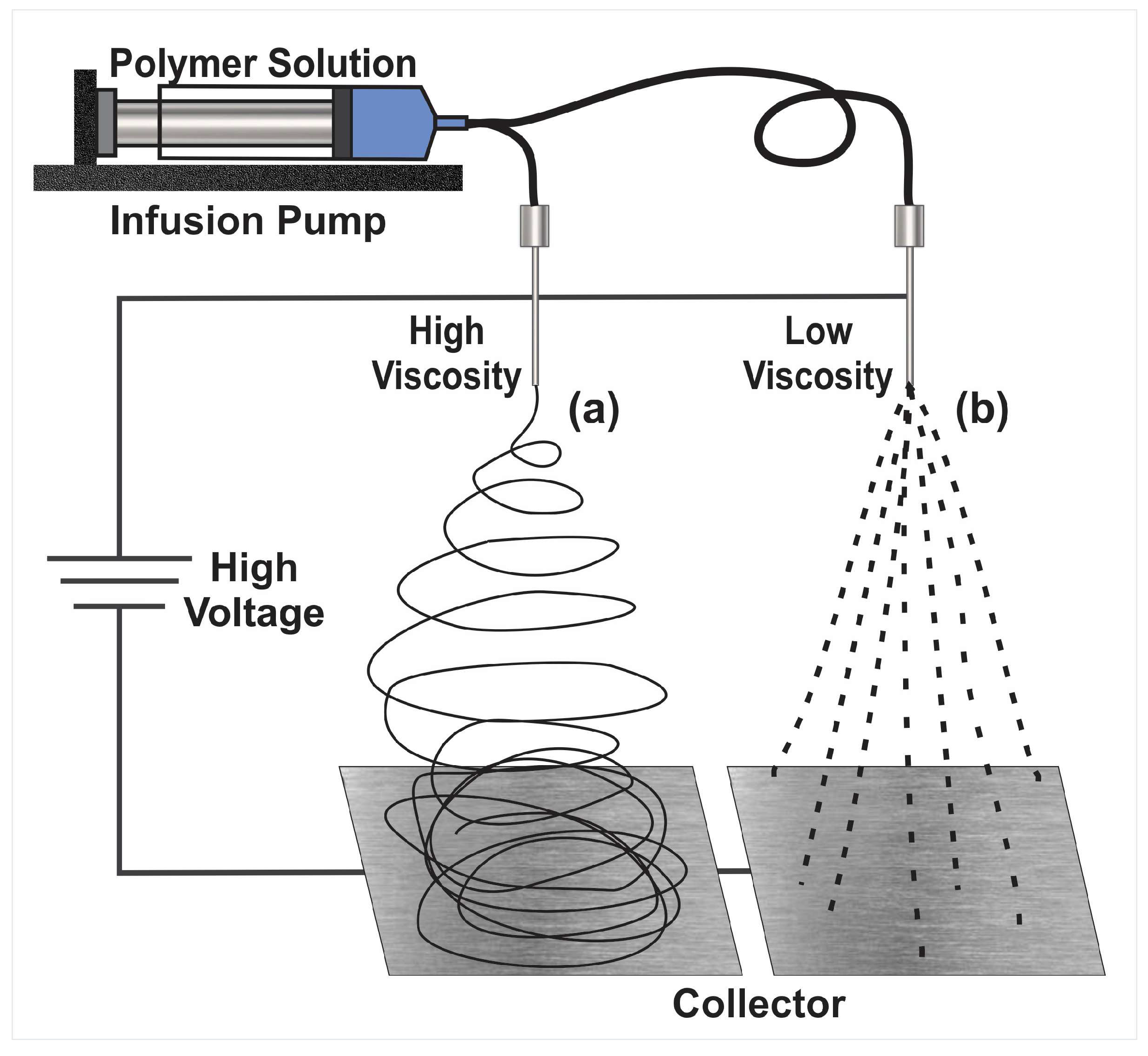
2. The Electrohydrodynamic Method

The electrodynamic methods consist of electrospinning for the creation of nanofibers and electrospray for the development of nanoparticles. These are similar techniques that take into account some experimental conditions but with different parameters and produce different material shapes, sizes, and characteristics [15]. The electrohydrodynamic method can make different types, kinds, and sizes of particles, mats, and a blend of both structure types [14,16]. The general setup for the electrodynamic method, either to form fibers (Figure 1a) or particles (Figure 1b), consists of applying high voltage to the tip of a needle of a syringe containing a polymer or biopolymer solution to a conductive metallic collector. First, the plunger is pushed by a syringe-pump, which produces the output of the solution at controlled parameters. Through the tip of the syringe, a cone is formed, called “Taylor’s cone”. The collector must be made of a conductive material that serves as a support for the material that is deposited in it, and the collector must be connected to the electric ground of the high-voltage system.
These particles and fibers have qualities that put them at an advantage against other types of materials. One of the most commonly mentioned are the small sizes, which range from micrometers to nanometers and qualifies them as nanotechnology; they also have very desirable characteristics, such as high porosity and small pore size. In addition, they have a high surface area to volume ratio, which means greater exposure to the environment [17]. The products of electrodynamic processes have a huge variety of applications in different areas, such as the food industry and the biomedical industry. Its applications include the encapsulation of substances, filtration, packaging development, and enzyme fixation, among others [18].
Electrospray follows the same principles as electrospinning. A charged solution passes through a needle, leading to the ionization and atomization of the solution, and produces microscopic, charged droplets. The solvent evaporates when nano- and micro-sized droplets are produced and collected on a grounded plate. Electrospray has great encapsulation capacity, especially for hydrophilic particles in hydrophobic carriers [19,20]. Nanoparticles formed by electrospray are suitable for drug-carrying applications. Different morphologies can be obtained in the nanoparticles, such as solid spheres, ellipses, or hollow spheres. In addition to morphology, size can also be controlled [21]. Another use for electrospray is the formation of membranes in conjunction with nanofibers, a useful configuration for filtering technologies [22,23].

2.1. Electrospinning

One of the products of the electrodynamic process is nanofibers. The electrospun nanofibers made from polymeric materials can be biocompatible, biodegradable, have a high load capacity, and encapsulation efficiency. Another advantage is that they can be produced cost-effectively. Additional features can be added to these electrospinning mats by using copolymers or functionalizing the solution with bioactive molecules. Coating the nanofiber’s surface with layers of polymeric materials, for example, which have desired physicochemical properties such as pH sensitivity. This can help to control the degradation of the nanofibers at different pH levels and provide stability in the cellular environment [15]. The electrospinning technique is easy to use, simple, unique, and versatile. With it, nanofibers of different polymeric materials can be created; about 100 different electrospun systems have already been reported, including organic and inorganic polymers. With the electrospinning technique, different strategies can be used to form nanostructures with the necessary qualities to be used as targeted delivery systems. These delivery systems can provide advantages such as increasing the therapeutic efficacy and reducing the toxicity of a drug [24]. Electrospinning nanofibers are networks capable of guiding and promoting cell migration. The properties of the nanofibers allow for cell proliferation in cellular microenvironments, which promotes angiogenesis, aids in healing, and reduces the inflammation response [25].
Nanofibers from the electrodynamic method can be used in cancer treatments, especially in solid tumors, where the implantation of these devices aids in the elimination of remaining cancer cells after the removal of a tumor as well as the regeneration of tissue in the cavity resulting from the tumor removal process. Since they have a nanostructure similar to the cellular matrix, the surface morphology can be modified for the desired properties to solve these needs [26].
Through the electrospinning method, hydrophobic and hydrophilic polymers can be mixed to modulate the rate of drug release according to the pharmacotherapeutic requirements. It is well known that prolonged release of hydrophilic drugs is difficult to maintain since they tend to dissolve very quickly in aqueous media. Through electrospinning, a hydrophilic drug can be loaded onto the nanofibers, and release can be controlled with the help of polymers with hydrophobic characteristics. Combined with the nanofibers, they minimize the drug delivery in the initial stage and keep it continuous, avoiding the typical explosive release of hydrophilic drugs (“burst release”). Another electrodynamic method that can be used when this approach does not work is coaxial electrospinning. In this method, a coaxial syringe is used for the mixture of two concentric fluids, which results in coaxial mats, obtaining nanofibers with a core-shell structure [27]. This method is applied to obtain encapsulated drugs within the nanofiber. These nanofibers, called core-sheath types, can be developed precisely. The interior of the nanofiber (core) is composed of the drug and a hydrophilic polymer, while the exterior (shell) consists of a hydrophobic polymer and controls the rate of release [27,28,29].
Electrospinning can also be used to prepare multilayer systems inspired by the treatment of skin wounds. The systems consist of layers with different physicochemical characteristics (Janus systems). The exterior layer is superhydrophobic to avoid bacterial proliferation, while the internal layer is superhydrophilic to release antibiotics and wound healing drugs, with a middle layer that delays exudate loss [30].
Reactive electrospinning is one of the varieties of electrospinning used to obtain nanofiber mats that resemble and share structural characteristics with biological tissue. Modified electrospinning techniques are used to copy or approximate biological qualities, obtaining anisotropy of the fibers, rigidity, mechanical properties, and porosity similar to living tissue. Functionalized joints are created for the interconnection of the fibers by combining electrospinning with a crosslinking process, either during electrospinning or after the formation of the nanofibers [31,32].
Electrospinning can be photoreactive if the fluid jet of the polymer solution is exposed to radiation, UV light, or laser, giving rise to photocrosslinking. When the use of chemical crosslinkers is necessary in the process, it is referred to as chemical reactive electrospinning [31].
Li-Li Wu et al., developed a nanocomposite of polyaniline and polyacrylonitrile that was electrospun with polymeric materials based on L-lysine due to their large number of amino groups and is used as an ammonium sensor. The nanocomposite is prepared by electrospinning a homogeneous solution of poly(L-Lysine) with base polymers to form uniform nanofibers as compared to fibers prepared with nanogels based on L-lysine. The nanocomposite with nanogels showed a better response to ammonia because the nanostructure of the nanogels resulted in a rougher surface that increases the surface area of the membrane [33].

2.2. Limitations in the Electrohydrodynamic Processes

The synthetic polymers used in electrospinning meet the necessary specifications to be electrospun, such as forming stable solutions in different solvents or having an adequate molecular weight. In contrast, natural polymers that are more focused on meeting needs in the field of biomedicine have more complications to meet qualities that facilitate the electrospinning process. Natural polymers show difficulties in being electrospun. Among these polymers are chitosan, collagen, gelatin, silk fibroins, and fibrinogen. They present instability in their mechanical qualities, low solubility, and high viscosity. This limits their ability to be electrospun [34,35,36]. The mechanical structure of electrospun fibers is of great importance as it influences the expected performance of devices and systems made from these nanofibers. Since these mechanical properties have an effect, for example, on cell growth and migration [37].
Systems and devices are developed for different purposes as required by different needs and individual characteristics. When the design of the nanofibers is projected to be a drug delivery system, another limitation that arises is the incompatibility of drugs and hydrophilic polymers, where the drug can concentrate on the surface of the nanofibers. The uneven distribution can cause an undesired quick release.
Regarding the mechanical properties, they must not only comply with a series of fixed qualities because they are generally developed in dry atmospheres. It is necessary to adjust and test their mechanical changes in environments similar to where they will be used, (generally humid environments). If they are exposed to fluids, for instance, the mechanical properties must be flexible, soft, and resistant at their working conditions [38,39,40].

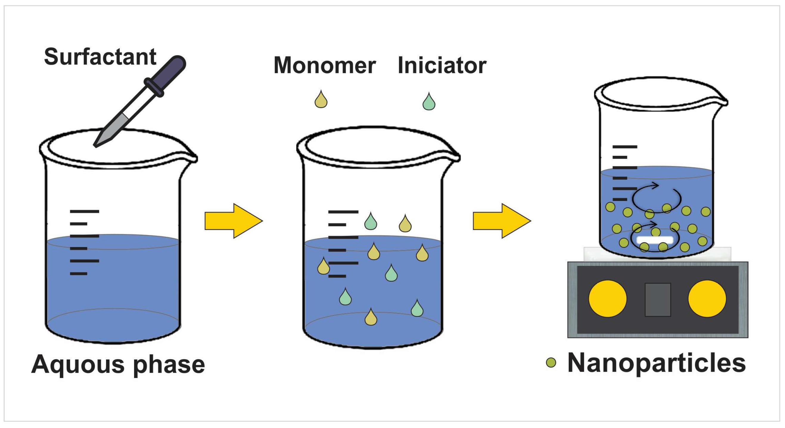
3. Nanoparticles’ Preparation and Drug Loading

Nanoparticles have great potential for use in many different applications. These include manufacturing, materials, energy harvesting, electronics, the environment, mechanical industries, and medications. The biomedicine research field in particular takes advantage of the different physicochemical properties of the nanoparticles [41].
The structures of these nanoparticles are more complex than a molecule; their composition is organized into three layers: the surface, the shell, and the core. This structure provides the nanoparticles with desirable qualities, such as the combination of different materials and the association of molecules at the surface layer [42,43]. Combining more than one material can provide the nanoparticles with unique properties. Additionally, the variety of blend compositions and formulations make nanoparticles with variable drug delivery capabilities to incorporate hydrophobic and hydrophilic molecules with different molecular weights to control their release [44,45].
Polymeric nanoparticles have been developed with different antineoplastic drugs, such as paclitaxel, docetaxel, cisplatin, 5-fluorouracil, and doxorubicin, among others. Depending on the required needs of the polymeric nanocarrier and the drug to be loaded, the most appropriate type of method for the development of the nanoparticles is chosen. There are several methods to prepare organic nanoparticles. They can be prepared from preformed polymers or by polymerization of monomers [46,47].
As mentioned previously, the electrodynamic method can be used to prepare nanoparticles. However, the most frequently used techniques for the preparation of nanoparticles from polymeric materials are the emulsion-evaporation and emulsion-diffusion techniques [47,48,49,50].
Regarding the synthesis of nanoparticles from polymerization of monomers, emulsion and suspension polymerizations are of great importance in industrial applications as they do not harm the environment, it is easy to remove the heat of reaction during polymerization, and they ensure feasible handling of the final product with low viscosity [51,52] (Figure 2). Table 1 presents the methods of synthesis and characteristics of the nanoparticles studied in the hybrid systems reported.
The current limitations of nanoparticles include their high operational cost, limited suitability (for laboratory use only), and expertise requirement. The characteristics of polymeric materials used to develop nanoparticles, such as the acidity of poly(D,L-lactide-co-glycolide) (PLGA), make it less suitable for carrying drugs and bioactive molecules [53].
Some polymers chosen as raw material for the encapsulation of bioactive compounds are poly(ethylene oxide) (PEO), poly(vinyl alcohol) (PVA), poly(caprolactone) (PCL), and poly(vinyl pyrrolidone) (PVP), which have low toxicity.
Encapsulated compounds are of interest to the food industry, as well as to the pharmaceutical industry, such as the polyphenols. Their antioxidant activities make them attractive to be encapsulated to maintain their bioactivity [53,54].
Substances that are prime candidates for encapsulation are unstable compounds, as they tend to oxidize easily, especially if they are exposed to light, heat, or oxygen. Hydrophobic compounds are also candidates for encapsulation since they have some limitations regarding integration with formulations of either food or drug delivery systems. Encapsulation with the electrospinning technique seeks to protect substances from an aggressive environment, to be released over a long period or at specific sites [18,27].
Another use of encapsulation is in the administration of different drugs at the same time and in the same place.
There are different techniques for the encapsulation of bioactive molecules, such as spray drying, phase separation, emulsion processes, and direct blend electrospinning. They all have different drawbacks. To overcome these barriers, researchers have developed nanofibers for encapsulation made from coaxial electrospinning tailored to drug specificities and delivery system requirements [55]. This is the case with the dual drug delivery system for tissue regeneration used by Mohamady Hussein et al. They developed core-shell type nanofibers using coaxial electrospinning, incorporating phenytoin into the shell, and using the drug as a proliferative agent to help cell regeneration. PCL was chosen for the shell material due to the hydrophobic nature of the drug, with the intention of controlled release and improved mechanical properties. A hydrophilic core composed of PVA was selected for the nanofibers. Since the humidity of the microenvironment is proposed to increase the efficiency of wound healing, silver nanoparticles with chitosan were incorporated as biocidal agents [56,57]. Chitosan also has the quality of promoting the formation of collagen when it breaks down in contact with wounds, which also helps wound healing [58]. The latter is an innovative scaffold system with multiple applications and specific functions.
Table 1. Summary of the nanoparticles in hybrid systems. Applications, synthesis, size, and loading method used.
NanoparticlesApplication (Advantage) Synthesis MethodSize Diameter NanoparticlesLoading Method and Drug LoadedReferences
Bacterial cellulose whisker (BCW NP) Increase the filler density in composite nanofibersPlasma treatment using a microwave ovenNanoparticles of an average size of 49.1 ± 13 nmWithout drug[59]
functionalized bacterial cellulose whisker (f-BCW NP)
Increase the filler density in composite nanofibers. Increased the glass transition temperaturePlasma treatment using a microwave ovenNanoparticles of an average size of 49.1 ± 13 nmWithout drug[59]
L-lysine based nanogelIncreased in varying degrees. The response to ammoniaIn vitro enzymatic biodegradation of 4-Lys-macrogelNanogels size was about 60 nm with a narrow size distributionWithout drug[33]
Chitosan (CS)Antibacterial activity Oil-in-water (o/w) type emulsion, ionic gelation Average particle size of the five nanoparticles ranged from 94.3 ± 2.1 to 246.1 ± 6.3 nm with the in- crease of chitosan concentration.Oil-in-water (o/w) type emulsion.
Moringa oil
[60]
ChitosanImprovement in the morphology, thermal, mechanical, antibacterial properties and cytobiocompatibiliy
Biocidal agents to potentiate the wound healing process
Nanoparticles thermally synthesized with chitosan acting as both a capping and reducing agentAverage particle size of 53.6 ± 20.5 nmThermally synthesized
Silver
[56]
ChitosanWound dressing, wound healing with antibacterial, antioxidant, and cell proliferation propertiesElectrosprayedCurcumin (CUR) loaded into chitosan nanoparticles average diameter: 32.17 nmNanoencapsulation of curcumin by emulsified curcumin-chitosan solution [57]
Poly(N-isopropylacrylamide-acrylic acid) Promote the wound healing process to achieve higher wound healing efficacyFree radical nanoprecipitation polymerizationAverage size ~100 nmWithout drug [61]
LigninCell viability and differentiation, along with neurite length extension were promotedCommercially availableAverage size ~90 nmWithout drug [62]
LigninBone tissue engineeringPrecipitation, via a simultaneous pH and solvent shifting technologyAverage size count at 100–200 nm rangeWithout drug [63]
KeratinPotential neural tissue applicationsElectrospray depositionAverage size 250–350 nm rangeWithout drug [64]
Carboxymethyl-hexanoyl/chitosanIncreased its cytotoxic effect in melanoma cellsIonotropic gelationAverage size of 32.6 ± 1.2 nmPyrazoline. In situ [65]
Chitosan-aniline nanogelsBactericideComplexation-reductionAverage size of 78 ± 19 nmSilver nanoparticles
In situ
[66]
Chitosan-tripolyphosphateEncapsulation and release of therapeutic proteinsIonotropic gelation Average size 194 ± 3 nmTransforming Growth Factor β3 (TGF-β3) In situ [67]
Carboxymethyl chitosanExhibits antibacterial properties and promotes skin wound healingElectrostatic dropletAverage size 164.6 ± 5 nmEncapsulates antibacterial peptide (OH-CATH30). In situ[23]
Chitosan Vaginal controlled release of benzydamineIonic gelationAverage size 128–710 nm range. (Depending on the formulation)Benzydamine. In situ [68]
Chitosan Antibacterial. Encapsulation of antibacterial agentIonic gelationAverage size 10–25 nm rangeAntibiotics (e.g., ciprofloxacin, ofloxacin, levoxacin, gemifloxacin). Absorption [69]
Poly (methacrylic acid)Peripheral nerve regeneration.Suspension polymerization. Molecular imprintedAverage size of 80–115 nm range4-aminopyridine (4-AP).
Entrapping the 4-AP molecules via hydrogen bond formation.
[54]
Poly (lactic-co-glycolide) Topical vaginal drug deliveryNanoprecipitation. Passive PEGylation with Pluronic®Average size 172 ± 19 nmRhodamine, directly conjugated.
Antiretroviral drug etravirine (ETR). In situ
[70]
ChitosanAntibacterial, mucoadhesive, encapsulation of drugIonic gelationAverage size ~300–400 nmEpinephrine. In situ [71]

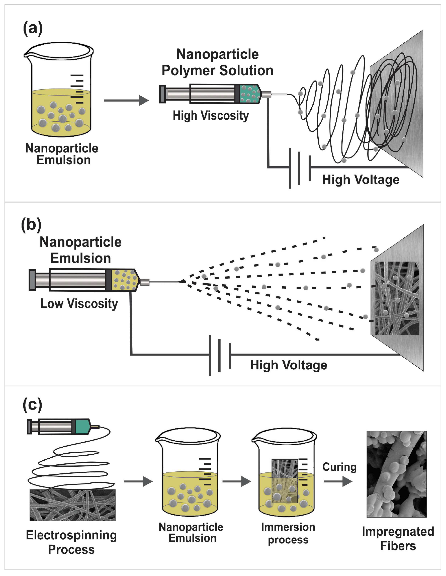
4. Incorporation of Nanoparticles into Nanofibers

The first nanofiber systems with polymeric nanoparticles mentioned in the literature was reported by Yoon, who studied hybrid systems of bacterial cellulose nanoparticles (BCW NPs) and functionalized bacterial cellulose nanoparticles (f-BCWNPs) in poly(ethylene oxide (PEO) nanofibers. TEM analysis demonstrated the embedding of nanoparticles in the nanofibers, but there was agglomeration and deformation of the embedded nanoparticles [59].
For the incorporation of nanoparticles in nanofibers, three main techniques were identified. The most commonly mentioned one is direct blending electrospinning, where the nanoparticles are encapsulated and/or entrapped in the nanofibers. For this purpose, the nanoparticles are incorporated by mixing them in the solution together with the base polymer solution before being electrospun (Figure 3a). Another way to encapsulate nanoparticles is through coaxial electrospinning, in which core-shell type nanofibers are created. These follow the same principle as the previous ones; the nanoparticles are mixed in the solution before being electrospun, but the difference is that two solutions are spun simultaneously using a needle with two different capillaries [72]. Another approach is to attach the nanoparticles to the surface of nanofiber mats by directly depositing them on previously prepared nanofiber mats by means of the electrospray technique (Figure 3b). The third option is to impregnate the nanoparticles in the preformed nanofibers’ mats by immersing the scaffolds in solutions containing nanoparticles (Figure 3c) [71,73].

5. Hybrid Systems for Control of Reactive Oxygen Species for Tissue Regeneration

The enzymes responsible for maintaining a balanced and adequate homeostasis in the skin are particular, providing focused mechanisms for redox mediation in homeostasis. This redox mediation system involves some antioxidants of organic origin and enzymes such as glutathione, peroxidases, catalase, superoxide dismutase, and thioredoxins [74]. Homeostasis is an essential process that keeps cell health optimal. Reactive oxygen species (ROS), such as hydroxyl radicals, singlet oxygen, peroxides, and superoxide, are actively involved in biochemical processes such as angiogenesis, collagen synthesis, epithelialization, and body protection against infectious pathogens [75].
In damaged tissue, a series of inflammatory processes occur that lead to tissue repair, but if the tissue is pathological, such as in cases of immunosuppression, diabetes, or advanced age, the proinflammatory cytokines generated by immune cells do not generate ROS appropriately. Since ROS also participates in the translation of signals in response to cellular oxidative stress, they can generate an imbalance in the cellular microenvironment. If ROS levels are inadequate, this can be counterproductive for tissue healing. Excessive ROS for prolonged periods is also harmful and generates signals that lead to cell apoptosis. One alternative to improving the regeneration of damaged tissue is to control ROS levels through biomaterials and nanotechnology [76,77,78].
Zhang et al., developed a hybrid system made of nanofibers with nanogels evenly distributed in the membrane. The system is fabricated using the airbrush technique with poly (L-lactic acid) (PLLA) nanofibers and poly (N-isopropylacrylamide-acrylic acid) (PNA) redox-sensitive nanogels that act as a reservoir of disulfide bonds for the mediation of redox potential to promote the wound healing process. They demonstrated that the incorporation of nanogels helped the healing of skin wounds, both in healthy tissue and diabetic tissue [61].
The excessive generation of ROS is detrimental to the regeneration of nervous tissue; it has been shown that materials such as PCL induce an inflammatory response that generates too much ROS, causing tissue damage. To solve this problem, it is necessary that the material used in the nanofibers have antioxidant properties, although PCL showed inferior antioxidant activity [79]. Nerve cells would create oxidases of nicotinamide adenine dinucleotide phosphate and ROS, resulting in DNA necrosis. Nervous tissue, involved in a microenvironment with H2O2, increases the amounts of ROS intracellularly, which increases oxidative stress and apoptosis. The oxidation diffusion reaction can end with some functional groups, such as methoxyl and hydroxyl, that provide hydrogen, inhibiting cell death by increasing ROS [80].
Some of the research focused on tissue engineering and wound healing have incorporated lignin into their studies due to its antioxidant capacity. This is because it scavenges oxygen free radicals and can stabilize the reactions initiated by these radicals [81]. With this, the issue of low antioxidant capacity in PCL nanofiber systems can be overcome. The project by Amini et al., focused on the regeneration of nervous tissue. They found in vivo and in vitro that PCL nanofibers with lignin nanoparticles in different concentrations promoted viability and cell differentiation by increasing the percentage of lignin nanoparticles in the PCL nanofibers. The expression of neuronal markers for differentiated cells was also increased with a higher percentage of lignin nanoparticles incorporated into the PCL nanofibers. Regarding the in vivo groups that showed better regeneration, 15% lignin nanoparticles were incorporated, demonstrating that their hybrid nanofiber/nanoparticle system has excellent potential to be used in the cellular regeneration of nervous tissue [62]. The potential of hybrid systems of PCL/lignin nanofibers and nanoparticles, respectively, is not only reduced to the regeneration of nervous tissue. In the work by Haider et al., the properties of lignin were exploited, synthesizing nanoparticles to take advantage of its antimicrobial qualities since the oxygen contained in the functional groups of its phenolic parts (–OH, –CO, COOH) present resistance against multiple microorganisms [82]. They developed PCL nanofiber scaffolds with lignin nanoparticles and observed a significant improvement in osteogenic cell differentiation versus pure PCL scaffolds. They integrated the antibacterial and osteostimulative capacities of nanoparticles with the mechanical properties of PCL nanofibers, demonstrating the potential of this type of hybrid nanodevice for bone tissue engineering [63].

6. Materials Used in Hybrid Systems

Several materials have been used as base materials to form the nanofiber mats and incorporate nanoparticles for diverse applications. Findings in the literature are summarized in Table 2 and surveyed in the present section. The structure of the polymers is presented in Figure 4.

6.1. Poly(D,L-Lactide-co-Glycolide) (PLGA)

PLGA nanofibers made by the electrospinning method have a morphology that contributes to nanoparticle retention. PLGA nanofiber plates have been produced to improve efficacy and safety in extended-release systems for analgesics, especially for local analgesia [83]. Another way in which nanofibers can be used to improve the bioavailability of a drug is in gastroretentive delivery systems. This is due to properties such as mucoadhesion, which provides longer exposure time of the drug within the intestinal tract [84]. The goals of an effective delivery system include the delivery of a specific amount of drug to a target site for a defined period. Despite the many advantages of nanofiber mats created by electrospinning, in a delivery system based on nanofibers, the drug mixed or encapsulated in the nanofiber matrix has one drawback when it comes to delivery: the “burst release” of the drug in the pharmacokinetic profile. This occurs when the drug is weakly anchored to the large surface of the polymeric mat. This problem can be solved by encapsulating the drugs within nanoparticles and then loading the nanoparticles into electrospun mats [85,86].

6.2. Keratin/Poly (Vinyl Alcohol) (PVA)

Biocompatibility is another highly desirable characteristic in delivery systems. Keratin combined with polymeric nanofibers is one option that has been investigated for this purpose. Another option involves coating nanofibers with keratose, a biomaterial that is extracted from human hair fibers by protein oxidation [87]. Oxidized keratins or keratose nanoparticles negatively compromised the mechanical properties of keratin/PVA blend nanofibers, keratins exhibited serious deficiencies, including low viscosity and complications with the electrospinning process. A new method of keratin modification was developed: PVA nanofibers coated with keratin nanoparticles were prepared by electrospray deposition after electrospinning, resulting in a simultaneous improvement of their mechanical properties and biocompatibility [64,88].

6.3. Chitosan

The use of natural polymers (also known as polysaccharides) offers antimicrobial, antioxidant, and anti-inflammatory features that help cell proliferation and decrease infections, all of which provides an improvement in the wound healing process. They are excellent biomimetics due to their biocompatibility and null response from the immune system [89].
Chitin and its deacetylated form, chitosan, have biocompatible and biodegradable properties, among other desirable characteristics in the field of biomedicine. Chitosan’s characteristics include non-toxicity, antibacterial activity, the ability to form films, and the chelation of metal ions. This is attributed to the presence of free protonatable amino groups along the chitosan skeleton, which provides rigidity due to the repulsive forces between the protonated -NH2 groups. Unfortunately, this property limits its electrospinning capacity. For this reason, chitosan nanofibers are combined with other more flexible materials, such as PEO, to form electrospun mats through coaxial-electrospinning [90,91].
The rigid backbone and high molecular weights make chitosan solutions very viscous, so the electric field is stretched. To reduce surface tension, researchers use acetic acid (90%) as a concentrated solvent and a combination of solvents such as 1,1,1,3,3,3-hexafluoro-2-propanol, trifluoroacetic, and dichloromethane; these are used for the development of chitosan-based electrospun nanofibers. To crosslink the nanofibers, the authors use glutaraldehyde in steam after electrospinning, which helps preserve the fibers’ structure when the scaffolds are immersed in aqueous media. Another of the strategies used to produce soft nanofibers was to use blends of chitin or chitosan with other polymers such as PVA or PEO [92]. The change in the surface of the nanofibers, either with the immobilization or functionalization of other nanomaterials (nanogels, nanocapsules, liposomes, and nanoparticles in general), results in scaffolds with new capacities, features, properties, or functions. These intelligent nanomaterials can have photocatalytic, self-cleaning, or antimicrobial characteristics that are activated by stimuli; light absorption, for example [93]. Ballesteros et al., developed, characterized, and compared different combinations of smart nanomaterials. They used PCL nanofibers as a base, which were functionalized with a combination of photosensitive aniline-chitosan nanogels and silver nanoparticles to obtain a hybrid device with antimicrobial characteristics and the ability to control release response after being exposed to laser light [66]. As the literature indicates, chitosan nanoparticles can be formed by ionotropic gelation. Well-defined nanogels have also been reported, using tripolyphosphate as a crosslinker, with potential for the development of drug delivery systems. Combining the positive zeta potential with polyanionic biopolymers can form hydrogels by deposition of the nanogels layer by layer for a multilayer coating that dries into a homogeneous film [69].
Hydrogels with a porous structure that allow a large volume of water have also been reported, which provide a more favorable environment to encapsulate biological products such as cells and proteins [94]. The composite systems also provide protection against degradation [95].
A different system for supplying drugs was developed, protecting active peptides from proteases and properly releasing these peptides. It was reported that a ready-made biodegradable drug nanoparticle delivery system based on carboxymethyl chitosan (CMCS) can promote the sustained release of the antibacterial peptide OH-CATH30 (OH-30). The investigation results also showed that the prepared CMCS-OH-30 nanoparticles sped healing and reduced skin injury time in mice studies [23].
Fatmanur Tuğcu-Demiröz et al., developed a hybrid system of nanofibers loaded with chitosan nanoparticles for controlled vaginal release of benzydamine. They obtained the controlled release of the drug with formulations loaded with nanoparticles. Although gel formulations had better mucoadhesive properties, the nanoparticle-loaded nanofiber formulations provided greater drug penetration through vaginal tissue. The results showed that the increased surface area, high solubility, high wettability, and porosity of nanofibers containing dispersed nanoparticles were parameters important for the effectivity of the system [68].
Felipe et al., developed a chitosan/PEO nanofiber drug delivery scaffold system with carboxymethyl-hexanoyl chitosan-dodecyl sulfate (CHC-SDS-H3) polymeric nanoparticles loaded with H3TM04 (1-(5-(naphthalen-2-yl)-3-(3,4,5-trimethoxyphenyl)-4,5-dihydro-1H-pyrazol-1-yl) ethenone). The literature mentions that pyrazoline compounds have a wide spectrum of pharmacological activity as anti-inflammatory, antibacterial, antifungal, antipyretic, analgesic, antiviral, antioxidant, anti-angiogenic, and anti-tumor compounds. The drug is of great importance for skin cancer therapy; studies have shown that it induces apoptosis in tumor cells, which results in the reduction or elimination of the tumor. In conjunction with nanofiber systems, which exhibit similar properties to the natural fibrillar structure, hybrid systems could be used for the controlled release of drugs in the treatment of local anti-melanoma chemotherapy [65,96].

6.4. Poly(Vinyl Alcohol) (PVA)

Pour Khalili et al., directed their contributions toward supporting the reduction of risks associated with the inappropriate use of antibiotics that create bacterial resistance. They opted to work with nanofibers made of polyvinyl alcohol (PVA), which were functionalized with tripolyphosphate (TPP)/chitosan nanogels. These nanogels were loaded with the antibiotic gemifloxacin. The nanogels were electrosprayed to be immobilized on the nanofiber mat, obtaining a hybrid release system in which the drug is encapsulated with the aim of being used as an antibacterial for wound dressing with a constant and controlled release of the drug [68,69,97].

6.5. Poly(Vinyl Pyrrolidone) (PVP)

Materials such as polyvinylpyrrolidone are used for their biocompatibility in the electrodynamic method to create nanomaterials. This polymer can be mixed with other biomaterials, for example, PCL, in such a way that nanofibers can be developed for healing tissues, helping to dress wounds and supporting their healing [97,98].
PVP nanofibers with mucoadhesive properties can increase the interaction with the vaginal mucosa and release the drug over the required time. One of its qualities widely mentioned in the literature is mucoadhesion, which is used to overcome issues such as low drug availability in vaginal applications since its mucoadhesive characteristic provides a great advantage by increasing interaction with the vaginal mucosa and releasing the drug over the required time. Hybrid nanofiber/nanoparticle systems are considered more suitable for vaginal formulations, as they achieve high permeability through the vaginal mucosa and controlled release.
Krogstad et al., studied models of hybrid delivery systems of PVP nanofibers and PVA nanofibers incorporating PLGA nanoparticles loaded with a fluorescent compound. Their study confirms that these delivery systems have the advantage of avoiding problems in topical vaginal application, such as low retention and leakage [70].

6.6. Poly(Caprolactone) (PCL)

Poly (caprolactone) (PCL) is another synthetic polymer, and was used in the work of Sundhari et al., where created PCL nanofibers proved to be a good scaffold for the promotion of cell differentiation. The structure of these nanofibers presented a smooth and uniform diameter, and with certain wettability of the nanofibers, there is potential to facilitate neurite outgrowth, promoting cell viability by including lignin nanoparticles in the PCL nanofibers [99].

6.7. Poly(Vinylidene Fluoride) (PVDF)

Piezoelectric scaffolds implanted at a bone defect or injury site trigger electrical stress-induced stimulus through physiological loads. Such hyperpolarization activates calcium channels with cell membrane voltage. Intracellular Ca2+ ions play an essential role in cell proliferation.
Polyvinylidene fluoride (PVDF) and its composites are biocompatible piezoelectric materials that show high efficiency for bone regeneration. The beta phase is the crystalline phase of PVDF with the most piezoelectric properties. Research showed that the electrospinning method is the most efficient for fabricating the 3D porous structure of the piezoelectric b-PVDF. The porous structure formed and the polarization improves biocompatibility growth in PVDF scaffolds. Different nanoparticles have also been incorporated in PVDF scaffolds to improve osteoinductive and mechanical properties. These include antimicrobial, antioxidant, and anti-inflammatory functionalities. Among the materials studied are hydroxyapatite, graphene oxide, ZnO, and polyhedral oligomeric silsesquioxane–epigallocatechin gallate conjugate [100].

6.8. Polymer Mixtures

Nanofibrous scaffolds based on mixtures of poly (L-lactide-D, L-lactide)/poly (acrylic acid) [PLDLLA/PAAc] in the presence of poly (2-hydroxyethyl methacrylate) have been prepared as molecularly imprinted polymer nanoparticles to enhance osteogenesis. By adding 10% by weight of PAAc to PLDLLA and using the response surface technique, the average diameter of the electrospun nanofibers obtained was 237 nm and increased the osteogenesis yield of optimized nanofiber scaffolds. In general, by minimizing the diameter of nanofibers, their specific surface area increases. Furthermore, the minimal size, together with a uniform morphology, of the non-beaded nanofibers leads to increased cell adhesion, improved drug loading, and controlled release [54,101,102].

7. Conclusions

Hybrid systems of polymeric nanofibers and nanoparticles represent the potential to improve the characteristics required for their biomedical use. They are also of interest in the food industry since they can be prepared with biocompatible and biodegradable polymeric materials.
The combination of these nanometric-dimensioned materials (nanofibers and polymer-based nanoparticles) provides advantages for being used together as hybrid systems and enhancing or developing properties that are not present individually. These properties include increases in surface-volume ratio, drug encapsulation, protection of proteins and bioactive substances sensitive to degradation, and improved cost-benefits of their development, among others. In the case of bio-drugs, the hybrid systems exhibit unique pharmacokinetics and pharmacodynamics.
The main application of nanoparticle/nanofiber hybrid systems is the avoidance of drug “burst release,” which occurs in systems using individual technologies. Most hybrid systems studied have applications for achieving sustained release of hydrophilic drugs.
Electrospun mats have mucoadhesive properties that improve residence times at the delivery site. Wound healing and tissue engineering scaffolds help cell proliferation and contribute to an improvement in both aesthetic and functional healing. The combination of mats with drug-loaded nanoparticles helps with greater penetration into the tissue of interest, such as vaginal or buccal delivery. Some materials are also useful due to their antibacterial properties, as in the case of chitosan nanoparticles.
Techniques used for mixing or incorporating polymeric nanoparticles into polymeric nanofiber mats, such as electrospinning, electrospray, and coaxial electrospinning, are widely studied techniques. This new information provides advantages when developing this type of hybrid nanotechnology.
A feasible combination of both techniques and materials must be sought. One complication is that some polymeric materials are difficult to electrospin. In the same way, other difficulties arise in investigating these systems; for example, the change in the mechanical properties of the nanofibers, the agglomeration of nanoparticles at the time of their incorporation into the mats, and irregular distribution issues. These limitations must be overcome in order to reveal their great potential for future research.
Researchers can take advantage of the characteristics of different polymeric materials that are used in these hybrid systems and may be of interest for future research in which the most convenient combination of polymeric materials is chosen for the desired purpose.
Electrospun mats share properties with hydrogels (three-dimensional crosslinked polymeric networks), such as acting as templates, exudate-absorbents, breathable barriers, and controlling drug diffusion. Hydrogels, for instance, are widely used in biomedical applications such as tissue engineering and drug delivery. Hydrogels can be produced more quickly compared to electrospun mats. Hydrogels can also be responsive to temperature, solvent, and bioelectricity, which makes them suitable for bionic skin. Recent studies relate to the 3D printing of hydrogels to be used in robotics, with promising results. This is not the case with electrospun mats, where applications are limited to tissue engineering and drug delivery. Setup for electrospinning is simple, however, and cost-efficient compared to 3D printing. One disadvantage of hydrogels is that they are prepared by the polymerization of monomers and crosslinkers, which can leach if unreacted and cause toxicity. For the preparation of hydrogels from biopolymers, the latter must be derivatized with polymerizable moieties for crosslinking. On the other hand, electrospun mats are prepared from prefabricated biocompatible and biodegradable polymers [103,104,105,106,107].
The incorporation of nanoparticles in hydrogels produces entanglement of the nanoparticles within the hydrogel network. Nanoparticles in hydrogels improve the mechanical properties of the material, but the advantages of nanoparticles are not observed in these systems, such as avoiding burst release of loaded drugs or improving penetration into tissues [108,109,110].
One of the major drawbacks for the application of nanofibrous mats in nanomedicine is the reduced number of in vivo studies in humans [111]. We were unable to find studies of biocompatibility and/or effectivity in volunteers for nanoparticle/nanofiber systems. Most of the reported systems require further investigations before products are ready for commercialization.

Author Contributions

Conceptualization, A.S.-M. and J.M.C.-B.; methodology, H.Y.V.-M. and G.L.P.-G.; software, G.L.P.-G.; validation, K.P.-V., E.A.L.-M. and L.J.V.-G.; formal analysis, J.M.C.-B.; investigation, K.P.-V.; resources, A.S.-M.; data curation, L.J.V.-G.; writing—original draft preparation, H.Y.V.-M.; writing—review and editing, J.M.C.-B. and L.J.V.-G.; visualization, K.P.-V.; supervision, E.A.L.-M.; project administration, A.S.-M.; funding acquisition, A.S.-M. All authors have read and agreed to the published version of the manuscript.

Funding

This research was funded by 23rd Internal Call for Research Projects Support UABC, grant number 4295.

Institutional Review Board Statement

Not applicable.

Informed Consent Statement

Not applicable.

Data Availability Statement

Not applicable.

Acknowledgments

H.Y.V.M. thanks CONACYT for the Ph.D. studies fellowship (No. 765709), and FCQI-UABC. Thanks to Albert L. Brown for proofreading the manuscript.

Conflicts of Interest

The authors declare that they have no known competing financial interests or personal relationships that could have appeared to influence the work reported in this paper.

References

  1. Li, J.; Zhao, J.; Tan, T.; Liu, M.; Zeng, Z.; Zeng, Y.; Zhang, L.; Fu, C.; Chen, D.; Xie, T. Nanoparticle drug delivery system for glioma and its efficacy improvement strategies: A comprehensive review. Int. J. Nanomedicine 2020, 15, 2563–2582. [Google Scholar] [CrossRef] [PubMed] [Green Version]
  2. Nikam, A.P.; Ratnaparkhiand, M.P.; Chaudhari, S.P. Nanoparticles—An overview. Int. J. Res. Dev. Pharm. Life Sci. 2017, 3, 1121–1127. [Google Scholar]
  3. Vlachopoulos, A.; Karlioti, G.; Balla, E.; Daniilidis, V.; Kalamas, T.; Stefanidou, M.; Bikiaris, N.D.; Christodoulou, E.; Koumentakou, I.; Karavas, E.; et al. Poly(Lactic Acid)-Based Microparticles for Drug Delivery Applications: An Overview of Recent Advances. Pharmaceutics 2022, 14, 359. [Google Scholar] [CrossRef] [PubMed]
  4. Villarreal-Gómez, L.J.; Cornejo-Bravo, J.M.; Vera-Graziano, R.; Grande, D. Electrospinning as a powerful technique for biomedical applications: A critically selected survey. J. Biomater. Sci. Polym. Ed. 2016, 27, 157–176. [Google Scholar] [CrossRef] [PubMed]
  5. Cornejo-Bravo, J.M.; Villarreal-Gomez, L.J.; Serrano-Medina, A. Electrospinning for Drug Delivery Systems: Drug Incorporation Techniques. In Electrospinning: Material, Techniques, and Biomedical Applications; InTech: Rijeka, Croatia, 2016. [Google Scholar] [CrossRef] [Green Version]
  6. Puttananjegowda, K.; Takshi, A.; Thomas, S. Silicon carbide nanoparticles electrospun nanofibrous enzymatic glucose sensor. Biosens. Bioelectron. 2021, 186, 113285. [Google Scholar] [CrossRef]
  7. Goyal, R.; Macri, L.K.; Kaplan, H.M.; Kohn, J. Nanoparticles and nano fi bers for topical drug delivery. J. Control. Release 2016, 240, 77–92. [Google Scholar] [CrossRef] [Green Version]
  8. Jafarpour, M.; Aghdam, A.S.; Koşar, A.; Cebeci, F.Ç.; Ghorbani, M. Electrospinning of ternary composite of PMMA-PEG-SiO2 nanoparticles: Comprehensive process optimization and electrospun properties. Mater. Today Commun. 2021, 29, 102865. [Google Scholar] [CrossRef]
  9. Zhang, M.; Song, W.; Tang, Y.; Xu, X.; Huang, Y.; Yu, D. Citation: Polymer-Based Nanofiber-Nanoparticle Hybrids and Their Medical Applications. Polymers 2022, 14, 351. [Google Scholar] [CrossRef]
  10. Chen, L.; Li, W.; Fang, K.; Liu, S.; Wu, J.; Luo, J.; Tyler, R.; Zhang, S. OQL036 topical gel inhibits the skin toxicity associated with 5-fluorouracil/capecitabine: Results from in vitro and in vivo preclinical studies. Eur. J. Cancer 2022, 174, S77–S78. [Google Scholar] [CrossRef]
  11. Liu, W.; Bi, W.; Sun, Y.; Wang, L.; Yu, X.; Cheng, R.; Yu, Y.; Cui, W. Biomimetic organic-inorganic hybrid hydrogel electrospinning periosteum for accelerating bone regeneration. Mater. Sci. Eng. C 2020, 110, 110670. [Google Scholar] [CrossRef]
  12. Abid, S.; Hussain, T.; Raza, Z.A.; Nazir, A. Current applications of electrospun polymeric nanofibers in cancer therapy. Mater. Sci. Eng. C 2019, 97, 966–977. [Google Scholar] [CrossRef] [PubMed]
  13. Soares, D.C.F.; Poletto, F.; Eberhardt, M.J.; Domingues, S.C.; De Sousa, F.B.; Tebaldi, M.L. Polymer-hybrid nanosystems for antiviral applications: Current advances. Biomed. Pharmacother. 2021, 146, 112249. [Google Scholar] [CrossRef] [PubMed]
  14. Rojas-Aguirre, Y.; Aguado-Castrejón, K.; González-Méndez, I. La nanomedicina y los sistemas de liberación de fármacos: ¿la (r)evolución de la terapia contra el cáncer? Educ. Quim. 2016, 27, 286–291. [Google Scholar] [CrossRef] [Green Version]
  15. Sayin, S.; Tufani, A.; Emanet, M.; Genchi, G.G.; Sen, O.; Shemshad, S.; Ozdemir, E.; Ciofani, G.; Ozaydin Ince, G. Electrospun Nanofibers With pH-Responsive Coatings for Control of Release Kinetics. Front. Bioeng. Biotechnol. 2019, 7, 309. [Google Scholar] [CrossRef]
  16. Drosou, C.; Krokida, M.; Biliaderis, C.G. Encapsulation of β-carotene into food-grade nanofibers via coaxial electrospinning of hydrocolloids: Enhancement of oxidative stability and photoprotection. Food Hydrocoll. 2022, 133, 107949. [Google Scholar] [CrossRef]
  17. Sharif, N.; Golmakani, M.T.; Niakousari, M.; Ghorani, B.; Lopez-Rubio, A. Food-grade gliadin microstructures obtained by electrohydrodynamic processing. Food Res. Int. 2019, 116, 1366–1373. [Google Scholar] [CrossRef] [PubMed]
  18. Khoshnoudi-Nia, S.; Sharif, N.; Jafari, S.M. Loading of phenolic compounds into electrospun nanofibers and electrosprayed nanoparticles. Trends Food Sci. Technol. 2020, 95, 59–74. [Google Scholar] [CrossRef]
  19. Batty, C.J.; Gallovic, M.D.; Williams, J.; Ross, T.M.; Bachelder, E.M.; Ainslie, K.M. Multiplexed electrospray enables high throughput production of cGAMP microparticles to serve as an adjuvant for a broadly acting influenza vaccine. Int. J. Pharm. 2022, 622, 121839. [Google Scholar] [CrossRef]
  20. Chen, N.; Gan, Y.; Luo, Y.; Jiang, Z. A review on the technology development and fundamental research of electrospray combustion of liquid fuel at small-scale. Fuel Process. Technol. 2022, 234, 107342. [Google Scholar] [CrossRef]
  21. Zhou, H.; Modi, S.; Biswas, P. Controlled synthesis of charged lignin nanocarriers by electrospray. Colloids Surfaces A Physicochem. Eng. Asp. 2022, 648, 129314. [Google Scholar] [CrossRef]
  22. Ma, Z.; Ren, L.; Ying, D.; Jia, J.; Shao, J. Sustainable electrospray polymerization fabrication of thin-film composite polyamide nanofiltration membranes for heavy metal removal. Desalination 2022, 539, 115952. [Google Scholar] [CrossRef]
  23. Zou, P.; Lee, W.H.; Gao, Z.; Qin, D.; Wang, Y.; Liu, J.; Sun, T.; Gao, Y. Wound dressing from polyvinyl alcohol/chitosan electrospun fiber membrane loaded with OH-CATH30 nanoparticles. Carbohydr. Polym. 2020, 232, 115786. [Google Scholar] [CrossRef] [PubMed]
  24. Haider, A.; Haider, S.; Kang, I.K. A comprehensive review summarizing the effect of electrospinning parameters and potential applications of nanofibers in biomedical and biotechnology. Arab. J. Chem. 2018, 11, 1165–1188. [Google Scholar] [CrossRef]
  25. ur Rehman Khan, A.; Huang, K.; Khalaji, M.S.; Yu, F.; Xie, X.; Zhu, T.; Morsi, Y.; Jinzhong, Z.; Mo, X. Multifunctional bioactive core-shell electrospun membrane capable to terminate inflammatory cycle and promote angiogenesis in diabetic wound. Bioact. Mater. 2021, 6, 2783–2800. [Google Scholar] [CrossRef]
  26. Khodadadi, M.; Alijani, S.; Montazeri, M.; Esmaeilizadeh, N.; Sadeghi-Soureh, S.; Pilehvar-Soltanahmadi, Y. Recent advances in electrospun nanofiber-mediated drug delivery strategies for localized cancer chemotherapy. J. Biomed. Mater. Res. Part A 2020, 108, 1444–1458. [Google Scholar] [CrossRef]
  27. Christy, P.N.; Basha, S.K.; Kumari, V.S.; Bashir, A.K.H.; Maaza, M.; Kaviyarasu, K.; Arasu, M.V.; Al-Dhabi, N.A.; Ignacimuthu, S. Biopolymeric nanocomposite scaffolds for bone tissue engineering applications—A review. J. Drug Deliv. Sci. Technol. 2020, 55, 101452. [Google Scholar] [CrossRef]
  28. Pant, B.; Park, M.; Park, S.J. Drug delivery applications of core-sheath nanofibers prepared by coaxial electrospinning: A review. Pharmaceutics 2019, 11, 305. [Google Scholar] [CrossRef] [Green Version]
  29. Villarreal-Gómez, L.J.; Serrano-Medina, A.; Torres-Martínez, E.J.; Perez-González, G.L.; Cornejo-Bravo, J.M. Polymeric advanced delivery systems for antineoplasic drugs: Doxorubicin and 5-fluorouracil. e-Polymers 2018, 18, 359–372. [Google Scholar] [CrossRef]
  30. Zhang, W.; Guan, X.; Qiu, X.; Gao, T.; Yu, W.; Zhang, M.; Song, L.; Liu, D.; Dong, J.; Jiang, Z.; et al. Bioactive composite Janus nanofibrous membranes loading Ciprofloxacin and Astaxanthin for enhanced healing of full-thickness skin defect wounds. Appl. Surf. Sci. 2023, 610, 155290. [Google Scholar] [CrossRef]
  31. Ismail, H.M.; Ali-Adib, S.; Younes, H.M. Reactive and functionalized electrospun polymeric nanofibers for drug delivery and tissue engineering applications. Ther. Deliv. 2019, 10, 397–399. [Google Scholar] [CrossRef]
  32. Xu, Q.; Ensign, L.M.; Boylan, N.J.; Schön, A.; Gong, X.; Yang, J.C.; Lamb, N.W.; Cai, S.; Yu, T.; Freire, E.; et al. Impact of Surface Polyethylene Glycol (PEG) Density on Biodegradable Nanoparticle Transport in Mucus ex Vivo and Distribution in Vivo. ACS Nano 2015, 9, 9217–9227. [Google Scholar] [CrossRef] [PubMed] [Green Version]
  33. Wu, D.Q.; Wu, L.L.; Cui, H.C.; Zhang, H.N.; Yu, J.Y. A rapid ammonia sensor based on lysine nanogel-sensitized PANI/PAN nanofibers. J. Mater. Chem. B 2016, 4, 1520–1527. [Google Scholar] [CrossRef] [PubMed]
  34. Juncos Bombin, A.D.; Dunne, N.J.; McCarthy, H.O. Electrospinning of natural polymers for the production of nanofibres for wound healing applications. Mater. Sci. Eng. C 2020, 114, 110994. [Google Scholar] [CrossRef] [PubMed]
  35. Xue, J.; Wu, T.; Dai, Y.; Xia, Y. Electrospinning and electrospun nanofibers: Methods, materials, and applications. Chem. Rev. 2019, 119, 5298–5415. [Google Scholar] [CrossRef] [PubMed]
  36. Mele, E. Electrospinning of natural polymers for advanced wound care: Towards responsive and adaptive dressings. J. Mater. Chem. B 2016, 4, 4801–4812. [Google Scholar] [CrossRef] [Green Version]
  37. Rao, S.S.; Nelson, M.T.; Xue, R.; DeJesus, J.K.; Viapiano, M.S.; Lannutti, J.J.; Sarkar, A.; Winter, J.O. Mimicking white matter tract topography using core-shell electrospun nanofibers to examine migration of malignant brain tumors. Biomaterials 2013, 34, 5181–5190. [Google Scholar] [CrossRef] [Green Version]
  38. Carlisle, C.R.; Coulais, C.; Namboothiry, M.; Carroll, D.L.; Hantgan, R.R.; Guthold, M. The mechanical properties of individual, electrospun fibrinogen fibers. Biomaterials 2009, 30, 1205–1213. [Google Scholar] [CrossRef] [Green Version]
  39. Ren, X.; Han, Y.; Wang, J.; Jiang, Y.; Yi, Z.; Xu, H.; Ke, Q. An aligned porous electrospun fibrous membrane with controlled drug delivery—An efficient strategy to accelerate diabetic wound healing with improved angiogenesis. Acta Biomater. 2018, 70, 140–153. [Google Scholar] [CrossRef]
  40. Sharpe, J.M.; Lee, H.; Hall, A.R.; Bonin, K.; Guthold, M. Mechanical properties of electrospun, blended fibrinogen: PCL nanofibers. Nanomaterials 2020, 10, 1843. [Google Scholar] [CrossRef]
  41. Ventola, C.L.; Bharali, D.J.; Mousa, S.A. The Nanomedicine Revolution: Part 1: Emerging Concepts. Pharmacy and Therapeutics. Pharmacol. Ther. 2010, 128, 512–525. [Google Scholar]
  42. Sultana, F.; Manirujjaman; Imran-Ul-Haque; Arafat, M.; Sharmin, S. An overview of nanogel drug delivery system. J. Appl. Pharm. Sci. 2013, 3, 95–105. [Google Scholar] [CrossRef]
  43. Khan, I.; Saeed, K.; Khan, I. Nanoparticles: Properties, applications and toxicities. Arab. J. Chem. 2019, 12, 908–931. [Google Scholar] [CrossRef]
  44. Peres, L.B.; Peres, L.B.; Faria, T.J.; De Assis, J.V.; de Almeida, M.V.; Gonçalves, O.H.; de Araújo, P.H.; Sayer, C. PLLA/PMMA blend in polymer nanoparticles: Influence of processing methods. Colloid Polym. Sci. 2017, 295, 1621–1633. [Google Scholar] [CrossRef]
  45. Mitchell, M.J.; Billingsley, M.M.; Haley, R.M.; Wechsler, M.E.; Peppas, N.A.; Langer, R. Engineering precision nanoparticles for drug delivery. Nat. Rev. Drug Discov. 2021, 20, 101–124. [Google Scholar] [CrossRef] [PubMed]
  46. Zielinska, A.; Carreiró, F.; Oliveira, A.M.; Neves, A.; Pires, B.; Nagasamy Venkatesh, D.; Durazzo, A.; Lucarini, M.; Eder, P.; Silva, A.M.; et al. Polymeric Nanoparticles: Production, Characterization, Toxicology and Ecotoxicology. Molecules 2020, 25, 3731. [Google Scholar] [CrossRef]
  47. Mirchandani, Y.; Patravale, V.B.; Brijesh, S. Solid lipid nanoparticles for hydrophilic drugs. J. Control. Release 2021, 335, 457–464. [Google Scholar] [CrossRef] [PubMed]
  48. Vauthier, C.; Bouchemal, K. Methods for the Preparation and Manufacture of Polymeric Nanoparticles. Pharm. Res. 2009, 26, 1025–1058. [Google Scholar] [CrossRef]
  49. Choukaife, H.; Doolaanea, A.A.; Alfatama, M. Alginate nanoformulation: Influence of process and selected variables. Pharmaceuticals 2020, 13, 335. [Google Scholar] [CrossRef]
  50. Urbaniak, T.; Musiał, W. Influence of solvent evaporation technique parameters on diameter of submicron lamivudine-poly-ε-caprolactone conjugate particles. Nanomaterials 2019, 9, 1240. [Google Scholar] [CrossRef] [Green Version]
  51. Lima, A.L.; Gratieri, T.; Cunha-Filho, M.; Gelfuso, G.M. Polymeric nanocapsules: A review on design and production methods for pharmaceutical purpose. Methods 2022, 199, 54–66. [Google Scholar] [CrossRef]
  52. Elzayat, A.; Adam-Cervera, I.; Álvarez-Bermúdez, O.; Muñoz-Espí, R. Nanoemulsions for synthesis of biomedical nanocarriers. Colloids Surf. B Biointerfaces 2021, 203, 111764. [Google Scholar] [CrossRef] [PubMed]
  53. Ahlawat, J.; Henriquez, G.; Narayan, M. Enhancing the delivery of chemotherapeutics: Role of biodegradable polymeric nanoparticles. Molecules 2018, 23, 2157. [Google Scholar] [CrossRef] [PubMed] [Green Version]
  54. Fallah-Darrehchi, M.; Zahedi, P.; Safarian, S.; Ghaffari-Bohlouli, P.; Aeinehvand, R. Conductive conduit based on electrospun poly (L-lactide-co-D, L-lactide) nanofibers containing 4-aminopyridine-loaded molecularly imprinted poly (methacrylic acid) nanoparticles used for peripheral nerve regeneration. Int. J. Biol. Macromol. 2021, 190, 499–507. [Google Scholar] [CrossRef]
  55. Li, J.; Liu, Y.; Abdelhakim, H.E. Drug Delivery Applications of Coaxial Electrospun Nanofibres in Cancer Therapy. Molecules 2022, 27, 1803. [Google Scholar] [CrossRef]
  56. Mohamady Hussein, M.A.; Guler, E.; Rayaman, E.; Cam, M.E.; Sahin, A.; Grinholc, M.; Sezgin Mansuroglu, D.; Sahin, Y.M.; Gunduz, O.; Muhammed, M.; et al. Dual-drug delivery of Ag-chitosan nanoparticles and phenytoin via core-shell PVA/PCL electrospun nanofibers. Carbohydr. Polym. 2021, 270, 118373. [Google Scholar] [CrossRef]
  57. Fahimirad, S.; Abtahi, H.; Satei, P.; Ghaznavi-rad, E.; Moslehi, M.; Ganji, A. Wound healing performance of PCL / chitosan based electrospun nanofiber electrosprayed with curcumin loaded chitosan nanoparticles. Carbohydr. Polym. 2021, 259, 117640. [Google Scholar] [CrossRef]
  58. Ahmed, R.; Tariq, M.; Ali, I.; Asghar, R.; Noorunnisa Khanam, P.; Augustine, R.; Hasan, A. Novel electrospun chitosan/polyvinyl alcohol/zinc oxide nanofibrous mats with antibacterial and antioxidant properties for diabetic wound healing. Int. J. Biol. Macromol. 2018, 120, 385–393. [Google Scholar] [CrossRef] [PubMed]
  59. Yoon, O.J. Thermal Characteristics of Polyethylene Oxide and Functionalized Bacterial Cellulose Whisker Nanoparticle Composite Nanofibers. Macromol. Res. 2016, 24, 973–979. [Google Scholar] [CrossRef]
  60. Lin, L.; Gu, Y.; Cui, H. Moringa oil/chitosan nanoparticles embedded gelatin nanofibers for food packaging against Listeria monocytogenes and Staphylococcus aureus on cheese. Food Packag. Shelf Life 2019, 19, 86–93. [Google Scholar] [CrossRef]
  61. Zhang, S.; Li, Y.; Qiu, X.; Jiao, A.; Luo, W.; Lin, X.; Zhang, X.; Zhang, Z.; Hong, J.; Cai, P.; et al. Bioactive Materials Incorporating redox-sensitive nanogels into bioabsorbable nanofibrous membrane to acquire ROS-balance capacity for skin regeneration. Bioact. Mater. 2021, 6, 3461–3472. [Google Scholar] [CrossRef]
  62. Amini, S.; Saudi, A.; Amirpour, N.; Jahromi, M.; Najafabadi, S.S.; Kazemi, M.; Rafienia, M.; Salehi, H. Application of electrospun polycaprolactone fibers embedding lignin nanoparticle for peripheral nerve regeneration: In vitro and in vivo study. Int. J. Biol. Macromol. 2020, 159, 154–173. [Google Scholar] [CrossRef] [PubMed]
  63. Haider, M.K.; Kharaghani, D.; Sun, L.; Ullah, S.; Sarwar, M.N.; Ullah, A.; Khatri, M.; Yoshiko, Y.; Gopiraman, M.; Kim, I.S. Synthesized bioactive lignin nanoparticles/polycaprolactone nanofibers: A novel nanobiocomposite for bone tissue engineering. Biomater. Adv. 2023, 144, 213203. [Google Scholar] [CrossRef] [PubMed]
  64. Guo, T.; Yang, X.; Deng, J.; Zhu, L.; Wang, B.; Hao, S. Keratin nanoparticles-coating electrospun PVA nano fi bers for potential neural tissue applications. J. Mater. Sci. Mater. Med. 2019, 30, 9. [Google Scholar] [CrossRef] [PubMed]
  65. Rengifo, A.F.; Stefanes, N.M.; Toigo, J.; Mendes, C.; Argenta, D.F.; Dotto, M.E.; da Silva, M.C.; Nunes, R.J.; Caon, T.; Parize, A.L.; et al. PEO-chitosan nano fi bers containing carboxymethyl-hexanoyl chitosan/dodecyl sulfate nanoparticles loaded with pyrazoline for skin cancer treatment. Eur. Polym. J. 2019, 119, 335–343. [Google Scholar] [CrossRef]
  66. Ballesteros, C.A.S.; Correa, D.S.; Zucolotto, V. Polycaprolactone nanofiber mats decorated with photoresponsive nanogels and silver nanoparticles: Slow release for antibacterial control. Mater. Sci. Eng. C 2020, 107, 110334. [Google Scholar] [CrossRef]
  67. Sydow, S.; De Cassan, D.; Hänsch, R.; Gengenbach, T.R.; Easton, C.D.; Thissen, H.; Menzel, H. Layer-by-layer deposition of chitosan nanoparticles as drug-release coatings for PCL nanofibers. Biomater. Sci. 2019, 7, 233–246. [Google Scholar] [CrossRef]
  68. Tuğcu-Demiröz, F.; Saar, S.; Kara, A.A.; Yıldız, A.; Tunçel, E.; Acartürk, F. Development and characterization of chitosan nanoparticles loaded nanofiber hybrid system for vaginal controlled release of benzydamine. Eur. J. Pharm. Sci. 2021, 161, 105801. [Google Scholar] [CrossRef]
  69. Pour Khalili, N.; Parsa, M.; Moradi, R. Polyvinyl alcohol nanofibers encompass Chitosan/Tripolyphosphate nanogels for controlled release of gemifloxacin antibiotic. Mater. Today Proc. 2022, 65, 2920–2925. [Google Scholar] [CrossRef]
  70. Krogstad, E.A.; Ramanathan, R.; Nhan, C.; Kraft, J.C.; Blakney, A.K.; Cao, S.; Ho, R.J.Y.; Woodrow, K.A. Nanoparticle-releasing nanofiber composites for enhanced in vivo vaginal retention. Biomaterials 2017, 144, 1–16. [Google Scholar] [CrossRef]
  71. Atashgahi, M.; Ghaemi, B.; Valizadeh, A.; Moshiri, A.; Nekoofar, M.H.; Amani, A. Epinephrine-entrapped chitosan nanoparticles covered by gelatin nanofibers: A bi-layer nano-biomaterial for rapid hemostasis. Int. J. Pharm. 2021, 608, 121074. [Google Scholar] [CrossRef]
  72. Lan, T.; Shao, Z.Q.; Wang, J.Q.; Gu, M.J. Fabrication of hydroxyapatite nanoparticles decorated cellulose triacetate nanofibers for protein adsorption by coaxial electrospinning. Chem. Eng. J. 2015, 260, 818–825. [Google Scholar] [CrossRef]
  73. Shetty, K.; Bhandari, A.; Yadav, K.S. Nanoparticles incorporated in nanofibers using electrospinning: A novel nano-in-nano delivery system. J. Control. Release 2022, 350, 421–434. [Google Scholar] [CrossRef] [PubMed]
  74. He, L.; He, T.; Farrar, S.; Ji, L.; Liu, T.; Ma, X. Antioxidants Maintain Cellular Redox Homeostasis by Elimination of Reactive Oxygen Species. Cell. Physiol. Biochem. 2017, 44, 532–553. [Google Scholar] [CrossRef] [PubMed]
  75. Ellis, S.; Lin, E.J.; Tartar, D. Immunology of Wound Healing. Curr. Dermatol. Rep. 2018, 7, 350–358. [Google Scholar] [CrossRef] [Green Version]
  76. Rather, H.A.; Thakore, R.; Singh, R.; Jhala, D.; Singh, S.; Vasita, R. Antioxidative study of Cerium Oxide nanoparticle functionalised PCL-Gelatin electrospun fibers for wound healing application. Bioact. Mater. 2018, 3, 201–211. [Google Scholar] [CrossRef]
  77. Huang, Q.; Gao, B.; Jie, Q.; Wei, B.Y.; Fan, J.; Zhang, H.Y.; Zhang, J.K.; Li, X.J.; Shi, J.; Luo, Z.J.; et al. Ginsenoside-Rb2 displays anti-osteoporosis effects through reducing oxidative damage and bone-resorbing cytokines during osteogenesis. Bone 2014, 66, 306–314. [Google Scholar] [CrossRef] [Green Version]
  78. Gorain, B.; Pandey, M.; Leng, N.H.; Yan, C.W.; Nie, K.W.; Kaur, S.J.; Marshall, V.; Sisinthy, S.P.; Panneerselvam, J.; Molugulu, N.; et al. Advanced drug delivery systems containing herbal components for wound healing. Int. J. Pharm. 2022, 617, 121617. [Google Scholar] [CrossRef]
  79. Wang, J.; Tian, L.; Luo, B.; Ramakrishna, S.; Kai, D.; Loh, X.J.; Yang, I.H.; Deen, G.R.; Mo, X. Engineering PCL/lignin nanofibers as an antioxidant scaffold for the growth of neuron and Schwann cell. Colloids Surf. B Biointerfaces 2018, 169, 356–365. [Google Scholar] [CrossRef]
  80. Ghasemi-Mobarakeh, L.; Prabhakaran, M.P.; Morshed, M.; Nasr-Esfahani, M.H.; Ramakrishna, S. Electrospun poly(ε-caprolactone)/gelatin nanofibrous scaffolds for nerve tissue engineering. Biomaterials 2008, 29, 4532–4539. [Google Scholar] [CrossRef]
  81. Xu, J.; Xu, J.J.; Lin, Q.; Jiang, L.; Zhang, D.; Li, Z.; Ma, B.; Zhang, C.; Li, L.; Kai, D.; et al. Lignin-Incorporated Nanogel Serving As an Antioxidant Biomaterial for Wound Healing. ACS Appl. Bio Mater. 2021, 4, 3–13. [Google Scholar] [CrossRef]
  82. Hérou, S.; Bailey, J.J.; Kok, M.; Schlee, P.; Jervis, R.; Brett, D.J.L.; Shearing, P.R.; Ribadeneyra, M.C.; Titirici, M. High-Density Lignin-Derived Carbon Nanofiber Supercapacitors with Enhanced Volumetric Energy Density. Adv. Sci. 2021, 8, 2100016. [Google Scholar] [CrossRef]
  83. He, Y.; Qin, L.; Fang, Y.; Dan, Z.; Shen, Y.; Tan, G.; Huang, Y.; Ma, C. Electrospun PLGA nanomembrane: A novel formulation of extended- release bupivacaine delivery reducing postoperative pain. Mater. Des. 2020, 193, 108768. [Google Scholar] [CrossRef]
  84. Malik, R.; Garg, T.; Goyal, A.K.; Rath, G. Polymeric nanofibers: Targeted gastro-retentive drug delivery systems. J. Drug Target. 2015, 23, 109–124. [Google Scholar] [CrossRef]
  85. Han, C.; Cai, N.; Chan, V.; Liu, M.; Feng, X.; Yu, F. Enhanced drug delivery, mechanical properties and antimicrobial activities in poly(lactic acid) nanofiber with mesoporous Fe3O4-COOH nanoparticles. Colloids Surfaces A Physicochem. Eng. Asp. 2018, 559, 104–114. [Google Scholar] [CrossRef]
  86. Rathinamoorthy, R. Nanofiber for drug delivery system—Principle and application. Pakistan Text. J. 2012, 16, 45–48. [Google Scholar]
  87. de Guzman, R.C.; Merrill, M.R.; Richter, J.R.; Hamzi, R.I.; Greengauz-Roberts, O.K.; Van Dyke, M.E. Mechanical and biological properties of keratose biomaterials. Biomaterials 2011, 32, 8205–8217. [Google Scholar] [CrossRef] [PubMed]
  88. Torres-Martinez, E.J.; Cornejo-Bravo, J.M.; Serrano-Medina, A.; Pérez-González, G.L.; Villarreal-Gómez, L.J. A Summary of Electrospun Nanofibers as Drug Delivery System: Drugs Loaded and Biopolymers Used as Matrices. Curr. Drug Deliv. 2018, 15, 1360–1374. [Google Scholar] [CrossRef]
  89. Gruppuso, M.; Turco, G.; Marsich, E.; Porrelli, D. Polymeric wound dressings, an insight into polysaccharide-based electrospun membranes. Appl. Mater. Today 2021, 24, 101148. [Google Scholar] [CrossRef]
  90. Pakravan, M.; Heuzey, M.; Ajji, A. Core—Shell Structured PEO-Chitosan Nanofibers by Coaxial Electrospinning. Biomacromolecules 2012, 13, 412–421. [Google Scholar] [CrossRef]
  91. de Sousa Victor, R.; Marcelo da Cunha Santos, A.; Viana de Sousa, B.; de Araújo Neves, G.; Navarro de Lima Santana, L.; Rodrigues Menezes, R. A Review on Chitosan’ s Uses as Biomaterial: Tissue Engineering, Drug Delivery Systems and. Materials 2020, 13, 4995. [Google Scholar] [CrossRef]
  92. Fadil, F.; Affandi, N.D.N.; Misnon, M.I.; Bonnia, N.N.; Harun, A.M.; Alam, M.K. Review on electrospun nanofiber-applied products. Polymers 2021, 13, 2087. [Google Scholar] [CrossRef]
  93. Severyukhina, A.N.; Petrova, N.V.; Yashchenok, A.M.; Bratashov, D.N.; Smuda, K.; Mamonova, I.A.; Yurasov, N.A.; Puchinyan, D.M.; Georgieva, R.; Bäumler, H.; et al. Light-induced antibacterial activity of electrospun chitosan-based material containing photosensitizer. Mater. Sci. Eng. C 2017, 70, 311–316. [Google Scholar] [CrossRef] [PubMed]
  94. Khanmohammadi, M.; Zolfagharzadeh, V.; Bagher, Z.; Soltani, H.; Ai, J. Cell encapsulation in core-shell microcapsules through coaxial electrospinning system and horseradish peroxidase-catalyzed crosslinking. Biomed. Phys. Eng. Express 2020, 6, 015022. [Google Scholar] [CrossRef] [PubMed]
  95. Jiang, T.; Ma, Y.; Xu, X.; Ji, Q.; Feng, M.; Cheng, C.; Feng, Y.; He, B.; Mo, R. Enzyme-instructed hybrid nanogel/nanofiber oligopeptide hydrogel for localized protein delivery. Acta Pharm. Sin. B 2021, 11, 2070–2079. [Google Scholar] [CrossRef] [PubMed]
  96. Antiqueira-Santos, P.; Teixeira, W.K.; Flores, A.F.; Piovesan, L.A.; Nery, L.E.; de Souza Votto, A.P. Synthesis of pyrazoline fatty chain derivatives and its effects on melanoma cells. Bioorg. Med. Chem. Lett. 2021, 41, 127988. [Google Scholar] [CrossRef] [PubMed]
  97. Cao, L.; Shao, G.; Ren, F.; Yang, M.; Nie, Y.; Peng, Q.; Zhang, P. Cerium oxide nanoparticle-loaded polyvinyl alcohol nanogels delivery for wound healing care systems on surgery. Drug Deliv. 2021, 28, 390–399. [Google Scholar] [CrossRef]
  98. Li, R.; Cheng, Z.; Yu, X.; Wang, S.; Han, Z.; Kang, L. Preparation of antibacterial PCL/PVP-AgNP Janus nanofibers by uniaxial electrospinning. Mater. Lett. 2019, 254, 206–209. [Google Scholar] [CrossRef]
  99. Sundhari, D.; Dhineshbabu, N.R.; Sutha, S.; Raja Saravanan, M.E. Encapsulation of bioactive agent (Curcumin, Moringa) in electrospun nanofibers—Some insights into recent research trends. Mater. Today Proc. 2021, 46, 2682–2685. [Google Scholar] [CrossRef]
  100. Samadi, A.; Salati, M.A.; Safari, A.; Jouyandeh, M.; Barani, M.; Singh Chauhan, N.P.; Golab, E.G.; Zarrintaj, P.; Kar, S.; Seidi, F.; et al. Comparative review of piezoelectric biomaterials approach for bone tissue engineering. J. Biomater. Sci. Polym. Ed. 2022, 33, 1555–1594. [Google Scholar] [CrossRef]
  101. Ghaffari-Bohlouli, P.; Zahedi, P.; Shahrousvand, M. Enhanced osteogenesis using poly (L-lactide-co-D,L-lactide)/poly (acrylic acid) nanofibrous scaffolds in presence of dexamethasone-loaded molecularly imprinted polymer nanoparticles. Int. J. Biol. Macromol. 2020, 165, 2363–2377. [Google Scholar] [CrossRef]
  102. Philip, P.; Tomlal Jose, E.; Chacko, J.K.; Philip, K.C.; Thomas, P.C. Preparation and characterisation of surface roughened PMMA electrospun nanofibers from PEO-PMMA polymer blend nanofibers. Polym. Test. 2019, 74, 257–265. [Google Scholar] [CrossRef]
  103. Ge, G.; Wang, Q.; Zhang, Y.Z.; Alshareef, H.N.; Dong, X. 3D Printing of Hydrogels for Stretchable Ionotronic Devices. Adv. Funct. Mater. 2021, 31, 2107437. [Google Scholar] [CrossRef]
  104. Gong, Y.; Zhang, Y.Z.; Fang, S.; Sun, Y.; Niu, J.; Lai, W.Y. Wireless Human-Machine Interface Based on Artificial Bionic Skin with Damage Reconfiguration and Multisensing Capabilities. ACS Appl. Mater. Interfaces 2022, 14, 47300–47309. [Google Scholar] [CrossRef] [PubMed]
  105. Haghniaz, R.; Kim, H.J.; Montazerian, H.; Baidya, A.; Tavafoghi, M.; Chen, Y.; Zhu, Y.; Karamikamkar, S.; Sheikhi, A.; Khademhosseini, A. Tissue adhesive hemostatic microneedle arrays for rapid hemorrhage treatment. Bioact. Mater. 2023, 23, 314–327. [Google Scholar] [CrossRef]
  106. Hong, Y.; Zou, J.; Ge, G.; Xiao, W.; Gao, L.; Shao, J.; Dong, X. Finite element modeling simulation-assisted design of integrated microfluidic chips for heavy metal ion stripping analysis. J. Phys. D. Appl. Phys. 2017, 50, 415303. [Google Scholar] [CrossRef]
  107. Huang, X.; Ge, G.; She, M.; Ma, Q.; Lu, Y.; Zhao, W.; Shen, Q.; Wang, Q.; Shao, J. Self-healing hydrogel with multiple dynamic interactions for multifunctional epidermal sensor. Appl. Surf. Sci. 2022, 598, 153803. [Google Scholar] [CrossRef]
  108. Palomino, K.; Magaña, H.; Meléndez-López, S.G.; Cornejo-Bravo, J.M.; Serrano-Medina, A.; Alatorre-Meda, M.; Rodríguez-Velázquez, E.; Rivero, I. Loading and release of a model high-molecular-weight protein from temperature-sensitive micro-structured hydrogels. MRS Commun. 2019, 9, 1041–1045. [Google Scholar] [CrossRef]
  109. Palomino, K.; Cornejo-Bravo, J.M.; Magaña, H.; Serrano-Medina, A. Microstructured hydrogels with modulated transition temperature for positive control release. Dig. J. Nanomater. Biostructures 2018, 13, 141–154. [Google Scholar]
  110. Palomino, K.; Suarez-Meraz, K.A.; Serrano-Medina, A.; Olivas, A.; Samano, E.C.; Cornejo-Bravo, J.M. Microstructured poly(N-isopropylacrylamide) hydrogels with fast temperature response for pulsatile drug delivery. J. Polym. Res. 2015, 22, 199. [Google Scholar] [CrossRef]
  111. Torres-Martínez, E.J.; Pérez-González, G.L.; Serrano-Medina, A.; Grande, D.; Vera-Graziano, R.; Cornejo-Bravo, J.M.; Villarreal-Gómez, L.J. Drugs Loaded into Electrospun Polymeric Nanofibers for Delivery. J. Pharm. Pharm. Sci. 2019, 22, 313–331. [Google Scholar] [CrossRef]
Figure 1. Basic setup for the electrodynamic method to produce either nanoparticles or nanofibers. (a) Electrospinning and (b) electrospray.
Micromachines 14 00208 g001
Figure 2. Preparation of nanoparticles by emulsion polymerization.
Micromachines 14 00208 g002
Figure 3. Incorporation of loaded nanoparticles into electrospun mats: (a) incorporation of nanoparticles by mixing in the polymer solution; (b) depositing nanoparticles on nanofiber mat via electrospray; and (c) impregnation of nanofiber mats with nanoparticles via immersion.
Micromachines 14 00208 g003
Figure 4. Biopolymer structures used in nanofibers and nanoparticles.
Micromachines 14 00208 g004
Table 2. Summarized organic nanoparticles/nanofibers hybrid systems.
NanoparticlesNanofibersApplicationsMethodSize Diameter NanoparticlesSize Diameter NanofibersReferences
Bacterial cellulose whisker (BCW NP) and functionalized bacterial cellulose whisker (f-BCW NP)Poly (ethylene oxide) (PEO)Improving the thermal characteristics of bacterial cellulose whisker for biological applications. Electrospinning. Plasma treatment using a microwave oven.Nanoparticles of an average size of 49.1 ± 13 nm, concentrations of nanoparticles (0.05, 0.1, 0.3, and 0.5 wt%)The diameter of PEO nanofibers was 0.37 ± 0.09 μm. The diameter of PEO/BCW NP and PEO/f-BCW NP composite nanofibers was about 0.38 ± 0.12 μm and 0.46 ± 0.08 (0.05%) 0.41 ± 0.21 and 0.57 ± 0.07 μm (0.1%). Showed heterogeneous nanostructures with various sizes and sometimes bead-like structure (0.3% y 0.5%) [59]
L-lysine based nanogelPoly (aniline)/poly (acrylonitrile) (PANI/PAN)Ammonia sensor basedElectrospinning. Enzyme biodegradationNanogels size was about 60 nm with a narrow size distributionThe diameters of the fibers PANI/PAN 250–300 average[33]
Chitosan (CS)Gelatin (type B from porcine skin) Biocontrol of Listeria monocytogenes and Staphylococcus aureus on cheeseElectrospinning. Emulsion and ionic gelation Average particle size of the five nanoparticles ranged from 94.3 ± 2.1 to 246.1 ± 6.3 nm with the increase of chitosan concentrationThe thickness of the gelatin nanofibers was 0.102 ± 0.004 mm [60]
ChitosanCore-shell PVA/PCL Cell proliferation and antibacterial activity for tissue regeneration and wound healing.Coaxial electrospinning. Nanoparticles Thermally synthesized with chitosan acting as both a capping and reducing agentAverage particle size of 53.6 ± 20.5 nmThe diameters of the fibers 70 nm average [57]
Chitosan(PCL)/ChitosanWound dressing, wound healing with antibacterial, antioxidant, and cell proliferation properties.Electrospinning. ElectrosprayedChitosan nanoparticles average diameter: 25.46 nm and curcumin (CUR) loaded into chitosan nanoparticles average diameter: 32.17 nm.PCL nanofiber (mean diameter 214.9 nm) PCL/CS nanofiber (mean diameter 115.6 nm). PCL/CS/CUR nanofiber (mean diameter 100.08 nm). PCL/CS/CUR electrosprayed with CURCSNPs (mean diameter 99.84 nm). [57]
PNAPLLAPromote the wound healing process to achieve higher wound healing efficacyAirbrushing. Radical emulsion polymerizationAverage size ~100 nmPLLA nanofiber diameters of 100–500 nm [61]
LigninPCLPotential to be applied for nerve regenerationElectrospinning. Kraft lignin nanoparticles (Mw = 9000; Sigma-Aldrich; Germany)Average size ~90 nmThe mean diameter of 0, 5, 10 and 15% Lignin fibers was 269 ± 60, 291 ± 82, 334 ± 86, and 364 ± 97 nm, respectively [62]
LigninPCLBone tissue engineeringElectrospinning. Simultaneous pH and solvent shiftingAverage size count at 100–200 nm rangeDiameter of the nanofibers, ranging from 400 to 2200 nm [63]
KeratinPVAPotential neural tissue applicationselectrospinning and electrospray depositionAverage size 250–350 nm rangeNo information [64]
Carboxymethyl-hexanoyl chitosanPEO-chitosanSkin cancer treatmentElectrospinning. Ionotropic GelationAverage size of 32.6 ± 1.2 nmThe average size of the PEO-CS nanofibers was 157.1 ± 5.0 nm. With nanoparticles 197.8 ± 4.1 nm)[65]
Silver nanoparticles, Chitosan-aniline nanogelsPCLAntibacterial ApplicationsElectrospinning complexation-reduction, ionotropic GelationAverage size of 78 ± 19 nmThe nanofibers presented diameters of 240 ± 70 nm [66]
ChitosanPCLDrug delivery system: encapsulation and release of therapeutic proteins (Transforming Growth Factor β3)Electrospinning. Ionotropic GelationAverage size 194 ± 3 nmThe layer thickness of the multilayer systems in the dry state was determined as a function of the number of layers [67]
carboxymethyl-chitosanPVA/chitosanEncapsulates antibacterial peptide (OH-CATH30) Exhibits antibacterial properties and promotes skin wound healingElectrospinning, electrostatic dropletAverage size 164.6 ± 5 nmThe mean diameter of the control group (mean ± SD) was 357.34 ± 110.45 nm, which de- creased from 371.80 ± 110.31 nm to 327.48 ± 114.28 nm after the drug was loaded[23]
Chitosan Poly (vinylpyrrolidone) (PVP)Vaginal controlled release of benzydamineElectrospinning. Ionic gelationAverage size 128–710 nm range. (Depends of the formulation)Average size 436 ± 155 nm and 557 ± 221 nm [68]
Chitosan PVPPotential application as antibacterial fabrics for wound dressings. Mats are especially applicable for the treatment of diabetic woundsElectrospinning. Ionic gelationAverage size 10–25 nm rangeAverage size 150 nm to 250 nm range [69]
Methacrylic acidPoly (L-lactide-co-D, L- lactide) containing multi-walled carbon nanotubes Peripheral nerve regenerationElectrospinning, Emulsion polymerization. Molecular imprintedAverage size of 80–115 nm rangeAverage size 92 nm[54]
Poly lactic-co-glycolide PVP, PVATopical Vaginal Delivery Platform for nanoparticlesElectrospinnning nanoprecipitationAverage size 172 ± 19 nmThe diameter of the composite fiber and the diameter of the nanoparticles were around 200–300 nm, with PVA fibers having a mean diameter of 248 ± 88 nm, PVP fibers of 297 ± 125 nm [70]
ChitosanGelatinFor rapid hemostasisElectrospinning. Ionic gelationSize of ~300–400 nmThe diameter of the nanofibers is about 305 ± 50 nm[71]
Disclaimer/Publisher’s Note: The statements, opinions and data contained in all publications are solely those of the individual author(s) and contributor(s) and not of MDPI and/or the editor(s). MDPI and/or the editor(s) disclaim responsibility for any injury to people or property resulting from any ideas, methods, instructions or products referred to in the content.

