
Microfluidic Immobilized Enzymatic Reactors for Proteomic Analyses—Recent Developments and Trends (2017–2021)


Abstract
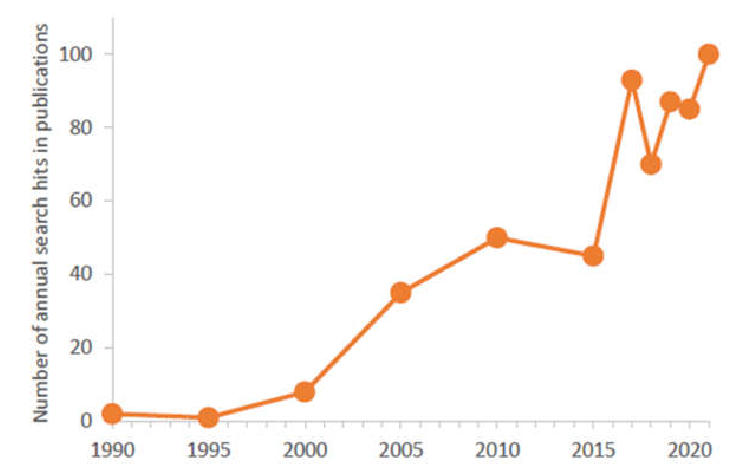
:1. Introduction
2. Fabrication of IMERs
2.1. Designs, Materials and Fabrication Technologies
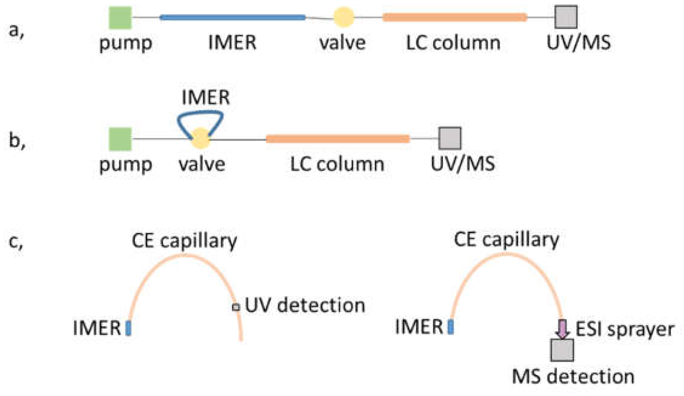
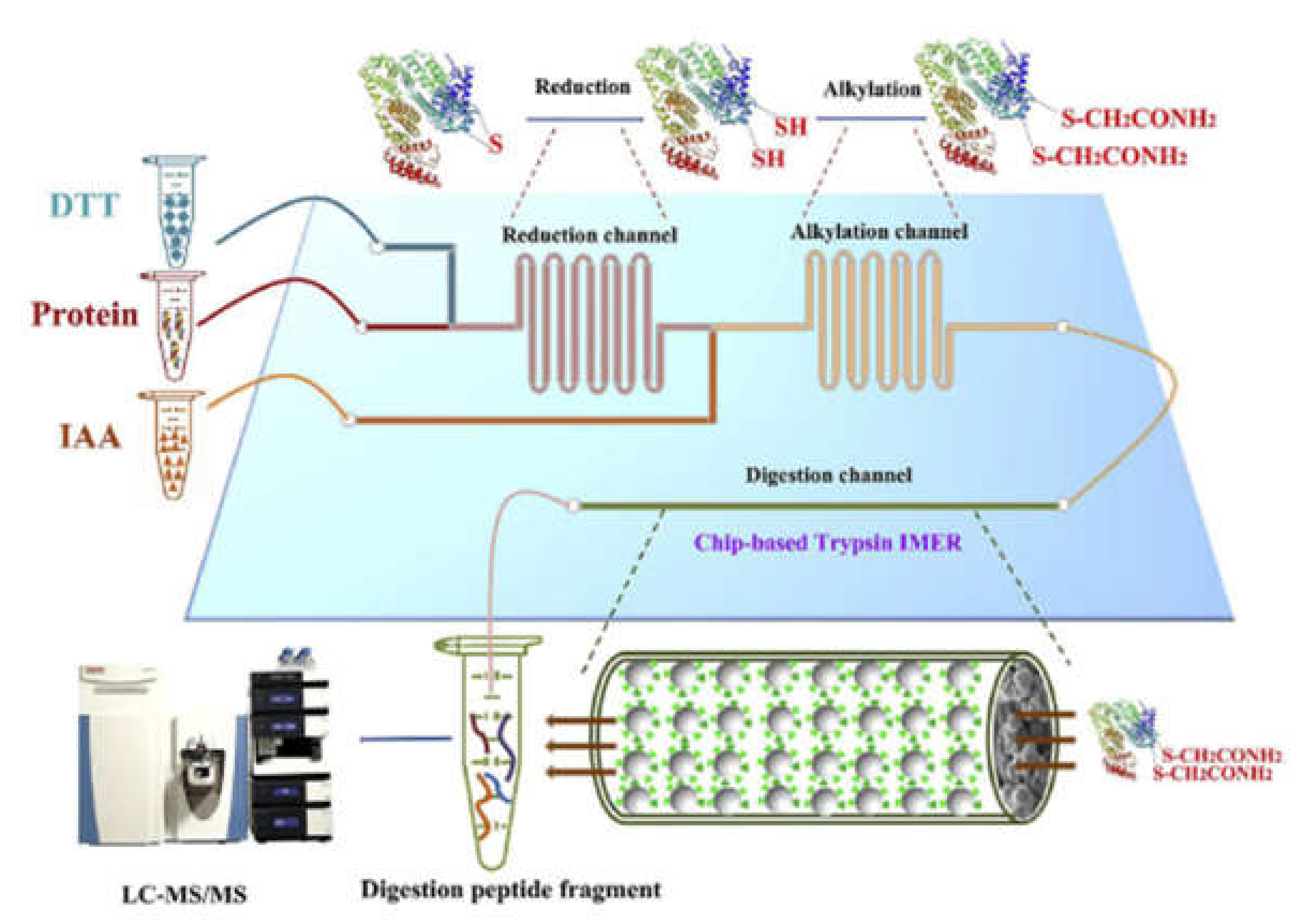
2.2. Coupling to Downstream Processing Units
3. Immobilization of Enzymes
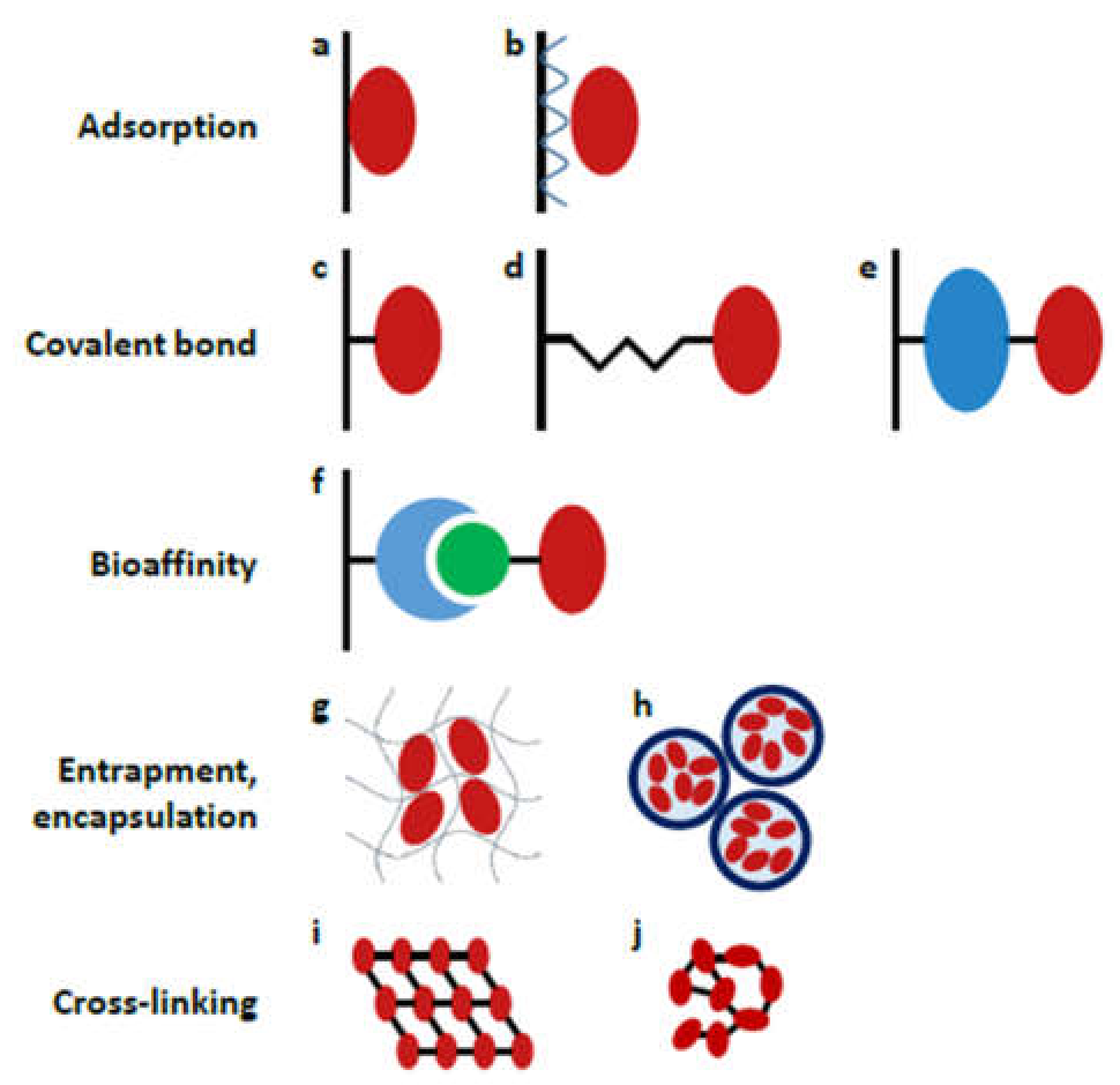
3.1. Modes of Immobilization
3.1.1. Adsorption
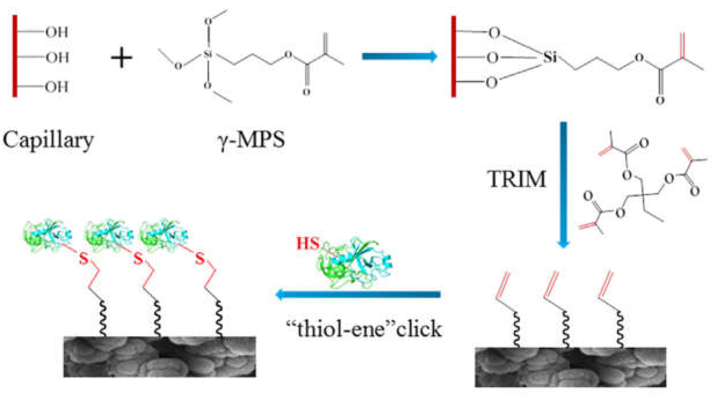
3.1.2. Covalent Coupling
3.1.3. Bioaffinity Linkage
3.2. Supports for the Immobilization
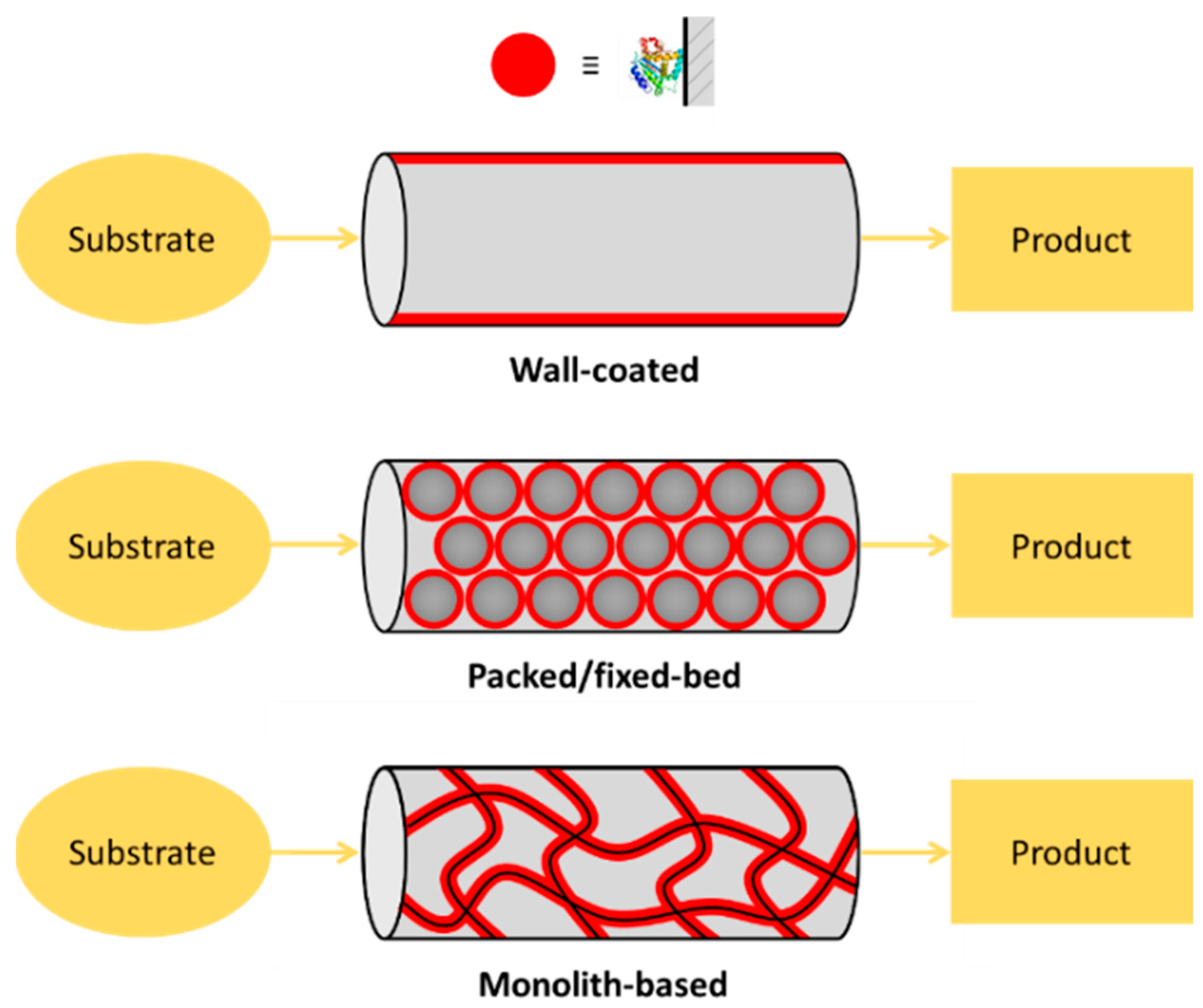
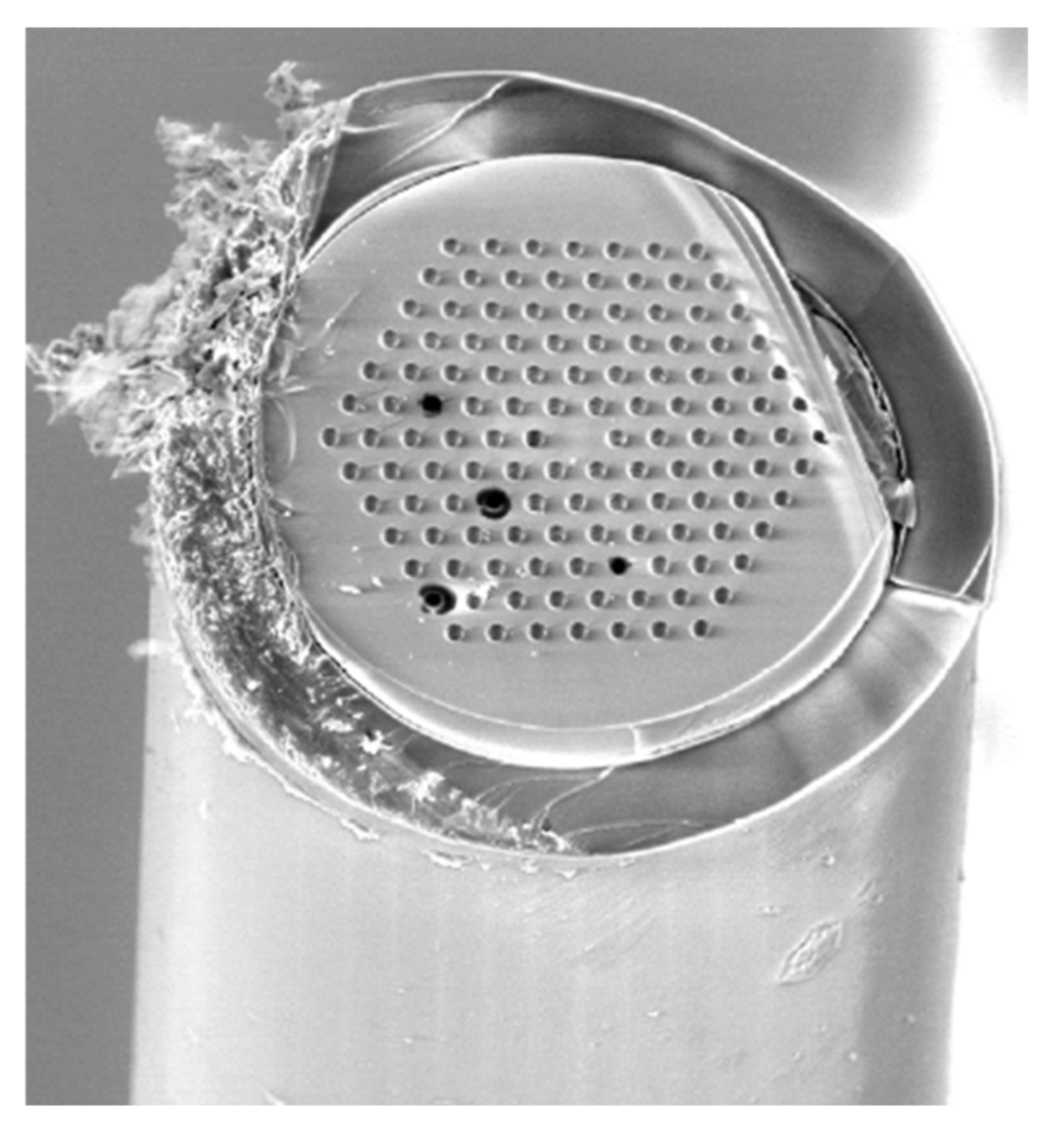
3.2.1. Open Tubular Channel/Capillary
3.2.2. Packed Channels
3.2.3. Monoliths
3.2.4. Other Supports
4. Proteomic Applications
5. Conclusions
Author Contributions
Funding
Institutional Review Board Statement
Data Availability Statement
Conflicts of Interest
References
- Licklider, L.; Kuhr, W.G.; Lacey, M.P.; Keough, T.; Purdon, M.P.; Takigiku, R. On-Line Microreactors/Capillary Electrophoresis/Mass Spectrometry for the Analysis of Proteins and Peptides. Anal. Chem. 1995, 67, 4170–4177. [Google Scholar] [CrossRef]
- Urban, P.L.; Goodall, D.M.; Bruce, N.C. Enzymatic microreactors in chemical analysis and kinetic studies. Biotechnol. Adv. 2006, 24, 42–57. [Google Scholar] [CrossRef] [PubMed]
- Elvira, K.S.; i Solvas, X.C.; Wootton, R.C.R.; DeMello, A.J. The past, present and potential for microfluidic reactor technology in chemical synthesis. Nat. Chem. 2013, 5, 905–915. [Google Scholar] [CrossRef] [PubMed]
- Miyazaki, M.; Maeda, H. Microchannel enzyme reactors and their applications for processing. Trends Biotechnol. 2006, 24, 463–470. [Google Scholar] [CrossRef] [PubMed]
- Tabeling, P. Introduction to Microfluidics; Oxford University Press: Oxford, UK, 2005. [Google Scholar]
- Ye, M.; Hu, S.; Schoenherr, R.M.; Dovichi, N.J. On-line protein digestion and peptide mapping by capillary electrophoresis with post-column labeling for laser-induced flourescence detection. Electrophoresis 2004, 25, 1319–1326. [Google Scholar] [CrossRef]
- Brás, E.J.S.; Chu, V.; Conde, J.P.; Fernandes, P. Recent developments in microreactor technology for biocatalysis applications. React. Chem. Eng. 2021, 6, 815–827. [Google Scholar] [CrossRef]
- De Mello, A.J.; Beard, N. Dealing with real samples: Sample pre-treatment in microfluidic systems. Lab Chip 2003, 3, 11N–20N. [Google Scholar] [CrossRef] [PubMed]
- Massolini, G.; Calleri, E. Immobilized trypsin systems coupled on-line to separation methods: Recent developments and analytical applications. J. Sep. Sci. 2005, 28, 7–21. [Google Scholar] [CrossRef]
- Cardoso Marques, M.P.; Lorente-Arevalo, A.; Bolivar, J.M. Biocatalysis in Continuous-Flow Microfluidic Reactors. In Advances in Biochemical Engineering/Biotechnology; Springer: Berlin/Heidelberg, Germany, 2021; pp. 1–36. [Google Scholar]
- Meller, K.; Szumski, M.; Buszewski, B. Microfluidic reactors with immobilized enzymes—Characterization, dividing, perspectives. Sens. Actuators B Chem. 2017, 244, 84–106. [Google Scholar] [CrossRef]
- Křenková, J.; Klepárník, K.; Foret, F. Capillary electrophoresis mass spectrometry coupling with immobilized enzyme electrospray capillaries. J. Chromatogr. A 2007, 1159, 110–118. [Google Scholar] [CrossRef]
- Kecskemeti, A.; Bako, J.; Csarnovics, I.; Csosz, E.; Gaspar, A. Development of an enzymatic reactor applying spontaneously adsorbed trypsin on the surface of a PDMS microfluidic device. Anal. Bioanal. Chem. 2017, 409, 3573–3585. [Google Scholar] [CrossRef] [PubMed]
- Nagy, C.; Kecskemeti, A.; Gaspar, A. Fabrication of immobilized enzyme reactors with pillar arrays into polydimethylsiloxane microchip. Anal. Chim. Acta 2020, 1108, 70–78. [Google Scholar] [CrossRef] [PubMed]
- Le Nel, A.; Krenkova, J.; Kleparnik, K.; Smadja, C.; Taverna, M.; Viovy, J.L.; Foret, F. On-chip tryptic digest with direct coupling to ESI-MS using magnetic particles. Electrophoresis 2008, 29, 4944–4947. [Google Scholar] [CrossRef] [PubMed]
- Kecskemeti, A.; Gaspar, A. Preparation and characterization of a packed bead immobilized trypsin reactor integrated into a PDMS microfluidic chip for rapid protein digestion. Talanta 2017, 166, 275–283. [Google Scholar] [CrossRef] [Green Version]
- Ma, J.; Liang, Z.; Qiao, X.; Deng, Q.; Tao, D.; Zhang, L.; Zhang, Y. Organic-inorganic hybrid silica monolith based immobilized trypsin reactor with high enzymatic activity. Anal. Chem. 2008, 80, 2949–2956. [Google Scholar] [CrossRef]
- Duan, J.; Liang, Z.; Yang, C.; Zhang, J.; Zhang, L.; Zhang, W.; Zhang, Y. Rapid protein identification using monolithic enzymatic microreactor and LC-ESI-MS/MS. Proteomics 2006, 6, 412–419. [Google Scholar] [CrossRef]
- Cooper, J.W.; Chen, J.; Li, Y.; Lee, C.S. Membrane-based nanoscale proteolytic reactor enabling protein digestion, peptide separation, and protein identification using mass spectrometry. Anal. Chem. 2003, 75, 1067–1074. [Google Scholar] [CrossRef]
- Xu, F.; Wang, W.H.; Tan, Y.J.; Bruening, M.L. Facile trypsin immobilization in polymeric membranes for rapid, efficient protein digestion. Anal. Chem. 2010, 82, 10045–10051. [Google Scholar] [CrossRef] [Green Version]
- Nadar, S.S.; Patil, P.D.; Tiwari, M.S.; Ahirrao, D.J. Enzyme embedded microfluidic paper-based analytic device (μPAD): A comprehensive review. Crit. Rev. Biotechnol. 2021, 41, 1046–1080. [Google Scholar] [CrossRef]
- Wang, C.; Oleschuk, R.; Ouchen, F.; Li, J.; Thibault, P.; Harrison, D.J. Integration of immobilized trypsin bead beds for protein digestion within a microfluidic chip incorporating capillary electrophoresis separations and an electrospray mass spectrometry interface. Rapid Commun. Mass Spectrom. 2000, 14, 1377–1383. [Google Scholar] [CrossRef]
- Jin, L.J.; Ferrance, J.; Sanders, J.C.; Landers, J.P. A microchip-based proteolytic digestion system driven by electroosmotic pumping. Lab Chip 2003, 3, 11–18. [Google Scholar] [CrossRef] [PubMed]
- Benítez-Mateos, A.I.; Contente, M.L.; Roura Padrosa, D.; Paradisi, F. Flow biocatalysis 101: Design, development and applications. React. Chem. Eng. 2021, 6, 599–611. [Google Scholar] [CrossRef]
- Slovakova, M.; Minc, N.; Bilkova, Z.; Smadja, C.; Faigle, W.; Fütterer, C.; Taverna, M.; Viovy, J.L. Use of self assembled magnetic beads for on-chip protein digestion. Lab Chip 2005, 5, 935–942. [Google Scholar] [CrossRef] [PubMed]
- Wang, C.; Jemere, A.B.; Harrison, D.J. Multifunctional protein processing chip with integrated digestion, solid-phase extraction, separation and electrospray. Electrophoresis 2010, 31, 3703–3710. [Google Scholar] [CrossRef]
- Le Nel, A.; Minc, N.; Smadja, C.; Slovakova, M.; Bilkova, Z.; Peyrin, J.M.; Viovy, J.L.; Taverna, M. Controlled proteolysis of normal and pathological prion protein in a microfluidic chip. Lab Chip 2008, 8, 294–301. [Google Scholar] [CrossRef]
- Nomura, A.; Shin, S.; Mehdi, O.O.; Kauffmann, J.M. Preparation, characterization, and application of an enzyme-immobilized magnetic microreactor for flow injection analysis. Anal. Chem. 2004, 76, 5498–5502. [Google Scholar] [CrossRef]
- Sheng, J.; Zhang, L.; Lei, J.; Ju, H. Fabrication of tunable microreactor with enzyme modified magnetic nanoparticles for microfluidic electrochemical detection of glucose. Anal. Chim. Acta 2012, 709, 41–46. [Google Scholar] [CrossRef]
- Richter, T.; Shultz-Lockyear, L.L.; Oleschuk, R.D.; Bilitewski, U.; Harrison, D. Bi-enzymatic and capillary electrophoretic analysis of non-fluorescent compounds in microfluidic devices—Determination of xanthine. Sens. Actuators B Chem. 2002, 81, 369–376. [Google Scholar] [CrossRef]
- Ito, T.; Kunimatsu, M.; Kaneko, S.; Ohya, S.; Suzuki, K. Microfluidic device for the detection of glucose using a micro direct methanol fuel cell as an amperometric detection power source. Anal. Chem. 2007, 79, 1725–1730. [Google Scholar] [CrossRef]
- Seong, G.H.; Crooks, R.M. Efficient mixing and reactions within microfluidic channels using microbead-supported catalysts. J. Am. Chem. Soc. 2002, 124, 13360–13361. [Google Scholar] [CrossRef] [Green Version]
- Boehm, C.R.; Freemont, P.S.; Ces, O. Design of a prototype flow microreactor for synthetic biology in vitro. Lab Chip 2013, 13, 3426–3432. [Google Scholar] [CrossRef] [PubMed]
- Liang, R.P.; Wang, X.N.; Liu, C.M.; Meng, X.Y.; Qiu, J.D. Construction of graphene oxide magnetic nanocomposites-based on-chip enzymatic microreactor for ultrasensitive pesticide detection. J. Chromatogr. A 2013, 1315, 28–35. [Google Scholar] [CrossRef] [PubMed]
- Bolivar, J.M.; López-Gallego, F. Characterization and evaluation of immobilized enzymes for applications in flow reactors. Curr. Opin. Green Sustain. Chem. 2020, 25, 100349. [Google Scholar] [CrossRef]
- Chen, X.; Shen, J. Review of membranes in microfluidics. J. Chem. Technol. Biotechnol. 2017, 92, 271–282. [Google Scholar] [CrossRef]
- Gkantzou, E.; Patila, M.; Stamatis, H. Magnetic Microreactors with Immobilized Enzymes—From Assemblage to Contemporary Applications. Catalysts 2018, 8, 282. [Google Scholar] [CrossRef] [Green Version]
- Kecskemeti, A.; Gaspar, A. Particle-based immobilized enzymatic reactors in microfluidic chips. Talanta 2018, 180, 211–228. [Google Scholar] [CrossRef] [Green Version]
- Kiani, M.R.; Meshksar, M.; Makarem, M.A.; Rahimpour, E. Catalytic Membrane Micro-Reactors for Fuel and Biofuel Processing: A Mini Review. Top. Catal. 2021, 1, 1–20. [Google Scholar] [CrossRef]
- Naldi, M.; Tramarin, A.; Bartolini, M. Immobilized enzyme-based analytical tools in the -omics era: Recent advances. J. Pharm. Biomed. Anal. 2018, 160, 222–237. [Google Scholar] [CrossRef]
- Shi, H.; Nie, K.; Dong, B.; Long, M.; Xu, H.; Liu, Z. Recent progress of microfluidic reactors for biomedical applications. Chem. Eng. J. 2019, 361, 635–650. [Google Scholar] [CrossRef]
- Wouters, B.; Currivan, S.A.; Abdulhussain, N.; Hankemeier, T.; Schoenmakers, P.J. Immobilized-enzyme reactors integrated into analytical platforms: Recent advances and challenges. TrAC Trends Anal. Chem. 2021, 144, 116419. [Google Scholar] [CrossRef]
- Zhang, H.; Bai, Y.; Zhu, N.; Xu, J. Microfluidic reactor with immobilized enzyme-from construction to applications: A review. Chin. J. Chem. Eng. 2021, 30, 136–145. [Google Scholar] [CrossRef]
- Zhu, Y.; Chen, Q.; Shao, L.; Jia, Y.; Zhang, X. Microfluidic immobilized enzyme reactors for continuous biocatalysis. React. Chem. Eng. 2020, 5, 9–32. [Google Scholar] [CrossRef]
- Wouters, B.; Pirok, B.W.J.; Soulis, D.; Garmendia Perticarini, R.C.; Fokker, S.; van den Hurk, R.S.; Skolimowski, M.; Peters, R.A.H.; Schoenmakers, P.J. On-line microfluidic immobilized-enzyme reactors: A new tool for characterizing synthetic polymers. Anal. Chim. Acta 2019, 1053, 62–69. [Google Scholar] [CrossRef] [PubMed]
- Cornelio, V.E.; de Moraes, M.C.; Domingues, V.d.C.; Fernandes, J.B.; da Silva, M.F.d.G.F.; Cass, Q.B.; Vieira, P.C. Cathepsin D immobilized capillary reactors for on-flow screening assays. J. Pharm. Biomed. Anal. 2018, 151, 252–259. [Google Scholar] [CrossRef] [PubMed]
- Liu, X.; Zhu, X.; Camara, M.A.; Qu, Q.; Shan, Y.; Yang, L. Surface modification with highly-homogeneous porous silica layer for enzyme immobilization in capillary enzyme microreactors. Talanta 2019, 197, 539–547. [Google Scholar] [CrossRef]
- Nagy, C.; Szabo, R.; Gaspar, A. Development of an in-line enzyme reactor integrated into a capillary electrophoresis system. Molecules 2021, 26, 5902. [Google Scholar] [CrossRef]
- Wu, N.; Wang, S.; Yang, Y.; Song, J.; Su, P.; Yang, Y. DNA-directed trypsin immobilization on a polyamidoamine dendrimer-modified capillary to form a renewable immobilized enzyme microreactor. Int. J. Biol. Macromol. 2018, 113, 38–44. [Google Scholar] [CrossRef]
- Wei, Z.H.; Zhang, X.; Zhao, X.; Jiao, Y.J.; Huang, Y.P.; Liu, Z.S. Construction of a microfluidic platform integrating online protein fractionation, denaturation, digestion, and peptide enrichment. Talanta 2021, 224, 121810. [Google Scholar] [CrossRef]
- Jiao, Y.J.; Yuan, F.F.; Fan, P.R.; Wei, Z.H.; Huang, Y.P.; Liu, Z.S. Macroporous monolithic enzyme microreactor based on high internal phase emulsion functionalized with gold nanorods for enzymatic hydrolysis of protein. Chem. Eng. J. 2021, 407, 127061. [Google Scholar] [CrossRef]
- Zhang, Y.; Wang, Y.; Tang, Y.; Li, R.; Ji, Y. An online immobilized pepsin microreactor based on polymer monoliths for screening inhibitors from natural products. Anal. Methods 2019, 11, 2465–2472. [Google Scholar] [CrossRef]
- Cheng, M.; Wang, R.; Zhang, B.; Mao, Z.; Chen, Z. Rapid proteolytic digestion and peptide separation using monolithic enzyme microreactor coupled with capillary electrophoresis. J. Pharm. Biomed. Anal. 2019, 165, 129–134. [Google Scholar] [CrossRef] [PubMed]
- Fan, P.R.; Zhao, X.; Wei, Z.H.; Huang, Y.P.; Liu, Z.S. Robust immobilized enzyme reactor based on trimethylolpropane trimethacrylate organic monolithic matrix through “thiol-ene” click reaction. Eur. Polym. J. 2020, 124, 109456. [Google Scholar] [CrossRef]
- Meller, K.; Pomastowski, P.; Szumski, M.; Buszewski, B. Preparation of an improved hydrophilic monolith to make trypsin-immobilized microreactors. J. Chromatogr. B Anal. Technol. Biomed. Life Sci. 2017, 1043, 128–137. [Google Scholar] [CrossRef] [PubMed]
- Olsen, C.; Skottvoll, F.S.; Brandtzaeg, O.K.; Schnaars, C.; Rongved, P.; Lundanes, E.; Wilson, S.R. Investigating Monoliths (Vinyl Azlactone-co-Ethylene Dimethacrylate) as a Support for Enzymes and Drugs, for Proteomics and Drug-Target Studies. Front. Chem. 2019, 7, 835. [Google Scholar] [CrossRef]
- Zhao, X.; Fan, P.R.; Mo, C.E.; Huang, Y.P.; Liu, Z.S. Green synthesis of monolithic enzyme microreactor based on thiol-ene click reaction for enzymatic hydrolysis of protein. J. Chromatogr. A 2020, 1611, 460618. [Google Scholar] [CrossRef]
- Wei, Z.; Fan, P.; Jiao, Y.; Wang, Y.; Huang, Y.; Liu, Z. Integrated microfluidic chip for on-line proteome analysis with combination of denaturing and rapid digestion of protein. Anal. Chim. Acta 2020, 1102, 1–10. [Google Scholar] [CrossRef]
- Yamamoto, K.; Morikawa, K.; Imanaka, H.; Imamura, K.; Kitamori, T. Picoliter enzyme reactor on a nanofluidic device exceeding the bulk reaction rate. Analyst 2020, 145, 5801–5807. [Google Scholar] [CrossRef]
- Bataille, J.; Viodé, A.; Pereiro, I.; Lafleur, J.P.; Varenne, F.; Descroix, S.; Becher, F.; Kutter, J.P.; Roesch, C.; Poüs, C.; et al. On-a-chip tryptic digestion of transthyretin: A step toward an integrated microfluidic system for the follow-up of familial transthyretin amyloidosis. Analyst 2018, 143, 1077–1086. [Google Scholar] [CrossRef]
- Gaspar, A.; Gomez, F.A. Application of surface plasmon resonance spectroscopy for adsorption studies of different types of components on poly(dimethylsiloxane). Anal. Chim. Acta 2013, 777, 72–77. [Google Scholar] [CrossRef]
- Tähkä, S.; Sarfraz, J.; Urvas, L.; Provenzani, R.; Wiedmer, S.K.; Peltonen, J.; Jokinen, V.; Sikanen, T. Immobilization of proteolytic enzymes on replica-molded thiol-ene micropillar reactors via thiol-gold interaction. Anal. Bioanal. Chem. 2019, 411, 2339–2349. [Google Scholar] [CrossRef] [Green Version]
- Lu, N.; Sticker, D.; Kretschmann, A.; Petersen, N.J.; Kutter, J.P. A thiol-ene microfluidic device enabling continuous enzymatic digestion and electrophoretic separation as front-end to mass spectrometric peptide analysis. Anal. Bioanal. Chem. 2020, 412, 3559–3571. [Google Scholar] [CrossRef] [PubMed]
- Jönsson, A.; Svejdal, R.R.; Bøgelund, N.; Nguyen, T.T.T.N.; Flindt, H.; Kutter, J.P.; Rand, K.D.; Lafleur, J.P. Thiol-ene Monolithic Pepsin Microreactor with a 3D-Printed Interface for Efficient UPLC-MS Peptide Mapping Analyses. Anal. Chem. 2017, 89, 4573–4580. [Google Scholar] [CrossRef] [PubMed] [Green Version]
- Wouters, B.; Dapic, I.; Valkenburg, T.S.E.; Wouters, S.; Niezen, L.; Eeltink, S.; Corthals, G.L.; Schoenmakers, P.J. A cyclic-olefin-copolymer microfluidic immobilized-enzyme reactor for rapid digestion of proteins from dried blood spots. J. Chromatogr. A 2017, 1491, 36–42. [Google Scholar] [CrossRef]
- Brandtzaeg, O.K.; Røen, B.T.; Enger, S.; Lundanes, E.; Wilson, S.R. Multichannel Open Tubular Enzyme Reactor Online Coupled with Mass Spectrometry for Detecting Ricin. Anal. Chem. 2017, 89, 8667–8673. [Google Scholar] [CrossRef] [PubMed]
- Yuan, H.; Zhang, S.; Zhao, B.; Weng, Y.; Zhu, X.; Li, S.; Zhang, L.; Zhang, Y. Enzymatic Reactor with Trypsin Immobilized on Graphene Oxide Modified Polymer Microspheres to Achieve Automated Proteome Quantification. Anal. Chem. 2017, 89, 6324–6329. [Google Scholar] [CrossRef] [PubMed]
- Zhang, S.; Yuan, H.; Zhao, B.; Zhang, L.; Zhang, Y. Integrated platform with combination of on-line protein digestion, isotope dimethyl labeling and multidimensional peptide separation for high-throughput proteome quantification. Anal. Chim. Acta 2018, 1000, 172–179. [Google Scholar] [CrossRef]
- Villegas, L.; Pero-Gascon, R.; Benavente, F.; Barbosa, J.; Sanz-Nebot, V. On-line protein digestion by immobilized enzyme microreactor capillary electrophoresis-mass spectrometry. Talanta 2019, 199, 116–123. [Google Scholar] [CrossRef]
- Currivan, S.A.; Chen, W.Q.; Wilson, R.; Sanz Rodriguez, E.; Upadhyay, N.; Connolly, D.; Nesterenko, P.N.; Paull, B. Multi-lumen capillary based trypsin micro-reactor for the rapid digestion of proteins. Analyst 2018, 143, 4944–4953. [Google Scholar] [CrossRef]
- Kim, D.; Herr, A.E. Protein immobilization techniques for microfluidic assays. Biomicrofluidics 2013, 7, 41501. [Google Scholar] [CrossRef] [Green Version]
- Homaei, A.A.; Sariri, R.; Vianello, F.; Stevanato, R. Enzyme immobilization: An update. J. Chem. Biol. 2013, 6, 185–205. [Google Scholar] [CrossRef] [Green Version]
- Nagy, C.; Huszank, R.; Gaspar, A. Study of the geometry of open channels in a layer-bed-type microfluidic immobilized enzyme reactor. Anal. Bioanal. Chem. 2021, 413, 6321–6332. [Google Scholar] [CrossRef]
- Kecskemeti, A.; Nagy, C.; Csosz, E.; Kallo, G.; Gaspar, A. The application of a microfluidic reactor including spontaneously adsorbed trypsin for rapid protein digestion of human tear samples. Prot. Clin. Appl. 2017, 11, 1700055. [Google Scholar] [CrossRef] [PubMed]
- Comamala, G.; Krogh, C.C.; Nielsen, V.S.; Kutter, J.P.; Voglmeir, J.; Rand, K.D. Hydrogen/Deuterium Exchange Mass Spectrometry with Integrated Electrochemical Reduction and Microchip-Enabled Deglycosylation for Epitope Mapping of Heavily Glycosylated and Disulfide-Bonded Proteins. Anal. Chem. 2021, 93, 16330–16340. [Google Scholar] [CrossRef] [PubMed]
- Hata, K.; Izumi, Y.; Hara, T.; Matsumoto, M.; Bamba, T. In-Line Sample Processing System with an Immobilized Trypsin-Packed Fused-Silica Capillary Tube for the Proteomic Analysis of a Small Number of Mammalian Cells. Anal. Chem. 2020, 92, 2997–3005. [Google Scholar] [CrossRef] [PubMed]
- Dong, J.; Ning, W.; Liu, W.; Bruening, M.L. Limited proteolysis in porous membrane reactors containing immobilized trypsin. Analyst 2017, 142, 2578–2586. [Google Scholar] [CrossRef]
- Han, X.; Xie, Y.; Wu, Q.; Wu, S. The Effect of Monolith Properties on the Digestion Performance of Monolith-Based Immobilized Enzyme Microreactor. J. Chromatogr. Sci. 2019, 57, 116–121. [Google Scholar] [CrossRef]
- Kjellander, M.; Billinger, E.; Ramachandraiah, H.; Boman, M.; Bergström Lind, S.; Johansson, G. A flow-through nanoporous alumina trypsin bioreactor for mass spectrometry peptide fingerprinting. J. Proteom. 2018, 172, 165–172. [Google Scholar] [CrossRef]
- Jiang, H.; Yuan, H.; Liang, Y.; Xia, S.; Zhao, Q.; Wu, Q.; Zhang, L.; Liang, Z.; Zhang, Y. A hydrophilic immobilized trypsin reactor with N-vinyl-2-pyrrolidinone modified polymer microparticles as matrix for highly efficient protein digestion with low peptide residue. J. Chromatogr. A 2012, 1246, 111–116. [Google Scholar] [CrossRef]
- Xie, S.; Svec, F.; Fréchet, J.M.J. Design of reactive porous polymer supports for high throughput bioreactors: Poly(2-vinyl-4;4 dimethylazlactone-co-acrylamide-co-ethylene dimethacrylate) monoliths. Biotechnol. Bioeng. 1999, 62, 30–35. [Google Scholar] [CrossRef]
- Benavente, F.; Medina-Casanellas, S.; Giménez, E.; Sanz-Nebot, V. On-Line Solid-Phase Extraction Capillary Electrophoresis Mass Spectrometry for Preconcentration and Clean-Up of Peptides and Proteins. In Methods in Molecular Biology; Humana Press: New York, NY, USA, 2016; Volume 1466, pp. 67–84. [Google Scholar]
- Pereiro, I.; Tabnaoui, S.; Fermigier, M.; Du Roure, O.; Descroix, S.; Viovy, J.L.; Malaquin, L. Magnetic fluidized bed for solid phase extraction in microfluidic systems. Lab Chip 2017, 17, 1603–1615. [Google Scholar] [CrossRef]
- Liu, B.; Wu, L.; Zhou, X.; Wu, H.; Zheng, B. Porous polydimethylsiloxane monolith for protein digestion. J. Mater. Chem. B 2018, 6, 824–829. [Google Scholar] [CrossRef] [PubMed]
- Messner, L.; Antink, M.H.; Guo, T.; Maas, M.; Beutel, S. A versatile ceramic capillary membrane reactor system for continuous enzyme-catalyzed hydrolysis. Eng. Life Sci. 2021, 21, 527–538. [Google Scholar] [CrossRef]
- Hoog Antink, M.M.; Sewczyk, T.; Kroll, S.; Árki, P.; Beutel, S.; Rezwan, K.; Maas, M. Proteolytic ceramic capillary membranes for the production of peptides under flow. Biochem. Eng. J. 2019, 147, 89–99. [Google Scholar] [CrossRef]
- Sewczyk, T.; Hoog Antink, M.; Maas, M.; Kroll, S.; Beutel, S. Flow rate dependent continuous hydrolysis of protein isolates. AMB Express 2018, 8, 1–9. [Google Scholar] [CrossRef] [PubMed] [Green Version]
- Dias, A.A.; Cardoso, T.M.G.; Cardoso, R.M.; Duarte, L.C.; Muñoz, R.A.A.; Richter, E.M.; Coltro, W.K.T. Paper-based enzymatic reactors for batch injection analysis of glucose on 3D printed cell coupled with amperometric detection. Sens. Actuators B Chem. 2016, 226, 196–203. [Google Scholar] [CrossRef]
- Carrell, C.; Kava, A.; Nguyen, M.; Menger, R.; Munshi, Z.; Call, Z.; Nussbaum, M.; Henry, C. Beyond the lateral flow assay: A review of paper-based microfluidics. Microelectron. Eng. 2019, 206, 45–54. [Google Scholar] [CrossRef]
- Liang, S.; Wu, X.-L.; Xiong, J.; Zong, M.-H.; Lou, W.-Y. Metal-organic frameworks as novel matrices for efficient enzyme immobilization: An update review. Coord. Chem. Rev. 2020, 406, 213149. [Google Scholar] [CrossRef]


| Ref. | Title | Keywords (max. 4) * |
|---|---|---|
| [35] | Characterization and evaluation of immobilized enzymes for applications in flow reactors | biocatalysis, protein immobilization, advanced materials, packed-bed reactors |
| [7] | Recent developments in microreactor technology for biocatalysis applications | enzymatic microreactor; biocatalysis; monolith; multiphase systems |
| [36] | Review on membranes in microfluidics | membranes; manufacturing methods; applications; mass transfer |
| [37] | Magnetic microreactors with immobilized enzymes—from assemblage to contemporary applications | enzymatic microreactors; magnetic particles; nanomaterials; immobilization |
| [38] | Particle-based immobilized enzymatic reactors in microfluidic chips | enzyme reactor; particle; enzyme immobilization; protein digestion |
| [39] | Catalytic membrane microreactors for fuel and biofuel processing | membrane; catalytic membrane microreactors; microchannels; catalytic processes |
| [10] | Biocatalysis in continuous-flow microfluidic reactors | enzyme immobilization; flow biocatalysis; microfluidic reactors; miniaturization |
| [11] | Microfluidic reactors with immobilized enzymes—characterization, dividing, perspectives | immobilized enzyme microreactor; miniaturization; immobilization strategies; biocatalysis |
| [40] | Immobilized enzyme-based analytical tools in the -omics era: recent advances | immobilized enzyme reactors; proteomics; glycomics; dual IMERs |
| [41] | Recent progress of microfluidic reactors for biomedical applications | microreactor; PCR; ELISA; hybridization |
| [42] | Immobilized enzyme reactors integrated into analytical platforms: recent advances and challenges | hyphenation; enzymatic reaction; immobilization; liquid chromatography |
| [43] | Microfluidic reactor with immobilized enzyme—from construction to applications | microfluidic IMER; immobilization strategies; biocatalysis; bioconversion |
| [44] | Microfluidic immobilized enzyme reactors for continuous biocatalysis | in vitro biocatalysis; microfluidic reactor; enzyme immobilization; multi-enzyme systems |
| [45] | On-line microfluidic immobilized enzyme reactors: A new tool for characterizing synthetic polymers | biodegradable polymer; enzymatic degradation; polyesters; lipase |
| [21] | Enzyme embedded microfluidic paper-based analytic device (μPAD): a comprehensive review | microfluidic devices; hybrid nanoflowers; design and fabrication; point-of-care |
| Immobilized Enzyme | Reactor Type | Type of Solid Support | Enzyme Immobilization Strategy | Coupled Detector-Analyzer | Application | Ref. |
|---|---|---|---|---|---|---|
| trypsin | fused silica capillary | monolith | TE click-reaction | LC-MS | protein extract digestion, breast cancer (MCF-7) cells | [50] |
| trypsin | glass microchip, fused silica capillary | monolith | TE click-reaction | LC-MS | protein extract digestion, mouse liver | [58] |
| TPCK-trypsin | PDMS chip (microfluidized bed) TE microchip | magnetic bead monolith | covalent (carbodiimide) TE click-reaction | LC-MS | protein standard digestion | [60] |
| pepsin | TE microchip | monolith | TE click-reaction | SDS-PAGE, LC-MS | protein standard digestion | [64] |
| trypsin | PDMS microchip | channel wall | adsorption | CE-UV, LC-MS | protein standard digestion | [13] |
| trypsin | PDMS microchip | silica particles | covalent (carbodiimide) | CE-UV, LC-MS | protein extract digestion, human serum | [16] |
| pepsin | TE microchip | monolith | TE click-reaction | FFE, ESI-MS | peptide digestion | [63] |
| α-chymotrypsin | TE microchip | GNPs | thiol-gold interaction | ESI-MS | peptide digestion | [62] |
| trypsin | COC microchip | monolith | covalent (azlactone chemistry) | nanoLC-MS | protein extract digestion, dried blood spots | [65] |
| TPCK-trypsintrypsinogen | glass microchip | derivatized channel wall | covalent (glutaraldehyde) | substrate digestion | [59] | |
| trypsin | PDMS microchip | channel wall | adsorption | CE-UV, CE-MS | protein extract digestion, snake venom | [14] |
| trypsin | PDMS microchip | channel wall | adsorption | CE-UV, CE-MS | protein extract digestion, saliva | [73] |
| trypsin | PDMS microchip | channel wall | adsorption | CE-UV, CE-MS | protein extract digestion, tear | [74] |
| PNGase A, Dj, H+ | TE microchip | monolith | TE click-reaction | LC-HDX-MS | deglycosylation | [75] |
| trypsin | fused silica capillary | PSDVB particles | commercial immobilized beads (covalent) | nanoLC-MS | protein extract digestion, HeLa cells | [76] |
| trypsin | capillary | GNR- functional-ized monolith | thiol-gold interaction | nanoLC-MS | protein extract digestion, rat liver | [51] |
| pepsin | capillary | polymer monolith | covalent (glutaraldehyde) | CE-UV | substrate digestion, inhibitor screening | [52] |
| trypsin/Lys-C | MCR | polymer layer | covalent (azlactone chemistry) | nanoLC-MS Q-Ex | protein extract digestion, castor bean | [66] |
| trypsin | capillary | polymer monolith | covalent | CE-UV, HPLC-UV | protein standard digestion | [53] |
| cathepsin D | capillary | derivatized channel wall | covalent (glutaraldehyde) | HPLC-FD | peptide digestion, inhibitor screening | [46] |
| trypsin | MCR | GNPs | covalent | capLC-UV, nanoLC-MS | protein standard digestion | [70] |
| trypsin | capillary | monolith | TE click-reaction | nanoLC-MS | protein extract digestion, egg white, mouse liver | [54] |
| TPCK-trypsin | capillary | porous layer | covalent (glutaraldehyde) | nanoLC-MS | protein extract digestion, HeLa cells | [47] |
| trypsin | capillary | polymer monolith | covalent | nanoLC-UV, MALDI-TOF MS | protein standard digestion | [55] |
| trypsin | capillary | channel wall | adsorption | CE-UV, CE-MS | protein extract digestion, tear | [48] |
| TPCK-trypsin | capillary | polymer monolith | covalent | LC-UV | protein standard digestion | [56] |
| trypsin | capillary | cellulose resin | commercial immobilized beads | CE-MS | protein extract digestion, E. coil | [69] |
| trypsin | capillary | channel wall | DNA-directed | CE-UV, MALDI-TOF MS | protein standard digestion | [49] |
| TPCK-trypsin | capillary | GO-modified polymer microsphere | electrostatic interaction | nanoLC-MS, MALDI-TOF MS | protein extract digestion, E. coil, Hca-F and Hca-P cells | [67] |
| trypsin | capillary | GO-modified polymer microsphere | electrostatic interaction | 2D nanoLC-MS, MALDI-TOF MS | protein extract digestion, E. coil, Hca-F and Hca-P cells | [68] |
| trypsin | capillary | monolith | TE click-reaction | CE-UV | protein extract digestion, rat liver | [57] |
| trypsin | membrane holder | porous membrane | adsorption, covalent (carbodiimide) | UV, SDS-PAGE, ESI-MS | protein standard digestion | [77] |
| trypsin | capillary | monolith | covalent | LC-UV, LC-MS | protein standard mixture digestion | [78] |
| trypsin | membrane holder | nanoporous alumina membrane | covalent (CDI) | SDS-PAGE, MALDI-TOF MS, ESI-MS, nanoLC-MS | protein extract digestion, human plasma | [79] |
Publisher’s Note: MDPI stays neutral with regard to jurisdictional claims in published maps and institutional affiliations. |
© 2022 by the authors. Licensee MDPI, Basel, Switzerland. This article is an open access article distributed under the terms and conditions of the Creative Commons Attribution (CC BY) license (https://creativecommons.org/licenses/by/4.0/).
Share and Cite
Nagy, C.; Szabo, R.; Gaspar, A. Microfluidic Immobilized Enzymatic Reactors for Proteomic Analyses—Recent Developments and Trends (2017–2021). Micromachines 2022, 13, 311. https://doi.org/10.3390/mi13020311
Nagy C, Szabo R, Gaspar A. Microfluidic Immobilized Enzymatic Reactors for Proteomic Analyses—Recent Developments and Trends (2017–2021). Micromachines. 2022; 13(2):311. https://doi.org/10.3390/mi13020311
Chicago/Turabian StyleNagy, Cynthia, Ruben Szabo, and Attila Gaspar. 2022. "Microfluidic Immobilized Enzymatic Reactors for Proteomic Analyses—Recent Developments and Trends (2017–2021)" Micromachines 13, no. 2: 311. https://doi.org/10.3390/mi13020311
APA StyleNagy, C., Szabo, R., & Gaspar, A. (2022). Microfluidic Immobilized Enzymatic Reactors for Proteomic Analyses—Recent Developments and Trends (2017–2021). Micromachines, 13(2), 311. https://doi.org/10.3390/mi13020311