Article

Phase Formation Behavior and Thermoelectric Transport Properties of S-Doped FeSe2−xSx Polycrystalline Alloys

Department of Materials Science and Engineering, University of Seoul, Seoul 02504, Republic of Korea
*
Author to whom correspondence should be addressed.
These authors contributed equally to this work.
Micromachines 2022, 13(12), 2066; https://doi.org/10.3390/mi13122066
Submission received: 14 October 2022 / Revised: 16 November 2022 / Accepted: 22 November 2022 / Published: 25 November 2022

Abstract

:
Some transition-metal dichalcogenides have been actively studied recently owing to their potential for use as thermoelectric materials due to their superior electronic transport properties. Iron-based chalcogenides, FeTe2, FeSe2 and FeS2, are narrow bandgap (~1 eV) semiconductors that could be considered as cost-effective thermoelectric materials. Herein, the thermoelectric and electrical transport properties FeSe2–FeS2 system are investigated. A series of polycrystalline samples of the nominal composition of FeSe2−xSx (x = 0, 0.2, 0.4, 0.6, and 0.8) samples are synthesized by a conventional solid-state reaction. A single orthorhombic phase of FeSe2 is successfully synthesized for x = 0, 0.2, and 0.4, while secondary phases (Fe7S8 or FeS2) are identified as well for x = 0.6 and 0.8. The lattice parameters gradually decrease gradually with S content increase to x = 0.6, suggesting that S atoms are successfully substituted at the Se sites in the FeSe2 orthorhombic crystal structure. The electrical conductivity increases gradually with the S content, whereas the positive Seebeck coefficient decreases gradually with the S content at 300 K. The maximum power factor of 0.55 mW/mK2 at 600 K was seen for x = 0.2, which is a 10% increase compared to the pristine FeSe2 sample. Interestingly, the total thermal conductivity at 300 K of 7.96 W/mK (x = 0) decreases gradually and significantly to 2.58 W/mK for x = 0.6 owing to the point-defect phonon scattering by the partial substitution of S atoms at the Se site. As a result, a maximum thermoelectric figure of merit of 0.079 is obtained for the FeSe1.8S0.2 (x = 0.2) sample at 600 K, which is 18% higher than that of the pristine FeSe2 sample.

1. Introduction

Thermoelectric materials have been widely studied in recent years, owing to their ability to convert waste thermal gradients into electrical energy [1]. The energy-conversion efficiency of thermoelectric materials can be evaluated using the dimensionless thermoelectric figure of merit, zT, which is expressed by the following Equation (1).
z T = σ S 2 κ t o t T
where σ, S, T, and κtot are the electrical conductivity, Seebeck coefficient, absolute temperature, and total thermal conductivity, respectively [2,3,4]. Generally, κtot is divided into two terms:
κ t o t = κ e l e c + κ l a t t
where κelec and κlatt are the electrical and lattice thermal conductivities, respectively. zT can be improved by increasing the power factor (σS2) or reducing κtot. However, the trade-off between σ and S and the proportionate relationship between σ and κelec make it difficult to improve zT. One strategy to obtain a high zT is to reduce κlatt via point-defect phonon scattering, which can be achieved by doping or using a partial solid solution [5,6]. For example, Asfandiyar et al. investigated the thermoelectric properties of alloyed samples in an SnS–SnSe system, and they reported that the κlatt of the alloyed samples was lower than that of the nonalloyed samples, and that the zT of the alloyed samples was greatly enhanced [7].
Transition-metal dichalcogenides (TMDCs) such as HfSe2 [8,9], HfTe2 [10], MoSe2 [11], and SnSe2 [12,13] have attracted significant attention because of their high potential for use as thermoelectric materials. Generally, TMDCs have a large effective mass owing to the presence of local d- or f-orbital electrons, leading to a large magnitude of S [14]. Among these TMDCs, FeSe2 and FeS2 have been actively investigated as promising thermoelectric materials. FeSe2 is a p-type semiconductor with a narrow direct bandgap of ~1 eV [15]. FeSe2 is known to be thermally and structurally stable, as previous studies have reported [16,17]. It has a high carrier concentration, of 1018–1019 cm−3, and is considered a good candidate for thermoelectric applications [17,18]. FeS2 is a semiconductor with a narrow bandgap (~1 eV) and is one of the most abundant sulfides in the Earth’s crust [19]. It is nontoxic, inexpensive, and is considered a promising cost-effective thermoelectric material. By combining the first-principles calculations with the Boltzmann transport theory, Harran et al. predicted that the zT of FeS2 could reach approximately 0.45 [20,21].
In this study, the electrical and thermoelectric transport properties of a series of polycrystalline FeSe2−xSx (x = 0, 0.2, 0.4, 0.6, and 0.8) samples of the FeSe2–FeS2 system are investigated. The S substitution for the Se site was successful up to S content of x = 0.6. The σ increased gradually with the S content, whereas the positive S decreased gradually with the S content at 300 K. The weighted mobility of the samples is calculated and analyzed to understand the electrical transport properties better. With an increase in S content, the κlatt decreases gradually and significantly.

2. Experimental Section

Polycrystalline FeSe2−xSx (x = 0, 0.2, 0.4, 0.6, and 0.8) samples were synthesized via a conventional solid-state reaction in vacuum-sealed quartz tubes. High-purity raw materials: Fe (99.9%, Kojundo Chemical Laboratory Co., Ltd., Tolya, Japan), Se (99.999%, 5 N Plus), and S (99.995%, Sigma-Aldrich, St. Louis, MO, USA) were weighed stoichiometrically and heated at 833 K for 48 h. The obtained ingots were pulverized into powder using a ball-milling machine (SPEX 8000D, SPEX, Costa Mesa, CA, USA). Each powder sample was densified through spark plasma sintering (SPS-1030, Sumitomo Coal Mining Co. Ltd., Japan) at 803 K for 7 min under a pressure of 75 MPa. The crystalline structures of the sintered samples were identified through X-ray diffraction (XRD, D8 Discover, Bruker, Billerica, MA, USA) using Cu Kα1 radiation. Energy-dispersive X-ray spectroscopy (EDS) and EDS mapping were measured by secondary electron microscopy (SEM). The thermoelectric transport properties (σ and S) of the samples were measured using a thermoelectric evaluation system (ZEM-3M8, Advance Riko, Kanagawa prefecture, Japan) in the temperature range of 300–600 K. Hall measurements were conducted in the van der Pauw configuration using a Hall measurement system (HMS-5300, Ecopia, Korea) at 300 K. The thickness of the specimen for Hall measurement were 0.75, 0.73, 0.70 and 0.70 mm for x = 0, 0.2, 0.4, and 0.6, respectively, and the applied electric current and magnitude of the magnetic field were 20 mA and 0.553 T, respectively. The κtot of each sample was calculated as follows:
κ t o t = α ρ C p
where α, ρ, and Cp are the thermal diffusivity, density, and specific heat capacity, respectively. The α of the samples were measured in the range of 300–600 K through laser flash analysis (LFA457, Netzsch, Germany). Theoretical densities (Dx) of FeSe2 and FeS2 are 7.125 and 4.882 g/cm3 (JCPDS #01-079-1892 and JCPDS #00-037-0375) respectively, and the Dx for the alloyed samples were considered to be the average value according to the solid solution ratio; the Dx values for the samples were 7.13, 6.90, 6.68, 6.45, and 6.23 g/cm3 for x = 0, 0.2, 0.4, 0.6, and 0.8, respectively. The bulk densities for the samples were measured using the Archimedes method, and the relative densities were obtained from Dx and bulk densities. The calculated relative density values were 99.7, 98.9, 98.3, 98.3, and 97.8% for x = 0, 0.2, 0.4, 0.6, and 0.8, respectively. The Cp for the samples was measured using a differential scanning calorimeter (DSC8000, Perkin Elmer, Waltham, MA, USA).

3. Results and Discussion

Figure 1a shows the XRD patterns of the sintered samples of FeSe2−xSx (x = 0, 0.2, 0.4, 0.6, and 0.8). The samples with x = 0, 0.2, and 0.4 exhibited a single orthorhombic phase (FeSe2, JCPDS #01-079-1892) without any impurity. However, for the samples with higher S content (x = 0.6 and 0.8), a secondary phase (Fe7S8, JCPDS #01-089-1954) was identified. For the sample with x = 0.8, the peak intensity of the Fe7S8 secondary phase was increased and a small amount of FeS2 was observed. The relative peak intensities remain similar for the samples with x = 0, 0.2, 0.4, and 0.6 (See Table S1 in Supplementary Materials). In addition, the grain sizes D for the samples (x = 0, 0.2, 0.4, and 0.6) were estimated using the Scherrer equation [22]:
D = K λ β cos θ
where K, λ, θ, and β are the Scherrer constant, the wavelength of the X-ray beam, Bragg angle, and full width at half maximum, respectively. The K value of 0.94 was used, assuming the spherical crystallites. The calculated D values for the samples were 39.8, 39.2, 32.2, and 24.8 nm for x = 0, 0.2, 0.4, and 0.6, respectively. Even though the grain size of x = 0.6 is a bit smaller (possibly due to secondary phase formation), it can be known that there is no large difference in microstructures between samples of x = 0, 0.2, 0.4, and 0.6. The lattice parameters a, b, and c for the FeSe2−xSx (x = 0, 0.2, 0.4, 0.6, and 0.8) crystal structures were calculated and are shown with error bars in Figure 1b. All lattice parameters decreased gradually with an increase in S content when x < 0.8, which confirmed that S atoms were successfully substituted at Se sites in the FeSe2 crystal structure (the ionic radii of S2− and Se2− are 170 and 184 pm, respectively). However, the lattice parameters a, b, and c for x = 0.8, exhibited values similar to that for x = 0.6, suggesting that further S substitution at Se sites was limited. The EDS-SEM results are shown in Figure S1 and the atomic ratios measured by EDS-SEM are shown in Table S2 (Supplementary Information). The S-excess/Se-deficient regions are seen for x = 0.8, where the secondary phases started to be seen. The overall compositional ratios of S increases as S doping increases (Table S2 in Supplementary Information).
Figure 2a shows the σ values of the FeSe2−xSx (x = 0, 0.2, 0.4, and 0.6) samples. The sample with x = 0.8 with extensive secondary phases, which did not show the gradual decrease in lattice parameters, exhibited much higher σ values ~440 S/cm at 300 K (Not shown in Figure 2a). The thermoelectric measurement data of the x = 0.8 sample is not included due to the non-systematic change due to the existence of the secondary phases. The σ of the samples increased with temperature, exhibiting intrinsic semiconducting behavior. The σ values were 12.4, 15.3, 19.8, and 49.6 S/cm at 300 K, and 247, 315, 353, 442, and 1170 S/cm at 600 K for x = 0, 0.2, 0.4, and 0.6, respectively. The value of σ increased gradually with an increase in S content, over the entire temperature range. The relationship between σ and T can be expressed by the Arrhenius relationship [23]:
σ = σ 0 exp E a / k T
where k is the Boltzmann constant and Ea is the activation energy. The inset in Figure 2b shows the Arrhenius relationship (logarithmic σ as a function of 1000/T) for the samples and Figure 2b shows the calculated Ea with respect to x. Noticeable changes in the slope were observed at 400–450 K (Inset of Figure 2b). The calculated Ea values of the samples were 0.033, 0.032, 0.028, 0.013, and 0.006 eV in the low-temperature range and 0.123, 0.115, 0.114, 0.108, and 0.048 eV in the high-temperature range for x = 0, 0.2, 0.4, 0.6, and 0.8, respectively. The Ea decreased gradually with an increase in S content in both low- and high-temperature ranges.
Figure 2c shows S as a function of the temperature for the FeSe2−xSx (x = 0, 0.2, 0.4, and 0.6) samples. The S values of the samples at 300 K were 445, 146, 85, and 6 μV/K for x = 0, 0.2, 0.4, and 0.6, respectively. All the samples exhibited positive S values at 300 K, showing p-type conduction: however, the conduction changes to n-type at 400–500 K. At higher temperatures, the S values of all the samples became negative and the S values of the samples at 600 K were gradually decreases from −143 to −62 μV/K. Interestingly, the magnitude of S decreased gradually with increasing S content at both 300 and 600 K.
Figure 2d,e shows the calculated power factors of the samples where the conduction is p- and n-type, respectively, according to the temperature range. In Figure 2d, the power factor of p-type conduction was the highest for the pristine FeSe2 sample (x = 0), exhibiting a maximum value of 0.31 mW/mK2 at 350 K. The power factors of the alloyed samples (x = 0, 0.2, 0.4, and 0.6) were lower than that of the pristine sample, despite the increase in σ, mainly owing to the significant decrease in S at the low-temperature range. However, when considering the power factor of the n-type conduction, the power factor at 600 K of x = 0.2 and 0.4 was comparable to the pristine sample (the power factors of the samples at 600 K were 0.50, 0.55, 0.48, 0.17, and 0.051 mW/mK2 for x = 0, 0.2, 0.4, 0.6, and 0.8, respectively). The power factor of the sample with x = 0.2 was the highest, which could be attributed to an increase in σ and a small decrease in the magnitude of S. Therefore, a maximum power factor of 0.55 mW/mK2 was achieved for the sample with x = 0.2 at 600 K, which was 10% higher than that of pristine FeSe2. The sample of x = 0.6 exhibits lower power factors due to a large decrease in the magnitude of S.
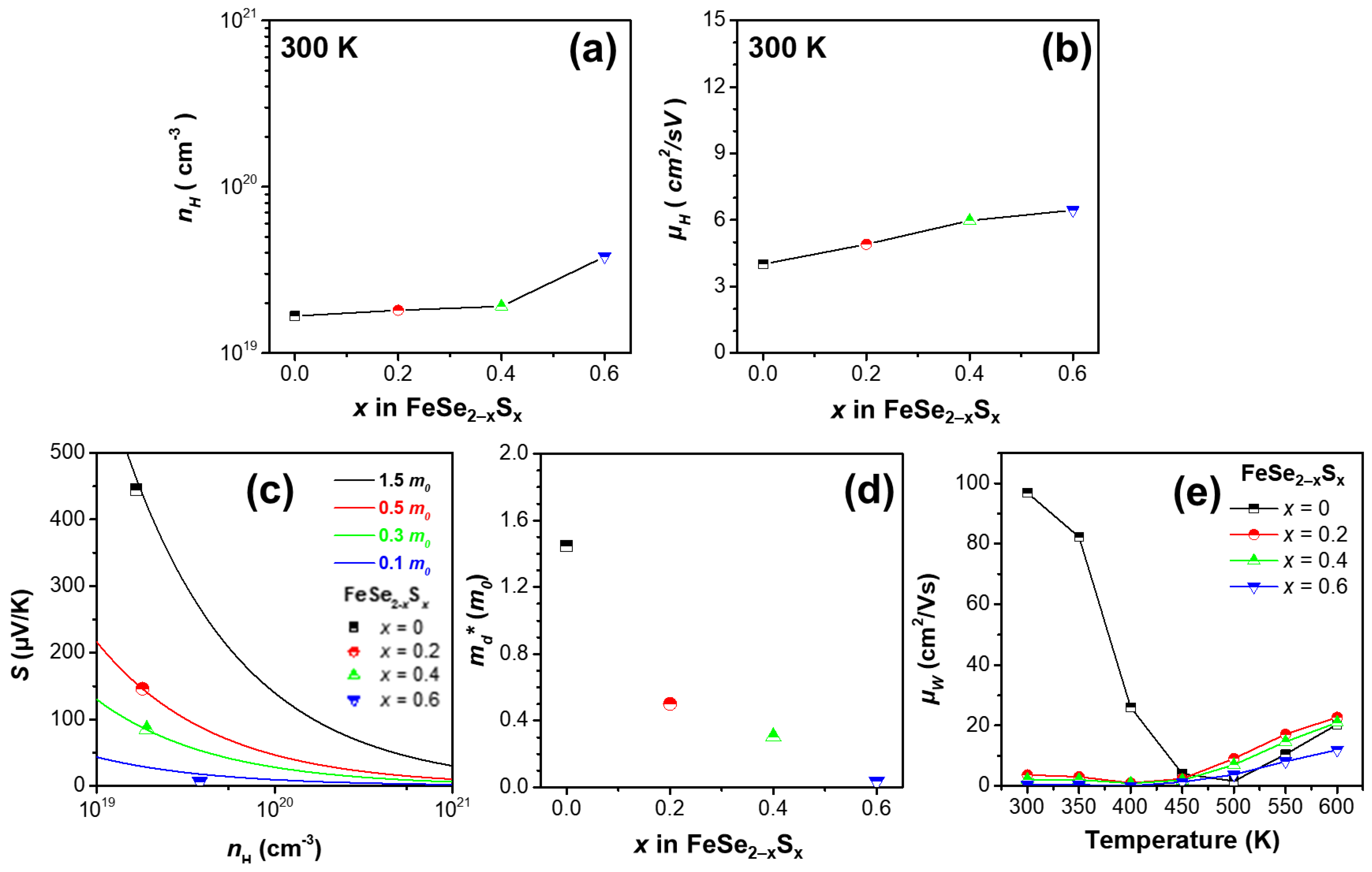
Figure 3a,b present the Hall carrier concentration (nH) and Hall mobility (μH) of the samples, measured at 300 K. The nH values were 1.67 × 10¹⁹, 1.81 × 10¹⁹, 1.91 × 10¹⁹, and 3.80 × 1019 cm−3 and the μH values were 4.01, 4.90, 5.97, and 6.44 cm2/Vs, for x = 0, 0.2, 0.4, and 0.6, respectively. All the samples exhibited positive nH values at 300 K, and nH and μH increased gradually with an increase in x. Thus, the increase in the σ of the samples could be due to the simultaneous increase in nH and μH. Furthermore, an increase in carrier concentration generally leads to a decrease in the magnitude of S, according to the Mott relationship [24]:
S = 8 π 2 k 2 3 e h 2 m d * T π 3 n 2 3 ,
where md*, e, and h are the density-of-state effective mass, elementary charge, and Planck’s constant, respectively. Therefore, the decrease in the magnitude of S for the alloyed samples at 300 K could be attributed to the increase in nH. Figure 3c,d show the md* values of the samples at 300 K calculated using the measured S and nH, based on the relationship in Equation (6). The md* values of the samples were 1.45, 0.50, 0.30, and 0.04 m0 for x = 0, 0.2, 0.4, and 0.6, respectively. The md* values decreased gradually with an increase in S content.
Figure 3e shows the weighted mobility (μw) of the samples, obtained for a better understanding of the thermoelectric properties of the samples. The μw was calculated using the measured σ and S from a simple analytic form that approximates the exact Drude–Sommerfeld free-electron model for |S| > 20 μV/K [25]:
μ w = 3 h 3 σ 8 π e 2 m e k T 3 / 2 exp S k / e 2 1 + exp 5 S k / e 1 + 3 π 2 S k / e 1 + exp 5 S k / e 1 ,
where me is the electron mass. μw is closely related to the theoretically optimum electrical performance of a thermoelectric material, and is relevant to the maximum power factor when nH is tuned properly [26]. Therefore, the μw trend was very similar to that of the power factor. The values of μw at 600 K were 20.3, 22.7, 20.9, and 11.9 cm2/Vs for x = 0, 0.2, 0.4, and 0.6, respectively. The μw increased initially at x = 0.2 and decreased gradually at x > 0.2, which was in agreement with the power factor trend at 600 K.
Figure 4a,b show the κtot and κlatt of the samples as functions of temperature. The inset of Figure 4a shows the temperature dependence of κelec. The κelec of the samples was calculated according to the Wiedemann–Franz law [27]:
κ e l e c = L σ T
where L is the Lorenz number. Subsequently, the κlatt was calculated by subtracting κelec from κtot. The electronic contribution to κtot was not very significant; thus, κtot and κlatt exhibited similar values. The κlatt values of the samples were 7.96, 6.07, 4.47, and 2.58 W/mK at 300 K and 4.29, 3.85, 3.51, and 2.10 W/mK at 600 K, for x = 0, 0.2, 0.4, and 0.6, respectively. The κlatt decreased gradually as the S content increased, over the entire temperature range, which was attributed to the point-defect phonon scattering caused by the partial substitution of S atoms at Se sites (the atomic masses of S and Se are 32.065 and 78.96 amu and the ionic radii of S2− and Se2− are 170 and 184 pm, respectively).
Figure 4c,d shows the calculated zT values of p- and n-type conduction, respectively. In Figure 4c, the maximum zT of the pristine sample was ~0.02, and the alloyed samples exhibited even lower zT values below 0.005. On the other hand, in Figure 4d of n-type conduction of high-temperature range, the samples with x = 0.2 and 0.4 exhibited zT values higher than that of the pristine sample, mainly owing to the decrease in κtot. Consequently, the FeSe1.8S0.2 (x = 0.2) sample exhibited a maximum zT value of 0.079, which was approximately 18% higher than that of the pristine FeSe2 sample.

4. Conclusions

A series of FeSe2−xSx (x = 0, 0.2, 0.4, 0.6, and 0.8) polycrystalline samples were synthesized by a conventional solid-state reaction, and their thermoelectric transport properties were examined in an effort to search for the cost-effective thermoelectric materials. A single orthorhombic FeSe2 phase was successfully synthesized for x = 0, 0.2, and 0.4; however, a secondary phase (Fe7S8 or FeS2) was identified for x = 0.6 and 0.8. The lattice parameters decreased gradually with an increase in S content for x < 0.8, suggesting that S atoms were substituted at the Se sites in the FeSe2 crystal structure. The electrical conductivity increased gradually with an increase in S content, whereas the magnitude of S decreased gradually with an increase in S content. As a result, the sample with x = 0.2 exhibited a maximum power factor of 0.55 mW/mK2 at 600 K. The total thermal conductivity decreased significantly with an increase in S content, and thus a maximum thermoelectric figure of merit value of 0.079 was obtained for the FeSe1.8S0.2 (x = 0.2) sample at 600 K, which was approximately 18% higher than that of the FeSe2 (x = 0) sample.

Supplementary Materials

The following supporting information can be downloaded at: https://www.mdpi.com/article/10.3390/mi13122066/s1, Figure S1: EDS-SEM results for FeSe2−xSx (x = 0, 0.2, 0.4, 0.6 and 0.8); Figure S2: (a) σ, (b) S, and (c) PF as a function of temperature for the FeSe2 sample, measured after ~180 days from the initial measurement for cycling test; Figure S3: Estimated Eg for the series of FeSe2−xSx (x = 0, 0.2, 0.4 and 0.6) samples using Goldsmid-Sharp empirical formular; Table S1: The relative peak intensities for (111), (012), (121), (011), (200) and (103) planes for FeSe2−xSx (x = 0, 0.2, 0.4, and 0.6) in the X-ray diffraction data; Table S2: Atomic ratios measured by EDS-SEM for FeSe2−xSx (x = 0, 0.2, 0.4, 0.6 and 0.8). References [16,28] are cited in the supplementary materials.

Author Contributions

Conceptualization, S.-i.K.; investigation, O.P. and S.W.L.; data curation, O.P. and S.W.L.; writing—original draft preparation, O.P., S.W.L., and S.J.P.; writing—review and editing, S.-i.K.; project administration, S.-i.K.; funding acquisition, S.-i.K. All authors have read and agreed to the published version of the manuscript.

Funding

This study was supported by the National Research Foundation of Korea (NRF-2019R1C1C1005254, and NRF-2022R1F1A1063054).

Data Availability Statement

Data available upon reasonable request.

Conflicts of Interest

The authors declare no conflict of interest.

References

  1. Flipse, J.; Bakker, F.L.; Slachter, A.; Dejene, F.K.; van Wees, B.J. Direct observation of the spin-dependent Peltier effect. Nat. Nanotechnol. 2012, 7, 166–168. [Google Scholar]
  2. Tritt, T.M.; Subramanian, M.A. Thermoelectric Materials, Phenomena, and Applications: A Bird’s Eye View. MRS Bull. 2006, 31, 188–198. [Google Scholar] [CrossRef] [Green Version]
  3. Gaultois, M.W.; Sparks, T.D.; Borg, C.K.H.; Seshadri, R.; Bonificio, W.D.; Clarke, D.R. Data-Driven Review of Thermoelectric Materials: Performance and Resource Considerations BT—Chemistry of Materials. Chem. Mater. 2013, 25, 2911–2920. [Google Scholar] [CrossRef] [Green Version]
  4. Zhao, K.; Guan, M.; Qiu, P.; Blichfeld, A.B.; Eikeland, E.; Zhu, C.; Ren, D.; Xu, F.; Iversen, B.B.; Shi, X.; et al. Thermoelectric Properties of Cu2Se1−xTex Solid Solutions. J. Mater. Chem. A 2018, 6, 6977–6986. [Google Scholar] [CrossRef] [Green Version]
  5. Skoug, E.J.; Cain, J.D.; Morelli, D.T. High thermoelectric figure of merit in the Cu3SbSe4-Cu3SbS4 solid solution. Appl. Phys. Lett. 2011, 98, 261911. [Google Scholar] [CrossRef]
  6. Xing, T.; Zhu, C.; Song, Q.; Huang, H.; Xiao, J.; Ren, D.; Shi, M.; Qiu, P.; Shi, X.; Xu, F.; et al. Ultralow Lattice Thermal Conductivity and Superhigh Thermoelectric Figure-of-Merit in (Mg, Bi) Co-Doped GeTe. Adv. Mater. 2021, 33, 2008773. [Google Scholar] [CrossRef]
  7. Asfandiyar, T.-R.W.; Li, Z.; Sun, F.-H.; Pan, Y.; Wu, C.-F.; Farooq, M.U.; Tang, H.; Li, F.; Li, B.; Li, J.-F. Thermoelectric SnS and SnS-SnSe solid solutions prepared by mechanical alloying and spark plasma sintering: Anisotropic thermoelectric properties. Sci. Rep. 2017, 7, 43262. [Google Scholar] [CrossRef] [Green Version]
  8. Bang, J.; Kim, H.-S.; Kim, D.H.; Lee, S.W.; Park, O.; Kim, S.-I. Phase formation behavior and electronic transport properties of HfSe2-HfTe2 solid solution system. J. Alloys Compd. 2022, 920, 166028. [Google Scholar] [CrossRef]
  9. Song, H.-Y.; Sun, J.-J.; Li, M. Enhancement of monolayer HfSe2 thermoelectric performance by strain engineering: A DFT calculation. Chem. Phys. Lett. 2021, 784, 139109. [Google Scholar] [CrossRef]
  10. Abdollah, H.M.; Mohammad, Y.; Mojtaba, Y.; Alireza, A. Investigation into thermoelectric properties of M (M = Hf, Zr) X2 (X = S, Se, Te) nanotubes using first-principles calculation. Solid State Commun. 2021, 336, 114289. [Google Scholar] [CrossRef]
  11. Kumar, S.; Schwingenschlögl, U. Thermoelectric Response of Bulk and Monolayer MoSe2 and WSe2. Chem. Mater. 2015, 27, 1278–1284. [Google Scholar] [CrossRef]
  12. Luo, Y.; Zheng, Y.; Luo, Z.; Hao, S.; Du, C.; Liang, Q.; Li, Z.; Khor, K.A.; Hippalgaonkar, K.; Xu, J.; et al. n-Type SnSe2 Oriented-Nanoplate-Based Pellets for High Thermoelectric Performance. Adv. Energy Mater. 2018, 8, 1702167. [Google Scholar] [CrossRef]
  13. Kim, S.-I.; Bang, J.; An, J.; Hong, S.; Bang, G.; Shin, W.H.; Kim, T.; Lee, K. Effect of Br substitution on thermoelectric transport properties in layered SnSe2. J. Alloys Compd. 2021, 868, 159161. [Google Scholar] [CrossRef]
  14. Mavrokefalos, A.; Nguyen, N.T.; Pettes, M.T.; Johnson, D.C.; Shi, L. In-plane thermal conductivity of disordered layered WSe2 and (W)x(WSe2)y superlattice films. Appl. Phys. Lett. 2007, 91, 171912. [Google Scholar] [CrossRef] [Green Version]
  15. Guo, Z.; Zhao, R.; Yan, S.; Xiong, W.; Zhu, J.; Lu, K.; Wang, X. Atomic Layer Deposition of FeSe2, CoSe2, and NiSe2. Chem. Mater. 2021, 33, 2478–2487. [Google Scholar] [CrossRef]
  16. Li, G.; Zhang, B.; Rao, J.; Herranz Gonzalez, D.; Blake, G.R.; de Groot, R.A.; Palstra, T.T. Effect of vacancies on magnetism, electrical transport, and thermoelectric performance of marcasite FeSe2−δ (δ = 0.05). Chem. Mat. 2015, 27, 8220. [Google Scholar] [CrossRef]
  17. Gudelli, V.K.; Kanchana, V.; Vaitheeswaran, G.; Valsakumar, M.C.; Mahanti, S.D. Thermoelectric properties of marcasite and pyrite FeX2 (X = Se, Te): A first principle study. RSC Adv. 2014, 4, 9424. [Google Scholar] [CrossRef] [Green Version]
  18. Park, O.; Kim, T.; Lee, S.W.; Kim, H.-S.; Shin, W.H.; Rahman, J.U.; Kim, S.-I. Study of Phase Formation Behavior and Electronic Transport Properties in the FeSe2-FeTe2 System. Korean J. Met. Mater. 2022, 60, 315–320. [Google Scholar] [CrossRef]
  19. Zuniga-Puelles, E.; Cardoso-Gil, R.; Bobnar, M.; Veremchuk, I.; Himcinschi, C.; Hennig, C.; Kortus, J.; Heide, G.; Gumeniuk, R. Structural stability and thermoelectric performance of high quality synthetic and natural pyrites (FeS2). Dalton Trans. 2019, 48, 10703–10713. [Google Scholar] [CrossRef]
  20. Harran, I.; Li, Y.; Wang, H.; Chen, Y.; Ni, Y. Iron Disulfide Compound: A Promising Thermoelectric Material. Mater. Res. Express 2017, 4, 105907. [Google Scholar] [CrossRef]
  21. Gronvold, F.; Westrum, E.F., Jr. Heat capacities of iron disulfides Thermodynamics of marcasite from 5 to 700 K, pyrite from 300 to 780 K, and the transformation of marcasite to pyrite. J. Chem. Thermodyn. 1976, 8, 1039–1048. [Google Scholar] [CrossRef]
  22. Patterson, A.L. The Scherrer Formula for X-Ray Particle Size Determination. Phys. Rev. 1939, 56, 978–982. [Google Scholar] [CrossRef]
  23. Yoo, J.; Kim, J.-I.; Cho, H.-J.; Choo, S.-S.; Kim, S.-I.; Lee, K.; Shin, W.H.; Kim, H.-S.; Roh, J.W. Electronic and Thermal Properties of Si-doped InSe Layered Chalcogenides. J. Korean Phys. Soc. 2018, 72, 775–779. [Google Scholar] [CrossRef]
  24. Ioffe, A.F. Physics of Semiconductors; Academic Press: New York, NY, USA, 1960. [Google Scholar]
  25. Snyder, G.J.; Snyder, A.H.; Wood, M.; Gurunathan, R.; Snyder, B.H.; Niu, C. Weighted mobility. Adv. Mater. 2020, 32, 2001537. [Google Scholar] [CrossRef]
  26. Kim, M.; Kim, S.-I.; Kim, S.W.; Kim, H.-S.; Lee, K.H. Weighted mobility ratio engineering for high-performance Bi-Te-based thermoelectric materials via suppression of minority carrier transport. Adv. Mater. 2021, 33, 2005931. [Google Scholar] [CrossRef]
  27. Lorenz, L. Determination of the degree of heat in absolute measure. Ann. Phys. Chem. 1872, 147, 429–451. [Google Scholar] [CrossRef] [Green Version]
  28. Goldsmid, H.J.; Sharp, J.W. Estimation of the thermal band gap of a semiconductor from seebeck measurements. J. Electron. Mater. 1999, 28, 869–872. [Google Scholar] [CrossRef]
Figure 1. (a) XRD patterns of the series of FeSe2−xSx (x = 0, 0.2, 0.4, 0.6, 0.8, and 1) samples. (b) Lattice parameters calculated using the XRD patterns.
Micromachines 13 02066 g001
Figure 2. (a) σ as a function of the temperature for the series of FeSe2−xSx (x = 0, 0.2, 0.4, 0.6, and 0.8) samples. (b) Ea of the samples calculated from σ for 300–400 K and 450–600 K. The inset of (b) shows logarithmic σ as a function of 1000/T for the samples. (c) S as a function of temperature for the samples. Power factors as functions of temperature for the samples in (d) p- and (e) n-type regions.
Micromachines 13 02066 g002
Figure 3. (a) nH and (b) μH of the series of FeSe2−xSx (x = 0, 0.2, 0.4, and 0.6) samples. (c) S as a function of nH (Pisarenko plot) at 300 K. (d) md* as a function of x in FeSe2−xSx (x = 0, 0.2, 0.4, and 0.6). (e) μw as a function of temperature for the samples.
Micromachines 13 02066 g003
Figure 4. (a) κtot and (b) κlatt as functions of temperature for the series of FeSe2−xSx (x = 0, 0.2, 0.4, and 0.6) samples. The inset of (a) shows κelec as a function of temperature for the samples. zT as a function of temperature for the samples in (c) p- and (d) n-type regions.
Micromachines 13 02066 g004
Publisher’s Note: MDPI stays neutral with regard to jurisdictional claims in published maps and institutional affiliations.