Review

Pseudomonas aeruginosa Type III Secretory Toxin ExoU and Its Predicted Homologs

Department of Anesthesiology, School of Medicine, Kyoto Prefectural University of Medicine, Kyoto 602-8566, Japan
*
Author to whom correspondence should be addressed.
Toxins 2016, 8(11), 307; https://doi.org/10.3390/toxins8110307
Submission received: 7 September 2016 / Revised: 20 October 2016 / Accepted: 21 October 2016 / Published: 26 October 2016
(This article belongs to the Section Bacterial Toxins)

Abstract

:
Pseudomonas aeruginosa ExoU, a type III secretory toxin and major virulence factor with patatin-like phospholipase activity, is responsible for acute lung injury and sepsis in immunocompromised patients. Through use of a recently updated bacterial genome database, protein sequences predicted to be homologous to Ps. aeruginosa ExoU were identified in 17 other Pseudomonas species (Ps. fluorescens, Ps. lundensis, Ps. weihenstephanensis, Ps. marginalis, Ps. rhodesiae, Ps. synxantha, Ps. libanensis, Ps. extremaustralis, Ps. veronii, Ps. simiae, Ps. trivialis, Ps. tolaasii, Ps. orientalis, Ps. taetrolens, Ps. syringae, Ps. viridiflava, and Ps. cannabina) and 8 Gram-negative bacteria from three other genera (Photorhabdus, Aeromonas, and Paludibacterium). In the alignment of the predicted primary amino acid sequences used for the phylogenetic analyses, both highly conserved and nonconserved parts of the toxin were discovered among the various species. Further comparative studies of the predicted ExoU homologs should provide us with more detailed information about the unique characteristics of the Ps. aeruginosa ExoU toxin.

1. Introduction

Bacterial pneumonia that occurs frequently in patients under artificial respiration is defined as ventilator-associated pneumonia (VAP) [1]. Pseudomonas aeruginosa (hereafter called Ps. aeruginosa) is one of the most frequent causative bacterial agents of VAP [2]. Additionally, when Ps. aeruginosa is the causative agent of pneumonia, acute lung injury and secondary sepsis can easily occur, leading to high fatality rates [3,4,5,6,7]. In animal models of Ps. aeruginosa pneumonia, once cytotoxic Ps. aeruginosa strains are administered to the lungs of an experimental animal, the animal immediately develops an acute lung injury followed by bacteremia and secondary sepsis [8,9,10]. Over the past 20 years, investigations to identify virulence factors and their related mechanisms of action in Ps. aeruginosa have led to the discovery of the type III secretory toxin, ExoU, as a major virulence factor responsible for acute lung injury and sepsis [10,11,12,13]. In addition, clinical associations with Ps. aeruginosa ExoU genotype isolates have recently been reported in various other infections, such as chronic otitis media, keratitis, burns and bloodstream infections, and skin infections in diabetic patients [14,15,16,17,18,19].
Until recently, there was no obvious homolog of ExoU, with the exception of Rickettsia prowazekii PR534 [20], and Ps. aeruginosa ExoU was considered to be virtually the sole patatin-like cytotoxin secreted by the type III secretory mechanism. However, recent advances in DNA sequencing technologies have facilitated identification of several predicted ExoU homologs in various bacterial species, and these homologs have been reported in the bacterial genome database of the National Center for Biotechnology Information [21]. In this review, our brief summary of the molecular biology of the ExoU cytotoxin as a patatin-like phospholipase (PLP) is augmented by a comparative study of the predicted ExoU homologs from various bacterial species.

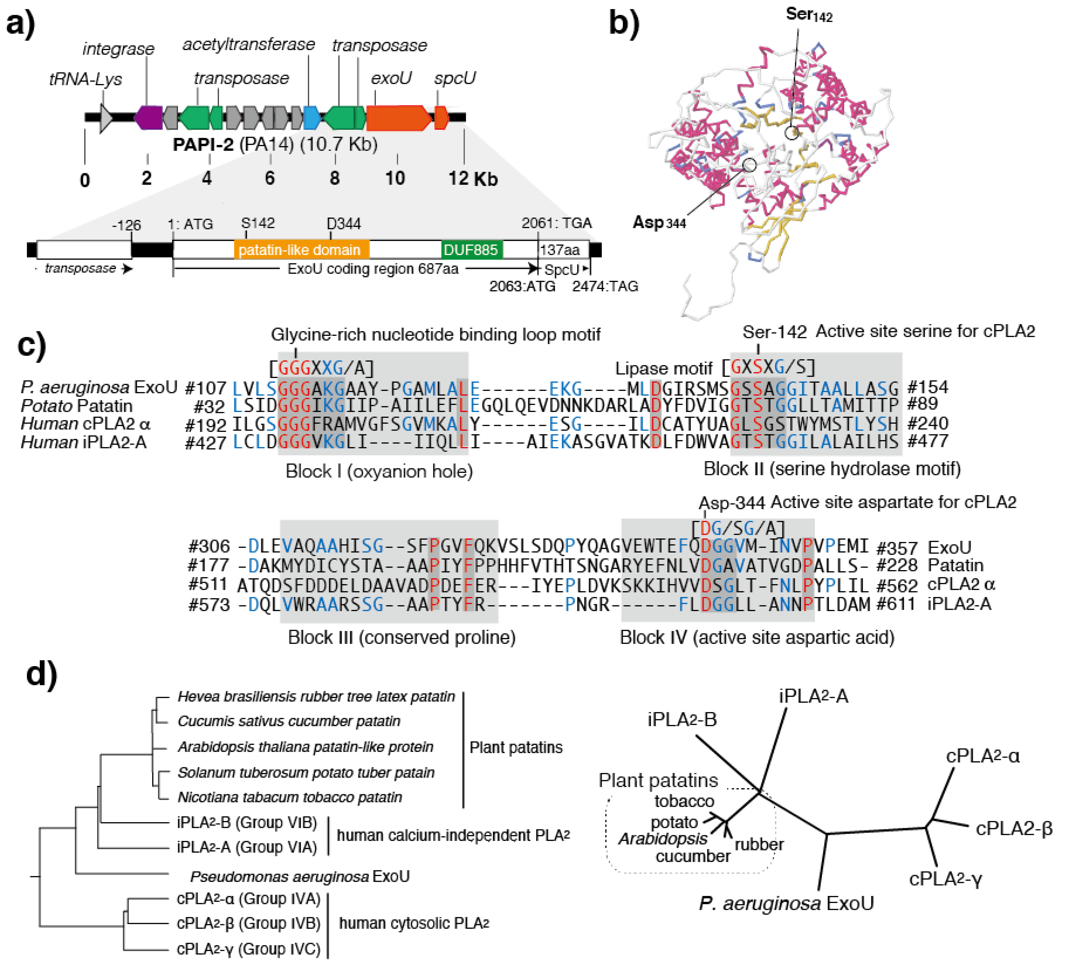
2. Molecular Biology of the ExoU Cytotoxin

In 1997, ExoU was first identified in the cytotoxic Ps. aeruginosa strain that causes severe acute lung injury and sepsis in animal models of pneumonia [11]. In that study, the 2064-bp open reading frame of the ExoU gene (exoU) was cloned, and the predicted 687 amino acid protein encoded by it was characterized (Figure 1a) [11]. The TGA stop codon of exoU overlaps with the ATG start codon of spcU, the latter of which encodes the SpcU chaperone for ExoU secretion consisting of 137 amino acids [22]. ExoU was simultaneously characterized as one of the type III secreted toxins that Ps. aeruginosa injects directly into the cytosol of targeted eukaryotic cells through its type III secretory apparatus [11]. Later, clinical studies revealed that Ps. aeruginosa isolates possessing exoU cause bacteremia, sepsis, and high mortality [3]. Genetic analyses also have shown that the exoU gene is located in the Ps. aeruginosa pathogenicity island-2, and was found as an insertional gene cluster in the chromosomal genome of cytotoxic Ps. aeruginosa isolates (Figure 1a) [23].
Virulent Ps. aeruginosa isolates secreting ExoU show cytotoxicity in cultured epithelial cells and macrophages [9,10,11]. In various animal models, ExoU has been characterized as a major virulence factor for acute lung injury [10,11,12]. Among the various phenotypes of Ps. aeruginosa isolates, the ExoU-positive phenotype is a major risk factor for poor clinical outcomes [3,4]. A correlation between the antimicrobial characteristics of the bacterium and an exoU-positive genotype has also been reported in recent clinical studies [13]. After its discovery in 1997, the mechanism of acute cell death induced by the ExoU cytotoxin remained unknown for some time. However, in 2003, the PLP structure and the enzymatic activity of activated ExoU were elucidated [24]. Both in vitro and in vivo studies have shown that the phospholipase A2 and lysophospholipase activities of ExoU are activated by unknown eukaryotic cell factor(s) [25,26]. The PLP domain of ExoU contains a catalytic dyad consisting of serine (Ser142) and aspartic acid (Asp344) residues (Figure 1a,b) [24]. This catalytic dyad structure is similar to those of mammalian cytosolic phospholipase A2 (cPLA2) and calcium2+-independent phospholipase A2 (iPLA2), both of which belong to a family of PLP-domain proteins (Figure 1c) [24]. In ExoU mutant strains, which were genetically modified by introducing missense point-mutations into the catalytic dyad, there was a loss of PLP activity and cytotoxicity to eukaryotic cells [24]. Therefore, the PLP activity of ExoU was characterized as a cell death factor that acts to destroy cellular lipid membranes [24].
One feature of the type III secretion system is that the enzymatic actions of each toxin it contains are activated by eukaryotic cell factor(s) after the toxins are translocated into the cytoplasm of the targeted eukaryotic cell. For example, the ADP-ribosyl transferase activity of the Ps. aeruginosa type III secretory toxin ExoS is activated by a factor that is a member of the 14-3-3 protein family [27], while the adenylate cyclase activity of ExoY is activated by unknown factor(s) derived from eukaryotic cells [28]. Similarly, the PLA2 action of ExoU requires the presence of an activator contained in the cytosol extract of eukaryotic cells. In 2006, ExoU was reported to be activated by superoxide dismutase (SOD1) [29]. In the same year, other research reported that lysine178 and the carboxyl-terminal amino acids 679–683 of SOD1 are ubiquitinated during the activation process [30]. Subsequent studies revealed that SOD1 acts as the ubiquitin donor, and the ubiquitination of the carboxyl terminal domain of ExoU is the mechanism of PLP activation [31,32]. The carboxyl terminal of ExoU contains a conserved domain called DUF885, and mutant proteins lacking this region cannot be activated [31,32]. The three-dimensional structural analysis using crystallized cPLA2 revealed that serine and asparagine residues in the enzymatic center of PLP are located very close to each other and form a catalytic dyad in the three-dimensional structure [33]. However, structural analysis of the nonactive form of ExoU revealed that Ser142 and Asp344, which form the catalytic dyad for PLP, are separated further away from each other in the predicted three-dimensional structure of the nonactivated ExoU [34]. Therefore, it can be assumed that ubiquitination of the carboxyl-terminal brings Ser142 and Asp344 closer to each other to produce an active form of the dyad structure.

3. ExoU and PLP A2 Activity

Patatins, which account for about 40% of the soluble protein in potatoes (Solanum tuberosum) and are present in cucumbers (Cucumis sativus) and Para rubber trees (Hevea brasiliensis), are plant storage glycoproteins with acyl hydrolase activities [36,37]. Patatins are also known to be cross-reactive latex allergens that can cause latex allergies (natural rubber fruit syndrome, latex-fruit syndrome). Conversely, it has been found that many latex allergens contain Hev b7, a protein with a structure similar to patatin [38,39]. Patatin exhibits insecticidal activity against plant pest larvae; it is also considered to be an immune system protein of the plant kingdom, and has become known as an infection-specific pathogenesis-related protein in the plant pathology field [40].
Lipid-degrading enzymes generally possess the triad structure characterized by the presence of asparagine (or glutamic acid), serine, and histidine. However, unlike lipid-degrading enzymes, PLPs such as potato patatin B2, human cPLA2/iPLA2, and Ps. aeruginosa ExoU all generate their acyl hydrolase activities through the enzyme active site of the dyad formed by serine and asparagine [36]. There is a possibility that, during the evolutionary process, the gene encoding the patatin-like protein with insecticidal activity in plants was somehow incorporated into the genomes of pathogenic bacteria through a horizontal gene transfer mechanism. Indeed, R. prowazekii RP534—which was the only obviously recognized homolog of ExoU until recently [20]—has a PLP structure, and the recombinant protein of RP534 has PLA2 and lysophospholipase activities. Therefore, there is a possibility that Rickettsia RP534 is the link between plant and bacterial patatins [20].

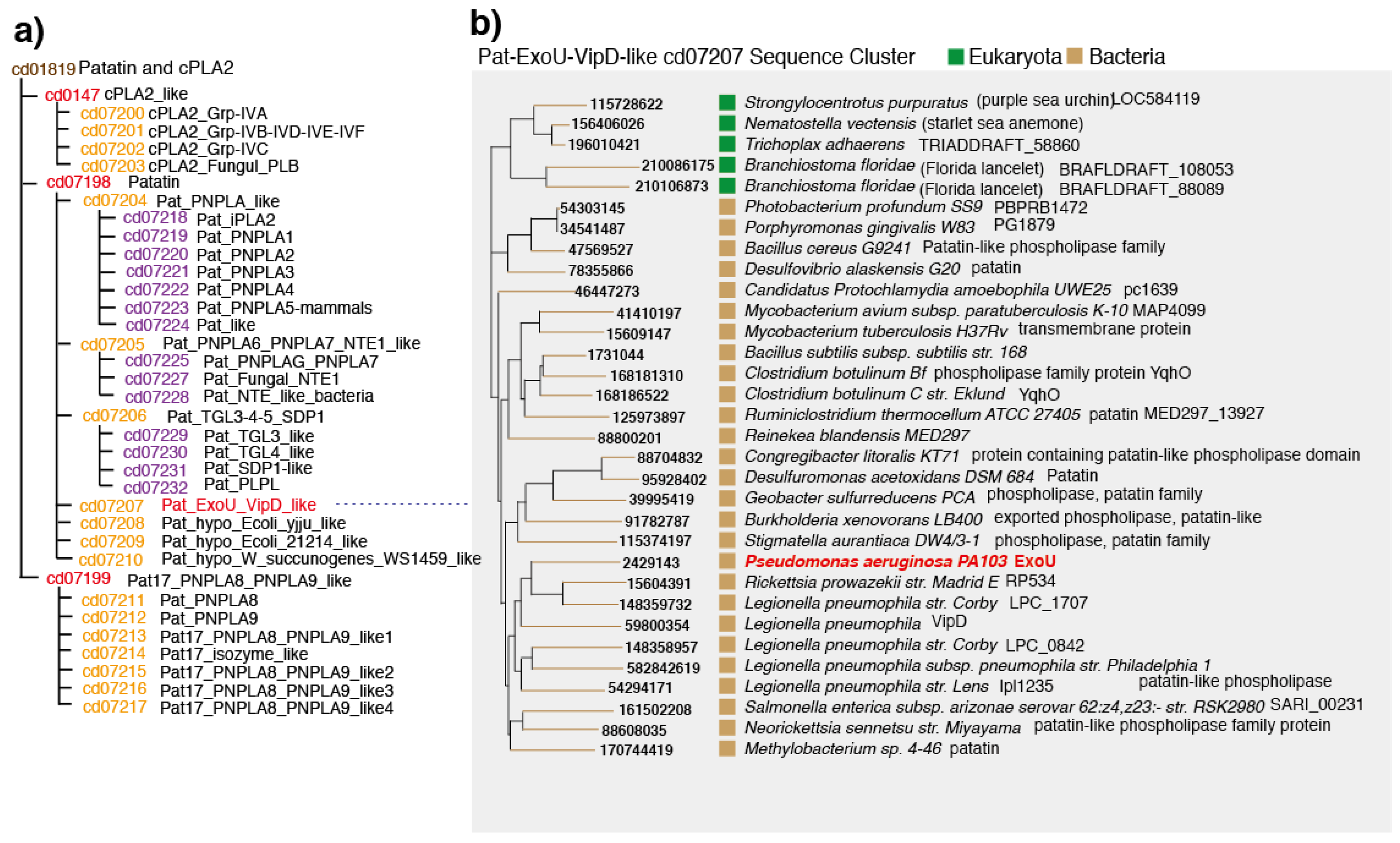
4. Conserved Domains in PLPs

The PLP protein family, which includes ExoU, contains four conserved domains (blocks I–IV) (Figure 1c) [41,42]. Block I is a glycine-rich region containing an arginine or lysine residue and consisting of an oxyanion hole. Block II, which is located 10–20 amino acids away from block I, contains a GxSxG (or GxSxS) hydrolase motif sequence and a serine residue at the PLP enzymatic activity center. Block III contains several well-conserved proline residues in the group, and these are considered to be critical for maintaining the three-dimensional structure. In block III, the bacterial PLPs have an ASxxxP sequence, while the eukaryotic PLPs have an AAP sequence. Block IV contains an Asp-residue that is a pair component of a catalytic dyad with the Ser-residue of block I.
The patatin-like motif in ExoU is located at amino acid positions 110–354 in the amino-terminal side of the protein. First, the 110–125 position in block I of ExoU has an oxyanion hole (a glycine-rich nucleotide binding loop) comprising a GGGAxxG/A motif. The 139–145 position of ExoU block II has a hydrolase motif GxSxG (or GxSxS) with a catalytic center Ser142 residue. ExoU block III contains a patatin-specific proline at position 320 within the 309–325 amino acid region. Position 341–352 of ExoU block IV contains a D-G/S-G/A motif containing the other catalytic center residue, Asp344. This block displays high sequence homology with the blocks of other PLPs (Figure 1c). In the phylogenetic diagram that was created based on the primary amino acid sequence of the region containing blocks I and II of the patatin-like domain, ExoU is located just between human cPLA2/iPLA2 and a plant PLP group (Figure 1d).

5. Patatins and Phospholipases

The patatin and cPLA2 domain family (cd01819) consists of various patatin glycoproteins from plants and mammalian cPLA2 proteins (EC 3.1.1.4) (Figure 2a). The three major subdomains in these proteins are cPLA2_like (cd0127), patatin (cd07198), and Pat17_PNPA8_PNPLA9_like (cd07199). The phospholipase A2 group of proteins is characterized by a lipid hydrolase catalytic dyad containing an active site serine and asparagine. cPLA2, which belongs to the domain family cd0147, is a group IV PLA2 that contains six intercellular enzymes (alpha, beta, gamma, delta, epsilon, and zeta). Vertebrate iPLA2 belongs to the domain family Pat17_PNPA8_PNPLA9_like (cd07199).
The Pat_ExoU_VipD_like domain family, which is one of seven patatin domain families, has a sequence cluster comprising ExoU of Ps. aeruginosa, VipD of Legionella pneumophila, several other L. pneumophila proteins, and proteins from Mycobacterium, Clostridium botulinum, Burkholderia xenovorans, and Salmonella enterica (Figure 2a). In addition, this family includes eukaryotic proteins from purple sea urchin, starlet sea anemone, Trichoplax adhaerens, and Florida lancelet. VipD was shown to have no phospholipase activity, even though it shares high sequence similarity with several functional regions of ExoU (e.g., oxyanion hole, active site serine, active site asparagine). Additionally, in the patatin-domain protein family, the closest homolog to Ps. aeruginosa ExoU is R. prowazekii PR534.

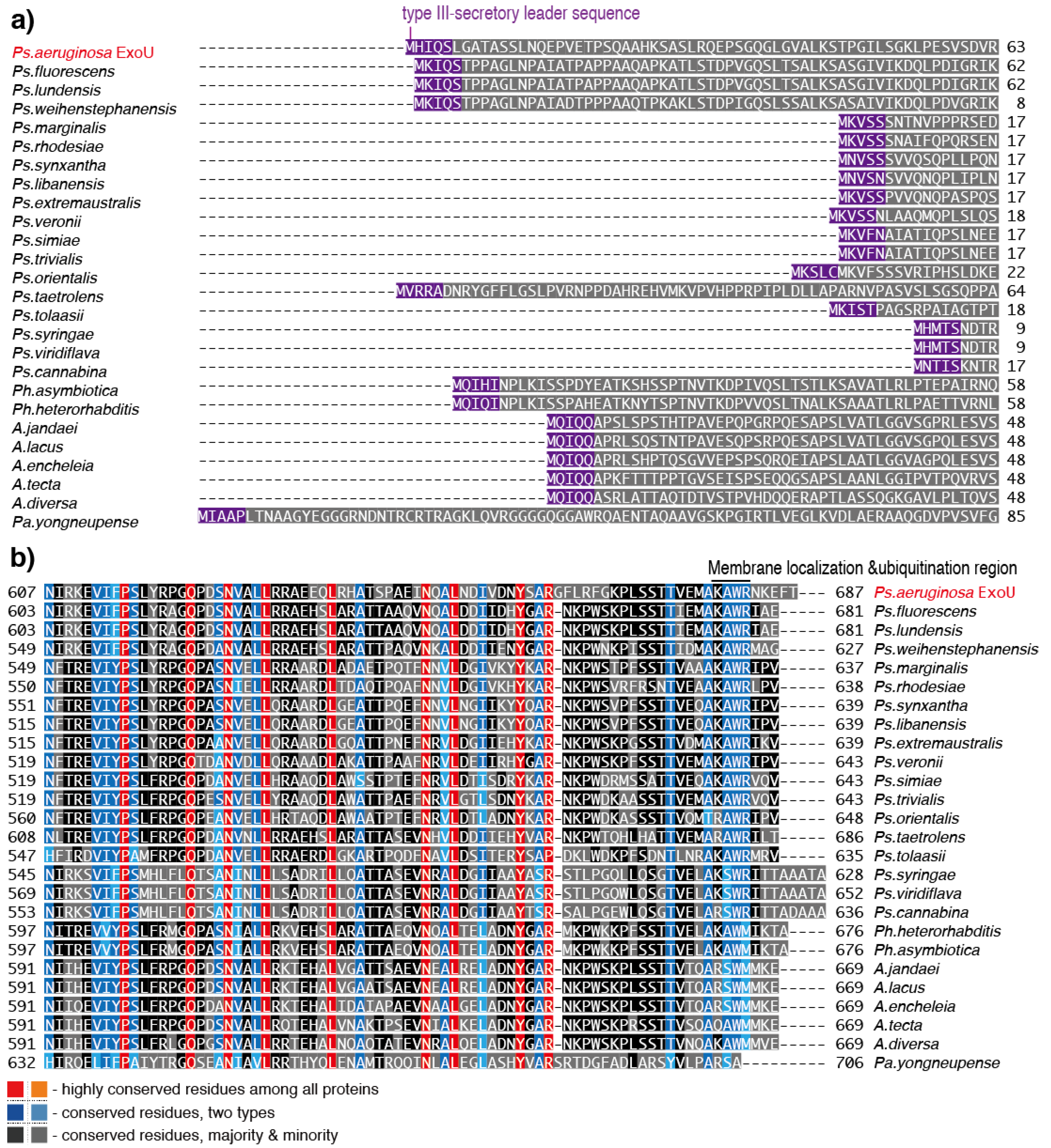
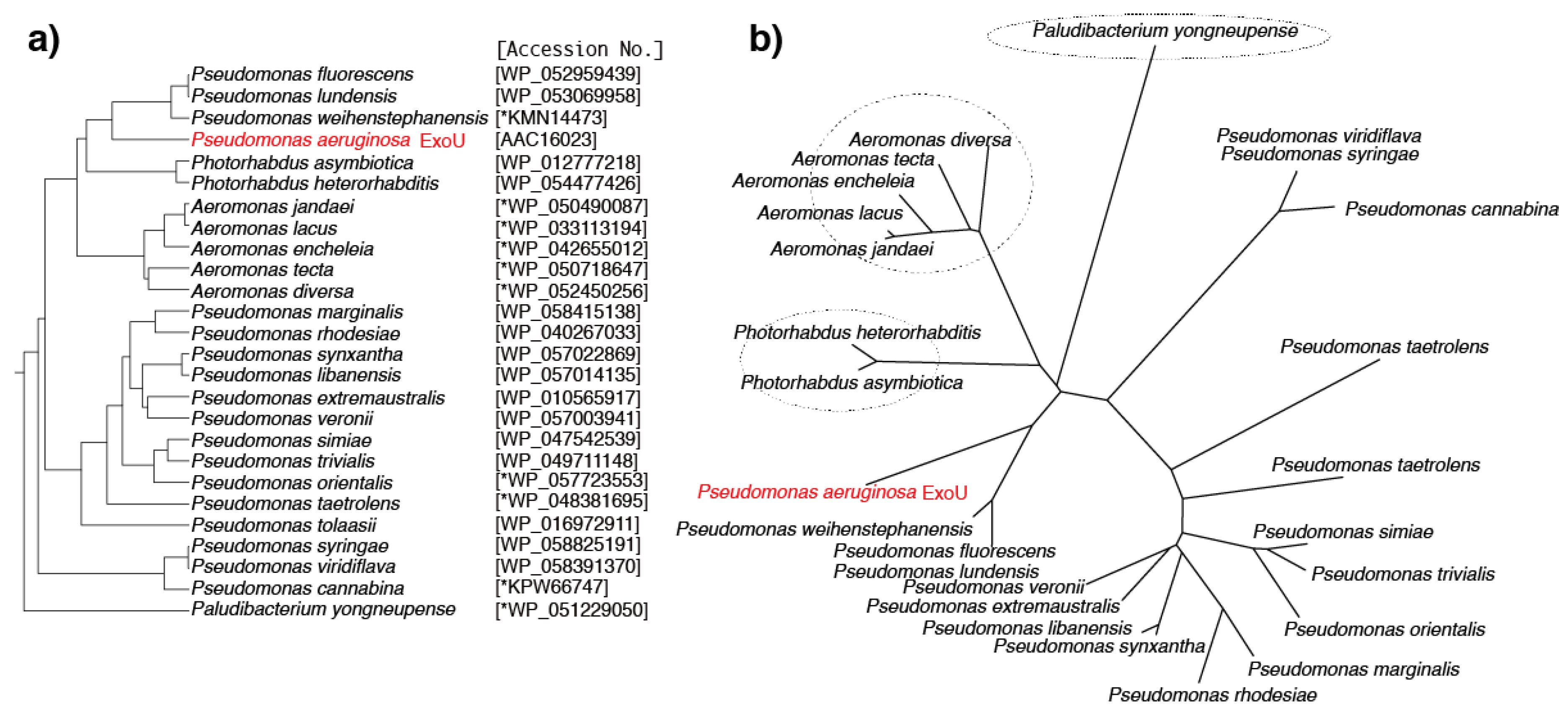
6. Predicted ExoU Homologs in Pseudomonas and Other Gram-Negative Bacteria

In our current search of the NCBI bacterial genome database we identified many predicted protein homologs of Ps. aeruginosa ExoU: 17 proteins from other Pseudomonas species and 8 proteins from other Gram-negative bacteria (Table 1, Figure 3a,b). Photorhabdus species are Gram-negative entomopathogenic bacteria of the family Enterobacteriaceae. Predicted ExoU homologs have been found in two Photorhabdus species. Among several members of this genus, Photorhabdus asymbiotica is a pathogen of both insects and humans. Ph. heterorhabditis, which was recently proposed to be a novel species of the genus Photorhabdus, was isolated from populations of the pathogenic insect nematode Heterorhabditis zealandica collected in South Africa [43]. It was reported that Ph. asymbiotica induces cellular death through the activation of caspases 8 and 9 and the executioner caspases 3 and 7 [44,45,46,47], and this suggests that the predicted ExoU homolog might be involved in the cell death mechanism.
Aeromonas, a Gram-negative, facultative anaerobic and rod-shaped bacterium, is ubiquitous in fresh and brackish water [48]. Most Aeromonas species are known to be associated with human diseases. Predicted ExoU homologs were found in five Aeromonas species. Aeromonas jandaei is a Gram-negative bacterium isolated from human feces in Oregon, USA [49]. A. lacus is one of three groups of Aeromonas strains isolated from lakes in Finland [50], and while A. encheleia was isolated from European eels in Valencia, Spain [51,52,53], A. tecta is a strain isolated from the stool of a child with diarrhea, from a healthy patient, and from environmental sources [54].
Paludibacterium yongneupense, a bacterium that falls within the family Neisseriaceae, was isolated from a Korean wetland area [55]. In the primary sequence of the predicted Pa. yongneupense ExoU, blocks I–IV of this ExoU homolog are a good match with those of Ps. aeruginosa ExoU. Figure 4 shows the amino acid sequences of eight predicted ExoU homologs from various species aligned against the Ps. aeruginosa ExoU sequence. The predicted ExoU homologs from the three non-Pseudomonas species (Paludibacterium, Photorhabdus, and Aeromonas) exhibit high sequence conservation in blocks I–IV of the ExoU patatin domain, implying that they should probably exhibit phospholipase A2 activities once they are properly secreted and activated in the targeted eukaryotic cells, although their functionalities have not been tested yet.
Predicted Ps. aeruginosa ExoU homologs were found in the following 17 Pseudomonas species: Ps. fluorescens, Ps. lundensis, Ps. weihenstephanensis, Ps. marginalis, Ps. rhodesiae, Ps. synxantha, Ps. libanensis, Ps. extremaustralis, Ps. veronii, Ps. simiae, Ps. trivialis, Ps. orientalis, Ps. tolaasii, Ps. taetrolens, Ps. syringae, Ps. Viridiflava, and Ps. cannabina. The primary amino acid sequence alignment scores for these homologs against Ps. aeruginosa ExoU varied from 41% to 60%, as shown in Table 1. We previously investigated the polymorphism levels in four toxin genes, exoS, exoT, exoU, and exoY, of the P aeruginosa type III secretion system in several clinical isolates. We reported that ExoU, the largest of the toxin genes (2064 base pairs long encoding a 687 amino acid protein), is the most highly conserved of these toxins at the nucleic acid and amino acid sequence levels [56]. When compared with the PA103 reference strain, ExoU gene single nucleotide polymorphisms (SNPs) are scattered throughout the nucleotide sequence, but there are no changes in the 359 amino acid N-terminal sequence of the protein encoded by it. Furthermore, the five amino acid sequence alterations found in the C-terminal portion of the protein occur in only 16% of the strains that were sequenced. Therefore, the sequence variations in the ExoU homologs among Ps. aeruginosa ExoU and other Pseudomonad homologs are definitely much greater than those among clinical isolates of Ps. aeruginosa. However, as shown in Figure 5, the amino acid sequences of four key blocks in the predicted PLP domain of the 17 predicted ExoU homologs from Pseudomonas species are quite conserved, implying that these homologs should act as phospholipase A2 molecules once they are properly activated via the type III secretion mechanism.
The predicted ExoU homologs from Pseudomonas genera, with the exception of Ps. fluorescens, Ps. lundensis, Ps. weihenstephanensis, and Ps. taetrolens, are 43–55 amino acids shorter at their amino-termini than that of Ps. aeruginosa ExoU (Figure 6a). The amino termini of the predicted homologs from Ps. fluorescens, Ps. lundensis, and Ps. weihenstephanensis each have a leader sequence (MKIQS) that is quite similar to the type III secretory leader sequence (MHIQS) of Ps. aeruginosa ExoU (Figure 6a). In contrast, the N-termini of the predicted homologs of other Pseudomonas species contain different leader sequence types such as MxVxx and MHxxS; however, we do not know whether they function as type III secretory leaders at this point in time.
Notably, amino acid sequence alignments show that the N-termini of the predicted ExoU homologs of Photorhabdus and Aeromonas contain ExoU-like leader sequences (MQIxI or MQIQQ), suggesting that these predicted proteins are probably secreted through the type III secretion mechanism (Figure 6a). In their C-termini, some variations are apparent in the primary sequences of the predicted ExoU homologs among all 22 bacterial species, although conserved regions exist among these homologs. Because the carboxyl terminal of ExoU is reported to be associated with enzymatic activation, further comparative analyses will help to resolve questions about the activation mechanism of this protein (Figure 6b). Such analyses are important because, until recently, no ExoU homologs had been tested for their potential in vitro enzymatic or cytotoxic activities against eukaryotic cells. Therefore, in the absence of information about such activities, we will need to wait for cytotoxicity testing of the ExoU homologs to be conducted to learn more about these proteins.

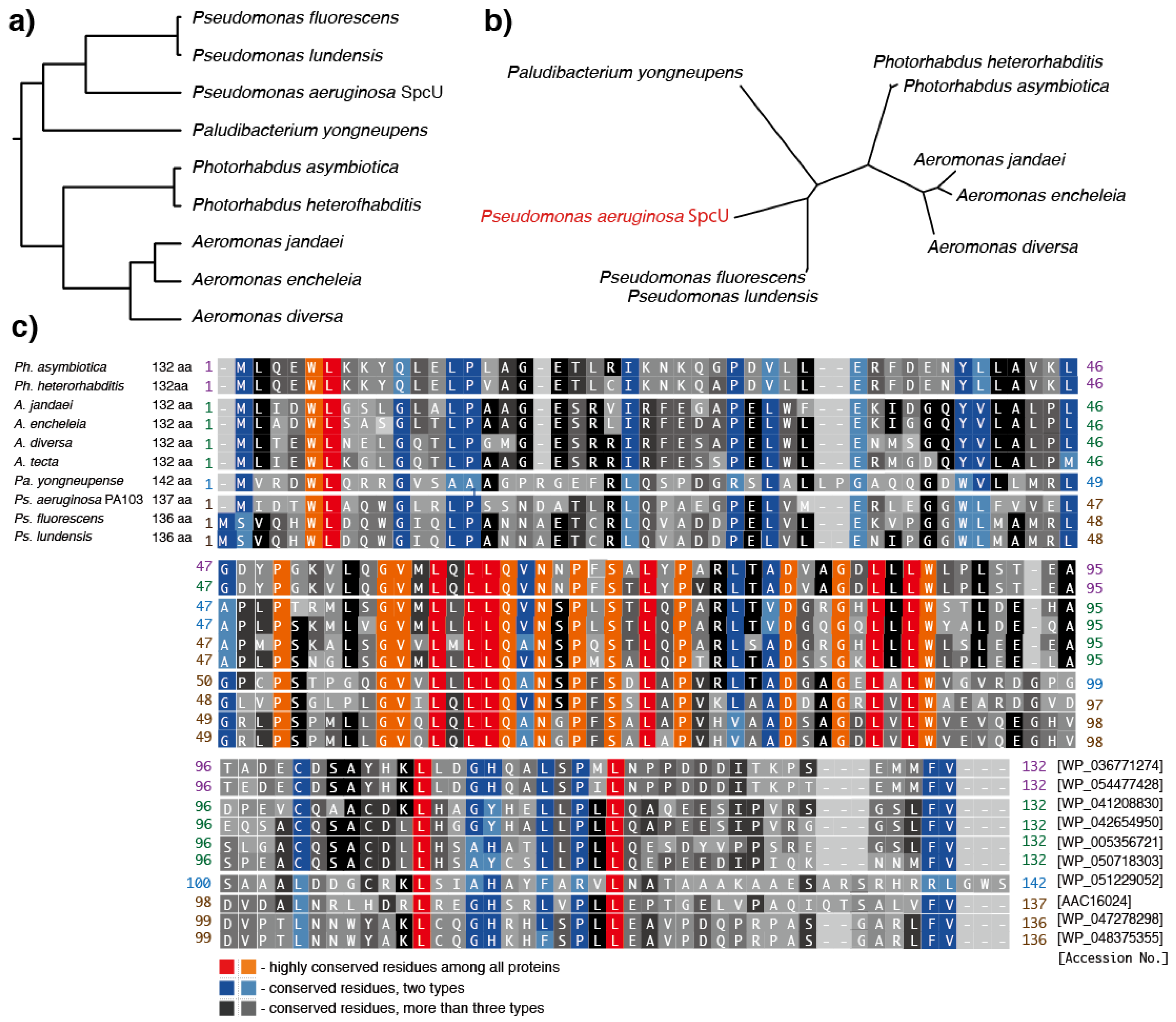
7. Predicted Homologs of the Ps. aeruginosa ExoU Chaperone, SpcU

Ps. aeruginosa possesses the ExoU-specific chaperone protein, SpcU [22]. The gene for SpcU (spcU) is located just downstream of exoU (Figure 1a). Therefore, detecting the presence or absence of specific chaperones for each predicted ExoU homolog might help to determine whether the homologs are type III secreted or not. In the NCBI bacterial genome database, eight predicted homologs of SpcU were found in Ps fluorescens, Ps. lundensis, Ph. heterorhabditis, Ph. asymbioitica, A. jandaei, A. encheleia, A. diversa, and Pa. yongneupense, (Table 2, Figure 7). SpcU homologs have well-conserved primary sequences, especially in the PLP blocks of the central regions. The positions corresponding to ExoU Lys178, which is a known ubiquitination region, are conserved in all Aeromonas species and four Pseudomonas species, but not in the remaining six Pseudomonas and Paludibacterium species. The region corresponding to the ExoU carboxyl terminal residues 679–683, which has also been reported as the region ubiquitinated for enzyme activation, is quite well-conserved among all the predicted homologs.

8. Conclusions

ExoU, a major virulence factor in cytotoxic Ps. aeruginosa, has gradually revealed its unique characteristics since its discovery. There are still many unsolved questions about the ExoU toxin, such as its evolutionary origin, its association with its chaperone, the activation mechanism underlying its PLP activity, and the association between its PLP activity and the cell death mechanism. In this review, we analyzed the primary sequences of eight predicted ExoU homologs from Gram-negative bacterial species, and 17 predicted homologs from Pseudomonas species other than Ps. aeruginosa. Both conserved and nonconserved regions were identified in the multiple alignments of the predicted primary sequences of the putative homologs. At this moment in time, the knowledge gap concerning the enzymatic functionality and cytotoxicity of the predicted homologs of ExoU will remain in place until in vitro and/or in vivo analyses are conducted on these proteins. Nevertheless, in the near future, we can probably obtain more knowledge about the unique characteristics of ExoU from cloning the genes from the predicted ExoU homologs of Ps. aeruginosa, from gene complementation studies, and investigation of the characteristics of recombinant proteins that share sequence homology with ExoU.

Acknowledgments

This work was supported by Japan Society for the Promotion of Science KAKENHI grants (No. 24390403, No. 26670791, & No. 15H05008) and The Ministry of Education, Culture, Sports, Science and Technology, Japan to Teiji Sawa.

Author Contributions

T.S. conceived and designed the review article; S.H., M.K. A.K. Y.N. K.A. and H.K. helped organize the data, drew figures and tables; T.S. wrote the paper.

Conflicts of Interest

The authors declare no conflict of interest.

References

  1. Ramirez-Estrada, S.; Borgatta, B.; Rello, J. Pseudomonas aeruginosa ventilator-associated pneumonia management. Infect. Drug Resist. 2016, 9, 7–18. [Google Scholar] [PubMed]
  2. Kollef, M.H.; Chastre, J.; Fagon, J.Y.; Francois, B.; Niederman, M.S.; Rello, J.; Torres, A.; Vincent, J.L.; Wunderink, R.G.; Go, K.W.; et al. Global prospective epidemiologic and surveillance study of ventilator-associated pneumonia due to Pseudomonas aeruginosa. Crit Care Med. 2014, 42, 2178–2187. [Google Scholar] [CrossRef] [PubMed]
  3. Roy-Burman, A.; Savel, R.H.; Racine, S.; Swanson, B.L.; Revadigar, N.S.; Fujimoto, J.; Sawa, T.; Frank, D.W.; Wiener-Kronish, J.P. Type III protein secretion is associated with death in lower respiratory and systemic Pseudomonas aeruginosa infections. J. Infect. Dis. 2001, 183, 1767–1774. [Google Scholar] [CrossRef] [PubMed]
  4. Hauser, A.R.; Cobb, E.; Bodi, M.; Mariscal, D.; Valles, J.; Engel, J.N.; Rello, J. Type III protein secretion is associated with poor clinical outcomes in patients with ventilator-associated pneumonia caused by Pseudomonas aeruginosa. Crit Care Med. 2002, 30, 521–528. [Google Scholar] [CrossRef] [PubMed]
  5. Wong-Beringer, A.; Wiener-Kronish, J.; Lynch, S.; Flanagan, J. Comparison of type III secretion system virulence among fluoroquinolone-susceptible and -resistant clinical isolates of Pseudomonas aeruginosa. Clin. Microbiol. Infect. 2008, 14, 330–336. [Google Scholar] [CrossRef] [PubMed]
  6. El-Solh, A.A.; Hattemer, A.; Hauser, A.R.; Alhajhusain, A.; Vora, H. Clinical outcomes of type III Pseudomonas aeruginosa bacteremia. Crit Care Med. 2012, 40, 1157–1163. [Google Scholar] [CrossRef] [PubMed]
  7. Sullivan, E.; Bensman, J.; Lou, M.; Agnello, M.; Shriner, K.; Wong-Beringer, A. Risk of developing pneumonia is enhanced by the combined traits of fluoroquinolone resistance and type III secretion virulence in respiratory isolates of Pseudomonas aeruginosa. Crit Care Med. 2014, 42, 48–56. [Google Scholar] [CrossRef] [PubMed]
  8. Fleiszig, S.M.; Wiener-Kronish, J.P.; Miyazaki, H.; Vallas, V.; Mostov, K.E.; Kanada, D.; Sawa, T.; Yen, T.S.; Frank, D.W. Pseudomonas aeruginosa-mediated cytotoxicity and invasion correlate with distinct genotypes at the loci encoding exoenzyme S. Infect. Immun. 1997, 65, 579–586. [Google Scholar] [PubMed]
  9. Sawa, T.; Ohara, M.; Kurahashi, K.; Twining, S.S.; Frank, D.W.; Doroques, D.B.; Long, T.; Gropper, M.A.; Wiener-Kronish, J.P. In vitro cellular toxicity predicts Pseudomonas aeruginosa virulence in lung infections. Infect Immun. 1998, 66, 3242–3249. [Google Scholar] [PubMed]
  10. Kurahashi, K.; Kajikawa, O.; Sawa, T.; Ohara, M.; Gropper, M.A.; Frank, D.W.; Martin, T.R.; Wiener-Kronish, J.P. Pathogenesis of septic shock in Pseudomonas aeruginosa pneumonia. J. Clin. Investig. 1999, 104, 743–750. [Google Scholar] [CrossRef] [PubMed]
  11. Finck-Barbancon, V.; Goranson, J.; Zhu, L.; Sawa, T.; Wiener-Kronish, J.P.; Fleiszig, S.M.; Wu, C.; Mende-Mueller, L.; Frank, D.W. ExoU expression by Pseudomonas aeruginosa correlates with acute cytotoxicity and epithelial injury. Mol. Microbiol. 1997, 25, 547–557. [Google Scholar] [CrossRef] [PubMed]
  12. Sawa, T. The molecular mechanism of acute lung injury caused by Pseudomonas aeruginosa: From bacterial pathogenesis to host response. J. Intensiv. Care 2014, 2. [Google Scholar] [CrossRef] [PubMed]
  13. Sawa, T.; Shimizu, M.; Moriyama, K.; Wiener-Kronish, J.P. Association between Pseudomonas aeruginosa type III secretion, antibiotic resistance, and clinical outcome: A review. Crit Care 2014, 18. [Google Scholar] [CrossRef] [PubMed]
  14. Park, M.H.; Kim, S.Y.; Roh, E.Y.; Lee, H.S. Difference of Type 3 secretion system (T3SS) effector gene genotypes (exoU and exoS) and its implication to antibiotics resistances in isolates of Pseudomonas aeruginosa from chronic otitis media. Auris. Nasus. Larynx 2016. [Google Scholar] [CrossRef] [PubMed]
  15. de Almeida Silva, K.C.; Calomino, M.A.; Deutsch, G.; de Castilho, S.R.; de Paula, G.R.; Esper, L.M.; Teixeira, L.A. Molecular characterization of multidrug-resistant (MDR) Pseudomonas aeruginosa isolated in a burn center. Burns 2016. [Google Scholar] [CrossRef] [PubMed]
  16. Peña, C.; Cabot, G.; Gómez-Zorrilla, S.; Zamorano, L.; Ocampo-Sosa, A.; Murillas, J.; Almirante, B.; Pomar, V.; Aguilar, M.; Granados, A.; et al. Influence of virulence genotype and resistance profile in the mortality of Pseudomonas aeruginosa bloodstream infections. Clin. Infect. Dis. 2015, 60, 539–548. [Google Scholar] [CrossRef] [PubMed]
  17. Borkar, D.S.; Acharya, N.R.; Leong, C.; Lalitha, P.; Srinivasan, M.; Oldenburg, C.E.; Cevallos, V.; Lietman, T.M.; Evans, D.J.; Fleiszig, S.M. Cytotoxic clinical isolates of Pseudomonas aeruginosa identified during the Steroids for Corneal Ulcers Trial show elevated resistance to fluoroquinolones. BMC Ophthalmol. 2014, 14, 54. [Google Scholar] [CrossRef] [PubMed]
  18. Zhang, J.; Chu, Y.; Wang, P.; Ji, X.; Li, X.; Wang, C.; Peng, Y. Clinical outcomes of multidrug resistant Pseudomonas aeruginosa infection and the relationship with type III secretion system in patients with diabetic foot. Int. J. Low Extrem. Wounds 2014, 13, 205–210. [Google Scholar] [CrossRef] [PubMed]
  19. Zhu, H.; Conibear, T.C.; Bandara, R.; Aliwarga, Y.; Stapleton, F.; Willcox, M.D. Type III secretion system-associated toxins, proteases, serotypes, and antibiotic resistance of Pseudomonas aeruginosa isolates associated with keratitis. Curr. Eye Res. 2006, 31, 297–306. [Google Scholar] [CrossRef] [PubMed]
  20. Housley, N.A.; Winkler, H.H.; Audia, J.P. The Rickettsia prowazekii ExoU homologue possesses phospholipase A1 (PLA1), PLA2, and lyso-PLA2 activities and can function in the absence of any eukaryotic cofactors in vitro. J. Bacteriol. 2011, 193, 4634–4642. [Google Scholar] [CrossRef] [PubMed]
  21. NCBI. Available online: http://www.ncbi.nlm.nih.gov/ (accessed on 2 October 2016).
  22. Finck-Barbancon, V.; Yahr, T.L.; Frank, D.W. Identification and characterization of SpcU, a chaperone required for efficient secretion of the ExoU cytotoxin. J. Bacteriol. 1998, 180, 6224–6231. [Google Scholar] [PubMed]
  23. He, J.; Baldini, R.L.; Deziel, E.; Saucier, M.; Zhang, Q.; Liberati, N.T.; Lee, D.; Urbach, J.; Goodman, H.M.; Rahme, L.G. The broad host range pathogen Pseudomonas aeruginosa strain PA14 carries two pathogenicity islands harboring plant and animal virulence genes. Proc. Natl. Acad. Sci. USA 2004, 101, 2530–2535. [Google Scholar] [CrossRef] [PubMed]
  24. Sato, H.; Frank, D.W.; Hillard, C.J.; Feix, J.B.; Pankhaniya, R.R.; Moriyama, K.; Finck-Barbancon, V.; Buchaklian, A.; Lei, M.; Long, R.M.; et al. The mechanism of action of the Pseudomonas aeruginosa-encoded type III cytotoxin, ExoU. EMBO J. 2003, 22, 2959–2969. [Google Scholar] [CrossRef] [PubMed]
  25. Pankhaniya, R.R.; Tamura, M.; Allmond, L.R.; Moriyama, K.; Ajayi, T.; Wiener-Kronish, J.P.; Sawa, T. Pseudomonas aeruginosa causes acute lung injury via the catalytic activity of the patatin-like phospholipase domain of ExoU. Crit Care Med. 2004, 32, 2293–2299. [Google Scholar] [CrossRef] [PubMed]
  26. Tamura, M.; Ajayi, T.; Allmond, L.R.; Moriyama, K.; Wiener-Kronish, J.P.; Sawa, T. Lysophospholipase A activity of Pseudomonas aeruginosa type III secretory toxin ExoU. Biochem. Biophys. Res. Commun. 2004, 316, 323–331. [Google Scholar] [CrossRef] [PubMed]
  27. Fu, H.; Coburn, J.; Collier, R.J. The eukaryotic host factor that activates exoenzyme S of Pseudomonas aeruginosa is a member of the 14–3-3 protein family. Proc. Natl. Acad. Sci. USA 1993, 90, 2320–2324. [Google Scholar] [CrossRef] [PubMed]
  28. Yahr, T.L.; Vallis, A.J.; Hancock, M.K.; Barbieri, J.T.; Frank, D.W. ExoY, an adenylate cyclase secreted by the Pseudomonas aeruginosa type III system. Proc. Natl. Acad. Sci. USA 1998, 95, 13899–13904. [Google Scholar] [CrossRef] [PubMed]
  29. Sato, H.; Feix, J.B.; Frank, D.W. Identification of superoxide dismutase as a cofactor for the Pseudomonas type III toxin, ExoU. Biochemistry 2006, 45, 10368–10375. [Google Scholar] [CrossRef] [PubMed]
  30. Stirling, F.R.; Cuzick, A.; Kelly, S.M.; Oxley, D.; Evans, T.J. Eukaryotic localization, activation and ubiquitinylation of a bacterial type III secreted toxin. Cell. Microbiol. 2006, 8, 1294–12309. [Google Scholar] [CrossRef] [PubMed]
  31. Schmalzer, K.M.; Benson, M.A.; Frank, D.W. Activation of ExoU phospholipase activity requires specific C-terminal regions. J. Bacteriol. 2010, 192, 1801–1812. [Google Scholar] [CrossRef] [PubMed]
  32. Anderson, D.M.; Schmalzer, K.M.; Sato, H.; Casey, M.; Terhune, S.S.; Haas, A.L.; Feix, J.B.; Frank, D.W. Ubiquitin and ubiquitin-modified proteins activate the Pseudomonas aeruginosa T3SS cytotoxin, ExoU. Mol. Microbiol. 2011, 82, 1454–1467. [Google Scholar] [CrossRef] [PubMed]
  33. Dessen, A.; Tang, J.; Schmidt, H.; Stahl, M.; Clark, J.D.; Seehra, J.; Somers, W.S. Crystal structure of human cytosolic phospholipase A2 reveals a novel topology and catalytic mechanism. Cell 1999, 97, 349–360. [Google Scholar] [CrossRef]
  34. Halavaty, A.S.; Borek, D.; Tyson, G.H.; Veesenmeyer, J.L.; Shuvalova, L.; Minasov, G.; Otwinowski, Z.; Hauser, A.R.; Anderson, W.F. Structure of the type III secretion effector protein ExoU in complex with its chaperone SpcU. PLoS ONE 2012, 7. [Google Scholar] [CrossRef] [PubMed]
  35. GenomeNet, Kyoto University Bioinformatics Center, Japan. Available online: http://www.genome.jp/tools/clustalw (accessed on 2 October 2016).
  36. Racusen, D. Lipid acyl hydrolase of patatin. Can. J. Bot. 1984, 62, 1640–1644. [Google Scholar] [CrossRef]
  37. Andrews, D.L.; Beames, B.; Summers, M.D.; Park, W.D. Characterization of the lipid acyl hydrolase activity of the major potato (Solanum tuberosum) tuber protein, patatin, by cloning and abundant expression in a baculovirus vector. Biochem. J. 1988, 252, 199–206. [Google Scholar] [CrossRef] [PubMed]
  38. Kostyal, D.A.; Hickey, V.L.; Noti, J.D.; Sussman, G.L.; Beezhold, D.H. Cloning and characterization of a latex allergen (Hev b7): Homology to patatin, a plant PLA2. Clin. Exp. Immunol. 1998, 112, 355–362. [Google Scholar] [CrossRef] [PubMed]
  39. Breiteneder, H.; Sowka, S.; Wagner, S.; Krebitz, M.; Hafner, C.; Kinaciyan, T.; Yeang, H.Y.; Scheiner, O. Cloning of the patatin-like latex allergen Hev b7, its expression in the yeast Pichia pastoris and its immunological characterization. Int. Arch. Allergy Immunol. 1999, 118, 309–310. [Google Scholar] [CrossRef] [PubMed]
  40. Van Loon, L.C. Pathogenesis-related proteins. Plant Mol. Biol. 1985, 4, 111–116. [Google Scholar] [CrossRef] [PubMed]
  41. Banerji, S.; Flieger, A. Patatin-Like proteins: A new family of lipolytic enzymes present in bacteria? Microbiology 2004, 150, 522–525. [Google Scholar] [CrossRef] [PubMed]
  42. Marchler-Bauer, A.; Derbyshire, M.K.; Gonzales, N.R.; Lu, S.; Chitsaz, F.; Geer, L.Y.; Geer, R.C.; He, J.; Gwadz, M.; Hurwitz, D.I.; et al. CDD: NCBI’s conserved domain database. Nucleic Acids Res. 2015, 43, D222–D226. [Google Scholar] [CrossRef] [PubMed]
  43. Ferreira, T.; van Reenen, C.A.; Endo, A.; Tailliez, P.; Pages, S.; Sproer, C.; Malan, A.P.; Dicks, L.M. Photorhabdus heterorhabditis sp. nov., a symbiont of the entomopathogenic nematode Heterorhabditis zealandica. Int. J. Syst. Evol. Microbiol. 2014, 64, 1540–1545. [Google Scholar] [CrossRef] [PubMed]
  44. Gerrard, J.; Waterfield, N.; Vohra, R.; Ffrench-Constant, R. Human infection with Photorhabdus asymbiotica: An emerging bacterial pathogen. Microbes Infect. 2004, 6, 229–237. [Google Scholar] [CrossRef] [PubMed]
  45. Tounsi, S.; Blight, M.; Jaoua, S.; de Lima Pimenta, A. From insects to human hosts: Identification of major genomic differences between entomopathogenic strains of Photorhabdus and the emerging human pathogen Photorhabdus asymbiotica. Int. J. Med. Microbiol. 2006, 296, 521–530. [Google Scholar] [CrossRef] [PubMed]
  46. Costa, S.C.; Girard, P.A.; Brehelin, M.; Zumbihl, R. The emerging human pathogen Photorhabdus asymbiotica is a facultative intracellular bacterium and induces apoptosis of macrophage-like cells. Infect. Immun. 2009, 77, 1022–1030. [Google Scholar] [CrossRef] [PubMed]
  47. Wilkinson, P.; Waterfield, N.R.; Crossman, L.; Corton, C.; Sanchez-Contreras, M.; Vlisidou, I.; Barron, A.; Bignell, A.; Clark, L.; Ormond, D.; et al. Comparative genomics of the emerging human pathogen Photorhabdus asymbiotica with the insect pathogen Photorhabdus luminescens. BMC Genomics 2009, 10. [Google Scholar] [CrossRef] [PubMed] [Green Version]
  48. Mashima, J.; Kodama, Y.; Kosuge, T.; Fujisawa, T.; Katayama, T.; Nagasaki, H.; Okuda, Y.; Kaminuma, E.; Ogasawara, O.; Okubo, K.; et al. DNA data bank of Japan (DDBJ) progress report. Nucleic Acids Res. 2016, 44, D51–D67. [Google Scholar] [CrossRef] [PubMed]
  49. Janda, J.M.; Abbott, S.L. The genus Aeromonas: Taxonomy, pathogenicity, and infection. Clin. Microbiol. Rev. 2010, 23, 35–73. [Google Scholar] [CrossRef] [PubMed]
  50. Beaz-Hidalgo, R.; Latif-Eugenin, F.; Hossain, M.J.; Berg, K.; Niemi, R.M.; Rapala, J.; Lyra, C.; Liles, M.R.; Figueras, M.J. Aeromonas aquatica sp. nov., Aeromonas finlandiensis sp. nov. and Aeromonas lacus sp. nov. isolated from Finnish waters associated with cyanobacterial blooms. Syst. Appl. Microbiol. 2015, 38, 161–168. [Google Scholar] [CrossRef] [PubMed]
  51. Esteve, C.; Gutierrez, M.C.; Ventosa, A. Aeromonas encheleia sp. nov., isolated from European eels. Int. J. Syst. Bacteriol. 1995, 45, 462–466. [Google Scholar] [CrossRef] [PubMed]
  52. Martinez-Murcia, A.J. Phylogenetic positions of Aeromonas encheleia, Aeromonas popoffii, Aeromonas DNA hybridization group 11 and Aeromonas group 501. Int. J. Syst. Bacteriol. 1999, 49, 1403–1408. [Google Scholar] [CrossRef] [PubMed]
  53. Novakova, D.; Svec, P.; Sedlacek, I. Characterization of Aeromonas encheleia strains isolated from aquatic environments in the Czech Republic. Lett. Appl. Microbiol. 2009, 48, 289–294. [Google Scholar] [CrossRef] [PubMed]
  54. Demarta, A.; Kupfer, M.; Riegel, P.; Harf-Monteil, C.; Tonolla, M.; Peduzzi, R.; Monera, A.; Jose Saavedra, M.; Martinez-Murcia, A. Aeromonas tecta sp. nov., isolated from clinical and environmental sources. Syst. Appl. Microbiol. 2008, 31, 278–286. [Google Scholar] [CrossRef] [PubMed]
  55. Kwon, S.W.; Kim, B.Y.; Kim, W.G.; Yoo, K.H.; Yoo, S.H.; Son, J.A.; Weon, H.Y. Paludibacterium yongneupense gen. nov., sp. nov., isolated from a wetland, Yongneup, in Korea. Int. J. Syst. Evol. Microbiol. 2008, 58, 190–194. [Google Scholar] [CrossRef] [PubMed]
  56. Ajayi, T.; Allmond, L.R.; Sawa, T.; Wiener-Kronish, J.P. Single-Nucleotide-Polymorphism mapping of the Pseudomonas aeruginosa type III secretion toxins for development of a diagnostic multiplex PCR system. J. Clin. Microbiol. 2003, 41, 3526–3531. [Google Scholar] [CrossRef] [PubMed]
Figure 1. The structures of the ExoU gene and ExoU protein, and the phylogenetic trees based on patatin-like phospholipases (PLPs). (a) The position of the ExoU gene in the Pseudomonas aeruginosa pathogenicity island-2 (PAPI-2). PAPI-2 has 14 open read frames including exoU and spcU. The SpcU chaperone gene for ExoU secretion is located next to exoU. (b) Three-dimensional structure of ExoU (MMDB ID: 99839 PDB ID: 3TU3). The catalytic dyad consists of serine142 and aspartic acid344. (c) Amino acid sequence alignment of ExoU, patatins, and human cytosolic/calcium2+-independent phospholipase A2 (cPLA2/iPLA2). (d) ExoU, patatins, and human cPLA2/iPLA2 phylogenetic trees. Left, rooted tree with branch lengths; right, unrooted tree with branch lengths. The phylogenetic trees and the sequence alignments were made by ClustalW with the BLOSUM protein weight matrix. GenomeNet, Kyoto University Bioinformatics Center, Japan [35].
Toxins 08 00307 g001
Figure 2. The conserved domain of patatins and cPLA2 according to the conserved domain database of the National Center for Biology Information [42]. (a) Patatin and cPLA2 domain (cd01819); (b) patatin and ExoU-VipD-like sequence clusters (cd07207).
Toxins 08 00307 g002
Figure 3. Phylogenetic trees of predicted ExoU homologs from the National Center for Biology Information GenBank database. (a) Rooted phylogenetic tree with branch lengths; (b) an unrooted phylogenetic tree with branch lengths. The phylogenetic trees were made by ClustalW with the BLOSUM protein weight matrix. GenomeNet, Kyoto University Bioinformatics Center, Japan [35]. * The ORF positions in GenBank were corrected in this study.
Toxins 08 00307 g003
Figure 4. Conserved blocks in the PLP domain of predicted ExoU homologs from Pseudomonas aeruginosa and other bacterial species. The sequence alignment was made by ClustalW with the BLOSUM protein weight matrix (DNA Data Bank of Japan) [48].
Toxins 08 00307 g004
Figure 5. Conserved blocks in the PLP domain of predicted ExoU homologs from Pseudomonas species. The sequence alignment was made by ClustalW with the BLOSUM protein weight matrix (DNA Data Bank of Japan) [48].
Toxins 08 00307 g005
Figure 6. Sequence alignment of predicted ExoU homologs. (a) The amino-terminal alignment for predicted ExoU homologs; (b) the carboxyl-terminal alignment for predicted ExoU homologs. The sequence alignment was made by ClustalW with the BLOSUM protein weight matrix (DNA Data Bank of Japan) [48].
Toxins 08 00307 g006
Figure 7. Sequence alignment of predicted SpcU homologs. (a) Rooted phylogenetic tree with branch lengths of the predicted SpcU homologs; (b) unrooted phylogenetic tree with branch lengths of the predicted SpcU homologs; (c) sequence alignment of predicted SpcU homologs. The phylogenetic trees and the sequence alignment were made by ClustalW with the BLOSUM protein weight matrix (DNA Data Bank of Japan) [48].
Toxins 08 00307 g007
Table 1. Predicted ExoU homologs.
SpeciesNCBI GenBank Accession No.DNA Sequence Length (bp)Protein Sequence Length (aa)Predicted Protein Size (kDa)Alignment Score against ExoU (%)
ProteinGenome
Pseudomonas aeruginosa (ExoU)AAC16023U97065206468773.9-
Pseudomonas weihenstephanensis*KMN14473JYLF01000003204668172.960
Pseudomonas fluorescensWP_052959439NZ_LCYS01000046204668172.760
Pseudomonas lundensisWP_053069958NZ_JYKY01000004204668172.759
Pseudomonas marginalisWP_058415138NZ_LKGY01000112191463767.943
Pseudomonas rhodesiaeWP_040267033NZ_CCYI01000012191763868.742
Pseudomonas synxanthaWP_057022869NZ_JYLJ01000004192063968.945
Pseudomonas libanensisWP_057014135NZ_JYLH01000017192063969.245
Pseudomonas extremaustralisWP_010565917NZ_AHIP01000021192063968.144
Pseudomonas veroniiWP_057003941NZ_JYLL01000003193264369.145
Pseudomonas simiaeWP_047542539NZ_JRMC01000004193264369.244
Pseudomonas trivialisWP_049711148NZ_CP011507193264369.044
Pseudomonas orientalis*WP_057723553NZ_JYLM01000004194764870.041
Pseudomonas taetrolens*WP_048381695NZ_JYLA01000004206168674.241
Pseudomonas tolaasiiWP_016972911NZ_AJXG01000265191163568.442
Pseudomonas syringaeWP_058825191NZ_LKEM01000062189062968.146
Pseudomonas viridiflavaWP_058391370NZ_LKEJ01000112195965270.644
Pseudomonas cannabina*KPW66747LJPX01000541188762868.145
Photorhabdus asymbioticaWP_012777218NC_012962206167673.552
Photorhabdus heterorhabditisWP_054477426NZ_LJCS01000013206167673.751
Aeromonas jandaei*WP_050490087NZ_JFDL01000003201066971.148
Aeromonas lacus*WP_033113194NZ_JRGM01000022201066971.248
Aeromonas encheleia*WP_042655012NZ_CDDI01000033201066971.248
Aeromonas tecta*WP_050718647NZ_CDCA01000025201066971.546
Aeromonas diversa*WP_052450256NZ_CDCE01000029201066971.747
Paludibacterium yongneupense*WP_051229050NZ_AUGZ01000008212170674.345
Alignment scores (as based on ExoU amino acid sequence similarities according to the BLOSUM protein weight matrix) are from the ClustalW multiple sequence alignment tool (DNA Data Bank of Japan) [48]. bp: base pairs, aa: amino acids, * The open reading frame (ORF) positions in GenBank were corrected in this study.
Table 2. Predicted SpcU homologs.
SpeciesNCBI GenBank Accession No.DNA Sequence Length (bp)Protein Sequence Length (aa)Predicted Protein Size (kDa)Alignment Score against SpcU (%)
ProteinGenome
Pseudomonas aeruginosa (SpcU)AAC16024U9706541413714.9-
Pseudomonas fluorescensWP_047278298NZ_LCYS0100004641113614.948.5294
Pseudomonas lundensisWP_048375355NZ_JYKY0100000441113614.947.7941
Photorhabdus asymbioticaWP_036771274NZ_JONO0100003239913214.831.8182
Photorhabdus heterorhabditisWP_054477428NZ_LJCS0100001339913214.931.0606
Aeromonas jandaeiWP_041208830NZ_JFDL0100000339913214.428.7879
Aeromonas encheleiaWP_042654950NZ_CDDI0100003339913214.231.0606
Aeromonas tectaWP_050718303NZ_CDCA0100002539913214.628.0303
Aeromonas diversaWP_005356721NZ_CDCE0100002939913214.428.7879
Paludibacterium yongneupenseWP_050718303NZ_AUGZ0100000842914215.033.5766
Alignment scores (as based on SpcU amino acid sequence similarities according to the BLOSUM protein weight matrix) are from the ClustalW multiple sequence alignment tool with the BLOSUM protein weight matrix (DNA Data Bank of Japan) [48]. bp: base pairs, aa: amino acids.

