From Neurophysiological Mechanisms to Rehabilitation After Botulinum Toxin Type A in Post-Stroke Spasticity

 , , ,
, , ,
Abstract
1. Introduction
2. Results
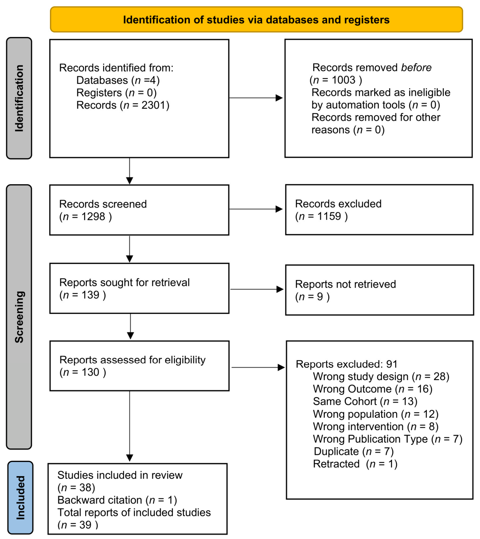
2.1. Study Selection
2.2. Study Characteristics
2.3. Risk of Bias Assessment
2.4. Certainty Assessment
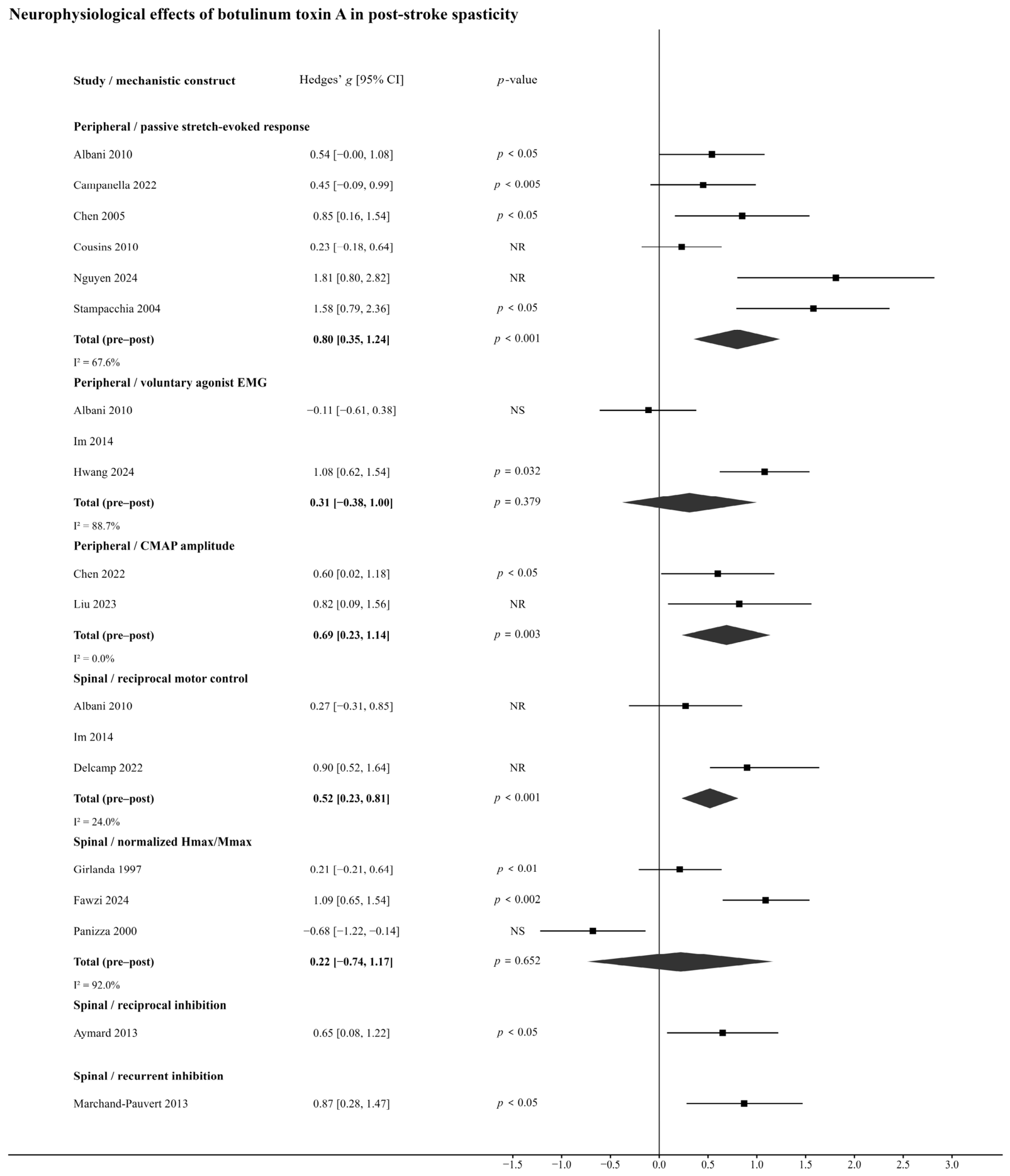
2.5. Quantitative Synthesis
2.5.1. Passive Stretch-Evoked Responses
2.5.2. Voluntary Agonist Muscle Activity
2.5.3. Compound Muscle Action Potential
2.5.4. Reciprocal Motor Control
2.5.5. Normalized H-Reflex Amplitude (Hmax/Mmax)
2.5.6. Reciprocal and Recurrent Inhibition
2.5.7. Sensitivity Analysis
2.6. Narrative Synthesis of Non-Pooled Neurophysiological Outcomes
2.6.1. Passive Stretch and Peripheral Neurophysiological Outcomes
2.6.2. Voluntary Activation and Motor Coordination
2.6.3. Mechanisms on Spinal Level
2.6.4. Supraspinal and Cortical Outcomes
3. Discussion
3.1. From Neurophysiological Mechanisms to Mechanism-Based Rehabilitation Following BoNT-A
3.2. Limitations
3.3. Future Research
4. Conclusions
5. Materials and Methods
5.1. Eligibility Criteria
5.2. Information Sources
5.3. Search Strategy
5.4. Selection Process
5.5. Data Collection Process
5.6. Data Items
5.7. Planned Methods of Analysis
5.8. Risk of Bias Assessment
5.9. Certainty Evidence
Author Contributions
Funding
Institutional Review Board Statement
Informed Consent Statement
Data Availability Statement
Acknowledgments
Conflicts of Interest
References
- Sacco, R.L.; Kasner, S.E.; Broderick, J.P.; Caplan, L.R.; Connors, J.J.; Culebras, A.; Elkind, M.S.; George, M.G.; Hamdan, A.D.; Higashida, R.T.; et al. An updated definition of stroke for the 21st century: A statement for healthcare professionals from the American Heart Association/American Stroke Association. Stroke 2013, 44, 2064–2089. [Google Scholar]
- Bourbonnais, D.; Noven, S.V. Weakness in patients with Hemiparesis. Am. J. Occup. Ther. 1989, 43, 313–319. [Google Scholar] [CrossRef] [Scilit]
- Santello, M.; Lang, C.E. Are movement disorders and sensorimotor injuries pathologic synergies? When normal multi-joint movement synergies become pathologic. Front. Hum. Neurosci. 2015, 8, 1050. [Google Scholar] [CrossRef] [Scilit]
- Lance, J.W. Spasticity: Disorder of Motor Control; Year Book Medical Publishers: Chicago, IL, USA, 1980; pp. 485–494. [Google Scholar]
- Pandyan, A.D.; Gregoric, M.; Barnes, M.P.; Wood, D.; Van Wijck, F.; Burridge, J.; Hermens, H.; Johnson, G.R. Spasticity: Clinical perceptions, neurological realities and meaningful measurement. Disabil. Rehabil. 2005, 27, 2–6. [Google Scholar] [CrossRef] [Scilit]
- Dressler, D.; Bhidayasiri, R.; Bohlega, S.; Chana, P.; Chien, H.F.; Chung, T.M.; Colosimo, C.; Ebke, M.; Fedoroff, K.; Frank, B.; et al. Defining spasticity: A new approach considering current movement disorders terminology and botulinum toxin therapy. J. Neurol. 2018, 265, 856–862. [Google Scholar] [CrossRef] [Scilit]
- Li, S.; Francisco, G.E.; Rymer, W.Z. A New Definition of Poststroke Spasticity and the Interference of Spasticity with Motor Recovery from Acute to Chronic Stages. Neurorehabil. Neural Repair 2021, 35, 601–610. [Google Scholar] [CrossRef] [Scilit]
- Sunnerhagen, K.S.; Opheim, A.; Alt Murphy, M. Onset, time course and prediction of spasticity after stroke or traumatic brain injury. Ann. Phys. Rehabil. Med. 2019, 62, 431–434. [Google Scholar] [CrossRef] [Scilit]
- Abbruzzese, G.; Berardelli, A. Neurophysiological effects of botulinum toxin type A. Neurotox. Res. 2006, 9, 109–114. [Google Scholar] [CrossRef] [Scilit]
- Elia, A.E.; Filippini, G.; Calandrella, D.; Albanese, A. Botulinum neurotoxins for post-stroke spasticity in adults: A systematic review. Mov. Disord. 2009, 24, 801–812. [Google Scholar] [CrossRef] [Scilit]
- Asimakidou, E.; Sidiropoulos, C. A Bayesian Network Meta-Analysis and Systematic Review of Guidance Techniques in Botulinum Toxin Injections and Their Hierarchy in the Treatment of Limb Spasticity. Toxins 2023, 15, 256. [Google Scholar] [CrossRef] [Scilit]
- Fleuren, J.F.; Voerman, G.E.; Erren-Wolters, C.V.; Snoek, G.J.; Rietman, J.S.; Hermens, H.J.; Nene, A.V. Stop using the Ashworth Scale for the assessment of spasticity. J. Neurol. Neurosurg. Psychiatry 2010, 81, 46–52. [Google Scholar] [CrossRef] [Scilit]
- Chowdhury, R.H.; Reaz, M.B.; Ali, M.A.; Bakar, A.A.; Chellappan, K.; Chang, T.G. Surface electromyography signal processing and classification techniques. Sensors 2013, 13, 12431–12466. [Google Scholar] [CrossRef] [Scilit]
- Shah, M.H.; Ganvir, S.S. Hoffmann reflex parameters as neurophysiological biomarkers for quantifying spasticity: A systematic review. Sri Ramachandra J. Health Sci. 2025, 5, 44–57. [Google Scholar] [CrossRef] [Scilit]
- Pulverenti, T.S.; Zaaya, M.; Grabowski, M.; Grabowski, E.; Islam, M.A.; Li, J.; Murray, L.M.; Knikou, M. Neurophysiological Changes After Paired Brain and Spinal Cord Stimulation Coupled with Locomotor Training in Human Spinal Cord Injury. Front. Neurol. 2021, 12, 627975. [Google Scholar] [CrossRef] [Scilit]
- Wissel, J.; Ward, A.B.; Erztgaard, P.; Bensmail, D.; Hecht, M.J.; Lejeune, T.M.; Schnider, P.; Altavista, M.C.; Cavazza, S.; Deltombe, T.; et al. European consensus table on the use of botulinum toxin type A in adult spasticity. J. Rehabil. Med. 2009, 41, 13–25. [Google Scholar] [CrossRef] [Scilit]
- Rosales, R.L.; Chua-Yap, A.S. Evidence-based systematic review on the efficacy and safety of botulinum toxin-A therapy in post-stroke spasticity. J. Neural Transm. 2008, 115, 617–623. [Google Scholar] [CrossRef] [Scilit]
- Andringa, A.; van de Port, I.; van Wegen, E.; Ket, J.; Meskers, C.; Kwakkel, G. Effectiveness of botulinum toxin treatment for upper limb spasticity poststroke over different ICF domains: A systematic review and meta-analysis. Arch. Phys. Med. Rehabil. 2019, 100, 1703–1725. [Google Scholar] [CrossRef] [Scilit]
- Weise, D.; Weise, C.M.; Naumann, M. Central effects of botulinum neurotoxin—Evidence from human studies. Toxins 2019, 11, 21. [Google Scholar] [CrossRef] [Scilit]
- Hok, P.; Veverka, T.; Hluštík, P.; Nevrlý, M.; Kaňovský, P. The central effects of botulinum toxin in dystonia and spasticity. Toxins 2021, 13, 155. [Google Scholar] [CrossRef] [Scilit]
- Albani, G.; Cimolin, V.; Galli, M.; Vimercati, S.; Bar, D.; Campanelli, L.; Gandolfi, R.; Lombardi, R.; Mauro, A. Use of surface EMG for evaluation of upper limb spasticity during botulinum toxin therapy in stroke patients. Funct. Neurol. 2010, 25, 103–107. [Google Scholar]
- Alvisi, E.; Serrao, M.; Conte, C.; Alfonsi, E.; Tassorelli, C.; Prunetti, P.; Cristina, S.; Perrotta, A.; Pierelli, F.; Sandrini, G. Botulinum toxin A modifies nociceptive withdrawal reflex in subacute stroke patients. Brain Behav. 2018, 8, e01069. [Google Scholar] [CrossRef] [Scilit]
- Aymard, C.; Giboin, L.-S.; Lackmy-Vallée, A.; Marchand-Pauvert, V. Spinal plasticity in stroke patients after botulinum neurotoxin A injection in ankle plantar flexors. Physiol. Rep. 2013, 1, e00173. [Google Scholar] [CrossRef] [Scilit]
- Bhakta, B.B.; O’Connor, R.J.; Cozens, J.A. Associated reactions after stroke: A randomized controlled trial of the effect of botulinum toxin type A. J. Rehabil. Med. 2008, 40, 36–41. [Google Scholar] [CrossRef] [Scilit]
- Boudarham, J.; Hameau, S.; Pradon, D.; Bensmail, D.; Roche, N.; Zory, R. Changes in electromyographic activity after botulinum toxin injection of the rectus femoris in patients with hemiparesis walking with a stiff-knee gait. J. Electromyogr. Kinesiol. 2013, 23, 1036–1043. [Google Scholar] [CrossRef] [Scilit]
- Campanella, W.; Corazza, A.; Puce, L.; Privitera, L.; Pedrini, R.; Mori, L.; Boccuni, L.; Turtulici, G.; Trompetto, C.; Marinelli, L. Shear wave elastography combined with electromyography to assess the effect of botulinum toxin on spastic dystonia following stroke: A pilot study. Front. Neurol. 2022, 13, 980746. [Google Scholar] [CrossRef] [Scilit]
- Chalard, A.; Amarantini, D.; Cormier, C.; Marque, P.; Gasq, D. Effect of the combination of botulinum toxin A injection in elbow flexor muscles and rehabilitation on cortical oscillatory activity in individuals with stroke. Ann. Phys. Rehabil. Med. 2021, 64, 101434. [Google Scholar] [CrossRef] [Scilit]
- Chandra, S.; Afsharipour, B.; Rymer, W.Z.; Suresh, N.L. Precise quantification of the time course of voluntary activation capacity following botulinum toxin injections in the biceps brachii muscles of chronic stroke survivors. J. Neuroeng. Rehabil. 2020, 17, 102. [Google Scholar] [CrossRef] [Scilit]
- Chandra, S.; Suresh, N.L.; Afsharipour, B.; Rymer, W.Z.; Holobar, A. Anomalies of motor unit amplitude and territory after botulinum toxin injection. J. Neural Eng. 2022, 19, 036041. [Google Scholar] [CrossRef] [Scilit]
- Chen, J.-J.J.; Wu, Y.-N.; Huang, S.-C.; Lee, H.-M.; Wang, Y.-L. The use of a portable muscle tone measurement device to measure the effects of botulinum toxin type A on elbow flexor spasticity. Arch. Phys. Med. Rehabil. 2005, 86, 1655–1660. [Google Scholar] [CrossRef] [Scilit]
- Chen, Y.-T.; Liu, Y.; Zhang, C.; Magat, E.; Zhou, P.; Zhang, Y.; Li, S. Comprehensive assessment of the time course of biomechanical, electrophysiological and neuro-motor effects after botulinum toxin injections in elbow flexors of chronic stroke survivors with spastic hemiplegia: A cross sectional observation study. Toxins 2022, 14, 104. [Google Scholar] [CrossRef] [Scilit]
- Cousins, E.; Ward, A.; Roffe, C.; Rimington, L.; Pandyan, A. Does low-dose botulinum toxin help the recovery of arm function when given early after stroke? A phase II randomized controlled pilot study to estimate effect size. Clin. Rehabil. 2010, 24, 501–513. [Google Scholar] [CrossRef] [Scilit]
- Delcamp, C.; Cormier, C.; Chalard, A.; Amarantini, D.; Gasq, D. Botulinum toxin combined with rehabilitation decrease corticomuscular coherence in stroke patients. Clin. Neurophysiol. 2022, 136, 49–57. [Google Scholar] [CrossRef] [Scilit]
- Fawzi, S.M.; Hamdan, F.B.; Jaafar, I.F.; Al Gawwam, G.A.A.S. Botulinum neurotoxin-A in a patient with post-stroke spasticity: A neurophysiological study. Folia Neuropathol. 2023, 61, 412–418. [Google Scholar] [CrossRef] [Scilit]
- Fujita, K.; Miaki, H.; Hori, H.; Kobayashi, Y.; Nakagawa, T. How effective is physical therapy for gait muscle activity in hemiparetic patients who receive botulinum toxin injections? Eur. J. Phys. Rehabil. Med. 2019, 55, 8–18. [Google Scholar] [CrossRef] [Scilit]
- Gandolfi, M.; Valè, N.; Dimitrova, E.K.; Mazzoleni, S.; Battini, E.; Filippetti, M.; Picelli, A.; Santamato, A.; Gravina, M.; Saltuari, L.; et al. Effectiveness of robot-assisted upper limb training on spasticity, function and muscle activity in chronic stroke patients treated with botulinum toxin: A randomized single-blinded controlled trial. Front. Neurol. 2019, 10, 41. [Google Scholar] [CrossRef] [Scilit]
- Girlanda, P.; Quartarone, A.; Sinicropi, S.; Nicolosi, C.; Roberto, M.L.; Picciolo, G.; Macaione, V.; Battaglia, F.; Ruggeri, M.; Messina, C. Botulinum toxin in upper limb spasticity: Study of reciprocal inhibition between forearm muscles. NeuroReport 1997, 8, 3039–3044. [Google Scholar] [CrossRef] [Scilit]
- Hesse, S.; Krajnik, J.; Luecke, D.; Jahnke, M.T.; Gregoric, M.; Mauritz, K.H. Ankle muscle activity before and after botulinum toxin therapy for lower limb extensor spasticity in chronic hemiparetic patients. Stroke 1996, 27, 455–460. [Google Scholar] [CrossRef] [Scilit]
- Hwang, I.-S.; Ryu, J.-W.; Jin, S.; Kim, S.-A.; Kim, M.-S. Long-term enhancement of botulinum toxin injections for post-stroke spasticity by use of stretching exercises—A randomized controlled trial. Toxins 2024, 16, 267. [Google Scholar] [CrossRef] [Scilit]
- Im, S.; Park, J.H.; Son, S.K.; Shin, J.-E.; Cho, S.H.; Park, G.-Y. Does botulinum toxin injection site determine outcome in post-stroke plantarflexion spasticity? Comparison study of two injection sites in the gastrocnemius muscle: A randomized double-blind controlled trial. Clin. Rehabil. 2014, 28, 604–613. [Google Scholar] [CrossRef] [Scilit]
- Kerzoncuf, M.; Bensoussan, L.; Delarque, A.; Durand, J.; Viton, J.-M.; Rossi-Durand, C. Plastic changes in spinal synaptic transmission following botulinum toxin A in patients with post-stroke spasticity. J. Rehabil. Med. 2015, 47, 910–916. [Google Scholar] [CrossRef] [Scilit]
- Kirazli, Y.; On, A.Y.; Kismali, B.; Aksit, R. Comparison of phenol block and botulinum toxin type A in the treatment of spastic foot after stroke: A randomized, double-blind trial. Am. J. Phys. Med. Rehabil. 1998, 77, 510–515. [Google Scholar]
- Lee, H.-M.; Chen, J.-J.J.; Wu, Y.-N.; Wang, Y.-L.; Huang, S.-C.; Piotrkiewicz, M. Time course analysis of the effects of botulinum toxin type A on elbow spasticity based on biomechanic and electromyographic parameters. Arch. Phys. Med. Rehabil. 2008, 89, 692–699. [Google Scholar] [CrossRef] [Scilit]
- Liu, Y.; Chen, Y.-T.; Zhang, C.; Zhou, P.; Li, S.; Zhang, Y. Motor unit number estimation in spastic biceps brachii muscles of chronic stroke survivors before and after BoNT injection. IEEE Trans. Biomed. Eng. 2023, 70, 1045–1052. [Google Scholar] [CrossRef] [Scilit]
- Marchand-Pauvert, V.; Aymard, C.; Giboin, L.-S.; Dominici, F.; Rossi, A.; Mazzocchio, R. Beyond muscular effects: Depression of spinal recurrent inhibition after botulinum neurotoxin A. J. Physiol. 2013, 591, 1017–1029. [Google Scholar] [CrossRef] [Scilit]
- Marvulli, R.; Mastromauro, L.; Romanelli, E.; Lopopolo, A.; Dargenio, M.; Fornarelli, F.; Conte, E.; Fiore, P.; Megna, M.; Ianieri, G. How botulinum toxin type A–occupational therapy (OT)–functional electrical stimulation (FES) modify spasticity and functional recovery in patients with upper limb spasticity post stroke. Clin. Immunol. Endocr. Metab. Drugs 2016, 3, 62–67. [Google Scholar] [CrossRef] [Scilit]
- Miscio, G.; Del Conte, C.; Pianca, D.; Colombo, R.; Panizza, M.; Schieppati, M.; Pisano, F. Botulinum toxin in post-stroke patients: Stiffness modifications and clinical implications. J. Neurol. 2004, 251, 189–196. [Google Scholar] [CrossRef] [Scilit]
- Nguyen, N.; Houston, M.; Liu, Y.; Chen, Y.-T.; Li, S.; Zhang, Y. High-density electromyography biomarkers for detecting and monitoring of spastic muscles during passive stretch. In 2024 46th Annual International Conference of the IEEE Engineering in Medicine and Biology Society (EMBC); IEEE: New York, NY, USA, 2024; pp. 1–4. [Google Scholar] [CrossRef] [Scilit]
- Pandyan, A.D.; Vuadens, P.; van Wijck, F.M.J.; Stark, S.; Johnson, G.R.; Barnes, M.P. Are we underestimating the clinical efficacy of botulinum toxin (type A)? Quantifying changes in spasticity, strength and upper limb function after injections of Botox® to the elbow flexors in a unilateral stroke population. Clin. Rehabil. 2002, 16, 654–660. [Google Scholar] [CrossRef] [Scilit]
- Panizza, M.; Castagna, M.; di Summa, A.; Saibene, L.; Grioni, G.; Nilsson, J. Functional and clinical changes in upper limb spastic patients treated with botulinum toxin (BTX). Funct. Neurol. 2000, 15, 147–155. [Google Scholar]
- Stampacchia, G.; Bradaschia, E.; Rossi, B. Change of stretch reflex threshold in spasticity: Effect of botulinum toxin injections. Arch. Ital. Biol. 2004, 142, 265–273. [Google Scholar]
- Tang, S.-F.; Hong, J.-P.; McKay, W.B.; Tang, C.-W.; Wu, P.-H.; Chu, N.-K. Modification of altered ankle motor control after stroke using focal application of botulinum toxin type A. Clin. Neurol. Neurosurg. 2012, 114, 498–501. [Google Scholar] [CrossRef] [Scilit]
- Trompetto, C.; Bove, M.; Avanzino, L.; Francavilla, G.; Berardelli, A.; Abbruzzese, G. Intrafusal effects of botulinum toxin in post-stroke upper limb spasticity. Eur. J. Neurol. 2008, 15, 367–370. [Google Scholar] [CrossRef] [Scilit]
- Veverka, T.; Hluštík, P.; Hok, P.; Otruba, P.; Krobot, A.; Kaňovský, P. Sensorimotor modulation by botulinum toxin A in post-stroke arm spasticity: Passive hand movement. J. Neurol. Sci. 2016, 362, 14–20. [Google Scholar] [CrossRef] [Scilit]
- Veverka, T.; Hluštík, P.; Otruba, P.; Hok, P.; Opavský, R.; Zapletalová, J.; Kaňovský, P. Cortical somatosensory processing after botulinum toxin therapy in post-stroke spasticity. Medicine 2021, 100, e26356. [Google Scholar] [CrossRef] [Scilit]
- Veverka, T.; Hok, P.; Trnečková, M.; Otruba, P.; Zapletalová, J.; Tüdös, Z.; Lotze, M.; Kaňovský, P.; Hluštík, P. Interhemispheric parietal cortex connectivity reflects improvement in post-stroke spasticity due to treatment with botulinum toxin-A. J. Neurol. Sci. 2023, 446, 120588. [Google Scholar] [CrossRef] [Scilit]
- Vinehout, K.; Tynes, K.; Sotelo, M.R.; Hyngstrom, A.S.; McGuire, J.R.; Schmit, B.D. Changes in cortical activity in stroke survivors undergoing botulinum neurotoxin therapy for treatment of focal spasticity. Front. Rehabil. Sci. 2021, 2, 735819. [Google Scholar] [CrossRef] [Scilit]
- Wang, R.; Gäverth, J.; Herman, P.A. Changes in the neural and non-neural related properties of the spastic wrist flexors after treatment with botulinum toxin A in post-stroke subjects: An optimization study. Front. Bioeng. Biotechnol. 2018, 6, 73. [Google Scholar] [CrossRef] [Scilit]
- Wu, T.; Yan, D.; Li, J.-H.; Shi, Z.-H. Gait improvement by low-dose botulinum toxin A injection treatment of the lower limbs in subacute stroke patients. J. Phys. Ther. Sci. 2015, 27, 759–762. [Google Scholar] [CrossRef] [Scilit]
- Esquenazi, A.; Novak, I.; Sheean, G.; Singer, B.J.; Ward, A.B. International consensus statement for the use of botulinum toxin treatment in adults and children with neurological impairments—Introduction. Eur. J. Neurol. 2010, 17, 1–8. [Google Scholar] [CrossRef] [Scilit]
- Francisco, G.E.; Balbert, A.; Bavikatte, G.; Bensmail, D.; Carda, S.; Deltombe, T.; Draulans, N.; Escaldi, S.; Gross, R.; Jacinto, J.; et al. A practical guide to optimizing the benefits of post-stroke spasticity interventions with botulinum toxin A: An international group consensus. J. Rehabil. Med. 2021, 53, jrm00134. [Google Scholar] [CrossRef] [Scilit]
- Turner-Stokes, L.; Baguley, I.J.; De Graaff, S.; Katrak, P.; Davies, L.; McCrory, P.; Hughes, A. Goal attainment scaling in the evaluation of treatment of upper limb spasticity with botulinum toxin: A secondary analysis from a double-blind placebo-controlled randomized clinical trial. J. Rehabil. Med. 2010, 42, 81–89. [Google Scholar] [CrossRef] [Scilit]
- Eeckhaut, B.; Truijen, S.; Leroij, C.; Dévillé, J.; Jacobs, L.; Saeys, W. Neurophysiological and structural-mechanical changes associated with dry needling in post-stroke spasticity: A systematic review. J. Clin. Med. 2026, 15, 4246. [Google Scholar] [CrossRef] [Scilit]
- Calota, A.; Levin, M.F. Tonic stretch reflex threshold as a measure of spasticity: Implications for clinical practice. Top. Stroke Rehabil. 2009, 16, 177–188. [Google Scholar] [CrossRef] [Scilit]
- Blanchette, A.K.; Bouyer, L.J.; Roy, J.S. Tonic Stretch Reflex Threshold as a Measure of Ankle Plantar-Flexor Spasticity After Stroke. Phys. Ther. 2016, 96, 687–695. [Google Scholar] [CrossRef] [Scilit]
- Mullick, A.A.; Musampa, N.K.; Feldman, A.G.; Levin, M.F. Tonic stretch reflex threshold as a measure of spasticity after stroke: Reliability, minimal detectable change and responsiveness. Clin. Neurophysiol. 2021, 132, 1226–1233. [Google Scholar] [CrossRef] [Scilit]
- Kinnear, B.Z.; Lannin, N.A.; Cusick, A.; Harvey, L.A.; Rawicki, B. Rehabilitation therapies after botulinum toxin-A injection to manage limb spasticity: A systematic review. Phys. Ther. 2014, 94, 1569–1581. [Google Scholar] [CrossRef] [Scilit]
- Hallett, M. Explanation of timing of botulinum neurotoxin effects, onset and duration, and clinical ways of influencing them. Toxicon 2015, 107, 64–67. [Google Scholar] [CrossRef] [Scilit]
- Baricich, A.; Wein, T.; Cinone, N.; Bertoni, M.; Picelli, A.; Chisari, C.; Molteni, F.; Santamato, A. BoNT-A for post-stroke spasticity: Guidance on unmet clinical needs from a Delphi panel approach. Toxins 2021, 13, 236. [Google Scholar] [CrossRef] [Scilit]
- Dressler, D.; Adib Saberi, F.; Reis Barbosa, E. Botulinum toxin: Mechanisms of action. Arq. Neuro-Psiquiatr. 2005, 63, 180–185. [Google Scholar] [CrossRef] [Scilit]
- Rosales, R.L.; Arimura, K.; Takenaga, S.; Osame, M. Extrafusal and intrafusal muscle effects in experimental botulinum toxin-A injection. Muscle Nerve 1996, 19, 488–496. [Google Scholar] [CrossRef] [Scilit]
- Reebye, R.; Jacinto, L.J.; Balbert, A.; Biering-Soerensen, B.; Carda, S.; Draulans, N.; Molteni, F.; O’dEll, M.W.; Picelli, A.; Santamato, A.; et al. Multimodal therapy and use of adjunctive therapies to BoNT-A in spasticity management: Defining terminology to help enhance spasticity treatment. Front. Neurol. 2024, 15, 1432330. [Google Scholar] [CrossRef] [Scilit]
- Page, M.J.; McKenzie, J.E.; Bossuyt, P.M.; Boutron, I.; Hoffmann, T.C.; Mulrow, C.D.; Shamseer, L.; Tetzlaff, J.M.; Akl, E.A.; Brennan, S.E.; et al. The PRISMA 2020 statement: An updated guideline for reporting systematic reviews. BMJ 2021, 372, n71. [Google Scholar] [CrossRef] [Scilit]
- Ouzzani, M.; Hammady, H.; Fedorowicz, Z.; Elmagarmid, A. Rayyan—A web and mobile app for systematic reviews. Syst. Rev. 2016, 5, 210. [Google Scholar] [CrossRef] [Scilit]
- Higgins, J.P.T.; Thomas, J.; Chandler, J.; Cumpston, M.; Li, T.; Page, M.; Welch, V. Cochrane Handbook for Systematic Reviews of Interventions; Wiley: Hoboken, NJ, USA, 2024. [Google Scholar]
- DerSimonian, R.; Laird, N. Meta-analysis in clinical trials. In Controlled Clinical Trials; Elsevier: Amsterdam, The Netherlands, 1986. [Google Scholar]
- Higgins, J.P.; Thompson, S.G.; Deeks, J.J.; Altman, D.G. Measuring inconsistency in meta-analyses. BMJ 2003, 327, 557–560. [Google Scholar] [CrossRef] [Scilit]
- Sterne, J.A.C.; Savović, J.; Page, M.J.; Elbers, R.G.; Blencowe, N.S.; Boutron, I.; Cates, C.J.; Cheng, H.-Y.; Corbett, M.S.; Eldridge, S.M.; et al. RoB 2: A revised tool for assessing risk of bias in randomised trials. BMJ 2019, 366, l4898. [Google Scholar] [CrossRef] [Scilit]
- Wells, G.A.; Shea, B.; O’Connell, D.; Peterson, J.; Welch, V.; Losos, M.; Tugwell, P. The Newcastle-Ottawa Scale (NOS) for Assessing the Quality of Nonrandomised Studies in Meta-Analyses; Ottawa Hospital Research Institute: Ottawa, ON, Canada, 2014. [Google Scholar]
- Schünemann, H.; Brożek, J.; Guyatt, G.; Oxman, A. (Eds.) GRADE Handbook for Grading Quality of Evidence and Strength of Recommendations; GRADE Working Group: Hamilton, ON, Canada, 2013. [Google Scholar]



| Study | Design | Participants | Time Post Stroke | BoNT-A Intervention | Neurophysiological Outcomes | Neurophysiological Outcome Domain | Clinical Outcomes | Results (Uniform, Compact) |
|---|---|---|---|---|---|---|---|---|
| Albani et al. (2010) [21] | Pre–post | n = 10 | 1–6 years (chronic) | BB 100U; FCR 50U; FDP 50U | EMG | Peripheral | MAS; GPS | MAS ↓ at 30d, return toward baseline at 180d; GPS ↓ at 30d & 180d (p < 0.05); EMG ↓ ~40–50% at rest/passive movement. |
| Alvisi et al. (2018) [22] | Pre–post | n = 14 | Subacute/established PSS; exact time NR here | 450–1680U (AD, PD, BB, TB, FCR, ECR) | EMG; NWR | Peripheral | MAS; FIM | MAS ↓ (p < 0.001); FIM ↑ (p < 0.001); EMG ↓ in AD/PD/BB/TB/FCR (p < 0.01); NWR ↓ (p = 0.015). |
| Aymard et al. (2013) [23] | Pre–post | n = 13 | Chronic PSS (≥6 months) | Soleus/MG/TP (300U) | EMG; H-reflex/reciprocal inhibition | Peripheral; spinal | MAS | MAS ↓ ~2 points; EMG ↓ during mid-swing (p < 0.05); reciprocal inhibitory/H-reflex measures changed after BoNT-A. |
| Bhakta et al. (2008) [24] | RCT | n = 40 | Median 2.7 years (chronic) | Dysport 1000 MU divided over elbow/wrist/finger flexors | Associated-reaction force; sEMG-related motor output | Spinal | MAS; ADL interference | Peak associated-reaction force ↓ vs. placebo at week 2 (p = 0.005) and week 6 (p < 0.01); effect attenuated by week 12. |
| Boudarham et al. (2013) [25] | Pre–post | n = 14 | Chronic hemiparetic stroke | RF (164 ± 50U) | EMG | Peripheral | MAS; gait | RF EMG ↓ (p = 0.013–0.004); MAS ↓ (p = 0.012); gait velocity/stride/cadence ↑ (p < 0.017). |
| Campanella et al. (2022) [26] | Pre–post | n = 14 | Chronic post-stroke hypertonia | BB, brachialis, GC, soleus | sEMG; SWE; MHS | Peripheral | MAS | MAS ↓ (p = 0.0018–0.018); sEMG ↓ (p < 0.005); SWE ↓ (p < 0.003); MHS NS. |
| Chalard et al. (2021) [27] | Prospective observational pre–post | n = 12 | ≥6 months post-stroke (chronic) | AboBoNT-A (Dysport) to individualized elbow flexors + usual PT/self-rehabilitation | EEG movement-related beta desynchronization; elbow-flexor EMG co-contraction | Spinal | Tardieu/catch angle; FMA-UE; WMFT; AROM | Movement-related beta desynchronization changed significantly at 4w and 16w vs. baseline; elbow-flexor overactivity/co-contraction and active elbow performance changed over follow-up. |
| Chandra et al. (2020) [28] | Longitudinal pre–post | n = 8 | 4–12 years post-stroke (chronic) | Routine-care BoNT-A to biceps brachii; individualized dose | Voluntary RMS-sEMG; sEMG–force relation; MU amplitude distribution | Peripheral | MAS; FMA; MVC force | Force and sEMG ↓ maximally ~2–4w after injection, followed by partial recovery; several participants remained below baseline at 12w. |
| Chandra et al. (2022) [29] | Pre–post | n = 7 | 4–12 years post-stroke (chronic) | BoNT-A to medial/lateral biceps; individualized dose | HD-sEMG MUAP peak-to-peak amplitude; motor-unit territory area | Spinal | MAS; FMA; MVC force | Median MUAP amplitude ↑ 47 ± 9% in 5/7 and MUTA ↓ ~20 ± 2% at 2–4w; 2 participants showed MUAP amplitude reduction. |
| Chen et al. (2005) [30] | Pre–post | n = 10 | Chronic post-stroke spasticity | BB/TB (57.5U) | Reflex EMG threshold | Spinal | MAS | MAS ↓ (p < 0.05); reflex EMG threshold maintained or ↑ in 7/10, consistent with reduced stretch-reflex excitability. |
| Chen et al. (2022) [31] | Longitudinal observation | n = 12 | Chronic stroke (≥6 months) | 100U BoNT-A to spastic biceps brachii | CMAP amplitude; reflex torque; voluntary force variability | Spinal | MAS; MVC force; non-reflex torque | At 3w, spasticity, strength, reflex torque and CMAP ↓ (all p < 0.05); values returned toward baseline by 3 months; force variability and non-reflex torque unchanged. |
| Cousins et al. (2010) [32] | RCT | n = 30 rand.; n = 23 ana. | Mean 23 ± 9 days; enrolled within 3 weeks of first stroke (early/subacute) | Half-dose BoNT-A, quarter-dose BoNT-A, or saline; elbow/wrist flexor muscles | sEMG during externally imposed slow passive stretch | Peripheral | ARAT; early arm recovery measures | At week 4, passive-stretch sEMG remained stable/decreased in BoNT-A arms while increasing in placebo; quantitatively usable mean ± SD data available for elbow and wrist. |
| Delcamp et al. (2022) [33] | Prospective cohort | n = 20 | Chronic stroke (≥6 months) | BB, BR, BRD (150U) + rehabilitation | EEG–EMG corticomuscular coherence; co-contraction | Spinal: Supraspinal | AROM | AROM ↑ during treatment course; co-contraction and corticomuscular coupling decreased during BoNT-A efficacy period. |
| Fawzi et al. (2023) [34] | Pre–post | n = 50 | Established/chronic PSS; exact time NR here | GC, soleus, BB, FCR (100U) | H-reflex | Spinal | MAS; MRC | MAS ↓ (p < 0.001); MRC ↑ (p < 0.001); H-reflex amplitude ↓ (p ≤ 0.002). |
| Fujita et al. (2019) [35] | Controlled | n = 34 | BoNT-A: 75.2 ± 51.2 m; BoNT-A+PT: 39.8 ± 37.7 m (chronic) | GC, soleus, TP, FDL, FHL (≤300U) | Gait EMG; co-activation | Peripheral; Spinal | MAS; gait; ROM; clonus | Soleus EMG ↓ after BoNT-A; broader gait-muscle reorganization with BoNT-A+PT; gait velocity/cadence/stride ↑ with PT; MAS/clonus ↓ and ROM ↑. |
| Gandolfi et al. (2019) [36] | RCT | n = 32 | Chronic stroke | BB, PM, TB, wrist flexors | sEMG | Peripheral | MAS; FMA; strength | MAS ↓ in both groups (p < 0.01); FMA ↑ in both groups (p < 0.001); biceps recruitment increased qualitatively. |
| Girlanda et al. (1997) [37] | Pre–post | n = 20 | Chronic post-stroke upper-limb spasticity | FCR, FCU, BR, FDP, FDS, FPL | EMG; H/M ratio; reciprocal inhibition | Peripheral; Spinal | MAS | MAS ↓ (p < 0.01); Hmax ↓ (p < 0.01); Mmax ↓ (p < 0.05); normalized spinal effects limited/variable. |
| Hesse et al. (1996) [38] | Pre–post | n = 12 | Chronic hemiparetic stroke | Soleus, TP, GC (400U) | EMG | Peripheral | MAS; gait; kinematics | MAS ↓ 1–2 grades; premature soleus EMG ↓ ~35%; gait velocity ↑ ~33%; dorsiflexion ROM ↑. |
| Hwang et al. (2024) [39] | RCT | n = 43 | Chronic PSS | BB, BR, FCU, FCR, FDS, FDP (≤300U) | EMG (RMS) | Peripheral | MAS; VAS; K-MBI; EQ-5D; FMA-UE | MAS ↓; VAS ↓ (p < 0.001); K-MBI and EQ-5D ↑; EMG showed significant group × time interaction (p = 0.032). |
| Im et al. (2014) [40] | Randomized controlled | n = 40 | Chronic stroke | GC (200U) | EMG (RMS) | Peripheral | MAS; MTS; clonus; gait | Injected-muscle RMS ↓; MAS markedly ↓; gait speed improved in one injection-site group. |
| Kerzoncuf et al. (2015) [41] | Longitudinal pre–post | n = 8 | 60 ± 45 months; range 29–168 months (chronic) | Botox to triceps surae; total ~250–300U; electrical-stimulation guidance | Soleus H-reflex post-activation depression | Spinal | MAS; clonus; ankle PROM; FIM; FAC; residual motor control | MAS ↓ 1 point at 3w; post-activation depression restored in participants with residual motor control but further reduced in those without; PROM improved in 6/8. |
| Kirazli et al. (1998) [42] | Controlled | n = 20 | Chronic post-stroke spastic foot | Soleus, TP, MG/LG (400U) | EMG (clonus) | Peripheral | MAS; ROM; ambulation | MAS ↓ (p < 0.05); clonus ↓ (p < 0.05); ROM ↑; BoNT-A compared favorably with phenol for several outcomes. |
| Lee et al. (2008) [43] | Before–after longitudinal | n = 8 | Chronic stroke; exact duration NR here | Botox to upper limb including biceps (50–100U); other flexors individualized | Reflex EMG threshold during passive stretch; viscosity index | Peripheral | MAS; biomechanical resistance | MAS and viscosity index ↓; reflex EMG threshold ↑ significantly after injection, with peak effects generally at 2–6w and variable relapse by 9w. |
| Liu et al. (2023) [44] | Pre–post | n = 8 | 73.1 ± 42.2 months; all ≥6 months (chronic) | 100U to spastic biceps brachii | CMAP; HDWA-MUNE; SMUP amplitude | Spinal | MAS; voluntary contraction measures | CMAP ↓ from 8.55 ± 2.34 to 6.57 ± 1.92 mV (p < 0.02); MUNE changes heterogeneous/non-significant overall; motor-unit size/distribution differed across sides and visits. |
| Marchand-Pauvert et al. (2013) [45] | Pre–post | n = 14 | Chronic stroke | Soleus, TP, GC | H/M reflex; Mmax; recurrent inhibition | Spinal | MAS | Mmax ↓ (p < 0.05); H-reflex amplitude largely NS; MAS ↓; recurrent inhibition was depressed after BoNT-A. |
| Marvulli et al. (2016) [46] | Controlled | n = 36 | Chronic post-stroke upper-limb spasticity | FDS (~118U) | CMAP | Spinal | MAS; ROM; ARAT | MAS ↓ (p < 0.001); ROM ↑ (p < 0.001); CMAP ↓ (p < 0.001); ARAT ↑ (p < 0.001). |
| Miscio et al. (2004) [47] | Pre–post | n = 18 | Chronic post-stroke spasticity | FCR, FCU, FDP, FDS | EMG; stiffness | Peripheral | MAS; BI; VAS | MAS ↓ (p < 0.05); stiffness ↓ (p < 0.001); ROM ↑ (p < 0.05); BI improved in 4 patients; pain improved in 3. |
| Nguyen et al. (2024) [48] | Pre–post | n = 10 | Chronic post-stroke spasticity | BB | HD-sEMG during passive stretch | Peripheral | MAS | MAS ↓ (p = 0.0238); passive-stretch HD-sEMG biomarkers changed, while EMG slope was NS. |
| Pandyan et al. (2002) [49] | Pre–post | n = 14 | Established unilateral post-stroke spasticity | BB 70U; BR 56.5U; FDL 83.3U | EMG | Peripheral | MAS; strength; ARAT | EMG ↓ (p < 0.05); MAS ↓ (p < 0.05); strength ↑ (p < 0.05); ARAT ↑ (p < 0.05). |
| Panizza et al. (2000) [50] | Pre–post | n = 15 | Mean 21 months; all ≥6 months (chronic) | Botox total 80–200 IU; individualized upper-limb muscles | FCR Hmax/Mmax; H-reflex presynaptic inhibition during vibration | Spinal | Ashworth; active ROM; task score | Ashworth ↓ 3.8 ± 0.9→2.5 ± 1.0 (p < 0.0001); task score ↑ (p < 0.0014); Hmax/Mmax and presynaptic inhibition showed no significant change. |
| Stampacchia et al. (2004) [51] | Pre–post | n = 20 | Chronic PSS | FDS, FDP, FCR, FCU, BB/BRD (100U) | Stretch-reflex threshold | Peripheral | MAS; ROM | Stretch-reflex threshold ↑ (p < 0.05); MAS ↓ ≥1 point in 16/20; ROM changes mixed. |
| Tang et al. (2012) [52] | Controlled | n = 25 | Chronic post-stroke lower-limb spasticity | GC, soleus, TP (400U) | PEMG/selective motor control | Peripheral | MAS; FMA | MAS ↓ (p < 0.01); EMG-derived selective motor control ↑ (p < 0.01); FMA ↑ at 12w (p < 0.05). |
| Trompetto et al. (2008) [53] | Longitudinal pre–post | n = 8 | Chronic post-stroke upper-limb spasticity | First BoNT-A injection; wrist/finger flexors 25–50U per muscle | Tonic vibration reflex; Mmax | Peripheral | Ashworth; MRC | Tonic vibration reflex relative to Mmax decreased after BoNT-A, supporting an intrafusal/spindle effect; clinical tone also decreased. |
| Veverka et al. (2016) [54] | Longitudinal fMRI pre–post | n = 7 | 7–28 months; median 10 months (chronic) | Botox 50U each to FCU, FCR, FDS, FDP + standardized physiotherapy | Task fMRI BOLD during passive wrist movement | Supraspinal | MAS; mMRC; NIHSS; BI; mRS | MAS ↓ transiently at 4w; additional bilateral cerebellar/contralesional activation emerged at 4w, with significant session contrasts in cerebellar, occipital and sensorimotor regions. |
| Veverka et al. (2021) [55] | Observational longitudinal | n = 31 | 3–139 months; median 10 months (chronic) | BoNT-A to affected upper limb + physiotherapy | Median-nerve cortical SEPs: P22/N30 and N20/P23 | Supraspinal | MAS | Postcentral SEP amplitudes lower over affected cortex at baseline; cortical SEP components showed no significant BoNT-related longitudinal change despite clinical MAS improvement. |
| Veverka et al. (2023) [56] | Observational | n = 22 (14 vs. 8) | Chronic; ≥3 months post-stroke | Upper-limb BoNT-A | Resting-state fMRI connectivity; SEP | Supraspinal | MAS | MAS ↓ at 4w; hIP3–contralesional superior parietal connectivity increased during peak BoNT effect; connectivity related to MAS at W4. |
| Vinehout et al. (2021) [57] | Longitudinal fMRI | n = 9 stroke + 8 controls | 1.1–11.9 years post-stroke (chronic) | Clinical BoNT-A for focal upper-limb spasticity; rehabilitation varied | Task fMRI BOLD activation and functional connectivity | Supraspinal | FMA; clinical spasticity measures | At 6w, activation ↑ in contralesional premotor cortex, cingulate, thalamus, superior cerebellum and ipsilesional sensory-integration cortex; connectivity related to FMA. |
| Wang et al. (2018) [58] | Pre–post | n = 21 | Chronic stroke | Wrist/finger flexors; BoNT-A | NeuroFlexor; neural component; elasticity; viscosity; modeled stretch-reflex parameters | Spinal | Passive ROM | Neural component ↓ at 4w and returned toward baseline at 12w; motoneuron-pool threshold ↑ at 4w; linear stiffness and viscosity NS; nonlinear stiffness ↑ at 12w; passive ROM ↓ at 12w |
| Wu-Tao et al. (2015) [59] | Controlled | n = 23 | Subacute stroke | GC 100U; soleus 50U; TP 50U | sEMG | Peripheral | MAS; FMA; gait; 6MWT | MAS ↓ (p < 0.05); sEMG ↓ (p < 0.05); gait parameters and FMA ↑ (p < 0.05). |
| Author | Year | Q1 | Q2 | Q3 | Q4 | Q5 | Q6 | Q7 | Q8 | Q9 | Q10 | Q11 | Q12 | Overall |
|---|---|---|---|---|---|---|---|---|---|---|---|---|---|---|
| Albani et al. [21] | 2010 | Y | Y | Y | NR | N | Y | Y | NR | Y | Y | N | NA | Fair |
| Alvisi et al. [22] | 2018 | Y | Y | Y | NR | N | Y | Y | NR | Y | Y | N | NA | Fair |
| Aymard et al. [23] | 2013 | Y | Y | Y | NR | N | Y | Y | NR | Y | Y | N | NA | Fair |
| Boudarham et al. [35] | 2013 | Y | Y | Y | NR | N | Y | Y | NR | Y | Y | N | NA | Fair |
| Campanella et al. [26] | 2022 | Y | Y | Y | NR | N | Y | Y | NR | Y | Y | N | NA | Fair |
| Chen et al. [30] | 2005 | Y | Y | Y | NR | N | Y | Y | NR | Y | Y | N | NA | Fair |
| Fawzi et al. [34] | 2023 | Y | Y | Y | NR | N | Y | Y | NR | Y | Y | N | NA | Fair |
| Girlanda et al. [37] | 1997 | Y | Y | Y | NR | N | Y | Y | NR | Y | Y | N | NA | Fair |
| Hesse et al. [38] | 1996 | Y | Y | Y | NR | N | Y | Y | NR | Y | Y | N | NA | Fair |
| Marchand-Pauvert et al. [45] | 2013 | Y | Y | Y | NR | N | Y | Y | NR | Y | Y | N | NA | Fair |
| Miscio et al. [47] | 2004 | Y | Y | Y | Y | N | Y | Y | NR | Y | Y | N | NA | Fair |
| Nguyen et al. [48] | 2024 | Y | Y | CD | NR | N | Y | Y | NR | Y | Y | N | NA | Fair |
| Pandyan et al. [49] | 2002 | Y | Y | Y | N | N | Y | Y | NR | Y | Y | N | NA | Fair |
| Stampacchia et al. [51] | 2004 | Y | Y | Y | NR | N | Y | Y | NR | Y | Y | N | NA | Fair |
| Chalard et al. [27] | 2021 | Y | Y | Y | NR | N | Y | Y | NR | Y | Y | N | NA | Fair |
| Chandra et al. [28] | 2020 | Y | Y | CD | NR | N | Y | Y | NR | CD | Y | N | NA | Fair |
| Chandra et al. [29] | 2022 | Y | Y | CD | NR | N | Y | Y | NR | Y | Y | N | NA | Fair |
| Chen et al. [31] | 2022 | Y | Y | Y | NR | N | Y | Y | NR | Y | Y | N | NA | Fair |
| Kerzoncuf et al. [41] | 2015 | Y | Y | CD | NR | N | Y | Y | NR | Y | Y | N | NA | Fair |
| Lee et al. [43] | 2008 | Y | Y | CD | NR | N | Y | Y | NR | Y | Y | N | NA | Fair |
| Liu et al. [44] | 2023 | Y | Y | CD | NR | N | Y | Y | NR | Y | Y | N | NA | Fair |
| Panizza et al. [50] | 2000 | Y | Y | CD | NR | N | CD | Y | NR | CD | Y | N | NA | Poor |
| Trompetto et al. [53] | 2008 | Y | Y | CD | NR | N | Y | Y | NR | Y | Y | N | NA | Fair |
| Veverka et al. [54] | 2016 | Y | Y | CD | NR | N | Y | Y | NR | Y | Y | N | NA | Fair |
| Veverka et al. [55] | 2021 | Y | Y | Y | NR | N | Y | Y | NR | Y | Y | N | NA | Fair |
| Wang et al. [58] | 2018 | Y | Y | Y | NR | N | Y | Y | NR | Y | Y | N | NA | Fair |
| Construct | Initial Certainty | Risk of Bias | Inconsistency | Indirectness | Imprecision | Publication Bias | Final Certainty |
|---|---|---|---|---|---|---|---|
| Passive stretch-evoked responses | Low | Serious | Serious | Not serious | Serious | Not assessable | Very low |
| Voluntary agonist activity | Low | Serious | Very serious | Not serious | Serious | Not assessable | Very low |
| CMAP amplitude | Low | Serious | Not serious | Not serious | Serious | Not assessable | Very low |
| Reciprocal motor control | Low | Serious | Not serious | Not serious | Serious | Not assessable | Very low |
| Hmax/Mmax | Low | Serious | Very serious | Not serious | Serious | Not assessable | Very low |
| Reciprocal inhibition | Low | Serious | Not assessable | Not serious | Serious | Not assessable | Very low |
| Recurrent inhibition | Low | Serious | Not assessable | Not serious | Serious | Not assessable | Very low |
| Outcome Construct | r = 0.30, g (95% CI) | r = 0.50, g (95% CI) | r = 0.70, g (95% CI) |
|---|---|---|---|
| Passive stretch-evoked responses | 0.80 (0.34–1.26) | 0.80 (0.35–1.24) | 0.79 (0.37–1.21) |
| Voluntary agonist activation | 0.32 (−0.39–1.02) | 0.31 (−0.38–1.00) | 0.31 (−0.35–0.97) |
| CMAP amplitude | 0.69 (0.16–1.22) | 0.69 (0.23–1.15) | 0.68 (0.30–1.07) |
| Reciprocal motor control | 0.54 (0.24–0.84) | 0.52 (0.23–0.81) | 0.50 (0.22–0.77) |
| Hmax/Mmax | 0.22 (−0.75–1.19) | 0.22 (−0.74–1.17) | 0.21 (−0.72–1.15) |
| Table Heading | Search Strategy | Results |
|---|---|---|
| PubMed | (“Stroke”[MeSH] OR stroke*[tiab] OR poststroke[tiab] OR “post-stroke”[tiab] OR “cerebrovascular accident*”[tiab] OR hemipleg*[tiab] OR hemipar*[tiab]) AND (“Botulinum Toxins”[MeSH] OR “Botulinum Toxins, Type A”[MeSH] OR “botulinum toxin”[tiab] OR “botulinum toxin type A”[tiab] OR “botulinum toxin A”[tiab] OR “botulinum neurotoxin”[tiab] OR “botulinum neurotoxin type A”[tiab] OR BoNT[tiab] OR “BoNT-A”[tiab] OR BTX[tiab] OR “BTX-A”[tiab] OR Botox[tiab] OR Dysport[tiab] OR Xeomin[tiab] OR onabotulinumtoxinA[tiab] OR abobotulinumtoxinA[tiab] OR incobotulinumtoxinA[tiab]) AND (EMG[tiab] OR electromyograph*[tiab] OR myoelectric*[tiab] OR “muscle activ*”[tiab] OR “muscular activ*”[tiab] OR “motor activ*”[tiab] OR “involuntary activ*”[tiab] OR “voluntary activ*”[tiab] OR coactivat*[tiab] OR “co-activat*”[tiab] OR cocontract*[tiab] OR “co-contract*”[tiab] OR “associated reaction*”[tiab] OR reflex*[tiab] OR “H reflex”[tiab] OR “H-reflex”[tiab] OR “Hoffmann reflex”[tiab] OR Hmax[tiab] OR Mmax[tiab] OR “H/M ratio”[tiab] OR “Hmax/Mmax”[tiab] OR “stretch reflex”[tiab] OR “stretch reflex threshold”[tiab] OR “reflex threshold”[tiab] OR “tonic stretch reflex threshold”[tiab] OR TSRT[tiab] OR “tendon reflex”[tiab] OR “Achilles reflex”[tiab] OR clonus[tiab] OR “tonic vibration reflex”[tiab] OR TVR[tiab] OR “post-activation depression”[tiab] OR “postactivation depression”[tiab] OR “presynaptic inhibition”[tiab] OR “reciprocal inhibition”[tiab] OR “recurrent inhibition”[tiab] OR Renshaw[tiab] OR “Ia inhibition”[tiab] OR “Ib inhibition”[tiab] OR “motor unit*”[tiab] OR CMAP[tiab] OR “compound muscle action potential*”[tiab] OR “M-wave”[tiab] OR “M wave”[tiab] OR motoneuron*[tiab] OR “motor neuron*”[tiab] OR excitability[tiab] OR “neural excitability”[tiab] OR afferent*[tiab] OR propriocept*[tiab] OR sensorimotor[tiab] OR “sensory input”[tiab] OR “sensory feedback”[tiab] OR “transcranial magnetic stimulation”[tiab] OR TMS[tiab] OR MEP[tiab] OR MEPs[tiab] OR “motor evoked potential*”[tiab] OR corticospinal[tiab] OR intracortical[tiab] OR “cortical excitability”[tiab] OR fMRI[tiab] OR “functional MRI”[tiab] OR “functional magnetic resonance imaging”[tiab] OR neuroimag*[tiab] OR “functional connectivity”[tiab] OR “brain activation”[tiab] OR “cortical activation”[tiab] OR plasticity[tiab] OR neuroplastic*[tiab] OR corticomuscular[tiab] OR “cortico-muscular”[tiab] OR coherence[tiab] OR neurophysiolog*[tiab] OR electrophysiolog*[tiab]) | 365 |
| Web of Science | (stroke OR poststroke OR “post-stroke” OR “cerebrovascular accident” OR hemiplegia OR hemiparesis) AND (“botulinum toxin” OR “botulinum toxin type A” OR “botulinum toxin A” OR “botulinum neurotoxin” OR BoNT OR BTX OR Botox OR Dysport OR Xeomin) AND (EMG OR electromyography OR electromyographic OR “muscle activation” OR “muscle activity” OR “associated reactions” OR reflex OR “H reflex” OR “Hoffmann reflex” OR Hmax OR Mmax OR “H/M ratio” OR “stretch reflex” OR “stretch reflex threshold” OR “reflex threshold” OR TSRT OR clonus OR “tendon reflex” OR “reciprocal inhibition” OR “recurrent inhibition” OR Renshaw OR “motor unit” OR CMAP OR “M wave” OR excitability OR afferent OR sensorimotor OR TMS OR “transcranial magnetic stimulation” OR MEP OR “motor evoked potential” OR corticospinal OR intracortical OR fMRI OR neuroimaging OR “functional connectivity” OR “brain activation” OR “cortical activation” OR plasticity OR neuroplasticity OR corticomuscular OR coherence OR neurophysiology OR neurophysiological OR electrophysiology OR electrophysiological) | 638 |
| Scopus | (TITLE-ABS-KEY (stroke) AND TITLE-ABS-KEY (“botulinum toxin” OR “botulinum toxin type A” OR “botulinum toxin A” OR “botulinum neurotoxin” OR Botox OR Dysport OR Xeomin OR onabotulinumtoxinA OR abobotulinumtoxinA OR incobotulinumtoxinA) AND TITLE-ABS-KEY (EMG OR electromyography OR electromyographic OR “muscle activation” OR “muscle activity” OR “motor activation” OR “motor activity” OR “voluntary activation” OR “involuntary activation” OR coactivation OR cocontraction OR “associated reaction” OR “associated reactions” OR reflex OR “H reflex” OR “H-reflex” OR “Hoffmann reflex” OR Hmax OR Mmax OR “H/M ratio” OR “Hmax/Mmax” OR “stretch reflex” OR “stretch reflex threshold” OR “reflex threshold” OR “tonic stretch reflex threshold” OR TSRT OR “tendon reflex” OR “Achilles reflex” OR clonus OR “tonic vibration reflex” OR “post-activation depression” OR “presynaptic inhibition” OR “reciprocal inhibition” OR “recurrent inhibition” OR Renshaw OR “motor unit” OR “motor units” OR CMAP OR “compound muscle action potential” OR “M wave” OR motoneuron OR “motor neuron” OR excitability OR “neural excitability” OR afferent OR afferents OR proprioception OR sensorimotor OR “sensory input” OR “sensory feedback” OR TMS OR “transcranial magnetic stimulation” OR MEP OR “motor evoked potential” OR corticospinal OR intracortical OR “cortical excitability” OR fMRI OR “functional MRI” OR “functional magnetic resonance imaging” OR neuroimaging OR “functional connectivity” OR “brain activation” OR “cortical activation” OR plasticity OR neuroplasticity OR corticomuscular OR coherence OR neurophysiology OR neurophysiological OR electrophysiology OR electrophysiological)) | 655 |
| Embase | (‘stroke’/exp OR stroke*:ti,ab,kw OR poststroke:ti,ab,kw OR ‘post-stroke’:ti,ab,kw OR ‘cerebrovascular accident’:ti,ab,kw OR hemipleg*:ti,ab,kw OR hemipar*:ti,ab,kw) AND (‘botulinum toxin’/exp OR ‘botulinum toxin type a’/exp OR ‘botulinum toxin’:ti,ab,kw OR ‘botulinum toxin type a’:ti,ab,kw OR ‘botulinum toxin a’:ti,ab,kw OR ‘botulinum neurotoxin’:ti,ab,kw OR botox:ti,ab,kw OR dysport:ti,ab,kw OR xeomin:ti,ab,kw OR onabotulinumtoxina:ti,ab,kw OR abobotulinumtoxina:ti,ab,kw OR incobotulinumtoxina:ti,ab,kw) AND (emg:ti,ab,kw OR electromyograph*:ti,ab,kw OR ‘surface emg’:ti,ab,kwOR semg:ti,ab,kw OR ‘muscle activation’:ti,ab,kw OR ‘muscle activity’:ti,ab,kw OR ‘associated reaction’:ti,ab,kw OR ‘associated reactions’:ti,ab,kw OR ‘h reflex’:ti,ab,kw OR ‘h-reflex’:ti,ab,kw OR ‘hoffmann reflex’:ti,ab,kw OR ‘stretch reflex’:ti,ab,kw OR ‘stretch reflex threshold’:ti,ab,kw OR ‘reflex threshold’:ti,ab,kw OR ‘reciprocal inhibition’:ti,ab,kw OR ‘recurrent inhibition’:ti,ab,kw OR ‘presynaptic inhibition’:ti,ab,kw OR hmax:ti,ab,kw OR mmax:ti,ab,kw OR ‘h/m ratio’:ti,ab,kw OR ‘motor unit’:ti,ab,kw OR ‘compound muscle action potential’:ti,ab,kw OR ‘m wave’:ti,ab,kw OR ‘transcranial magnetic stimulation’:ti,ab,kw OR tms:ti,ab,kw OR mep:ti,ab,kw OR ‘motor evoked potential’:ti,ab,kw OR corticospinal:ti,ab,kw OR intracortical:ti,ab,kw OR fmri:ti,ab,kw OR neuroimag*:ti,ab,kw OR ‘functional connectivity’:ti,ab,kw OR ‘brain activation’:ti,ab,kw OR sensorimotor:ti,ab,kwOR afferent*:ti,ab,kw OR neuroplastic*:ti,ab,kw OR neurophysiolog*:ti,ab,kw OR electrophysiolog*:ti,ab,kw) | 643 |
| Author | Year | D1 | D2 | D3 | D4 | D5 | Overall Risk of Bias |
|---|---|---|---|---|---|---|---|
| Bhakta et al. [24] | 2008 | ● | ◐ | ● | ◐ | ● | ◐ |
| Cousins et al. [32] | 2010 | ● | ◐ | ○ | ● | ◐ | ○ |
| Gandolfi et al. [36] | 2019 | ● | ● | ● | ● | ◐ | ◐ |
| Hwang et al. [39] | 2024 | ◐ | ◐ | ● | ◐ | ● | ◐ |
| Im et al. [40] | 2014 | ● | ● | ● | ◐ | ● | ◐ |
| Kirazli et al. [42] | 1998 | ● | ◐ | ● | ● | ● | ◐ |
| Wu Tao et al. [59] | 2015 | ● | ● | ● | ● | ● | ● |
| Risk of Bias Assessment Tool: Newcastle Ottawa Scale | |||||
|---|---|---|---|---|---|
| Author | Year | Selection | Comparability | Exposure/Outcome | Total |
| Delcamp et al. [33] | 2022 | ★★★★ | ★☆ | ★★★ | 8/9 |
| Fujita et al. [35] | 2019 | ★★★★ | ★☆ | ★★☆ | 7/9 |
| Marvulli et al. [46] | 2016 | ★★★★ | ★★ | ★★★ | 9/9 |
| Tang et al. [52] | 2012 | ★★★★ | ★★ | ★★★ | 9/9 |
| Veverka et al. [56] | 2023 | ★★★★ | ★★ | ★★★ | 9/9 |
| Vinehout et al. [57] | 2021 | ★★★★ | ★★ | ★★★ | 9/9 |
Disclaimer/Publisher’s Note: The statements, opinions and data contained in all publications are solely those of the individual author(s) and contributor(s) and not of MDPI and/or the editor(s). MDPI and/or the editor(s) disclaim responsibility for any injury to people or property resulting from any ideas, methods, instructions or products referred to in the content. |
© 2026 by the authors. Licensee MDPI, Basel, Switzerland. This article is an open access article distributed under the terms and conditions of the Creative Commons Attribution (CC BY) license.
Share and Cite
Eeckhaut, B.; Truijen, S.; Haghshenas, P.; Roussou, A.; Taflampas, P.; Saeys, W. From Neurophysiological Mechanisms to Rehabilitation After Botulinum Toxin Type A in Post-Stroke Spasticity. Toxins 2026, 18, 402. https://doi.org/10.3390/toxins18090402
Eeckhaut B, Truijen S, Haghshenas P, Roussou A, Taflampas P, Saeys W. From Neurophysiological Mechanisms to Rehabilitation After Botulinum Toxin Type A in Post-Stroke Spasticity. Toxins. 2026; 18(9):402. https://doi.org/10.3390/toxins18090402
Chicago/Turabian StyleEeckhaut, Bart, Steven Truijen, Parham Haghshenas, Antriana Roussou, Petros Taflampas, and Wim Saeys. 2026. "From Neurophysiological Mechanisms to Rehabilitation After Botulinum Toxin Type A in Post-Stroke Spasticity" Toxins 18, no. 9: 402. https://doi.org/10.3390/toxins18090402
APA StyleEeckhaut, B., Truijen, S., Haghshenas, P., Roussou, A., Taflampas, P., & Saeys, W. (2026). From Neurophysiological Mechanisms to Rehabilitation After Botulinum Toxin Type A in Post-Stroke Spasticity. Toxins, 18(9), 402. https://doi.org/10.3390/toxins18090402