Review

The Battle Against Pertussis: Discovery of Endogenous Human Proteins and Peptides as Toxin-Inhibitors

Institute of Experimental and Clinical Pharmacology, Toxicology and Pharmacology of Natural Products, Ulm University Medical Center, 89081 Ulm, Germany
*
Authors to whom correspondence should be addressed.
Toxins 2026, 18(5), 208; https://doi.org/10.3390/toxins18050208
Submission received: 25 March 2026 / Revised: 27 April 2026 / Accepted: 27 April 2026 / Published: 29 April 2026
(This article belongs to the Special Issue Bacterial Toxins and Immune System)

Abstract

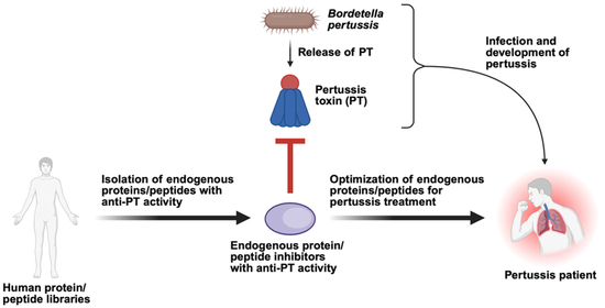
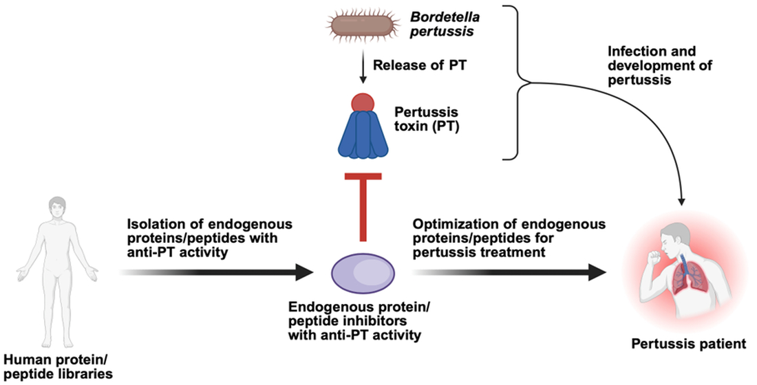
The life-threatening disease pertussis, also known as whooping cough, is caused by a complex interplay of several virulence factors produced by the bacterium Bordetella (B.) pertussis. These include the AB-type protein toxin pertussis toxin (PT), the main causative agent of pertussis. After infection with B. pertussis, PT is released and binds to its human target cells, which internalize PT. The enzyme subunit of PT is then taken up into the cytosol, where it catalyzes the ADP-ribosylation of the α-subunit of inhibitory GTP-binding proteins from the Gαi type. This ultimately leads to the development of the characteristic clinical symptoms associated with pertussis. Pertussis is a vaccine-preventable but highly infectious respiratory disease, and especially younger children are prone to develop severe pertussis. Despite the vaccination, over the past few years, increasing case numbers have been reported globally. Moreover, treatment options are strongly limited to antibiotics and symptomatic treatment. Therefore, novel therapies against toxin-mediated diseases are urgently required, while AB-type toxins such as PT are promising pharmacological targets to combat these associated diseases. To identify novel pharmacological inhibitors for AB-type toxins, huge potential lies within the human proteome/peptidome. Endogenous protein or peptide inhibitors for bacterial toxins might have evolved as part of the innate immunity and are awaited to be discovered. The scientific community is committed to identify potential candidates through targeted screening or explorative hypothesis-driven approaches. This review summarizes the recent efforts in the identification and characterization of the human body’s own proteins and peptides that inhibit PT. PT-inhibiting peptides were found by unbiased screening of peptide libraries from human hemofiltrate or hypothesis-driven evaluation, and PT-neutralizing mechanisms were discovered in cell-based approaches. The identification of endogenous peptides and proteins, e.g., defensins and α1-antitrypsin, as potent inhibitors of PT paves the way towards the development of novel therapeutic options against pertussis.
Key Contribution: This review highlights the potential of endogenous human proteins and peptides, specifically α-defensins and α1-antitrypsin, which directly and specifically target and neutralize pertussis toxin. This should offer novel pharmacological strategies to treat pertussis in the future.

Graphical Abstract

1. Introduction

Over the past few years, increasing case numbers for several highly infectious diseases have been reported. In particular, bacterial infections such as pertussis or diphtheria are on the rise. Despite high vaccination coverage, several outbreaks were reported by the European Center for Disease Prevention and Control (ECDC), also in Western countries [1,2].
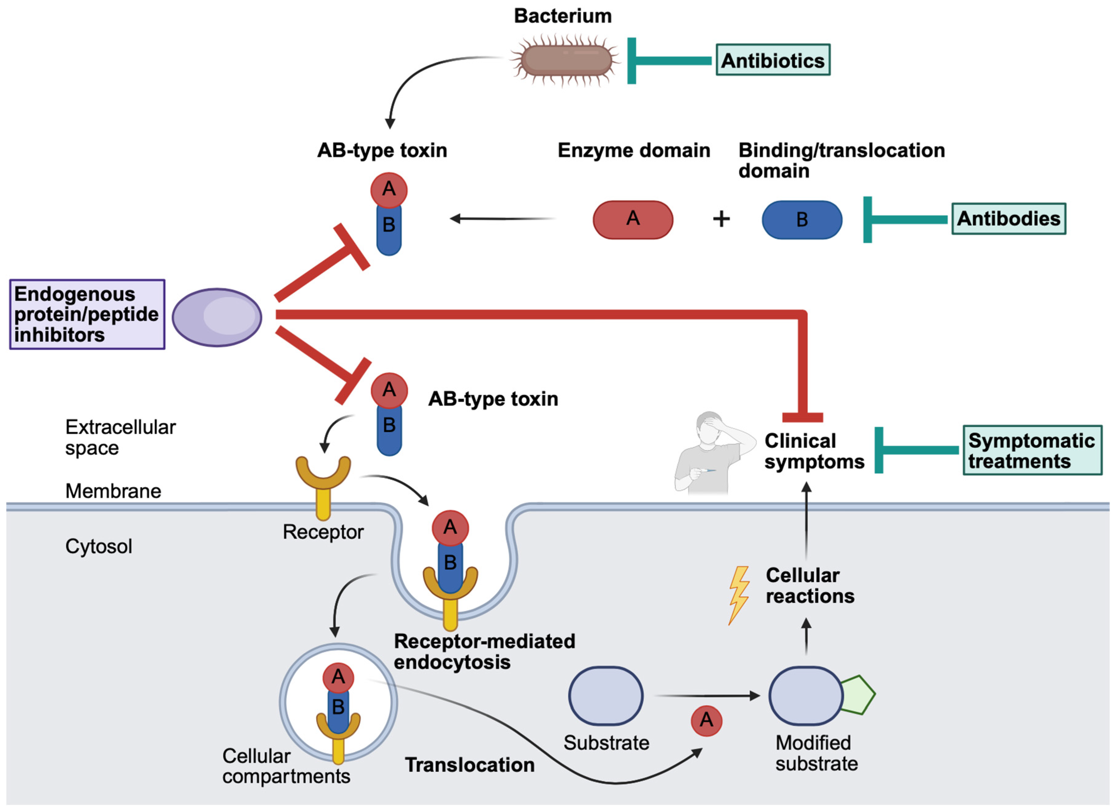
Infection of individuals with the bacteria often occurs through droplets or smears. Upon bacterial infection, the bacteria travel and attach using different virulence factors to the primary site of infection, e.g., the upper respiratory tract and lungs. For successful replication of the bacteria and thus colonization of the host, bacteria produce host-damaging virulence factors, amongst others, so-called exotoxins. Bacterial exotoxins can be classified into three different types (Type I–III) [3,4]. Type I exotoxins cause signaling dysfunction via receptors on cell surfaces. Type II exotoxins damage host cell membranes. Type III exotoxins act intracellularly and are able to enter target cells and directly modify cell functions. The medically highly relevant AB-type protein toxins are type III exotoxins [3,4]. AB-type toxins are highly potent and the causative agents of the clinical symptoms upon bacterial infection. Structurally, AB-type toxins consist of different functional subunits, including an enzymatically active (A) subunit and a binding/translocation (B) subunit (Figure 1). After binding of the AB-type toxin to specific cell surface receptors via its B-subunit, receptor-mediated endocytosis occurs, which results in the uptake of the toxin into cellular compartments. From there, the A-subunit translocates into the cytosol and modifies its specific substrate, in most cases a protein, with very high potency. The toxin-catalyzed substrate modification triggers cellular reactions, which in turn ultimately lead to the development of the typical clinical symptoms associated with the respective disease [5].
Typically, bacterial infections are treated with antibacterial drugs (“antibiotics”, Figure 1). However, the usage of antibiotics is often limited due to various reasons, including the evasion of the immune system through the development of antibiotic-resistant strains [6]. The therapeutic application of antibiotics, for example, in the case of pertussis, must occur as early as possible to successfully manage the infection and consequently the symptoms [7]. As soon as AB-type toxins are released, they are taken up into target cells, and subsequently, the clinical symptoms are caused by the enzymatic activity of the toxin. Since antibiotics are ineffective towards AB-type toxins themselves, they must be applied before the release of the toxin takes place. Anyhow, the early treatment using antibiotics requires early diagnosis and the identification of the pathogen. Regardless, early diagnosis is rare, and therefore, usually, at the start of the antibiotic treatment, the symptoms are already caused. Nonetheless, the application of antibiotics is essential to mitigate or prevent the spread of bacteria within the population.
Further treatment options for toxin-mediated disease are limited and comprise antibodies, which either derived from previous vaccination or due to the application of an antitoxin (e.g., in case of diphtheria antibodies produced in horses). For pertussis treatment, the monoclonal antibodies under evaluation in mice and baboon studies are Hu1B7 and Hu11E6. More detailed information, as well as the advantages and disadvantages of these antibodies, were described elsewhere [8]. However, the antibodies can only bind and thereby neutralize toxin molecules outside cells, since antibodies cannot enter cells and are therefore not able to catch already internalized AB-type toxins (Figure 1). Further clinical interventions are limited to supportive or intensive care to manage disease symptoms. For example, in severe pertussis cases in infants, limited success was reported for extra-corporeal membrane oxygenation or oscillatory ventilation [9,10]. As increasing case numbers are reported and treatment options are limited, toxin-associated diseases are still a matter of concern.
Consequently, novel therapeutic options for the treatment of toxin-mediated diseases are urgently required. These novel therapies could comprise the body’s own protein/peptide-based inhibitors, as the human body might already has developed strategies as part of the innate immune system to combat toxin-mediated diseases. Previously, numerous body’s own proteins and peptides that neutralize clinically relevant bacterial AB-type toxins, including pertussis toxin, diphtheria toxin, anthrax toxin, and clostridial enterotoxins, were identified [11]. This review highlights human proteins and peptides that might be exploited for the future treatment of pertussis.

2. The Uptake and Mode of Action of Pertussis Toxin Produced by Bordetella pertussis

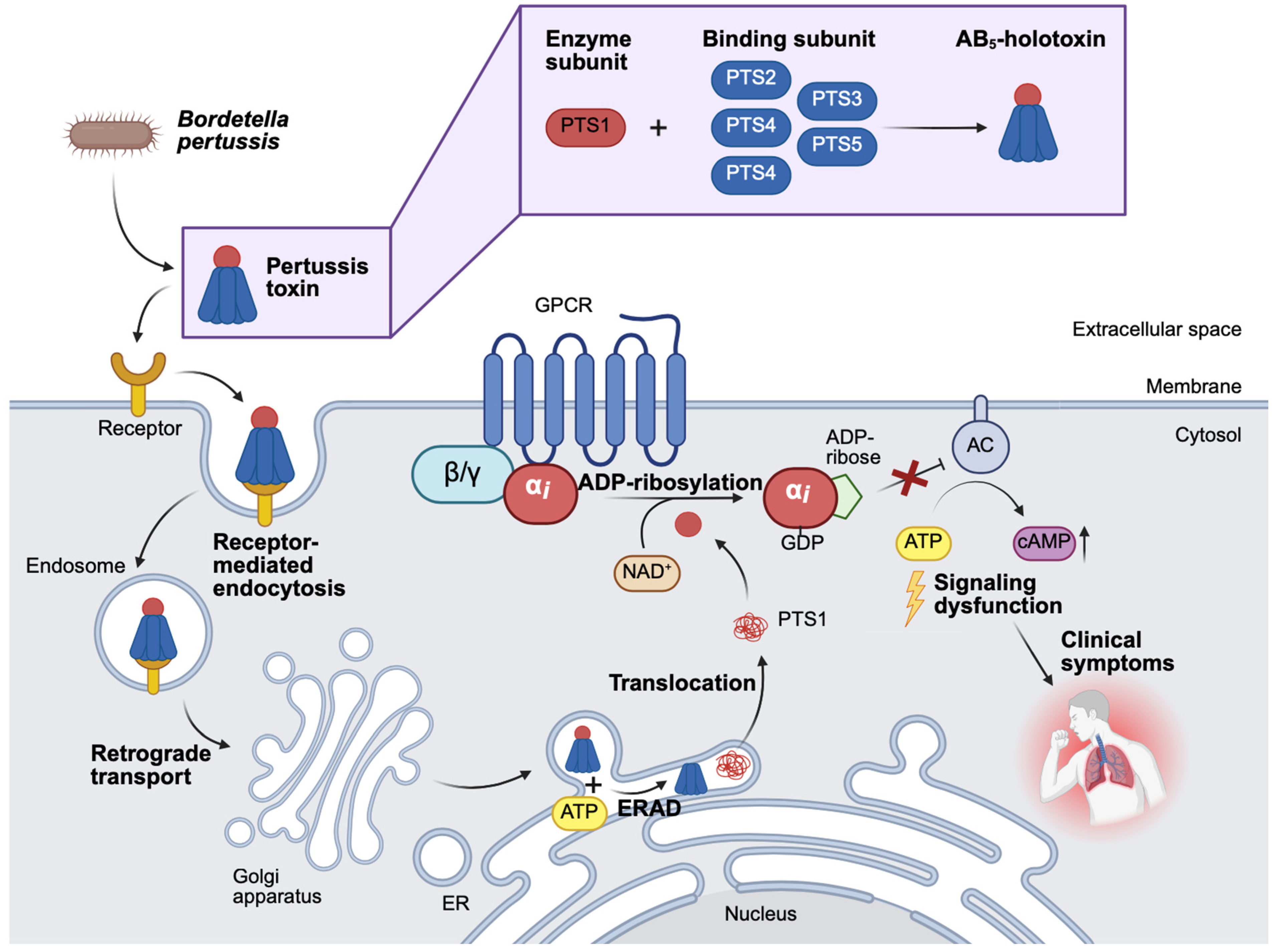
Bordetella (B.) pertussis is a Gram-negative, strictly human pathogen causing the respiratory disease pertussis, also known as whooping cough. Thereby, B. pertussis produces several virulence factors that are essential for the development of pertussis disease. One of the most important virulence factors of B. pertussis and essential for disease pathology and associated with severe pertussis is the ADP-ribosyltransferase (ART) pertussis toxin (PT) [7,12,13]. As an AB-type protein toxin, PT can be more specifically subclassified due to its structure as AB5-holotoxin. PT consists of the enzymatically active A-subunit PTS1 and a binding subunit, the B-pentamer PTS2–4 (Figure 2). In more detail, the B-pentamer is built by five subunits, PTS2, PTS3, PTS4, and PTS5, while PTS4 is present twice. Consequently, the stoichiometry of the PT-holotoxin with its subunits S1:S2:S3:S4:S5 is 1:1:1:2:1 [14,15,16].
After the binding of PT to sialic acid conjugates on cell surfaces, receptor-mediated endocytosis and uptake of PT into endosomes are enabled [17,18]. Subsequently, the holotoxin travels in a retrograde manner from the endosome to the Golgi apparatus and the ER [19,20]. Due to the physiological ATP stores of the ER, ATP attaches to the central pore of the B-subunit, which leads to the disassembly of PT and the release of PTS1. Once released, PTS1, which is thermally unstable, shifts into a disordered/unfolded state, which is recognized by the ER-associated degradation pathway (ERAD), mediating the translocation of PTS1 from the ER into the cytosol [21,22]. The translocation process of PTS1 into the cytosol is assisted by host cell chaperones [23,24,25,26]. Within the cytosol, PTS1 ADP-ribosylates the α-subunit of inhibitory G-proteins (Gαi) associated with G-protein-coupled receptors (GPCRs) using its co-substrate NAD+ [27]. Modified Gαi causes the inhibition of the Gαi-mediated inhibition of the adenylate cyclase (AC). Therefore, the AC is activated, and the production of cyclic AMP (cAMP) is enhanced [28]. Elevated cAMP levels are cell type dependent and influence two downstream signaling cascades, protein kinase A (PKA) and the exchange proteins directly activated by cAMP (EPAC) [8,29]. PKA and EPAC signaling are involved in diverse immune and metabolic pathways, causing systemic immunosuppression, lymphocytosis, and metabolic disruptions [29]. Therefore, the downstream signal transduction is altered, and specific symptoms associated with the pertussis disease occur.
Figure 2. The structure, uptake, and mode of action of PT from Bordetella pertussis. Bordetella (B.) pertussis produces, amongst other virulence factors, the AB-type protein toxin pertussis toxin (PT). The AB5-holotoxin has one enzyme subunit (PTS1) and five binding subunits S2, S3, S4, and S5 (PTS2–5) which assemble in a 1:1:1:2:1 stoichiometry [14]. Uptake of PT into endosomes occurs after the binding of PT to sialic acid on cell surfaces and receptor-mediated endocytosis. After the retrograde transport of PT to the endoplasmic reticulum (ER), it gets disassembled. Then, PTS1 is transported by the ER-associated degradation pathway (ERAD) into the cytosol, where PTS1 ADP-ribosylates its substrate, inhibitory G-proteins (Gαi) of G-protein-coupled receptors (GPCR), while NAD+ serves as a co-substrate. Consequently, Gαi is no longer able to downregulate the adenylate cyclase (AC) activity, causing the enhanced production of intracellular cAMP. Finally, the downstream signaling cascade is altered, and the typical clinical symptoms develop. (adenosine triphosphate (ATP), adenosine diphosphate (ADP), cyclic adenosine monophosphate (AMP) (cAMP), guanosine diphosphate (GDP)) [11,14,30,31,32,33]. (Created in BioRender. Lietz, S. (2026) https://BioRender.com/3th6gu2).
Toxins 18 00208 g002

3. The Relevance of Exploiting Human Endogenous Proteins and Peptides as Part of the Innate Immunity as Novel Inhibitors Against Pertussis Toxin

As previously introduced, despite the vaccination for pertussis, increasing case numbers were reported. The treatment of pertussis in the clinics is solely based on the application of antibiotics and symptomatic therapies, e.g., treating secondary complications of pertussis. Due to the adaptation of pathogens in general, but also B. pertussis, increasing antibiotic resistances occur. In the clinics, for the treatment of pertussis, antibiotics belonging to the group of macrolides, such as erythromycin, but also newer macrolides, e.g., azithromycin and clarithromycin, are used [7]. However, there have been reports of macrolide-resistant B. pertussis strains. Especially in certain parts of China, between 70 and 100% of clinical isolates showed a macrolide-resistance due to a specific mutation, a 2047 A-to-G mutation within the 23S rRNA gene [34,35,36]. In contrast, outside of China, there have been only sporadic reports on the appearance of macrolide-resistant B. pertussis strains [34,36]. Moreover, it is likely that those strains are additionally resistant towards other macrolide antibiotics [7] and that, in the future, further cases of macrolide-resistant B. pertussis strains will be reported.
In addition to antibiotic-resistant B. pertussis, there were reports of B. pertussis escape mutants lacking the virulence factor pertactin. Some acellular pertussis vaccines contain pertactin antigens, which, in addition, contribute to waning immunity after vaccination [6,37,38,39,40].
In general, vaccination is an essential strategy to prevent pertussis. However, infants (<6 months) are a population group at high risk for severe pertussis, since they are unimmunized or only partially immunized [2]. Here, the importance of maternal immunization vaccination programs for pertussis should be communicated early during pregnancy, to protect newborns from pertussis with maternal antibodies [2]. At the same time, for population groups at high risk, effective treatment strategies are urgently required.
Consequently, novel targeted strategies that address the treatment gap that has opened due to evolved B. pertussis strains and to protect vulnerable groups at high risk for severe pertussis are urgently required. These strategies are essential for the future treatment of toxin-mediated diseases such as pertussis in the clinics. Because AB-type toxins are the causative agents of the symptoms of the disease, their specific inactivation prevents the clinical manifestation of the disease. Therefore, novel strategies should be specifically directed towards bacterial AB-type protein toxins.
Novel strategies to exclusively target AB-type toxins could comprise endogenous proteins and peptides. Endogenous proteins and peptides are the first line of defense and belong to innate immunity. This involves, for example, antimicrobial peptides that target a diverse set of microbes, such as bacteria or viruses. Since the human body has evolved protein-/peptide-based first line defense strategies to combat, e.g., bacteria, it is likely that the body has also developed strategies against bacterial virulence factors, including AB-type protein toxins. Especially, in regard to the tremendous complexity of the human proteome/peptidome, the odds of identifying the body’s own peptides with anti-toxin activity are extremely high. Due to alternative promoters, splicing, post-translational modifications, and proteolytic processing, the human proteome/peptidome contains more than a million bioactive compounds and, as such, contains huge potential for the discovery of novel protein- and peptide-based therapeutics [41]. Moreover, endogenous proteins and peptides have several benefits regarding their human application, including lower immunogenicity, better safety profile, higher serum stability, better distribution within the human body, and lower production costs [42].
For the identification of novel endogenous human protein/peptide-based inhibitors for PT, two different approaches were exploited. Here, an explorative hypothesis-driven approach and a targeted screening approach were used. Through the explorative hypothesis-driven approach, the group of human α-defensins, specifically α-defensin-1 and -5, was identified as peptide inhibitors of PT [8,43,44]. Moreover, the screening of a human-derived hemofiltrate protein/peptide library allowed the identification of a human protease inhibitor of the SERPIN family, α1-antitrypsin (α1AT), as a potent inhibitor of PT [31].

4. Examples of Human Endogenous Proteins/Peptides and Their Derivatives with Anti-PT Activity

In the following, examples of human endogenous proteins and peptides or thereof derived peptide derivatives are given, which possess anti-PT activity (Table 1). Subsequently, the mode of action of the single proteins and peptides towards PT is described in more detail (Table 1 and Figure 3). Here, the special focus lies on human α-defensin-1 and -5, as well as human α1AT [11].
Table 1. Examples for human endogenous proteins and peptides with anti-toxin activity. Human-derived proteins and peptides that inhibit bacterial toxins are listed with the respective mode of action [11].
Human α- and β-Defensins (def)
InhibitorPhysiological FunctionRationale Behind Experimental TestingMechanism of Inhibition References
α-def-1,


α-def-2,


α-def-3,


α-def-4,


α-def-5
Antimicrobial peptides of the innate immune system are produced by different cell types and organs, depending on the defensin [45,46]Hypothesis-driven—inhibition of multiple bacterial AB-type protein toxins was previously reportedInhibition of enzyme activity and inhibition of toxin binding to cells

Toxin inhibition, mechanism not tested yet

Toxin inhibition, mechanism not tested yet

Inhibition of enzyme activity and inhibition of toxin binding to cells
[43,44]
α-def-6,
β-def-1,
β-def-2
No inhibition
No inhibition
No inhibition
[43,44]
Human α1-antitrypsin (α1AT)
InhibitorPhysiological functionRationale behind experimental testingMechanism of inhibitionReferences
α1ATSerin protease inhibitor of the SERPIN-family, activity regulation of serin proteases, e.g., neutrophil elastase within the lungs [47]ScreeningInhibition of binding[31]
Antithrombin, antithrombin with fondaparinuxProtease inhibitor of the SERPIN-family, Antithrombin, cleaves thrombin, and fondaparinux acts as an activator of the physiological function of antithrombin, a natural anticoagulant [47,48,49]Hypothesis-driven—Belongs to the same group of protease inhibitors as α1ATNo inhibition[31]
α1AT-HF,
α1AT-HF P8,
42,
64, (α1AT-derived peptides)
No physiological function is attributedHypothesis-driven screeningInhibition of intoxication, due to inhibition of binding (α1AT-HF) or a mechanism not yet tested[50]
VIRIP, and other α1AT-derived peptidesVIRIP was previously identified as an inhibitor of HIV [51]No inhibition [50]

4.1. Group of Defensins as Inhibitors for Pertussis Toxin

Earlier, the scientific community has shown that the group of human defensins has activity in vitro and in vivo against multiple AB-type protein toxins, including, e.g., iota toxin, Clostridioides (C.) difficile toxin (CDT), Toxin A (TcdA) and Toxin B (TcdB), diphtheria toxin (DT), and anthrax lethal toxin (LT) [52,53,54,55,56,57,58]. Here, the mechanism of action of the different defensins towards the bacterial toxins was often related to the formation of aggregates and binding of the bacterial toxin, as well as the inhibition of the enzyme activity of the toxin through the defensins. Therefore, testing whether the defensins have activity against PT was a hypothesis-driven approach, since a broad set of bacterial toxins had already been inhibited by the group of defensins.
Human endogenous defensins are antimicrobial peptides (AMPs) that are involved as part of the innate immune system in the first line defense in a non-specific way against a broad spectrum of pathogens, including bacteria, fungi, and viruses [45]. In more detail, human defensins showed antibacterial activity against a variety of Gram-negative and Gram-positive bacteria. Here, most often the mechanism of action of the human defensins towards the bacteria is related to the disruption of bacterial membranes or the inhibition of cell wall synthesis [46]. During these processes, the human defensins target structures such as the outer membrane proteins, LPS, peptidoglycan, lipid II, phosphatidylglycerol, and lipoteichoic acids, as well as bacterial DNA and RNA [46]. To reach their target, namely the pathogens in the human body, defensins need to be produced, stored, transported, and released when needed at specific pathogen entry sites. Human defensins are located alongside other antimicrobial factors within the azurophilic granules of neutrophils or are secreted by other cell types, e.g., epithelial cells, amongst others of the kidneys, pancreas, and female reproductive tract or by Paneth cells found in the small intestine at the bottom of the crypt [45,46]. A more comprehensive overview of the structure, expression sites of different defensins and the attributed function was summarized earlier by others and would exceed the scope of this review [45,46]. Briefly, the human defensins can be grouped according to their cysteine pairing and their structure into α-, β-, and θ-defensins [59,60]. They are small (16–50 amino acids), arginine-rich, cationic peptides with a β-sheet structure and three intramolecular disulfide bridges [45,59,60]. For the following, relevant defensins are the α-defensins (3–4 kDa, 29–35 amino acids) and the β-defensins (3–5 kDa, 38–50 amino acids) [45]. The α-defensins are further divided into α-defensin-1-4, also termed Human Neutrophil Peptides (HNPs) and the α-defensin-5 and 6, also termed Human (enteric) Defensins (HDs) [46], while the β-defensins are also known as human β-defensins (HBDs).
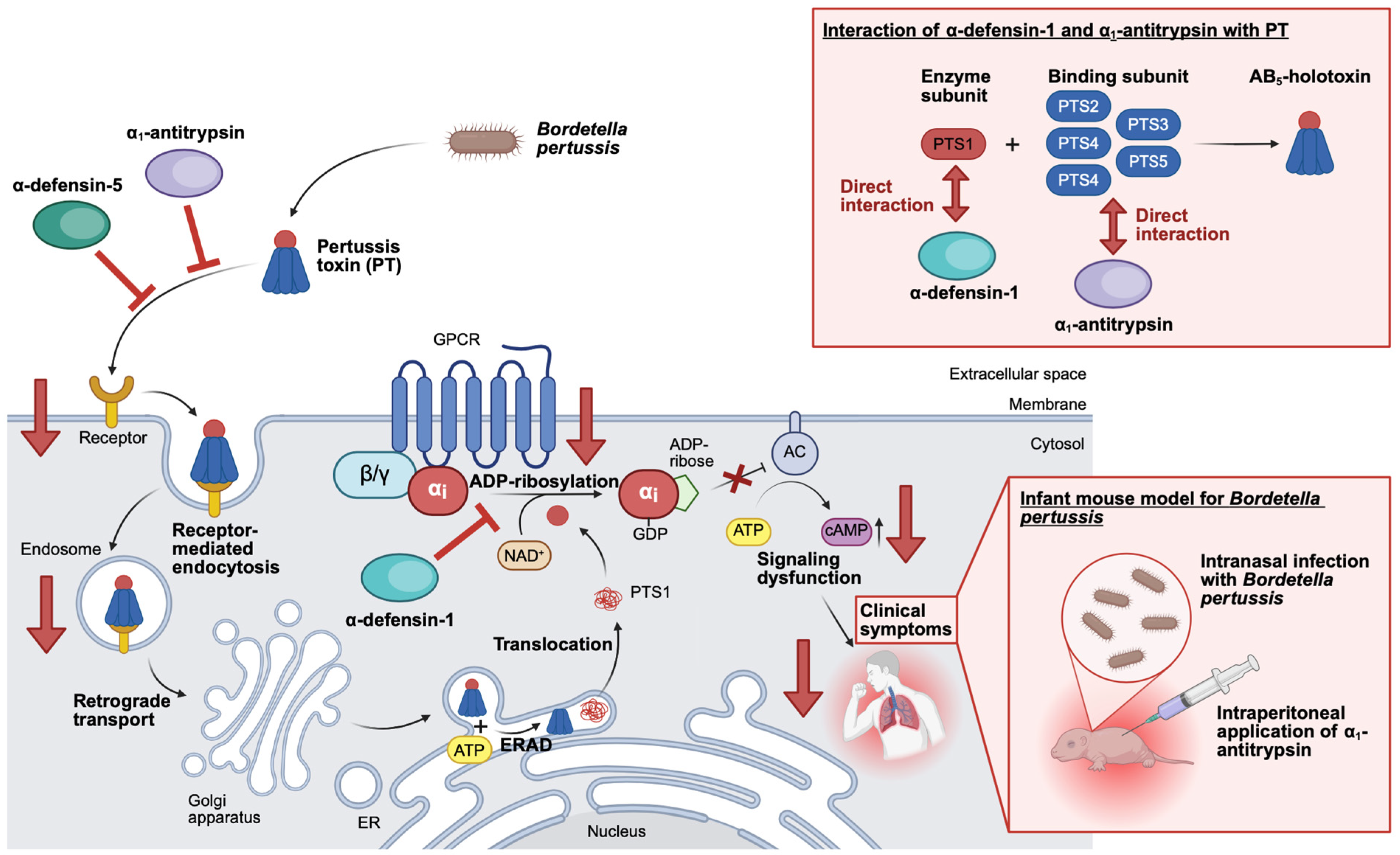
In our hypothesis-driven approach, selected α- and β-defensins were tested for their anti-PT activity by analyzing the ADP-ribosylation status of Gαi using CHO-K1 or A549 cells. When investigating the effect of α- and β-defensins towards PT in this assay, it was shown that α-defensin-1, -2, -3, -4, and -5 have inhibitory potential against PT, but not α-defensin-6 and β-defensin-1 and -2 [43,44]. Subsequently, α-defensin-1 and -5 were selected as lead candidates for further mechanistic evaluation, while the results of Kling and colleagues are briefly summarized in the following [8,43,44]. When α-defensin-1 and -5 were preincubated with PT before addition to cells, the inhibitory effect on ADP-ribosylation of Gαi was even stronger. Moreover, the inhibition of ADP-ribosylation of Gαi mediated by α-defensin-1 and -5 occurred in a concentration-dependent manner. An alternative endpoint to determine the PT intoxication of cells is the measurement of PT-mediated effects on cAMP signaling in the living cell-based interference in the Gαi-mediated signal transduction (iGIST) assay. The results of the iGIST assay further confirmed that α-defensin-1 and -5 are inhibitors of PT, since the two peptides caused a decrease in cAMP signaling in the presence of PT. Moreover, α-defensin-1 and -5 but not β-defensin-1 inhibited the enzyme activity of PTS1 in vitro using recombinant Gαi in a concentration-dependent manner. However, the inhibition of enzyme activity mediated by α-defensin-1 was stronger than the inhibition mediated by α-defensin-5. Most interestingly, the inhibition of ADP-ribosylation of Gαi in PT-treated living cells and of enzyme activity of PTS1 in vitro mediated by α-defensin-1 and -5 was lost when linearized versions of α-defensin-1 and -5 were used. The linearized versions of α-defensin-1 and -5 had no disulfide bonds as the cysteine residues were replaced with serine residues, which indicates that the three-dimensional structure of the defensins is important for mediating the inhibition of PT. Furthermore, the effect of α-defensin-1 and -5 on PT binding to cells was investigated using flow cytometry and fluorescence microscopy. Here, both defensins showed inhibition of binding and uptake of PT into cells, while the inhibitory effect mediated by α-defensin-5 was stronger compared to the effect mediated by α-defensin-1. In addition, it was shown that α-defensin-1 is taken up into different cell lines in a stronger manner than α-defensin-5. Intracellularly, the signal for α-defensin-1 and PTS1 but not α-defensin-5 and PTS1 are in proximity, as shown by a proximity ligation assay. The close proximity of α-defensin-1 and PTS1, but not of α-defensin-5 and PTS1, was further supported by in silico modeling, demonstrating that α-defensin-1 but not α-defensin-5 contains specific interaction interfaces with PTS1. Those results were additionally confirmed by in vitro interaction analysis using dot plot experiments. Collectively, the results showed that α-defensin-1 is able to enter cells, reach and interact with its target PTS1.
Consequently, the inhibitory effect of α-defensin-1 on PT was mainly mediated via inhibition of the enzyme activity of PTS1, while α-defensin-5 mainly inhibited cellular uptake of PT. Therefore, the inhibitory mode of action for α-defensin-1 and -5 slightly differs but complements each other. Since no adverse effects on cell viability by α-defensin-1 and -5 were detected, both human endogenous defensins are promising candidates for further evaluation regarding the treatment of pertussis in the clinics.

4.2. α1AT and Derived Peptides as Inhibitors for Pertussis Toxin

Through the screening of a human hemofiltrate peptide library and the application of bioassay-guided fractionation, as well as mass spectrometry analysis, α1AT was identified as an inhibitor of PT [31].
As mentioned above, α1AT (52 kDa) is a highly abundant human serin protease inhibitor of the SERPIN superfamily, which is mainly produced by hepatocytes of the liver, but also other cell types [61,62,63]. The physiological function of α1AT is to bind and therefore inhibit serine proteases, including neutrophil elastase, proteinase 3, cathepsin G, trypsin, and transmembrane serine protease 2 (TMPRSS2) [47,64,65,66]. For example, the serine protease neutrophil elastase targets components of the extracellular matrix, e.g., elastin and collagen [67]. The inhibition of neutrophil elastase by α1AT leads to the protection of cells and tissues from proteolytic activity and degradation, e.g., within the lungs [68]. Consequently, α1AT is a major regulator of protease activity and modulator of inflammatory responses in the human body, protecting the lungs from neutrophil elastase released by immune cells [69]. Therefore, the acute phase protein α1AT combats and prevents inflammation-induced tissue damage, while physiological plasma concentrations of ~17–38 μM can increase 4–5-fold upon acute inflammation [47,70]. Individuals suffering from the genetic disorder α1AT deficiency have lower serum levels of α1AT, which results in unregulated tissue degradation in the lower respiratory tract [71]. The α1AT deficiency can be treated by the intravenous augmentation of α1AT, which is obtained and purified from donor blood, using licensed drugs, e.g., Prolastin®. For such drugs, administration protocols and safety profiles are already well-established [72].
After the identification of α1AT through an unbiased screening approach, the inhibitory mode of action of α1AT towards PT was investigated in detail. This experimental validation involved different in silico, in vitro, and in vivo methods, while corresponding results are briefly summarized [31]. For these experiments, the approved drug Prolastin® was used as a source for α1AT, while being referred to as α1AT in the following. First, the effect of α1AT as an isolated compound was investigated in a cell-based assay. This assay analyzed the ADP-ribosylation status of Gαi, the specific substrate of PT, in PT-treated CHO-K1 cells and the more physiologically relevant lung adenoma A549 cells in FCS-free conditions. Here, a concentration-dependent inhibition of ADP-ribosylation of Gαi in PT-treated cells was observed for both cell lines. This effect was even stronger when α1AT was preincubated with PT before the addition to cells. Moreover, the inhibitory effect of α1AT towards PT was preserved when repeating the assay in FCS-containing conditions. Notably, α1AT had no adverse effects on the cell viability of CHO-K1 cells after 4 and 24 h and no effect on the detection of ADP-ribosylated Gαi. To analyze whether the protective effect of α1AT was related to its protease activity, another member of the SERPIN superfamily, antithrombin, was tested for its ability to inhibit PT. Anyhow, the protease activity of α1AT was most likely not mediating the inhibition of PT, as neither antithrombin alone nor the combination of antithrombin with its activator fondaparinux inhibited the ADP-ribosylation of Gαi in PT-treated CHO-K1 cells. Moreover, α1AT showed only minor influences on the enzyme activity of PTS1 in vitro, independent of whether recombinantly purified Gαi or CHO-K1 cell lysate was used as a source for Gαi. Subsequently, the uptake of PT into target cells was investigated in more detail, using flow cytometry and fluorescence microscopy. In those experiments, it was shown that α1AT inhibits the binding of PT to CHO-K1 and A549 cells in a concentration-dependent manner. Here, the preincubation of α1AT with PT before addition to cells increased the inhibition of PT-binding to CHO-K1 cells even more, indicating that the interaction of PT and α1AT during the preincubation might be relevant. Using high-resolution stimulated emission depletion (STED) microscopy, it was demonstrated that signals for PT and α1AT partially colocalize, particularly visible when higher α1AT concentrations were used. To investigate whether there is an interaction between α1AT and PT in vitro and in silico, interaction analysis was conducted. The dot blot analysis, where α1AT was vacuum-aspirated onto a membrane and overlayed with PT or PTS1, revealed that α1AT exclusively interacts with the PT holotoxin but not with its isolated enzyme subunit, PTS1. This suggests that the interaction of PT and α1AT is most likely mediated by the B-subunit of PT. This result is further strengthened by the in silico analysis, where a blind docking analysis was performed during which α1AT was allowed to explore the surface of PT. This analysis suggests as well that the B-pentameric ring of PT, especially subunit 3 (PTS3), is important for the α1AT-PT interaction. Finally, an infant mouse model for pertussis was employed to investigate the effect of α1AT in vivo. Upon infection of infant C57BL/6 mice with B. pertussis, the mice develop pertussis-associated leukocytosis, the hallmark of pertussis disease, which is also observed in human infants with pertussis. Using this animal model, expression levels for the murine α1AT genes (serpinA1a-e) were downregulated in B. pertussis-infected infant mice in direct comparison to non-infected mice. Furthermore, when B. pertussis-infected infant mice were treated with α1AT, the white blood cell count, used to assess leukocytosis, was reduced in comparison to infected vehicle-treated mice.
Consequently, α1AT inhibits intoxication of target cells by inhibiting the uptake of PT through direct interaction with PT. Thus, α1AT prevents subsequent steps of the intoxication cascade and ultimately reduces the development of pertussis-associated symptoms in an in vivo mouse model for pertussis. Further studies will examine α1AT levels in humans who have developed pertussis disease and whether augmentation of reduced or low α1AT levels might benefit the disease outcome.
A further study revealed that α1AT inhibits further PT-related bacterial AB-type protein toxins, including C2 toxin from C. botulinum, DT from C. diphtheriae, and a B. anthracis fusion toxin [73]. This further highlights the potential of α1AT as a pan-toxin inhibitor and the prospect of α1AT-containing drugs for the treatment of pertussis and other toxin-mediated diseases (diphtheria and anthrax) in repurposing approaches.
Finally, the amino acid region of full-length human endogenous α1AT that bears anti-PT activity was identified [50]. For that purpose, α1AT-derived peptides spanning different parts of the full-length α1AT were generated and tested for their ability to inhibit PT by analyzing the ADP-ribosylation status of Gαi in PT-treated CHO-K1 cells. Through this approach, three α1AT-derived peptides termed α1AT-HF, 42, and 64 were identified to inhibit the ADP-ribosylation of Gαi in PT-treated CHO-K1 cells. Another previously published peptide derived from α1AT, termed VIRIP [51] showed no inhibition of PT. The amino acid region of α1AT that bears anti-PT activity was confirmed by an overlap of a total of nine peptides of various lengths. The lead peptide α1AT-HF is endogenously present in human hemofiltrate and has the shortest sequence of α1AT with anti-PT activity. Interestingly, this peptide was shown to have no adverse effects on cell viability and in vivo in zebrafish embryos, rendering it a perfect candidate for further modification and optimization approaches for human application.
The findings with PT provided further proof-of-concept that the generation of peptides based on endogenous human protein precursors can result in the generation of peptides with anti-toxin activity, which has a scientific and clinical impact for various protein toxins from other medically highly relevant bacteria, e.g., C. difficile, which are also neutralized by human peptides and proteins [73,74].

5. Conclusions

This comprehensive review summarizes the recent efforts to identify and optimize the human body’s own peptides and proteins as novel inhibitors of PT, the highly potent and specific virulence factor of B. pertussis, responsible for the severe (childhood) disease pertussis. These results from basic research revealed novel aspects of human innate immunity, as the identification of human endogenous proteins and peptides as natural inhibitors of bacterial toxins underlines and highlights the role of the innate immune system to fight not only bacteria but also their produced virulence factors. Moreover, the novel findings should be transferred for the development of novel pharmacological strategies against pertussis. Such novel therapeutic approaches are urgently required as the current approaches, including vaccination, antibiotic treatments, and symptomatic treatment strategies, are losing their effectiveness, causing an increase in reported pertussis cases worldwide.

Author Contributions

Conceptualization and writing: S.L. and H.B.; funding acquisition, H.B. All authors have read and agreed to the published version of the manuscript.

Funding

This work was supported by the Deutsche Forschungsgemeinschaft (DFG, German Research Foundation)—project number 316249678-SFB1279 (A07, funding to H.B.).

Institutional Review Board Statement

Not applicable.

Informed Consent Statement

Not applicable.

Data Availability Statement

No new data were created or analyzed in this study.

Conflicts of Interest

The authors declare no conflicts of interest.

Abbreviations

α1ATα1-antitrypsin
Gαiα-subunit of inhibitory G-proteins
ARTADP-ribosyltransferase
ADPAdenosine diphosphate
AMPAdenosine monophosphate
ATPAdenosine triphosphate
ACAdenylate cyclase
AMPsAntimicrobial peptides
B. anthracisBacillus anthracis
B. pertussisBordetella pertussis
CHO-K1 cellsChinese hamster ovary cells strain K1
C. difficileClostridioides difficile
CDTClostridioides difficile toxin
cAMPCyclic AMP
defDefensin
DTDiphtheria toxin
EREndoplasmic reticulum
ERADER-associated degradation
ECDCEuropean Center for Disease Prevention and Control
EPACExchange proteins directly activated by cAMP
GDPGuanosine diphosphate
GPCRsG-protein-coupled receptors
HBDsHuman β-defensins
HDsHuman (enteric) Defensins
HNPsHuman Neutrophil Peptides
iGISTInterference in Gαi-mediated Signal Transduction
LTLethal toxin
PTPertussis toxin
PTS1Pertussis toxin enzyme subunit
PTS2–5Pertussis toxin binding subunits S2, S3, S4, and S5
PKAProtein kinase A
TcdAToxin A
TcdBToxin B

References

  1. European Centre for Disease Prevention and Control Epidemiological Update: Diphtheria Cases in Europe. Available online: https://www.ecdc.europa.eu/en/news-events/epidemiological-update-diphtheria-cases-europe (accessed on 9 January 2025).
  2. European Centre for Disease Prevention and Control. Increase of Pertussis Cases in the EU/EEA: 8 May 2024; European Centre for Disease Prevention and Control: Solna, Sweden, 2024. [Google Scholar]
  3. Ala’Aldeen, D.A.A.; Wooldridge, K.G. 13—Bacterial Pathogenicity. In Medical Microbiology, 8th ed.; Greenwood, D., Barer, M., Slack, R., Irving, W., Eds.; Churchill Livingstone: Edinburgh, UK, 2012; pp. 156–167. ISBN 978-0-7020-4089-4. [Google Scholar]
  4. Xiao, Y.; Yan, Z.; Ren, F.; Tan, Y. Bacterial Exotoxins in Medicine: Potential Value and Perspectives. Int. J. Med. Sci. 2025, 22, 2010–2019. [Google Scholar] [CrossRef] [Scilit]
  5. Aktories, K.; Barbieri, J.T. Bacterial Cytotoxins: Targeting Eukaryotic Switches. Nat. Rev. Microbiol. 2005, 3, 397–410. [Google Scholar] [CrossRef] [Scilit] [PubMed]
  6. Ma, L.; Caulfield, A.; Dewan, K.K.; Harvill, E.T. Pertactin-Deficient Bordetella Pertussis, Vaccine-Driven Evolution, and Reemergence of Pertussis. Emerg. Infect. Dis. 2021, 27, 1561–1566. [Google Scholar] [CrossRef] [Scilit] [PubMed]
  7. Mattoo, S.; Cherry, J.D. Molecular Pathogenesis, Epidemiology, and Clinical Manifestations of Respiratory Infections Due to Bordetella Pertussis and Other Bordetella Subspecies. Clin. Microbiol. Rev. 2005, 18, 326–382. [Google Scholar] [CrossRef] [Scilit]
  8. Ernst, K. Novel Strategies to Inhibit Pertussis Toxin. Toxins 2022, 14, 187. [Google Scholar] [CrossRef] [Scilit] [PubMed]
  9. Crichton, H.; Nkado, N.; Morrow, B.; Salie, S.; Procter, C. Critical Pertussis Infection in the Paediatric Intensive Care Unit: A Case Series in an Outbreak. S. Afr. J. Crit. Care 2025, 41, e2049. [Google Scholar] [CrossRef] [Scilit]
  10. Surridge, J.; Segedin, E.R.; Grant, C.C. Pertussis Requiring Intensive Care. Arch. Dis. Child. 2007, 92, 970–975. [Google Scholar] [CrossRef] [Scilit]
  11. Lietz, S.; Barth, H. Novel Endogenous Protein-Based Strategies to Inhibit Clinically Relevant Bacterial AB-Type Toxins Including Pertussis Toxin. Arch. Toxicol. 2026; online ahead of print. [CrossRef] [Scilit]
  12. Scanlon, K.; Skerry, C.; Carbonetti, N. Association of Pertussis Toxin with Severe Pertussis Disease. Toxins 2019, 11, 373. [Google Scholar] [CrossRef] [Scilit]
  13. Scanlon, K.; Skerry, C.; Carbonetti, N. Role of Major Toxin Virulence Factors in Pertussis Infection and Disease Pathogenesis. Adv. Exp. Med. Biol. 2019, 1183, 35–51. [Google Scholar] [CrossRef] [Scilit]
  14. Stein, P.E.; Boodhoo, A.; Armstrong, G.D.; Cockle, S.A.; Klein, M.H.; Read, R.J. The Crystal Structure of Pertussis Toxin. Structure 1994, 2, 45–57. [Google Scholar] [CrossRef] [Scilit]
  15. Tamura, M.; Nogimori, K.; Murai, S.; Yajima, M.; Ito, K.; Katada, T.; Ui, M.; Ishii, S. Subunit Structure of Islet-Activating Protein, Pertussis Toxin, in Conformity with the A-B Model. Biochemistry 1982, 21, 5516–5522. [Google Scholar] [CrossRef] [Scilit] [PubMed]
  16. Weiss, A.A.; Johnson, F.D.; Burns, D.L. Molecular Characterization of an Operon Required for Pertussis Toxin Secretion. Proc. Natl. Acad. Sci. USA 1993, 90, 2970–2974. [Google Scholar] [CrossRef] [Scilit] [PubMed]
  17. Armstrong, G.D.; Howard, L.A.; Peppler, M.S. Use of Glycosyltransferases to Restore Pertussis Toxin Receptor Activity to Asialoagalactofetuin. J. Biol. Chem. 1988, 263, 8677–8684. [Google Scholar] [CrossRef] [Scilit]
  18. Witvliet, M.H.; Burns, D.L.; Brennan, M.J.; Poolman, J.T.; Manclark, C.R. Binding of Pertussis Toxin to Eucaryotic Cells and Glycoproteins. Infect. Immun. 1989, 57, 3324–3330. [Google Scholar] [CrossRef] [Scilit]
  19. el Bayâ, A.; Linnemann, R.; von Olleschik-Elbheim, L.; Robenek, H.; Schmidt, M.A. Endocytosis and Retrograde Transport of Pertussis Toxin to the Golgi Complex as a Prerequisite for Cellular Intoxication. Eur. J. Cell Biol. 1997, 73, 40–48. [Google Scholar]
  20. Plaut, R.D.; Carbonetti, N.H. Retrograde Transport of Pertussis Toxin in the Mammalian Cell. Cell. Microbiol. 2008, 10, 1130–1139. [Google Scholar] [CrossRef] [Scilit]
  21. Banerjee, T.; Cilenti, L.; Taylor, M.; Showman, A.; Tatulian, S.A.; Teter, K. Thermal Unfolding of the Pertussis Toxin S1 Subunit Facilitates Toxin Translocation to the Cytosol by the Mechanism of Endoplasmic Reticulum-Associated Degradation. Infect. Immun. 2016, 84, 3388–3398. [Google Scholar] [CrossRef] [Scilit] [PubMed]
  22. Pande, A.H.; Moe, D.; Jamnadas, M.; Tatulian, S.A.; Teter, K. The Pertussis Toxin S1 Subunit Is a Thermally Unstable Protein Susceptible to Degradation by the 20S Proteasome. Biochemistry 2006, 45, 13734–13740. [Google Scholar] [CrossRef] [Scilit]
  23. Ernst, K.; Eberhardt, N.; Mittler, A.-K.; Sonnabend, M.; Anastasia, A.; Freisinger, S.; Schiene-Fischer, C.; Malešević, M.; Barth, H. Pharmacological Cyclophilin Inhibitors Prevent Intoxication of Mammalian Cells with Bordetella Pertussis Toxin. Toxins 2018, 10, 181. [Google Scholar] [CrossRef] [Scilit]
  24. Ernst, K.; Mittler, A.-K.; Winkelmann, V.; Kling, C.; Eberhardt, N.; Anastasia, A.; Sonnabend, M.; Lochbaum, R.; Wirsching, J.; Sakari, M.; et al. Pharmacological Targeting of Host Chaperones Protects from Pertussis Toxin in Vitro and in Vivo. Sci. Rep. 2021, 11, 5429. [Google Scholar] [CrossRef] [Scilit]
  25. Kellner, A.; Taylor, M.; Banerjee, T.; Britt, C.B.T.; Teter, K. A Binding Motif for Hsp90 in the A Chains of ADP-Ribosylating Toxins That Move from the Endoplasmic Reticulum to the Cytosol. Cell. Microbiol. 2019, 21, e13074. [Google Scholar] [CrossRef] [Scilit]
  26. Kellner, A.; Cherubin, P.; Harper, J.K.; Teter, K. Proline Isomerization as a Key Determinant for Hsp90-Toxin Interactions. Front. Cell. Infect. Microbiol. 2021, 11, 771653. [Google Scholar] [CrossRef] [Scilit]
  27. Bokoch, G.M.; Katada, T.; Northup, J.K.; Hewlett, E.L.; Gilman, A.G. Identification of the Predominant Substrate for ADP-Ribosylation by Islet Activating Protein. J. Biol. Chem. 1983, 258, 2072–2075. [Google Scholar] [CrossRef] [Scilit]
  28. Katada, T.; Ui, M. Direct Modification of the Membrane Adenylate Cyclase System by Islet-Activating Protein Due to ADP-Ribosylation of a Membrane Protein. Proc. Natl. Acad. Sci. USA 1982, 79, 3129–3133. [Google Scholar] [CrossRef] [Scilit]
  29. Tang, Q.; Chan, H.Y.; Huang, Y.; Wong, Y.H. Mechanisms of Pertussis Toxin Action: ADP-Ribosylation and Its Role in Pertussis Pathogenesis. Toxins 2026, 18, 148. [Google Scholar] [CrossRef] [Scilit]
  30. Hoonakker, M.E. In Vivo Models and In Vitro Assays for the Assessment of Pertussis Toxin Activity. Toxins 2021, 13, 565. [Google Scholar] [CrossRef] [Scilit] [PubMed]
  31. Lietz, S.; Sommer, A.; Sokolowski, L.-M.; Kling, C.; Rodríguez Alfonso, A.A.; Preising, N.; Alpízar-Pedraza, D.; King, J.; Streit, L.; Schröppel, B.; et al. Alpha-1 Antitrypsin Inhibits Pertussis Toxin. J. Biol. Chem. 2024, 300, 107950. [Google Scholar] [CrossRef] [Scilit] [PubMed]
  32. Nowakowska-Gołacka, J.; Sominka, H.; Sowa-Rogozińska, N.; Słomińska-Wojewódzka, M. Toxins Utilize the Endoplasmic Reticulum-Associated Protein Degradation Pathway in Their Intoxication Process. Int. J. Mol. Sci. 2019, 20, 1307. [Google Scholar] [CrossRef] [Scilit] [PubMed]
  33. Teter, K. Intracellular Trafficking and Translocation of Pertussis Toxin. Toxins 2019, 11, 437. [Google Scholar] [CrossRef] [Scilit]
  34. Ivaska, L.; Barkoff, A.-M.; Mertsola, J.; He, Q. Macrolide Resistance in Bordetella Pertussis: Current Situation and Future Challenges. Antibiotics 2022, 11, 1570. [Google Scholar] [CrossRef] [Scilit]
  35. Guillot, S.; Descours, G.; Gillet, Y.; Etienne, J.; Floret, D.; Guiso, N. Macrolide-Resistant Bordetella Pertussis Infection in Newborn Girl, France. Emerg. Infect. Dis. 2012, 18, 966–968. [Google Scholar] [CrossRef] [Scilit]
  36. Ma, K.; Sun, W.; Pan, G.; Xu, Y.; Shi, Y.; Chen, Y. Antimicrobial Resistance in Bordetella Pertussis: A Systematic Review and Meta-Analysis. Epidemiol. Infect. 2026, 154, e25. [Google Scholar] [CrossRef] [Scilit]
  37. Klein, N.P.; Bartlett, J.; Rowhani-Rahbar, A.; Fireman, B.; Baxter, R. Waning Protection after Fifth Dose of Acellular Pertussis Vaccine in Children. N. Engl. J. Med. 2012, 367, 1012–1019. [Google Scholar] [CrossRef] [Scilit]
  38. Mooi, F.R.; Van Der Maas, N.A.T.; Melker, H.E.D. Pertussis Resurgence: Waning Immunity and Pathogen Adaptation—Two Sides of the Same Coin. Epidemiol. Infect. 2014, 142, 685–694. [Google Scholar] [CrossRef] [Scilit]
  39. Tartof, S.Y.; Lewis, M.; Kenyon, C.; White, K.; Osborn, A.; Liko, J.; Zell, E.; Martin, S.; Messonnier, N.E.; Clark, T.A.; et al. Waning Immunity to Pertussis Following 5 Doses of DTaP. Pediatrics 2013, 131, e1047–e1052. [Google Scholar] [CrossRef] [Scilit] [PubMed]
  40. van Boven, M.; de Melker, H.E.; Schellekens, J.F.P.; Kretzschmar, M. Waning Immunity and Sub-Clinical Infection in an Epidemic Model: Implications for Pertussis in The Netherlands. Math. Biosci. 2000, 164, 161–182. [Google Scholar] [CrossRef] [Scilit]
  41. Bosso, M.; Ständker, L.; Kirchhoff, F.; Münch, J. Exploiting the Human Peptidome for Novel Antimicrobial and Anticancer Agents. Bioorg. Med. Chem. 2018, 26, 2719–2726. [Google Scholar] [CrossRef] [Scilit]
  42. Xiao, W.; Jiang, W.; Chen, Z.; Huang, Y.; Mao, J.; Zheng, W.; Hu, Y.; Shi, J. Advance in Peptide-Based Drug Development: Delivery Platforms, Therapeutics and Vaccines. Sig. Transduct. Target. Ther. 2025, 10, 74. [Google Scholar] [CrossRef] [Scilit] [PubMed]
  43. Kling, C.; Pulliainen, A.T.; Barth, H.; Ernst, K. Human Peptides α-Defensin-1 and -5 Inhibit Pertussis Toxin. Toxins 2021, 13, 480. [Google Scholar] [CrossRef] [Scilit] [PubMed]
  44. Kling, C.; Sommer, A.; Almeida-Hernandez, Y.; Rodríguez, A.; Perez-Erviti, J.A.; Bhadane, R.; Ständker, L.; Wiese, S.; Barth, H.; Pupo-Meriño, M.; et al. Inhibition of Pertussis Toxin by Human α-Defensins-1 and -5: Differential Mechanisms of Action. Int. J. Mol. Sci. 2023, 24, 10557. [Google Scholar] [CrossRef] [Scilit]
  45. Jarczak, J.; Kościuczuk, E.M.; Lisowski, P.; Strzałkowska, N.; Jóźwik, A.; Horbańczuk, J.; Krzyżewski, J.; Zwierzchowski, L.; Bagnicka, E. Defensins: Natural Component of Human Innate Immunity. Hum. Immunol. 2013, 74, 1069–1079. [Google Scholar] [CrossRef] [Scilit]
  46. Nagib, M.; Sayed, A.M.; Korany, A.H.; Abdelkader, K.; Shari, F.H.; Mackay, W.G.; Rateb, M.E. Human Defensins: Structure, Function, and Potential as Therapeutic Antimicrobial Agents with Highlights Against SARS CoV-2. Probiotics Antimicrob. Proteins 2025, 17, 1563–1583. [Google Scholar] [CrossRef] [Scilit]
  47. O’Brien, M.E.; Murray, G.; Gogoi, D.; Yusuf, A.; McCarthy, C.; Wormald, M.R.; Casey, M.; Gabillard-Lefort, C.; McElvaney, N.G.; Reeves, E.P. A Review of Alpha-1 Antitrypsin Binding Partners for Immune Regulation and Potential Therapeutic Application. Int. J. Mol. Sci. 2022, 23, 2441. [Google Scholar] [CrossRef] [Scilit]
  48. Schlömmer, C.; Brandtner, A.; Bachler, M. Antithrombin and Its Role in Host Defense and Inflammation. Int. J. Mol. Sci. 2021, 22, 4283. [Google Scholar] [CrossRef] [Scilit]
  49. Zhang, Y.; Zhang, M.; Tan, L.; Pan, N.; Zhang, L. The Clinical Use of Fondaparinux: A Synthetic Heparin Pentasaccharide. Prog. Mol. Biol. Transl. Sci. 2019, 163, 41–53. [Google Scholar] [CrossRef] [Scilit]
  50. Lietz, S.; Sokolowski, L.-M.; Beyschlag, A.; Rosenau, H.; Siewert, A.; Alfonso, A.A.R.; Preising, N.; Ständker, L.; Wiese, S.; Köhler, J.; et al. An Endogenous Human Peptide Derived from A1-Antitrypsin as a Novel Pharmacological Inhibitor of Bordetella Pertussis Toxin. Naunyn-Schmiedeberg’s Arch. Pharmacol. 2025, 399, 5987–6002. [Google Scholar] [CrossRef] [Scilit] [PubMed]
  51. Münch, J.; Ständker, L.; Adermann, K.; Schulz, A.; Schindler, M.; Chinnadurai, R.; Pöhlmann, S.; Chaipan, C.; Biet, T.; Peters, T.; et al. Discovery and Optimization of a Natural HIV-1 Entry Inhibitor Targeting the Gp41 Fusion Peptide. Cell 2007, 129, 263–275. [Google Scholar] [CrossRef] [Scilit] [PubMed]
  52. Barthold, L.; Heber, S.; Schmidt, C.Q.; Gradl, M.; Weidinger, G.; Barth, H.; Fischer, S. Human α-Defensin-6 Neutralizes Clostridioides Difficile Toxins TcdA and TcdB by Direct Binding. Int. J. Mol. Sci. 2022, 23, 4509. [Google Scholar] [CrossRef] [Scilit]
  53. Fischer, S.; Popoff, M.R.; Barth, H. Human Alpha-Defensin-1 Protects Cells from Intoxication with Clostridium Perfringens Iota Toxin. Pathog. Dis. 2018, 76, fty022. [Google Scholar] [CrossRef] [Scilit] [PubMed]
  54. Fischer, S.; Ückert, A.-K.; Landenberger, M.; Papatheodorou, P.; Hoffmann-Richter, C.; Mittler, A.-K.; Ziener, U.; Hägele, M.; Schwan, C.; Müller, M.; et al. Human Peptide α-Defensin-1 Interferes with Clostridioides Difficile Toxins TcdA, TcdB, and CDT. FASEB J. 2020, 34, 6244–6261. [Google Scholar] [CrossRef] [Scilit]
  55. Giesemann, T.; Guttenberg, G.; Aktories, K. Human α-Defensins Inhibit Clostridium Difficile Toxin B. Gastroenterology 2008, 134, 2049–2058. [Google Scholar] [CrossRef] [Scilit]
  56. Kim, C.; Gajendran, N.; Mittrücker, H.-W.; Weiwad, M.; Song, Y.-H.; Hurwitz, R.; Wilmanns, M.; Fischer, G.; Kaufmann, S.H.E. Human α-Defensins Neutralize Anthrax Lethal Toxin and Protect against Its Fatal Consequences. Proc. Natl. Acad. Sci. USA 2005, 102, 4830–4835. [Google Scholar] [CrossRef] [Scilit]
  57. Kim, C.; Slavinskaya, Z.; Merrill, A.R.; Kaufmann, S.H.E. Human α-Defensins Neutralize Toxins of the Mono-ADP-Ribosyltransferase Family. Biochem. J. 2006, 399, 225–229. [Google Scholar] [CrossRef] [Scilit]
  58. Korbmacher, M.; Fischer, S.; Landenberger, M.; Papatheodorou, P.; Aktories, K.; Barth, H. Human α-Defensin-5 Efficiently Neutralizes Clostridioides Difficile Toxins TcdA, TcdB, and CDT. Front. Pharmacol. 2020, 11, 1204. [Google Scholar] [CrossRef] [Scilit] [PubMed]
  59. Kim, C.; Kaufmann, S.H.E. Defensin: A Multifunctional Molecule Lives up to Its Versatile Name. Trends Microbiol. 2006, 14, 428–431. [Google Scholar] [CrossRef] [Scilit] [PubMed]
  60. Ganz, T. Defensins: Antimicrobial Peptides of Innate Immunity. Nat. Rev. Immunol. 2003, 3, 710–720. [Google Scholar] [CrossRef] [Scilit] [PubMed]
  61. Chan, E.D.; King, P.T.; Bai, X.; Schoffstall, A.M.; Sandhaus, R.A.; Buckle, A.M. The Inhibition of Serine Proteases by Serpins Is Augmented by Negatively Charged Heparin: A Concise Review of Some Clinically Relevant Interactions. Int. J. Mol. Sci. 2024, 25, 1804. [Google Scholar] [CrossRef] [Scilit]
  62. Rogers, J.; Kalsheker, N.; Wallis, S.; Speer, A.; Coutelle, C.H.; Woods, D.; Humphries, S.E. The Isolation of a Clone for Human α 1-Antitrypsin and the Detection of α 1-Antitrypsin in mRNA from Liver and Leukocytes. Biochem. Biophys. Res. Commun. 1983, 116, 375–382. [Google Scholar] [CrossRef] [Scilit]
  63. Strnad, P.; McElvaney, N.G.; Lomas, D.A. Alpha1-Antitrypsin Deficiency. N. Engl. J. Med. 2020, 382, 1443–1455. [Google Scholar] [CrossRef] [Scilit]
  64. Beatty, K.; Bieth, J.; Travis, J. Kinetics of Association of Serine Proteinases with Native and Oxidized Alpha-1-Proteinase Inhibitor and Alpha-1-Antichymotrypsin. J. Biol. Chem. 1980, 255, 3931–3934. [Google Scholar] [CrossRef] [Scilit]
  65. Duranton, J.; Bieth, J.G. Inhibition of Proteinase 3 by A1-Antitrypsin In Vitro Predicts Very Fast Inhibition In Vivo. Am. J. Respir. Cell Mol. Biol. 2003, 29, 57–61. [Google Scholar] [CrossRef] [Scilit]
  66. Wettstein, L.; Weil, T.; Conzelmann, C.; Müller, J.A.; Groß, R.; Hirschenberger, M.; Seidel, A.; Klute, S.; Zech, F.; Prelli Bozzo, C.; et al. Alpha-1 Antitrypsin Inhibits TMPRSS2 Protease Activity and SARS-CoV-2 Infection. Nat. Commun. 2021, 12, 1726. [Google Scholar] [CrossRef] [Scilit]
  67. Kelly-Robinson, G.A.; Reihill, J.A.; Lundy, F.T.; McGarvey, L.P.; Lockhart, J.C.; Litherland, G.J.; Thornbury, K.D.; Martin, S.L. The Serpin Superfamily and Their Role in the Regulation and Dysfunction of Serine Protease Activity in COPD and Other Chronic Lung Diseases. Int. J. Mol. Sci. 2021, 22, 6351. [Google Scholar] [CrossRef] [Scilit]
  68. Farady, C.J.; Craik, C.S. Mechanisms of Macromolecular Protease Inhibitors. Chembiochem 2010, 11, 2341–2346. [Google Scholar] [CrossRef] [Scilit] [PubMed]
  69. Travis, J.; Salvesen, G.S. Human Plasma Proteinase Inhibitors. Annu. Rev. Biochem. 1983, 52, 655–709. [Google Scholar] [CrossRef] [Scilit] [PubMed]
  70. Janciauskiene, S.; Welte, T. Well-Known and Less Well-Known Functions of Alpha-1 Antitrypsin. Its Role in Chronic Obstructive Pulmonary Disease and Other Disease Developments. Ann. Am. Thorac. Soc. 2016, 13, S280–S288. [Google Scholar] [CrossRef] [Scilit] [PubMed]
  71. Stoller, J.K.; Hupertz, V.; Aboussouan, L.S. Alpha-1 Antitrypsin Deficiency. In GeneReviews®; Adam, M.P., Feldman, J., Mirzaa, G.M., Pagon, R.A., Wallace, S.E., Amemiya, A., Eds.; University of Washington: Seattle, WA, USA, 1993. [Google Scholar]
  72. Gadek, J.E.; Klein, H.G.; Holland, P.V.; Crystal, R.G. Replacement Therapy of Alpha 1-Antitrypsin Deficiency. Reversal of Protease-Antiprotease Imbalance within the Alveolar Structures of PiZ Subjects. J. Clin. Investig. 1981, 68, 1158–1165. [Google Scholar] [CrossRef] [Scilit]
  73. Lietz, S.; Sokolowski, L.-M.; Barth, H.; Ernst, K. Alpha-1 Antitrypsin Inhibits Clostridium Botulinum C2 Toxin, Corynebacterium Diphtheriae Diphtheria Toxin and B. Anthracis Fusion Toxin. Sci. Rep. 2024, 14, 21257. [Google Scholar] [CrossRef] [Scilit]
  74. Lietz, S.; Sokolowski, L.-M.; Lindner, K.; Rodríguez, A.A.; Ständker, L.; Vogel, V.; Spellerberg, B.; Stenger, S.; Alpízar-Pedraza, D.; Ernst, K.; et al. The Antimicrobial Peptide Angie 5 Inhibits TcdA and TcdB from Clostridioides Difficile. Cell. Mol. Life Sci. 2025, 82, 265. [Google Scholar] [CrossRef] [Scilit]
Figure 1. Structure and uptake of AB-type toxins and targets of conventional and novel therapies to combat toxin-mediated diseases. Bacteria produce AB-type protein toxins that are virulence factors and the causative agents of the symptoms after bacterial infection. AB-type toxins are built of two domains, an enzyme A-domain and a binding/translocation B-domain. After binding of the toxin via its B-domain to cell surface receptors, receptor-mediated endocytosis takes place, and the toxin ends up in cellular compartments. From those, the A-domain translocates into the cytosol and modifies its corresponding substrate, which causes specific cellular reactions and ultimately, clinical symptoms. Conventional therapies used to combat toxin-mediated diseases comprise the application of antibiotics and antibodies. Aside, symptoms are treated. Novel strategies comprise toxin-targeted approaches based on endogenous proteins/peptides. (Created in BioRender. Lietz, S. (2026) https://BioRender.com/77qiisd).
Toxins 18 00208 g001
Figure 3. Mode of action of novel protein- and peptide-based inhibitors on PT. α1AT and α-defensin-1 and -5 were identified as potent inhibitors for PT. Here, α1AT and α-defensin-5 inhibit the binding of PT to cells and, therefore, inhibit subsequent steps of the intoxication cascade. In more detail, α1AT prevents the binding of PT to target cells via the direct interaction with the B-pentamer. Moreover, α1AT supplementation in pertussis-infected mice led to a reduction in leukocytosis, the hallmark of pertussis disease. In addition, α-defensin-1 inhibits the enzyme activity of PTS1 in vitro. (Created in BioRender. Lietz, S. (2026) https://BioRender.com/0ff7ele).
Toxins 18 00208 g003
Disclaimer/Publisher’s Note: The statements, opinions and data contained in all publications are solely those of the individual author(s) and contributor(s) and not of MDPI and/or the editor(s). MDPI and/or the editor(s) disclaim responsibility for any injury to people or property resulting from any ideas, methods, instructions or products referred to in the content.