Article

Regulation of FpvelC on Conidiation, Pathogenicity and Secondary Metabolism in Fusarium proliferatum

State Key Laboratory of Rice Biology and Breeding, China National Center for Rice Improvement, China National Rice Research Institute, Hangzhou 311401, China
*
Authors to whom correspondence should be addressed.
These authors contributed equally to this work.
Toxins 2025, 17(9), 433; https://doi.org/10.3390/toxins17090433 (registering DOI)
Submission received: 30 July 2025 / Revised: 25 August 2025 / Accepted: 28 August 2025 / Published: 30 August 2025

Abstract

The velvet complex is a master regulator of multiple physiological processes in filamentous fungi. In this study, we characterized the functions of velvet gene FpvelC in Fusarium proliferatum, which was the causative agent of rice spikelet rot disease. Compared with the wild-type Fp9 strain, deletion of FpvelC hindered conidiation, leading to a low level of trehalose content but excessive accumulation of chitin in conidia. Lack of FpvelC resulted in increased sensitivity to oxidative stress and decreased expression of antioxidant genes. Notably, ΔFpvelC exhibited attenuated pathogenicity on rice and maize, failure to produce invasive hyphae, and downregulation of genes encoding xylanases and xyloglucanases during infection processes. Nevertheless, disruption of FpvelC enhanced production of fumonisin B1 (FB1) and fusaric acid concomitantly; transcripts of the clustering genes responsible for the two mycotoxins’ biosynthesis were significantly increased. Additionally, the absence of FpvelC was displayed as more sensitive to rapamycin than the Fp9 strain, accompanied with less intracellular glutamine. Overall, FpvelC played versatile roles in conidiation, response to oxidative stress, pathogenicity and mycotoxins production in F. proliferatum.
Key Contribution: Our study revealed that FpvelC positively modulated asexual development and virulence in F. proliferatum; and clarified for the first time the negative regulation of FpvelC in the biosynthesis of fumonisin B1 and fusaric acid. These findings strongly suggest that FpvelC was crucial for the pathogenesis and mycotoxins production in F. proliferatum.

1. Introduction

Fusarium proliferatum (Matsushima) Nirenberg is a worldwide fungal pathogen, which can infect a wide range of important crops, including rice, wheat, maize, barley, oat, rye, millet and sorghum [1]. As a polyphagous fungus, F. proliferatum is able to survive on various crops for long periods, and it has the extraordinary adaptation ability of colonizing new environments. Under favorable climatic conditions, the infection often leads to substantial economic losses. More importantly, colonization of hosts by F. proliferatum impairs the security and quality of agricultural products due to mycotoxins contamination. The pathogen is a main producer of polyketide-derived fumonisins [2]. To date, more than 28 structural analogs of fumonisins have been identified, with fumonisin B1 (FB1) occupying a large proportion, with a percentage of over 70% [3]. FB1 can cause leukoencephalomalacia in horses, pulmonary edema syndrome in pigs and hepatic and renal damage in rodents [4]. High levels of FB1 exposure are associated with outbreaks of esophagus cancer and neural tube defects in humans, as evidenced from epidemiological surveys [5]. Strict legislations have been set to control the maximum acceptable levels for FB1 in food and feedstuffs by the European Commission, the World Health Organization, the Food and Agriculture Organization and the Codex Alimentarius Commission [6]. In addition to FB1, F. proliferatum can produce some other mycotoxins, such as fusarin acid, fusarin C, fusarubin, beauvericin, moniliformin and fusaproliferin [7]. Among them, fusaric acid, a pyridine carboxylic acid derivative, not only has strong phytotoxic properties to plants [8], but also exerts mild toxicity towards animals, causing neurotoxicity in rats and notochord malformation in zebrafish [9,10]. Cytotoxicity of fusaric acid is closely linked to mitochondrial dysfunction, DNA hypomethylation, cell apoptosis, membrane permeability and oxidative damage [11,12]. With respect to the economic and public health issues, it is necessary to take effective approaches to minimize or remove mycotoxins contamination in agricultural and food products.
In filamentous fungi, velvet family proteins consist of a class of regulatory proteins harboring a common velvet domain [13]. Four well-known members, veA (velvet A), velB (velvet-like B), velC (velvet-like C) and vosA (viability of spores A), have been characterized in the model organism Aspergillus nidulans [13,14]. Velvet proteins and laeA usually form a variety of homodimers, heterodimers or heterotrimers, such as velB-vosA, velC-vosA, velB-velB and velB-veA-laeA, thus participating in the regulation of secondary metabolites [15]. In toxin-producing fungi, veA was reported to be indispensable for the synthesis of sterigmatocystin in Aspergillus pachycristatus [16], aflatoxin B1 in Aspergillus flavus [17], ochratoxin A in Aspergillus ochraceus [18], alternariol in Alternaria alternata [19], and trichothecene in Fusarium graminearum [20]. VelB served as a positive activator for deoxynivalenol in F. graminearum [21] and Fusarium pseudograminearum [22] or for patulin in Penicillium expansum [23], whereas it negatively regulated the production of pigment melanin in Colletotrichum siamense [24]. However, the roles of velC and vosA proteins remain poorly defined in toxigenic fungal species.
Rice (Oryza sativa L.) is one of the most widely cultivated crops in the world. Rice spikelet rot disease (RSRD) caused by F. proliferatum is one of the prevalent fungal diseases in China [25]. Severe epidemics have occurred frequently under humid conditions in the main rice-growing regions, with estimated yield losses of up to 30% [25]. Meanwhile, the mycotoxins produced by the pathogen are harmful to the health of humans and animals. Chemical fungicides have been routinely used to manage RSRD due to lack of resistant rice cultivars [26]. Understanding the processes of biology and metabolism of F. proliferatum is crucial, as it is instrumental for the prevention and treatment of RSRD. The purpose of this study was to investigate the biological functions of velC orthologous (FpvelC) in F. proliferatum. Our findings indicate that FpvelC played key roles in asexual development, virulence and secondary metabolism. This was the first functional study to reveal the role of FpvelC in pathogenesis and mycotoxins biosynthesis in F. proliferatum.

2. Results

2.1. Identification and Deletion of FpvelC

The FpvelC protein was obtained from the genome of F. proliferatum through a BLASTP search using the velC ortholog of A. nidulans (ABQ17968) as a bait. Analysis of conserved domains showed that FpvelC contained a velvet domain at the C-terminus. The EMBOSS program identified a potential proline, glutamate, serine and threonine (PEST) region, which was a signal peptide for protein degradation (Figure 1A). Phylogenetic analysis indicated that the velC protein was evolutionarily conserved across filamentous ascomycota, and the FpvelC protein exhibited the closest similarity to homologous proteins of Fusarium species (Figure 1B).
To further clarify the roles of the FpvelC gene in F. proliferatum, a deletion mutant and a complementary strain were constructed by homologous recombination. Deletion mutant ΔFpvelC was generated by replacing the open reading frame (ORF) with a hygromycin resistance cassette (HYG) (Figure S1A). Positive transformants were verified by diagnostic PCR and Southern blot analysis (Figure S1B,C). Upstream and downstream regions of homologous recombination were further confirmed by Sanger sequencing. The full-length sequence of the FpvelC gene including its promoter region was reintroduced into ΔFpvelC to generate a complementary strain ΔFpvelC-C (Figure S2).

2.2. FpvelC Is Indispensable for Normal Conidiation

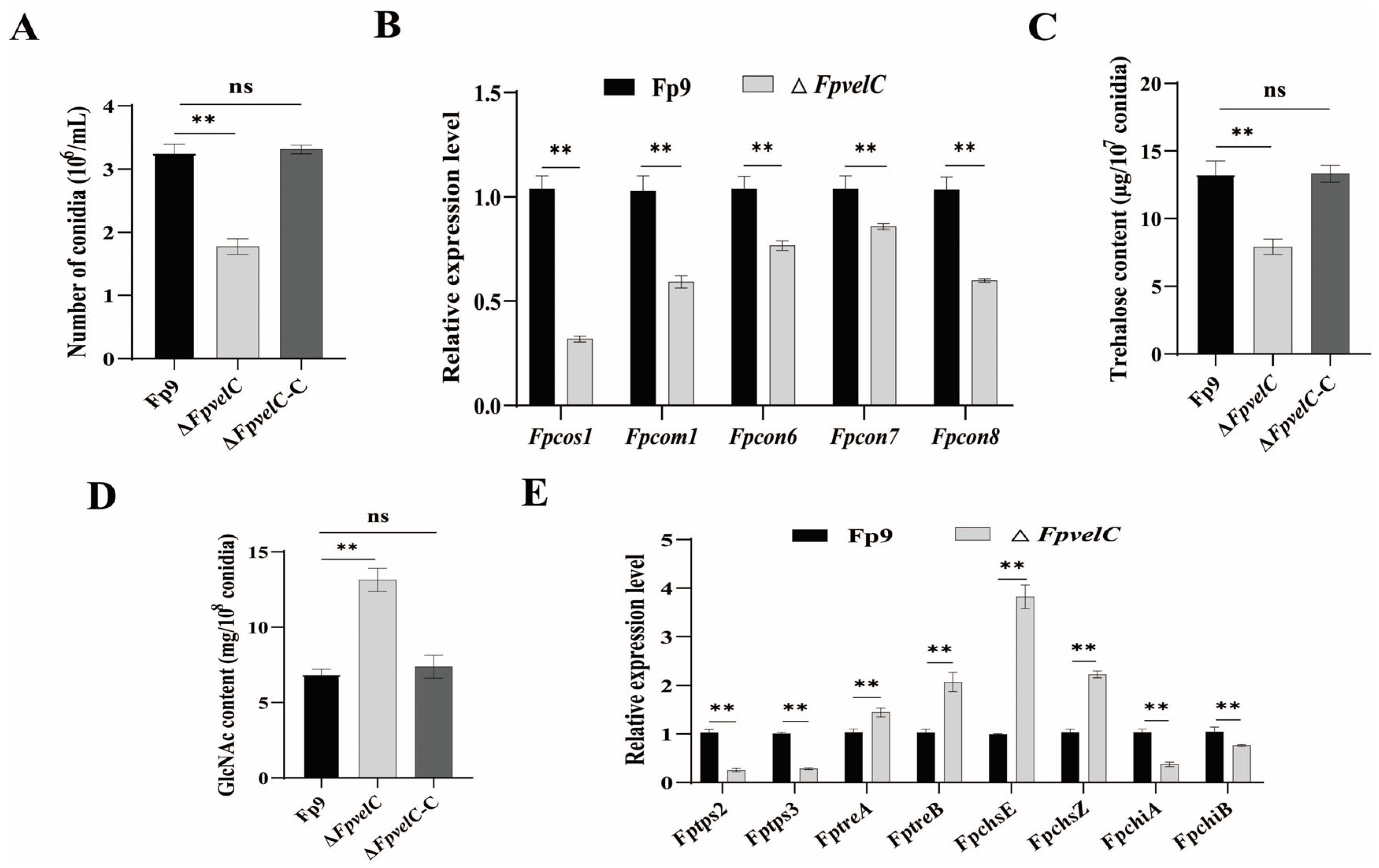
To elucidate the effect of FpvelC in asexual sporulation, all strains (Fp9, ΔFpvelC and ΔFpvelC-C) were cultured in yeast extract peptone dextrose (YEPD) liquid media. Compared with the Fp9 strain, the number of conidia was significantly lower in ΔFpvelC (Figure 2A). Transcript levels of sporogenesis-related genes, including Fpcos1, Fpcom1, Fpcon6, Fpcon7 and Fpcon8, were decreased in ΔFpvelC (Figure 2B). In conidia, trehalose amount was less in ΔFpvelC than that of the Fp9 strain, but chitin content was more in ΔFpvelC than that of the Fp9 strain (Figure 2C,D). The mRNA levels of genes associated with trehalose synthesis (Fptps2 and Fptps3) and chitin degradation (FpchiA and FpchiB) were downregulated, whereas the transcripts of genes involved in trehalose hydrolysis (FptreA and FptreB) and chitin synthesis (FpchsE and FpchsZ) were markedly elevated in the conidia of ∆FpvelC (Figure 2E). The reversal of conidiation phenomena was observed after reintroducing the FpvelC gene into the ∆FpvelC mutant. Collectively, these results demonstrate that FpvelC played a critical role in conidiation of F. proliferatum.

2.3. FpvelC Is Required for Responses to Stress Tolerance

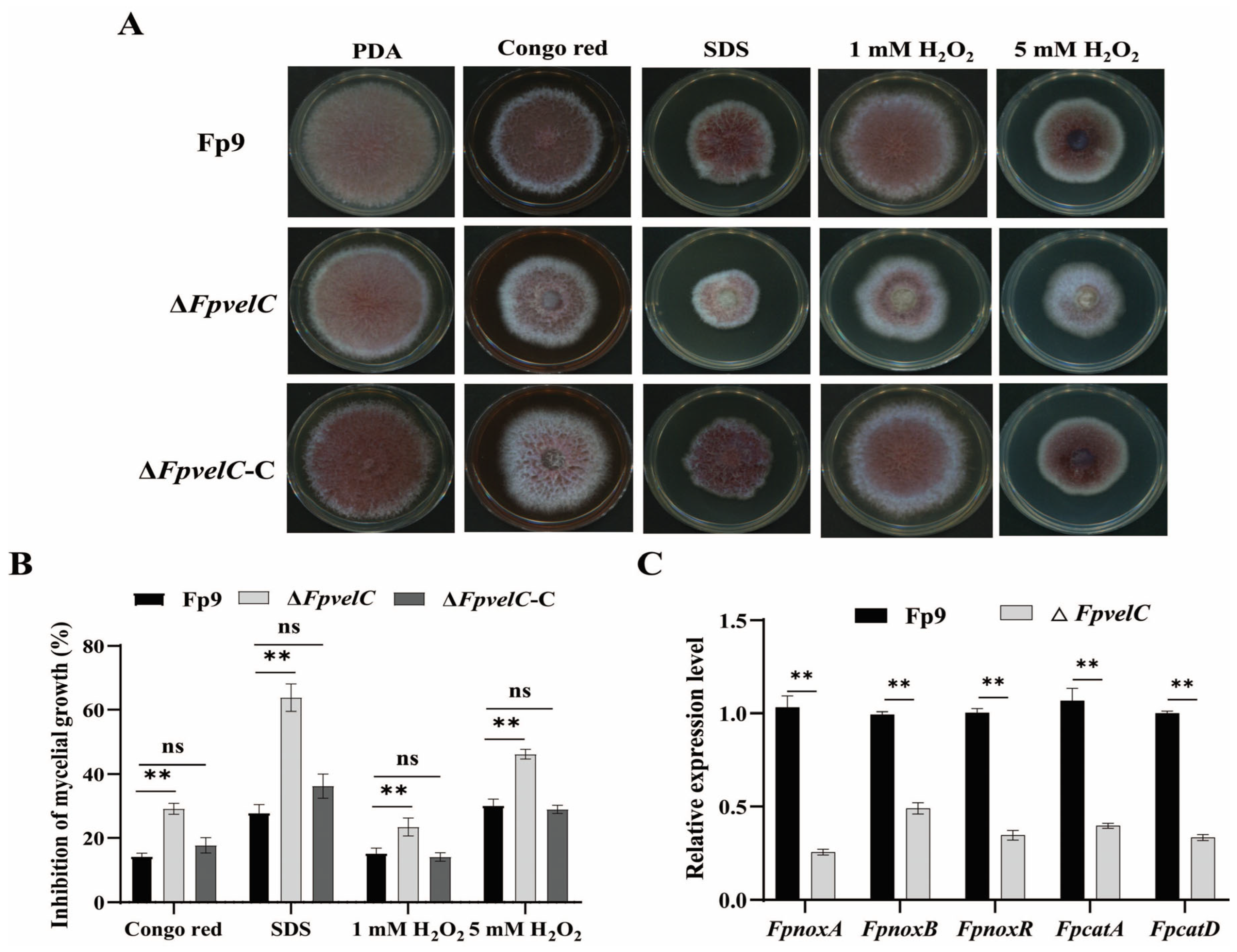
To evaluate the involvement of FpvelC in response to abiotic stimuli, all strains were cultured on potato dextrose agar (PDA) media supplemented with chemical reagents. Compared with the Fp9 strain, the growth of ΔFpvelC was significantly delayed when exposed to the cell wall-perturbing agents Congo red and cell membrane inhibitor sodium dodecyl sulphate (SDS) (Figure 3A,B). Furthermore, the relative inhibition of growth in ΔFpvelC was obviously higher than that of the Fp9 strain after being treated with different concentrations of hydrogen peroxide (H2O2), suggesting that ΔFpvelC was more sensitive to oxidative stress (Figure 3A,B). The levels of mRNA from oxidative responsive genes, NADPH oxidase subfamilies (FpnoxA, FpnoxB and FpnoxC) and catalase genes (FpcatA, FpcatD) were strongly repressed in ΔFpvelC after exposure to H2O2 (Figure 3C). Reintroduction of the FpvelC gene into ΔFpvelC rescued the stress tolerance. Taken together, these results imply that FpvelC was involved in maintenance of cell wall integrity and resistance to oxidative stress in F. proliferatum.

2.4. FpvelC Plays an Important Role in Full Virulence

To detect the potential function of FpvelC in plant colonization, susceptible rice and maize were inoculated with the conidial suspension of each strain. When infected with the Fp9 strain, rice spikelets exhibited wilting and rotten symptoms, whereas ΔFpvelC caused minor necrosis on spikelets at 21 days post-inoculation (dpi) (Figure 4A). The disease index of ΔFpvelC was much less than that of the Fp9 strain (Figure 4B). During the infection processes, the Fp9 strain developed copious hyphae along the epidermal cells of rice glumes at 48 h post-inoculation (hpi), and numerous hyphae gathered together at 72 hpi. Conversely, the fungal colonization was rarely detected in ΔFpvelC-inoculated glumes at 48 hpi, and sparse invasive hyphae were attached to the surfaces of glumes at 72 hpi (Figure 4C). Transcript levels of genes encoding xylanases and xyloglucanases of F. proliferatum were significantly decreased in ΔFpvelC-inoculated plants (Figure 4D). At the same time, starch grains of chloroplasts in glumes challenged with Fp9 appeared augmented, while starch grains in ΔFpvelC-infected glumes were obviously fewer (Figure 4E). In the case of corn infection assays, maize treated with the Fp9 strain showed dense aerial hyphae on kernels and necrotic lesions in stalks, but lesions were relatively smaller after being inoculated with ΔFpvelC (Figure 4F,G). The ability of ΔFpvelC to infect hosts was restored after complementation with a wild-type copy of FpvelC. These combined data substantiate that FpvelC contributed to pathogenicity in the initiation of infection events.

2.5. FpvelC Negatively Governs Production of Secondary Metabolites

In order to determine if FpvelC participated in specialized metabolism, metabolite extracts from cultures of each strain in potato dextrose broth (PDB) were measured by high-performance liquid chromatography–tandem mass spectrometry (HPLC-MS/MS). Evidently, ΔFpvelC yielded greatly increased amounts of FB1 when compared with the Fp9 strain (Figure 5A). Simultaneously, levels of mRNA from most genes (Fpfums) involved in fumonisin biosynthesis were dramatically higher in ΔFpvelC than those in the Fp9 strain (Figure 5B). Meanwhile, production of fusaric acid was markedly elevated in ΔFpvelC cultures (Figure 5C). Correspondingly, expressions of the majority of backbone genes (Fpfubs) responsible for fusaric acid biosynthesis were upregulated in ΔFpvelC (Figure 5D). Reintroduction of the intact FpvelC allele into ΔFpvelC fully restored the levels, resembling those observed in the Fp9 strain. These results suggest that FpvelC played negative roles in regulating production of fumonisins and fusaric acid in F. proliferatum.

2.6. FpvelC Is Involved in Nitrogen Metabolism

To ascertain whether disruption of FpvelC affected nitrogen metabolism, expression of genes involved in nitrogen regulation was investigated in ΔFpvelC. Relative to the Fp9 strain, mRNA levels of nitrogen regulators (FpareA, FpmeaB and Fpnmr), ammonium permease (Fpmep3) and nitrate reductase (FpniiA) were markedly increased in ΔFpvelC (Figure 6A). The amount of intracellular glutamine was dramatically decreased in ΔFpvelC (Figure 6B). Moreover, ΔFpvelC was more sensitive to a series of concentrations of rapamycin than the Fp9 strain, when grown in minimal medium (MM) media with glutamine as a sole nitrogen source (Figure 6C,D). These results illustrate that FpvelC might be implicated in the nitrogen-sensing pathway.

3. Discussion

F. proliferatum is notorious for its capability of producing mycotoxins, which threaten the health of humans and domestic animals [27]. A diverse array of corn and corn-based food commodities were frequently contaminated by F. proliferatum due to growth of toxigenic fungi and/or mycotoxins production, especially in tropical and subtropical regions of Europe, Asia and North Africa [28,29]. Considering economic losses and food safety, it is urgent to develop promising drug targets for control strategies to prevent F. proliferatum infection and mycotoxin contamination in agricultural products. Velvet complex has been known to be critical for fungal development and differentiation [15]. The present study firstly characterized the functions of velvet gene FpvelC in pathogenicity and secondary metabolism in F. proliferatum.
Asexual spores (conidia) are the main propagules of filamentous fungi. Infection of F. proliferatum on crops was sustained by dissemination and adaptation of conidia in ecological niches. Absence of FpvelC resulted in decreased conidiation in F. proliferatum. Notably, transcript abundances of sporulation-related genes were reduced in ΔFpvelC, including Fpcos1, Fpcom1, Fpcon6, Fpcon7 and Fpcon8 (orthologs of cos1, com1, con6, con7 and con8 in Neurospora crassa). Of these, com1 and con7 genes governed phialides differentiation, while cos1, con6 and con8 genes were responsible for the formation and maturation of conidia [30,31]. Especially, con7 played a crucial role in controlling the expression of transcription factor abaA of the central developmental pathway for conidiation [32,33]. These results infer that FpvelC acted as a positive activator in conidiation during vegetative growth in F. proliferatum. This mode of action was similar to velC orthologs in other pathogenic fungi, including Verticillium dahliae [34], Ustilaginoidea virens [35] and Magnaporthe oryzae [36]. By contrast, there were several reports that velC negatively regulated conidiation in A. nidulans [37], Penicillium chrysogenum [38] and Fusarium oxysporum [39]. Apart from alteration in expression patterns of conidiation-specific genes, another reason for impaired conidiation might be attributed to the perturbed conidia structure. Deletion of FpvelC resulted in less trehalose through interference with trehalose synthesis in conidia, whereas accumulation of chitin was opposite to trehalose behaviors in ΔFpvelC. Moreover, ΔFpvelC displayed higher sensitivity towards cell wall stresses. The trehalose was a non-reducing disaccharide that was conducive to conidial viability and stress tolerance [40]. The chitin was the primary polysaccharide of spore walls, which was indispensable for cell wall integrity and rigidity [40]. Therefore, loss of FpvelC led to damage to the cell wall by influencing the synthesis and distribution of trehalose and chitin in conidia. Collectively, these results corroborate that FpvelC was required for conidiation and spore wall remodeling in F. proliferatum.
To ensure the rapid infection and survival on hosts, the phytopathogens have evolved antioxidant enzymes to detoxify and quench reactive oxygen species (ROS) generated from plants, such as superoxide dismutase (SOD), catalase (CAT), peroxidase (POD) and ascorbate peroxidase (APX) [41]. NADPH oxidases are capable of univalent reduction of molecular oxygen into superoxide anion (O2•−), which is dismutated to H2O2 by SOD and subsequently converted into water by CAT [42]. It is worth noting that ΔFpvelC displayed significant sensitivity toward oxidative stress. Moreover, transcripts of NADPH oxidase subfamilies and catalase genes were decreased in ΔFpvelC after exposure to H2O2. These results might account for the possibility of FpvelC in modulating ROS scavenging. A similar phenomenon was reported in the maize pathogen Fusarium verticillioides, where velvet members VeA and VelB positively regulated the expression of catalase-encoding gene FvCAT2 [43]. Once the encounter between hosts and pathogens occurred, plants produced ROS to eliminate pathogens or trigger immune responses [44]. How the pathogens utilize antioxidants to scavenge ROS from hosts might influence the outcomes of infections; the impaired capacity of the ROS scavenging system in ΔFpvelC cannot be ruled out, which was partially correlated with attenuated virulence against host challenges.
F. proliferatum employed a hemibiotrophic lifestyle to obtain host nutrients from living tissues before switching to a necrotrophic phase [27]. More notably, deletion of FpvelC in F. proliferatum displayed compromised colonization on rice and maize, which may be attributable to the pleiotropic factors. Firstly, the conidial production was dramatically decreased in ΔFpvelC compared with the Fp9 strain. Asexual spore was a primary inoculum that was extremely important for successful invasion of F. proliferatum. Upon landing on hosts, conidia germinated and subsequently colonized plant tissues. A similar observation was obtained from rice blast fungus M. oryzae, where velC positively regulated appressorial development; thus, loss of velC weakened the ability of appressorium to penetrate rice leaves [36]. By contrast, in the necrotrophic fungus Botrytis cinerea, disruption of velC did not affect infection on tomatoes and apples [45]. Secondly, the growth of invasive hyphae was found to be retarded in ΔFpvelC-infected rice glumes. After invasion into rice, F. proliferatum initially produced hyphal fragments to spread along the surfaces of plants [46]. Shortly thereafter, a switching of its lifestyle from biotrophy to necrotrophy occurred, thereby facilitating secondary hyphae to decompose or kill plant tissues [27]. As revealed, rice glumes inoculated with the Fp9 strain were filled with hyphae, causing the obvious breakage of cells, accompanied by abnormal accumulation of starch grains in chloroplasts. In contrast, FpvelC deficiency failed to form invasive hyphae; as a result, the development of starch grains was normal. Last but not least, expressions of genes encoding xylanases and xyloglucanases were downregulated in glumes infected with ΔFpvelC. Importantly, these enzymes were responsible for hydrolyzation of hemicellulosic components of plant cell walls, conferring an advantage for plant pathogens to hijack nutrient reservoir from the hosts [47], which were considered to be associated with pathogenicity in B. cinerea [48] and V. dahliae [49]. Therefore, we speculate that FpvelC regulated infection and host colonization by orchestrating conidia reproduction, development of invasive hyphae and secretion of extracellular enzymes in F. proliferatum.
The biosynthetic pathways of mycotoxins consisted of a cascade of intricate enzymatic reactions. These enzymes were encoded by genes in transcriptionally coregulated clusters, which tend to be located contiguously on the chromosome in F. proliferatum [27]. However, the elaborate mechanisms of how environmental and internal cues affected mycotoxin anabolism remain an enigma. Intriguingly, inactivation of FpvelC provoked production of FB1 and fusaric acid, which was in accordance with upregulation of biosynthetic clustering genes. To the best of our knowledge, this was the first report of negative regulation of FpvelC in fumonisins and fusaric acid in F. proliferatum. Nevertheless, in most fungal species, velC served as positive activators of the biosynthesis of secondary metabolites, such as kojic acid in Aspergillus oryzae [50], penicillin in P. chrysogenum [38] and patulin in P. expansum [23]. Peculiarly, lack of velC did not affect aflatoxins production in A. flavus or fumonisins biosynthesis in F. verticillioides [17,43]. Clearly, the discrepancies reveal that the roles of velC on secondary metabolism may be specific for each species. On the other hand, the phytotoxic activity of FB1 has been shown to contribute to virulence of F. proliferatum to rice [51]. However, loss of FpvelC enhanced FB1 production but did not accelerate exacerbation of disease development. Thus, we propose that FB1 was not essential for pathogenicity; it is plausible that additional determinants participated in the pathogenic diseases.
Nitrogen sources are indispensable elements for fungal growth and secondary metabolism. Nitrogen catabolite repression (NCR) allowed fungi to possess the capacity of preferential uptake of glutamine and ammonium [52]. In the rice bakanae disease pathogen Fusarium fujikuroi, the biosynthesis of fumonisins and fusaric acid was regulated by the quality and quantity of available nitrogen sources [52]. Unexpectedly, loss of function of FpvelC resulted in the upregulation of NCR regulators, ammonium permease and nitrate reductase, concomitant with a drastic reduction in the level of intracellular glutamine. This phenomenon can be interpreted by our previous study that FB1 production was specifically elicited by glutamine limitation, which was strictly dependent on NCR transcription factor FpareA in F. proliferatum [53]. The nucleus localization of FpareA responded to nitrogen starvation, leading to an elegant regulation of NCR-sensitive primary and secondary metabolism [53]. As a key metabolite in nitrogen metabolism, the signal of glutamine availability might be sensed by specific sensors. It is possible that loss of FpvelC gene resulted in glutamine limitation, which induced FpareA to be dephosphorylated and enter the nucleus, thereby driving expression of nitrogen catabolic genes. Moreover, GATA transcription factor AreA, AreA-binding protein Nmr and bZIP transcription factor MeaB were involved in the regulation of production of multiple secondary metabolites in F. fujikuroi [52]. Thus, we speculate that depletion of intracellular glutamine converged with FpvelC to coordinate FB1 biosynthesis. This idea is further supported by the fact that ΔFpvelC mutant exhibited apparent sensitivity after treatment with rapamycin (an inhibitor of TOR kinase). In eukaryotes, target of rapamycin (TOR) signaling responded to nutrients deficiency, and the TOR kinase activity became inactive under nitrogen starvation [54]. Thus, these findings provide evidence that FpvelC is involved in nitrogen sensing and signaling. A possible scenario is that FpvelC had inhibitory effects on biosynthesis of secondary metabolites, which might be connected with the crosstalk between nitrogen metabolism and TOR pathway.

4. Conclusions

In summary, this study highlighted that FpvelC positively governed conidiation, oxidative stress and virulence but negatively modulated production of fumonisins and fusaric acid. To the best of our knowledge, this was the first report where FpvelC was associated with mycotoxins biosynthesis in F. proliferatum. Our findings revealed the versatile roles of FpvelC in F. proliferatum, which contributed to the development of feasible strategies to mitigate the detrimental hazards of the pathogen on humans and plant hosts.

5. Materials and Methods

5.1. Fungal Strains and Growth Conditions

F. proliferatum strain Fp9 isolated from infected rice spikelets was used as the wild-type progenitor [27]. The Fp9 strain and its derivative transformants were stored as conidial suspensions at −80 °C with 30% (v/v) glycerol–water mixture. Fungal strains were cultured on potato dextrose agar (PDA, potato extract 200 g, dextrose 20 g and agar 15 g per liter) at 28 °C. For conidiation assays, the strains were incubated in yeast extract peptone dextrose (YEPD, yeast extract 10 g, peptone 20 g and dextrose 20 g per liter) on a rotary shaker for 3 days at 28 °C with 12 h light–dark cycle. The conidia were harvested by filtration and counted using a hemacytometer. For mycotoxin production, the strains were cultured at 28 °C in potato dextrose broth (PDB, potato extract 200 g and dextrose 20 g per liter). For determining the sensitivity of rapamycin, the strains were grown in minimal medium (MM, sucrose 30 g, glutamine 6 mM, KH2PO4 1 g, MgSO4·7H2O 0.5 g, KCl 0.5 g, trace elements 200 μL and agar 20 g per liter) supplemented with rapamycin (Sigma-Aldrich, St. Louis, MO, USA) to the desired final concentrations. All experiments were performed at least three times.

5.2. Sequence Analysis of FpvelC

The amino acid sequence of FpvelC protein in F. proliferatum was identified by a BLASTP search at the National Center for Biotechnology Information (NCBI) using velC protein (ABQ17968) of A. nidulans as a query. The velvet domain was verified through the Conserved Domain Database at NCBI (https://www.ncbi.nlm.nih.gov/Structure/cdd/wrpsb.cgi) (accessed on 26 March 2024). The PEST motif was predicted using EMBOSS “epestfind” (http://emboss.bioinformatics.nl/cgi-bin/emboss/epestfind) (accessed on 26 March 2024). The available velC orthologs of different filamentous fungi were downloaded from the NCBI database. Multiple sequences were aligned with CLUSTALW [55]. A phylogenetic tree was constructed with the neighbor-joining algorithm with a bootstrap value of 1000 repetitions using the MEGA 11 software package (Mega Limited, Auckland, New Zealand) [56].

5.3. Gene Deletion and Complementation

Deletion and complementation of FpvelC gene were constructed by homologous recombination, following previously established protocols [57]. For gene deletion construction, the 5′- and 3′-flanking sequences were amplified with primers F1/R1 and F2/R2 from the genomic DNA of the Fp9 strain, respectively. Primers F3/R3 were used for amplification of hygromycin phosphotransferase gene (HYG) using plasmid pFGL821 as a template. Deletion fragments were generated with primers F4/R4 and F5/R5 by a double-joint PCR approach [58] and transformed into the protoplasts of Fp9 strain by polyethylene glycol (PEG)-mediated transformation. Positive transformants were selected on PDA plates containing 250 µg/mL hygromycin B (Calbiochem, La Jolla, CA, USA) and further identified by diagnostic PCR and Southern hybridization. For complementation construction, the full-length sequence of FpvelC gene containing the open-reading frame (ORF) region and its native promoter regions was amplified with primers Com-F1/Com-R1 from the genomic DNA of Fp9 strain. This fragment was fused with the geneticin resistance gene (GEN) and introduced into the protoplasts of ΔFpvelC mutants. The transformants were screened with 250 µg/mL G418 disulfate salt (Sigma-Aldrich, St. Louis, MO, USA) and verified by PCR analysis. The procedures of gene deletion and complementation are conducted as detailed in Figure S1 and Figure S2, respectively. All primers are listed with brief descriptions in Table S1.

5.4. Quantitative Real-Time PCR (qRT-PCR)

The total RNA was extracted using the TRIzol reagent (Invitrogen, Waltham, MA, USA) according to the manufacturer’s instructions. The quality and quantity of RNA were determined by a NanoDrop ND-1000 spectrophotometer (Nanodrop Technologies, Wilmington, DE, USA). First-strand complementary DNA (cDNA) was synthesized from total RNA with the QuantiTect Reverse Transcription Kit (Qiagen, Hilden, Germany). qRT-PCR reactions were performed with the SYBR Premix Ex TaqTM II Kit (TaKaRa, Shiga, Japan) on a CFX96 Touch Real-Time PCR Detection system (Bio-Rad, Hercules, CA, USA). The β-tubulin (Fptub) gene of F. proliferatum was used as an internal control for normalization. The expression levels were calculated using the 2−ΔΔCT method [59]. Transcript levels in Fp9 strain were arbitrarily set to 1. The specific primers used for gene expression analysis are listed in Table S2. The experiment was repeated three times with three technical replicates.

5.5. Trehalose Assay

The trehalose content was measured by the acid trehalase method [60]. Two-day conidia were collected by centrifugation, resuspended in sterile water at 1 × 10⁷ conidia/mL, and disrupted with Bullet Blender Gold homogenizer (Next Advance, Inc., Averill Park, NY, USA) at 4 °C for 10 min. The lysate was boiled at 95 °C for 20 min and centrifuged at 10,000× g for 10 min. The supernatant was digested with 3 mU of trehalose (Sigma-Aldrich, St. Louis, MO, USA) in 0.2 M sodium citrate buffer (pH 5.5) at 37 °C for 8 h. The reactions were stopped by the addition of 0.12 mol/L sulfuric acid. The amount of glucose generated from trehalose was examined with the Glucose Assay Kit (Sigma-Aldrich, St. Louis, MO, USA). Sample untreated with trehalase was a negative control. All experiments were conducted in triplicates.

5.6. Chitin Assay

The chitin content was estimated by assaying the release of glucosamine by hydrolysis of chitin [61]. Two-day conidia (1 × 10⁸) were harvested by centrifugation and incubated with lysis buffer (3% SDS, 0.3 M β-mercaptoethanol, 1 mM EDTA, 50 mM Tris, pH 8.0) at 100 °C for 40 min. The samples were washed three times with sterile saline solution and sonicated for 1 min on ice. After centrifugation at 10,000× g at 4 °C for 10 min, the pellet was acidified in 6 M HCl and hydrolyzed by N-acetylglucosamine (GlcNAc, Sigma-Aldrich, St. Louis, MO, USA) at 100 °C for 17 h. Samples were dried and dissolved in sterile water. Then, an equal volume of 4% (v/v) acetylacetone in 1.5 M Na2CO3 was added, the preparation was heated at 100 °C for 20 min. The samples were diluted with 96% ethanol and incubated with Ehrlich’s reagent buffer (26 mg/L p-dimethylaminobenzaldehyde, 5.8 M HCl, 50% ethanol) for 1 h at 20 °C. The absorbance at 520 nm was measured using a spectrophotometer (Genesys 10uv, Spectronic Unicam, Rochester, NY, USA). The experiment was repeated in triplicate.

5.7. Stress Tolerance Analysis

To assess responses to various stresses, the strains were grown on PDA media supplemented with chemicals, 0.3 mg/mL Congo red (Sigma-Aldrich, St. Louis, MO, USA) and 0.01% SDS for cell wall stress; or 1 mM or 5 mM H2O2 for oxidative stress. Strains without treatment were incubated in parallel as internal controls. After incubation at 28 °C for 5 days, the colonies were recorded and measured. The inhibition of mycelia growth was calculated using the formula [(C − N)/(C)] × 100%, where C is the colony diameter of the control and N is the colony diameter of the treatment. Each treatment consisted of three replicates, and the experiment was repeated thrice.

5.8. Rice Infection Assay

The susceptible rice cultivar Jiahe218 (China National Rice Research Institute, Hangzhou, Zhejing, China) was used for pathogenicity tests. Conidia were harvested from 3-day-old cultures in YEPD media and resuspended in sterile water to a final concentration of 1 × 10⁶ conidia/mL. At the booting stage, 1 mL of conidia suspension was injected into rice spikelets. The inoculated plants were maintained at 26 °C with a relative humidity of 85% and a photoperiod of 14 h in a greenhouse. Disease severity was assessed at 21 days post-inoculation (dpi) according to our previous method [62]. To further evaluate the infection behavior in florets, a 10 μL aliquot of conidial suspension was dropped into a floret at the anthesis stage. Infected rice glumes were observed under a scanning electron micrograph (SEM) and transmission electron micrograph (TEM), respectively. Ten plants were used for each strain, and the experiment was repeated three times independently.

5.9. Maize Infection Assay

The susceptible maize cultivar Suyunuo2 (Jiangsu Academy of Agricultural Sciences, Nanjing, Jiangsu, China) was selected for infection tests. The maize was surface-sterilized by 2% sodium hypochlorite (NaClO) and rinsed with sterile water three times. Maize kernels were aseptically wounded with sterile needles and inoculated with 10 μL of conidial suspension at a concentration of 5 × 10⁶ conidia/mL. Infected kernels were kept in a growth chamber at 28 °C with a relative humidity of 70%. Lesions were photographed at 7 dpi. Maize stems were punctured by sterile toothpicks to approximately 1 cm depth, and 50 μL of conidial suspension (5 × 10⁶ conidia/mL) was injected into the wounded stalks. Infected stalks were placed on wet filter papers in a sealed box. Stalk segments were dissected longitudinally, and the necroses were photographed at 14 dpi. All treatments were performed with at least three replicates, and each experiment was repeated three times.

5.10. Microscopy Observation

For scanning electron microscopy (SEM) examination, the samples were fixed in 2.5% glutaraldehyde solution overnight and washed three times with 0.1 M phosphate buffered saline (PBS). Samples were post-fixed in 1% osmic acid (Merck, Schwalbach, Germany) for 3 h and dehydrated in graded ethanol solutions (30%, 50%, 70%, 80%, 95% and 100% [v/v]) for 15 min with each grade. After critical point drying with liquid carbon dioxide, the specimens were sputter-coated with gold using a sputter coater (Bal-Tec SCD 005, Bal-Tec AG, Balzers, Liechtenstein) and visualized with a scanning electron microscope (Hitachi Model SU-8010, Hitachi High-Technologies Corporation, Tokyo, Japan) at an accelerating voltage of 2 kV. Three independent experiments were conducted with ten samples in each treatment.
For transmission electron microscopy (TEM) observation, the samples were fixed with 2.5% glutaraldehyde in 0.1 M sodium cacodylate buffer (pH 7.4) at room temperature for 2 h, fixed with 2% osmium tetroxide (Merck, Darmstadt, Germany) for 1 h at 4 °C. The samples were dehydrated in a series of ethanol (30%, 50%, 70%, 90% and 100% [v/v]) for 10 min each and embedded in Spurr’s resin (PolySciences, Niles, IL, USA). Ultrathin sections (80 nm) were sliced using an ultramicrotome (Leica EM UC6, Lecia Microsystems, Nussloch, Germany) and stained with 2% uranyl acetate and 0.4% Reynold’s lead citrate. The sections were examined with a transmission electron microscope (Hitachi Model H-7650, Hitachi High-Technologies Corporation, Tokyo, Japan) at an accelerating voltage of 120 kV. Each treatment contained ten samples, and the experiment was performed three times independently.

5.11. Determination of FB1 Production

Quantification of FB1 production was determined by HPLC-MS/MS [63]. The samples were collected from the fermentation broth in PDB and extracted with acetonitrile/water/acetic acid (74:25:1, v/v/v). The organic phase was filtered through a 0.22 μm polypropylene membrane. The chromatographic separation was performed with an HPLC system (HPLC-1260, Agilent Technologies, Santa Clara, CA, USA) on a Zorbax Extend-C18 column (100 × 2.1 mm, 3.5 μm). Mobile phases were composed of solvent A (water with 0.1% formic acid) and solvent B (methanol with 0.1% formic acid). The gradient elution was performed as follows: 0 to 1 min, 30% solvent B; 1.01 to 6.0 min, where solvent B was increased linearly from 30% to 80%; 6.01 to 9.0 min, where solvent B was maintained at 80%; 9.01 to 10.0 min, where solvent B was decreased linearly from 80% to 30%; and 10.01 to 16 min, where solvent B was maintained at 30%. A volume of 2 μL was injected into the column. The column oven was set at 30 °C. The flow rate was 0.3 mL/min. Mass spectra were analyzed in a triple quadrupole mass spectrometer equipped with an electrospray ionization (ESI) source in a positive ionization mode. Capillary voltages were kept at 3500 V. Nitrogen was supplied as the nebulizing and drying gas, the curtain gas was 18 psi, the nebulizer gas was 40 psi, the heater gas was 40 psi, and the CAD gas was 4 psi. The temperature of drying gas was set at 350 °C. The experiment was repeated three times.

5.12. Quantification of Fusaric Acid Production

Detection of fusaric acid was performed with the HPLC-MS/MS system [64]. The samples were harvested from PDB cultures and extracted with acetonitrile/water/acetic acid (79:20:1, v/v/v). The crude extract was centrifuged at 5000× g for 10 min, and the supernatant was filtered with a 0.45 μm polypropylene membrane. The chromatographic separation was carried out with a reversed-phase Zorbax SB-C18 column (150 × 4.6 mm, 5 μm, Agilent Technologies, Santa Clara, CA, USA). Mobile phases consisted of solvent A (0.1% trifluoroacetic acid in water) and solvent B (acetonitrile). The gradient elution procedure was as follows: 0 to 2 min, 10% solvent B; 2.01 to 20 min, where solvent B was increased linearly from 10% to 100%; and 20.01 to 28 min, where solvent B was maintained at 100%. The flow rate was 1 mL/min. The injection volume was 2 μL. The column oven was set at 45 °C. Mass spectra were operated in an ESI source in a positive ionization mode. The capillary voltage was kept at 3500 V, and the fragmentor voltage was set at 130 V. Nitrogen served as the curtain gas at 30 psi, and the nebulizer gas was set at 45 psi. The temperature of the drying gas was set at 350 °C. The experiment was conducted three times.

5.13. Measurement of Intracellular Glutamine

Glutamine content was analyzed by an ultra-high performance liquid chromatography system (UHPLC, 1290 Infinity, Agilent Technologies, Santa Clara, CA, USA) coupled with a tandem mass spectrometer (QTRAP5500, Applied Biosystems, Sciex, Concorde, ON, Canada) [65]. The samples were collected from mycelia and extracted with acetonitrile/water (1:1, v/v). After centrifugation, the supernatant was filtered with a 0.22 μm polypropylene membrane. Chromatographic separation was performed with a Waters ACQUITY UPLC BEH C18 column (2.1 × 150 mm, 1.7 μm, Waters, Milford, MA, USA). The column temperature was set at 35 °C. The flow rate was 0.3 mL/min and the injection volume was 2 μL. The composition of the mobile phases was solvent A (25 mM ammonium acetate and 0.08% formic acid in water) and solvent B (0.1% formic acid in acetonitrile). The linear gradient elution was as follows: 0 to 1 min, 85% solvent B; 1.01 to 11 min, where solvent B was decreased linearly from 85% to 50%; 11.01 to 12 min, where solvent B was maintained at 40%; 12.01 to 12.1 min, where solvent B was increased linearly from 40% to 75%; and 12.11 to 19 min, where solvent B was maintained at 75%. Mass spectrometry was equipped with an ESI source in positive ionization mode. The parameters were as follows: source temperature, 500 °C; ion source gas1, 55 psi; ion source gas2, 50 psi; curtain gas, 30 psi; ion spray voltage floating, 4500 V. The concentration of glutamine was expressed as μmol per g of dry weight of mycelia. The experiment was repeated thrice.

5.14. Statistical Analysis

All data are presented as the mean ± standard deviation of three individual replicates. Statistical analysis was performed using GraphPad Prism Version 7 (GraphPad Software Inc., San Diego, CA, USA). Significance was determined by a one-way analysis of variance (ANOVA) followed by Student’s t-test. Differences were considered to be statistically significant when the p-value was below 0.05.

Supplementary Materials

The following supporting information can be downloaded at: https://www.mdpi.com/article/10.3390/toxins17090433/s1, Figure S1: Schematic representation for the construction of the deletion mutant ΔFpvelC; Figure S2: Schematic representation of the construction of the complementation strain ∆FpvelC-C; Table S1: Primers used for gene deletion and complementation in this study; Table S2: Primers for qRT-PCR in this study.

Author Contributions

Conceptualization, P.H., S.T. and L.W.; methodology, L.W., W.L. and G.J.; software, S.H., Z.S., G.S. and L.X.; validation, L.W. and S.T.; formal analysis, L.W. and S.T.; investigation, L.W. and W.L.; resources, P.H. and S.T.; data curation, L.W. and S.T.; writing—original draft preparation, L.W.; writing—review and editing, P.H. and S.T.; visualization, P.H.; supervision, P.H. and S.T.; project administration, P.H.; funding acquisition, P.H. All authors have read and agreed to the published version of the manuscript.

Funding

This research was funded by National Natural Science Foundation of China (32188102, 31800133), the Biological Breeding-National Science and Technology Major Project of China (2022ZD04003), the Project of Laboratory of Advanced Agricultural Sciences, Heilongjiang Province (ZY04JD05-005), the Science and Technology Project of Yunnan Province (202402AE090036, 202405AF140064), Natural Science Foundation of Zhejiang Province, China (LQ18C140005).

Institutional Review Board Statement

Not applicable.

Informed Consent Statement

Not applicable.

Data Availability Statement

The original contributions presented in the study are included in the article/Supplementary Materials; further inquiries can be directed to the corresponding authors.

Conflicts of Interest

The authors declare no conflicts of interest.

References

  1. Yu, H.; Hwang, S.F.; Strelkov, S.E. The host range of Fusarium proliferatum in western Canada. Pathogens 2024, 13, 407. [Google Scholar] [CrossRef]
  2. Braun, M.S.; Wink, M. Exposure, occurrence, and chemistry of fumonisins and their cryptic derivatives. Compr. Rev. Food Sci. Food Saf. 2018, 17, 769–791. [Google Scholar] [CrossRef] [PubMed]
  3. Ponce-García, N.; Serna-Saldivar, S.O.; Garcia-Lara, S. Fumonisins and their analogues in contaminated corn and its processed foods—A review. Food Addit. Contam. Part A Chem. Anal. Control Expo. Risk Assess. 2018, 35, 2183–2203. [Google Scholar] [CrossRef]
  4. Yang, D.; Ye, Y.; Sun, J.; Wang, J.-S.; Huang, C.; Sun, X. Occurrence, transformation, and toxicity of fumonisins and their covert products during food processing. Crit. Rev. Food Sci. Nutr. 2024, 64, 3660–3673. [Google Scholar] [CrossRef] [PubMed]
  5. Anumudu, C.K.; Ekwueme, C.T.; Uhegwu, C.C.; Ejileugha, C.; Augustine, J.; Okolo, C.A.; Onyeaka, H. A review of the mycotoxin family of fumonisins, their biosynthesis, metabolism, methods of detection and effects on humans and animals. Int. J. Mol. Sci. 2024, 26, 184. [Google Scholar] [CrossRef]
  6. Kamle, M.; Mahato, D.K.; Devi, S.; Lee, K.E.; Kang, S.G.; Kumar, P. Fumonisins: Impact on agriculture, food, and human health and their management strategies. Toxins 2019, 11, 328. [Google Scholar] [CrossRef]
  7. Guo, Z.; Pfohl, K.; Karlovsky, P.; Dehne, H.-W.; Altincicek, B.; Marion-Poll, F. Dissemination of Fusarium proliferatum by mealworm beetle Tenebrio molitor. PLoS ONE 2018, 13, e0204602. [Google Scholar] [CrossRef] [PubMed]
  8. Palmieri, D.; Segorbe, D.; López-Berges, M.S.; De Curtis, F.; Lima, G.; Di Pietro, A.; Turrà, D. Alkaline pH, low iron availability, poor nitrogen sources and CWI MAPK signaling are associated with increased fusaric acid production in Fusarium oxysporum. Toxins 2023, 15, 50. [Google Scholar] [CrossRef]
  9. Porter, J.K.; Bacon, C.W.; Wray, E.M.; Hagler, W.M. Fusaric acid in Fusarium moniliforme cultures, corn, and feeds toxic to livestock and the neurochemical effects in the brain and pineal gland of rats. Nat. Toxins 1995, 3, 91–100. [Google Scholar] [CrossRef]
  10. Yin, E.S.; Rakhmankulova, M.; Kucera, K.; de Sena Filho, J.G.; Portero, C.E.; Narváez-Trujillo, A.; Holley, S.A.; Strobel, S.A. Fusaric acid induces a notochord malformation in zebrafish via copper chelation. BioMetals 2015, 28, 783–789. [Google Scholar] [CrossRef]
  11. Shi, W.T.; Yao, C.P.; Liu, W.H.; Cao, W.Y.; Shao, W.; Liao, S.Q.; Yu, T.; Zhu, Q.F.; Chen, Z.; Zang, Y.J.; et al. An fusaric acid-based CRISPR library screen identifies MDH2 as a broad-spectrum regulator of Fusarium toxin-induced cell death. J. Hazard. Mater. 2024, 480, 135937. [Google Scholar] [CrossRef] [PubMed]
  12. Mamur, S.; Ünal, F.; Yılmaz, S.; Erikel, E.; Yüzbaşıoğlu, D. Evaluation of the cytotoxic and genotoxic effects of mycotoxin fusaric acid. Drug Chem. Toxicol. 2018, 43, 149–157. [Google Scholar] [CrossRef] [PubMed]
  13. Bayram, Ö.; Braus, G.H. Coordination of secondary metabolism and development in fungi: The VELVET family of regulatory proteins. FEMS Microbiol. Rev. 2012, 36, 1–24. [Google Scholar] [CrossRef] [PubMed]
  14. Bayram, Ö.; Krappmann, S.; Ni, M.; Bok, J.W.; Helmstaedt, K.; Valerius, O.; Braus-Stromeyer, S.; Kwon, N.; Keller, N.P.; Yu, J.; et al. VelB/VeA/LaeA complex coordinates light signal with fungal development and secondary metabolism. Science 2008, 320, 1504–1506. [Google Scholar] [CrossRef]
  15. Yu, W.; Pei, R.; Zhang, Y.; Tu, Y.; He, B. Light regulation of secondary metabolism in fungi. J. Biol. Eng. 2023, 17, 57. [Google Scholar] [CrossRef]
  16. Lan, N.; Yue, Q.; An, Z.; Bills, G.F. Apc.LaeA and Apc.VeA of the velvet complex govern secondary metabolism and morphological development in the echinocandin-producing fungus Aspergillus pachycristatus. J. Ind. Microbiol. Biotechnol. 2020, 47, 155–168. [Google Scholar] [CrossRef]
  17. Eom, T.J.; Moon, H.; Yu, J.H.; Park, H.S. Characterization of the velvet regulators in Aspergillus flavus. J. Microbiol. 2018, 56, 893–901. [Google Scholar] [CrossRef]
  18. Wang, G.; Zhang, H.; Wang, Y.; Liu, F.; Li, E.; Ma, J.; Yang, B.; Zhang, C.; Li, L.; Liu, Y. Requirement of LaeA, VeA, and VelB on asexual development, ochratoxin A biosynthesis, and fungal virulence in Aspergillus ochraceus. Front. Microbiol. 2019, 10, 2759. [Google Scholar] [CrossRef]
  19. Estiarte, N.; Lawrence, C.; Sanchis, V.; Ramos, A.; Crespo-Sempere, A. LaeA and VeA are involved in growth morphology, asexual development, and mycotoxin production in Alternaria alternata. Int. J. Food Microbiol. 2016, 238, 153–164. [Google Scholar] [CrossRef]
  20. Merhej, J.; Urban, M.; Dufresne, M.; Hammond-Kosack, K.E.; Richard-Forget, F.; Barreau, C. The velvet gene, FgVe1, affects fungal development and positively regulates trichothecene biosynthesis and pathogenicity in Fusarium graminearum. Mol. Plant Pathol. 2012, 13, 363–374. [Google Scholar] [CrossRef]
  21. Jiang, J.; Yun, Y.; Liu, Y.; Ma, Z. FgVELB is associated with vegetative differentiation, secondary metabolism and virulence in Fusarium graminearum. Fungal Genet. Biol. 2012, 49, 653–662. [Google Scholar] [CrossRef] [PubMed]
  22. Wu, Y.; Han, S.; Wang, Y.; Li, Q.; Kong, L. Velvet family protein FpVelB affects virulence in association with secondary metabolism in Fusarium pseudograminearum. Cells 2024, 13, 950. [Google Scholar] [CrossRef] [PubMed]
  23. Li, B.; Chen, Y.; Zong, Y.; Shang, Y.; Zhang, Z.; Xu, X.; Wang, X.; Long, M.; Tian, S. Dissection of patulin biosynthesis, spatial control and regulation mechanism in Penicillium expansum. Environ. Microbiol. 2019, 21, 1124–1139. [Google Scholar] [CrossRef]
  24. Gao, J.; Zhou, S.; Tang, W.; Wang, J.; Liu, H.; Zhang, Y.; Wang, L.; Li, X.; Liu, Z. The velvet proteins CsVosA and CsVelB coordinate growth, cell wall integrity, sporulation, conidial viability and pathogenicity in the rubber anthracnose fungus Colletotrichum siamense. Microbiol. Res. 2023, 268, 127290. [Google Scholar] [CrossRef]
  25. Wang, L.; Liu, Q.; Ge, S.; Liang, W.; Liao, W.; Li, W.; Jiao, G.; Wei, X.; Shao, G.; Xie, L.; et al. Genomic footprints related with adaptation and fumonisins production in Fusarium proliferatum. Front. Microbiol. 2022, 13, 1004454. [Google Scholar] [CrossRef]
  26. Zhu, Q.; Fei, Y.J.; Wu, Y.B.; Luo, D.L.; Chen, M.; Sun, K.; Zhang, W.; Dai, C.C. Endophytic fungus reshapes spikelet microbiome to reduce mycotoxin produced by Fusarium proliferatum through altering rice metabolites. J. Agric. Food Chem. 2023, 71, 11350–11364. [Google Scholar] [CrossRef]
  27. Wang, L.; Ge, S.; Liang, W.; Liao, W.; Li, W.; Jiao, G.; Wei, X.; Shao, G.; Xie, L.; Sheng, Z.; et al. Genome-wide characterization reveals variation potentially involved in pathogenicity and mycotoxins biosynthesis of Fusarium proliferatum causing spikelet rot disease in rice. Toxins 2022, 14, 568. [Google Scholar] [CrossRef]
  28. Corrêa, J.A.F.; Orso, P.B.; Bordin, K.; Hara, R.V.; Luciano, F.B. Toxicological effects of fumonisin B1 in combination with other Fusarium toxins. Food Chem. Toxicol. 2018, 121, 483–494. [Google Scholar] [CrossRef]
  29. Tsehaye, H.; Brurberg, M.B.; Sundheim, L.; Assefa, D.; Tronsmo, A.; Tronsmo, A.M. Natural occurrence of Fusarium species and fumonisin on maize grains in Ethiopia. Eur. J. Plant Pathol. 2017, 147, 141–155. [Google Scholar] [CrossRef]
  30. Park, H.S.; Yu, J.H. Genetic control of asexual sporulation in filamentous fungi. Curr. Opin. Microbiol. 2012, 15, 669–677. [Google Scholar] [CrossRef] [PubMed]
  31. Ruger-Herreros, C.; Corrochano, L.M. Conidiation in Neurospora crassa: Vegetative reproduction by a model fungus. Int. Microbiol. 2020, 23, 97–105. [Google Scholar] [CrossRef]
  32. Cho, H.J.; Son, S.H.; Chen, W.; Son, Y.E.; Lee, I.; Yu, J.H.; Park, H.S. Regulation of conidiogenesis in Aspergillus flavus. Cells 2022, 11, 2796. [Google Scholar] [CrossRef]
  33. Zhou, S.; Liu, S.; Guo, C.; Wei, H.; He, Z.; Liu, Z.; Li, X. The C2H2 transcription factor Con7 regulates vegetative growth, cell wall integrity, oxidative stress, asexual sporulation, appressorium and hyphopodium formation, and pathogenicity in Colletotrichum graminicola and Colletotrichum siamense. J. Fungi 2024, 10, 495. [Google Scholar] [CrossRef] [PubMed]
  34. Höfer, A.M.; Harting, R.; Aßmann, N.F.; Gerke, J.; Schmitt, K.; Starke, J.; Bayram, Ö.; Tran, V.-T.; Valerius, O.; Braus-Stromeyer, S.A.; et al. The velvet protein Vel1 controls initial plant root colonization and conidia formation for xylem distribution in Verticillium wilt. PLoS Genet. 2021, 17, e1009434. [Google Scholar] [CrossRef]
  35. Yu, M.; Song, T.; Yu, J.; Cao, H.; Pan, X.; Qi, Z.; Du, Y.; Liu, W.; Liu, Y. UvVelC is important for conidiation and pathogenicity in the rice false smut pathogen Ustilaginoidea virens. Virulence 2024, 15, 2301243. [Google Scholar] [CrossRef]
  36. Kim, H.-J.; Han, J.-H.; Kim, K.S.; Lee, Y.-H. Comparative functional analysis of the velvet gene family reveals unique roles in fungal development and pathogenicity in Magnaporthe oryzae. Fungal Genet. Biol. 2014, 66, 33–43. [Google Scholar] [CrossRef]
  37. Park, H.S.; Nam, T.Y.; Han, K.H.; Kim, S.C.; Yu, J.H.; Yun, S.H. VelC positively controls sexual development in Aspergillus nidulans. PLoS ONE 2014, 9, e89883. [Google Scholar] [CrossRef] [PubMed]
  38. Kopke, K.; Hoff, B.; Bloemendal, S.; Katschorowski, A.; Kamerewerd, J.; Kück, U. Members of the Penicillium chrysogenum velvet complex play functionally opposing roles in the regulation of penicillin biosynthesis and conidiation. Eukaryot. Cell 2013, 12, 299–310. [Google Scholar] [CrossRef] [PubMed]
  39. López-Berges, M.S.; Hera, C.; Sulyok, M.; Schäfer, K.; Capilla, J.; Guarro, J.; Di Pietro, A. The velvet complex governs mycotoxin production and virulence of Fusarium oxysporum on plant and mammalian hosts. Mol. Microbiol. 2012, 87, 49–65. [Google Scholar] [CrossRef]
  40. Arroyo, J.; Farkaš, V.; Sanz, A.B.; Cabib, E. Strengthening the fungal cell wall through chitin-glucan cross-links: Effects on morphogenesis and cell integrity. Cell. Microbiol. 2016, 18, 1239–1250. [Google Scholar] [CrossRef]
  41. Wang, Y.; Ji, D.; Chen, T.; Li, B.; Zhang, Z.; Qin, G.; Tian, S. Production, signaling, and scavenging mechanisms of reactive oxygen species in fruit–pathogen interactions. Int. J. Mol. Sci. 2019, 20, 2994. [Google Scholar] [CrossRef]
  42. Segal, L.M.; Wilson, R.A. Reactive oxygen species metabolism and plant-fungal interactions. Fungal Genet. Biol. 2018, 110, 1–9. [Google Scholar] [CrossRef]
  43. Lan, N.; Zhang, H.; Hu, C.; Wang, W.; Calvo, A.M.; Harris, S.D.; Chen, S.; Li, S. Coordinated and distinct functions of velvet proteins in Fusarium verticillioides. Eukaryot. Cell 2014, 13, 909–918. [Google Scholar] [CrossRef]
  44. Camejo, D.; Guzmán-Cedeño, Á.; Moreno, A. Reactive oxygen species, essential molecules, during plant-pathogen interactions. Plant Physiol. Biochem. 2016, 103, 10–23. [Google Scholar] [CrossRef]
  45. Müller, N.; Leroch, M.; Schumacher, J.; Zimmer, D.; Könnel, A.; Klug, K.; Leisen, T.; Scheuring, D.; Sommer, F.; Mühlhaus, T.; et al. Investigations on VELVET regulatory mutants confirm the role of host tissue acidification and secretion of proteins in the pathogenesis of Botrytis cinerea. New Phytol. 2018, 219, 1062–1074. [Google Scholar] [CrossRef] [PubMed]
  46. Lei, S.; Wang, L.; Liu, L.; Hou, Y.; Xu, Y.; Liang, M.; Gao, J.; Li, Q.; Huang, S. Infection and colonization of pathogenic fungus Fusarium proliferatum in rice spikelet rot disease. Rice Sci. 2019, 26, 60–68. [Google Scholar] [CrossRef]
  47. Rafiei, V.; Vélëz, H.; Tzelepis, G. The role of glycoside hydrolases in phytopathogenic fungi and Oomycetes virulence. Int. J. Mol. Sci. 2021, 22, 9359. [Google Scholar] [CrossRef] [PubMed]
  48. García, N.; González, M.A.; González, C.; Brito, N. Simultaneous silencing of xylanase genes in Botrytis cinerea. Front. Plant Sci. 2017, 8, 2174. [Google Scholar] [CrossRef] [PubMed]
  49. Gui, Y.; Chen, J.; Zhang, D.; Li, N.; Li, T.; Zhang, W.; Wang, X.; Short, D.P.G.; Li, L.; Guo, W.; et al. Verticillium dahliae manipulates plant immunity by glycoside hydrolase 12 proteins in conjunction with carbohydrate-binding module 1. Environ. Microbiol. 2017, 19, 1914–1932. [Google Scholar] [CrossRef]
  50. Sano, M. Regulation of kojic acid production in Aspergillus oryzae. JSM Mycotoxins 2022, 72, 39–42. [Google Scholar] [CrossRef]
  51. Sun, L.; Chen, X.; Gao, J.; Zhao, Y.; Liu, L.; Hou, Y.; Wang, L.; Huang, S. Effects of disruption of five FUM genes on fumonisin biosynthesis and pathogenicity in Fusarium proliferatum. Toxins 2019, 11, 327. [Google Scholar] [CrossRef] [PubMed]
  52. Niehaus, E.-M.; Münsterkötter, M.; Proctor, R.H.; Brown, D.W.; Sharon, A.; Idan, Y.; Oren-Young, L.; Sieber, C.M.; Novák, O.; Pěnčík, A.; et al. Comparative “omics” of the Fusarium fujikuroi species complex highlights differences in genetic potential and metabolite synthesis. Genome Biol. Evol. 2016, 8, 3574–3599. [Google Scholar] [CrossRef] [PubMed]
  53. Sun, L.; Chen, X.; Zhou, Q.Y.; Zhang, T.L.; Yu, Q.; Liu, L.M.; Huang, S.W.; Wang, L. Effects of nitrogen-regulating gene AreA on growth, pathogenicity, and fumonisin synthesis of Fusarium proliferatum. Rice Sci. 2024, 31, 10–13. [Google Scholar]
  54. Conrad, M.; Schothorst, J.; Kankipati, H.N.; Van Zeebroeck, G.; Rubio-Texeira, M.; Thevelein, J.M. Nutrient sensing and signaling in the yeast Saccharomyces cerevisiae. FEMS Microbiol. Rev. 2014, 38, 254–299. [Google Scholar] [CrossRef]
  55. Larkin, M.A.; Blackshields, G.; Brown, N.P.; Chenna, R.; McGettigan, P.A.; McWilliam, H.; Valentin, F.; Wallace, I.M.; Wilm, A.; Lopez, R.; et al. Clustal W and Clustal X version 2.0. Bioinformatics 2007, 23, 2947–2948. [Google Scholar] [CrossRef] [PubMed]
  56. Tamura, K.; Stecher, G.; Kumar, S.; Battistuzzi, F.U. MEGA11: Molecular Evolutionary Genetics Analysis Version 11. Mol. Biol. Evol. 2021, 38, 3022–3027. [Google Scholar] [CrossRef]
  57. Wang, L.; Li, W.; Ge, S.; Sheng, Z.; Hu, S.; Jiao, G.; Shao, G.; Xie, L.; Tang, S.; Hu, P. The role of FpfetC from Fusarium proliferatum in iron acquisition, fumonisin B1 production, and virulence. Int. J. Mol. Sci. 2025, 26, 2883. [Google Scholar] [CrossRef]
  58. Yu, J.H.; Hamari, Z.; Han, K.H.; Seo, J.A.; Reyes-Domínguez, Y.; Scazzocchio, C. Double-joint PCR: A PCR-based molecular tool for gene manipulations in filamentous fungi. Fungal Genet. Biol. 2004, 41, 973–981. [Google Scholar] [CrossRef]
  59. Livak, K.J.; Schmittgen, T.D. Analysis of relative gene expression data using real-time quantitative PCR and the 2−ΔΔCT method. Methods 2001, 25, 402–408. [Google Scholar] [CrossRef]
  60. Park, H.S.; Ni, M.; Jeong, K.C.; Kim, Y.H.; Yu, J.H. The role, interaction and regulation of the velvet regulator VelB in Aspergillus nidulans. PLoS ONE 2012, 7, e45935. [Google Scholar] [CrossRef]
  61. Selvaggini, S.; Munro, C.A.; Paschoud, S.; Sanglard, D.; Gow, N.A.R. Independent regulation of chitin synthase and chitinase activity in Candida albicans and Saccharomyces cerevisiae. Microbiology 2004, 150, 921–928. [Google Scholar] [CrossRef] [PubMed]
  62. Huang, S.W.; Wang, L.; Liu, L.M.; Tang, S.Q.; Zhu, D.F.; Savary, S. Rice spikelet rot disease in China—2. Pathogenicity tests, assessment of the importance of the disease, and preliminary evaluation of control options. Crop. Prot. 2011, 30, 10–17. [Google Scholar] [CrossRef]
  63. de Oliveira, G.B.; de Castro Gome Vieira, C.M.; Orlando, R.M.; Faria, A.F. Simultaneous determination of fumonisins B1 and B2 in different types of maize by matrix solid phase dispersion and HPLC-MS/MS. Food Chem. 2017, 233, 11–19. [Google Scholar] [CrossRef]
  64. Dong, X.; Ling, J.; Li, Z.; Jiao, Y.; Zhao, J.; Yang, Y.; Mao, Z.; Xie, B.; Lai, D.; Li, Y. Insights into the pathogenic role of fusaric acid in Fusarium oxysporum infection of Brassica oleracea through the comparative transcriptomic, chemical, and genetic analyses. J. Agric. Food Chem. 2025, 73, 9559–9569. [Google Scholar] [CrossRef]
  65. Bheemanapally, K.; Napit, P.R.; Ibrahim, M.M.H.; Briski, K.P. UHPLC–electrospray ionization–mass spectrometric analysis of brain cell-specific glucogenic and neurotransmitter amino acid content. Sci. Rep. 2021, 11, 16079. [Google Scholar] [CrossRef] [PubMed]
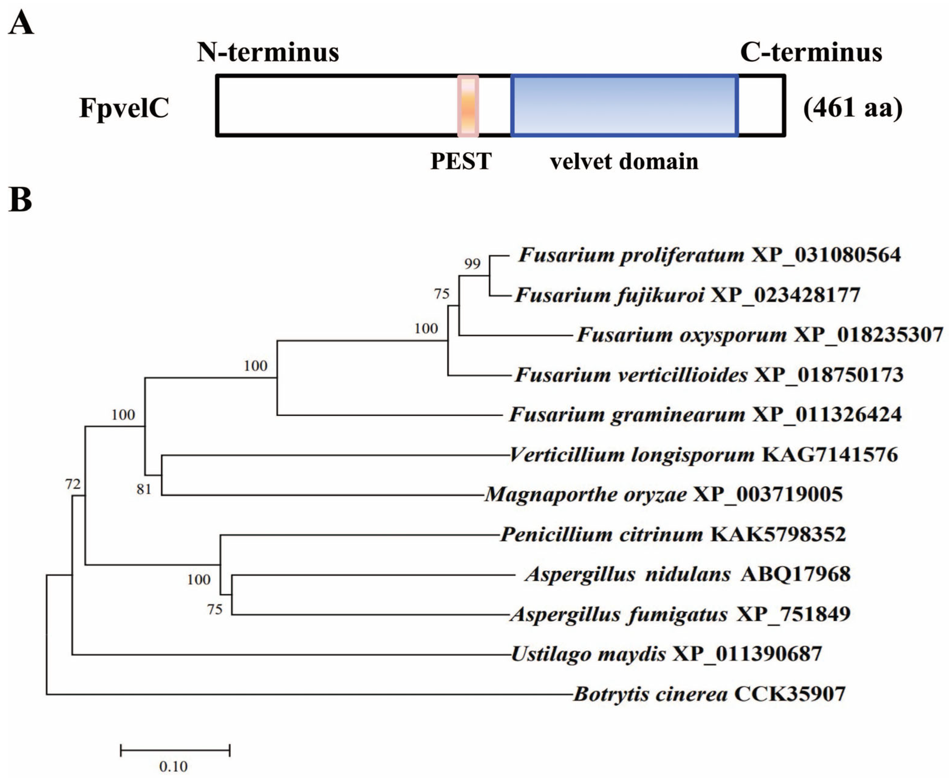
Figure 1. The structure and phylogenetic tree of F. proliferatum FpvelC protein. (A) Domain architecture of the FpvelC protein in F. proliferatum. The length of amino acid was noted in parentheses. Blue indicates a velvet domain between amino acids 245 and 426, while pink indicates a PEST domain between amino acids 198 and213. (B) Phylogenetic analysis of FpvelC protein and the orthologs in different filamentous fungi. A phylogenetic tree was constructed based on the amino acid sequences using MEGA 11 using the neighbor-joining method with 1000 bootstrap repetitions. Numbers on the branches represent the percentage of replicates supporting each branch. The fungal species and GenBank accession numbers are labeled on the right.
Toxins 17 00433 g001
Figure 2. Influence of FpvelC on conidiation in F. proliferatum. (A) The number of conidia of ΔFpvelC in YEPD liquid media. (B) Relative expression levels of conidiation-related genes in conidia of ΔFpvelC after being cultured in YEPD media for 48 h. Fpcos1 and Fpcom1 genes encoded transcription factors; Fpcon6, Fpcon7 and Fpcon8 genes encoded conidiation-specific proteins. (C) The amounts of trehalose per 107 conidia of ΔFpvelC. (D) The amounts of N-acetylglucosamine (GlcNAc) per 108 conidia of ΔFpvelC. (E) Relative expression levels of genes associated with trehalose and chitin synthesis in conidia of ΔFpvelC after being cultured in YEPD media for 48 h. The Fptps2 and Fptps3 genes encoding alpha, alpha-trehalose phosphate synthases were involved in trehalose synthesis; the FptreA gene encoding alpha, alpha-trehalose glucohydrolase and the FptreB gene encoding trehalase were involved in trehalose hydrolysis; the FpchsE and FpchsZ genes encoding chitin synthases were involved in chitin synthesis; and the FpchiA gene encoding class III chitinase and the FpchiB gene encoding class V chitinase were involved in chitin degradation. The bars represent the standard deviation from three replicates. Significant differences were determined by Student’s t-test (**, p < 0.01; ns, not significant). All experiments were repeated three times with three replicates each time.
Toxins 17 00433 g002
Figure 3. Effect of FpvelC on stress responses in F. proliferatum. (A) Colony phenotypes of ΔFpvelC on PDA media supplemented with 0.3 mg/mL Congo red, 0.01% SDS, or 1 mM or 5 mM H2O2 at 28 °C for 5 days. (B) The inhibition of mycelial growth of ΔFpvelC under different stresses. (C) Relative expression levels of antioxidant genes in ΔFpvelC. After being grown in PDB media for 3 days, ΔFpvelC was treated with 5 mM H2O2 for 1 h. FpnoxA, FpnoxB and FpnoxR encoded NADPH oxidases; FpcatA and FpcatD encoded catalases. The bars represent the standard deviations from three replicates. Significant differences were determined by Student’s t-test (**, p < 0.01; ns, not significant). All experiments were repeated three times with three replicates each time.
Toxins 17 00433 g003
Figure 4. Involvement of FpvelC in the pathogenicity of F. proliferatum. (A) Disease symptoms of rice spikelets inoculated with ΔFpvelC at 21 dpi. (B) Disease index of rice spikelets inoculated with ΔFpvelC at 21 dpi. (C) Invasive hyphae of ΔFpvelC on the epidermis of rice glumes under scanning electron microscopy at 48 hpi and 72 hpi. Infectious hyphae are denoted with yellow arrows. Scale bars = 100 μm. (D) Relative expression levels of the genes encoding xylanases and xyloglucanases in infected glumes challenged by ΔFpvelC at 72 hpi. Fpxyn10A, Fpxyn10B, Fpxyn11B and Fpxyn11C genes encoded xylanases, and Fpxeg1, Fpxeg2, Fpxeg3 and Fpxeg4 genes encoded xyloglucanases. (E) Ultrastructure of rice glumes inoculated with ΔFpvelC under transmission electron microscopy at 72 hpi. Sg represents starch grain. Chl represents chloroplast. Scale bars = 2 μm. (F) Disease symptoms of maize kernels inoculated with ΔFpvelC at 7 dpi. (G) Disease symptoms of maize stalks inoculated with ΔFpvelC at 14 dpi. The bars represent the standard deviation from three replicates. Significant differences were determined by Student’s t-test (**, p < 0.01; ns, not significant). All experiments were repeated three times with three replicates each time.
Toxins 17 00433 g004
Figure 5. Impact of FpvelC on production of FB1 and fusaric acid in F. proliferatum. (A) Quantification of FB1 produced by ΔFpvelC in PDB cultures at 28 °C for 10 days. (B) Relative expression levels of genes (Fpfums) responsible for fumonisin biosynthesis in ΔFpvelC after being cultured in PDB for 72 h. (C) Quantification of fusaric acid produced by ΔFpvelC in PDB cultures at 28 °C for 10 days. (D) Relative expression levels of genes (Fpfubs) responsible for fusaric acid biosynthesis in ΔFpvelC after being cultured in PDB for 72 h. The bars represent the standard deviation from three replicates. Significant differences were determined by Student’s t-test (*, p < 0.05; **, p < 0.01; ns, not significant). The experiments were performed in triplicate with three replicates each time.
Toxins 17 00433 g005
Figure 6. The role of FpvelC in nitrogen metabolism in F. proliferatum. (A) Relative expression levels of genes associated with nitrogen regulation in ΔFpvelC. The FpareA gene encoded the GATA transcription factor, the FpmeaB gene encoded the bZIP transcription factor, the FpnmrA gene encoded the NmrA family protein, the Fpmep3 gene encoded ammonium permease, and the Fpnii1 gene encoded nitrite reductase. (B) Quantification of intracellular glutamine in ΔFpvelC. (C) Colony morphology of ΔFpvelC on MM media amended with TOR kinase inhibitor rapamycin at final concentrations of 100, 200, 300 or 400 nM at 28 °C for 4 days. (D) The inhibition of mycelial growth of ΔFpvelC treated with different concentrations of rapamycin. The bars represent the standard deviation from three replicates. Significant differences were determined by Student’s t-test (**, p < 0.01; ns, not significant). The experiments were performed in triplicate with three replicates each time.
Toxins 17 00433 g006
Disclaimer/Publisher’s Note: The statements, opinions and data contained in all publications are solely those of the individual author(s) and contributor(s) and not of MDPI and/or the editor(s). MDPI and/or the editor(s) disclaim responsibility for any injury to people or property resulting from any ideas, methods, instructions or products referred to in the content.