Article

Modulation of Broiler Intestinal Changes Induced by Clostridium perfringens and Deoxynivalenol through Probiotic, Paraprobiotic, and Postbiotic Supplementation

by
Marielen de Souza
1,2,3,
Ana Angelita Sampaio Baptista
2,
Maísa Fabiana Menck-Costa
2,
Larissa Justino
2,
Eduardo Micotti da Glória
4,
Gabriel Danilo Shimizu
5,
Camila Rodrigues Ferraz
6,
Waldiceu A. Verri
6,
Filip Van Immerseel
3 and
Ana Paula Frederico Rodrigues Loureiro Bracarense
1,*
1
Laboratory of Animal Pathology (LAP), Department of Preventive Veterinary Medicine, Universidade Estadual de Londrina, Londrina 86057-970, Brazil
2
Laboratory of Avian Medicine (LAM), Department of Preventive Veterinary Medicine, Universidade Estadual de Londrina, Londrina 86057-970, Brazil
3
Livestock Gut Health Team (LiGHT), Department of Pathobiology, Pharmacology and Zoological Medicine, Faculty of Veterinary Medicine, Ghent University, 9820 Merelbeke, Belgium
4
Biological Science Department, Luiz de Queiroz College of Agriculture, University of São Paulo, Piracicaba 13418-900, Brazil
5
Department of Statistics, Universidade Estadual de Londrina, Londrina 86057-970, Brazil
6
Laboratory of Pain, Inflammation, Neuropathy and Cancer, Department of General Pathology, Universidade Estadual de Londrina, Londrina 86057-970, Brazil
*
Author to whom correspondence should be addressed.
Toxins 2024, 16(1), 46; https://doi.org/10.3390/toxins16010046
Submission received: 21 November 2023 / Revised: 28 December 2023 / Accepted: 7 January 2024 / Published: 14 January 2024

Abstract

:
Deoxynivalenol (DON) is a predisposing factor for necrotic enteritis. This study aimed to investigate the effects of a DON and Clostridium perfringens (CP) challenge on the intestinal morphology, morphometry, oxidative stress, and immune response of broilers. Additionally, we evaluated the potential of a Lactobacillus spp. mixture as an approach to mitigate the damage induced by the challenge. One-day-old broiler chickens (n = 252) were divided into seven treatment groups: Control, DON, CP, CP + DON, VL (DON + CP + viable Lactobacillus spp. mixture), HIL (DON + CP + heat-inactivated Lactobacillus spp. mixture), and LCS (DON + CP + Lactobacillus spp. mixture culture supernatant). Macroscopic evaluation of the intestines revealed that the CP + DON group exhibited the highest lesion score, while the VL and HIL groups showed the lowest scores. Microscopically, all Lactobacillus spp. treatments mitigated the morphological changes induced by the challenge. DON increased levels of reactive oxygen species (ROS) in the jejunum, and CP increased ROS levels in the jejunum and ileum. Notably, the Lactobacillus spp. treatments did not improve the antioxidant defense against CP-induced oxidative stress. In summary, a Lactobacillus spp. mixture, whether used as a probiotic, paraprobiotic, or postbiotic, exerted a partially protective effect in mitigating most of the intestinal damage induced by DON and CP challenges.
Key Contribution: The combined challenge of deoxynivalenol (DON) and Clostridium perfringens exacerbates necrotic enteritis lesions in broilers, yet supplementation with a Lactobacillus spp. mixture effectively mitigates the resulting intestinal changes, highlighting its potential as a protective measure.

1. Introduction

Intestinal health plays a key role in broiler performance and productivity [1]. The growing concern regarding the emergence of multi-resistant bacteria from poultry has led many markets to prohibit or restrict the use of antimicrobials as growth promoters [2,3]. Consequently, maintaining intestinal health in poultry flocks has become increasingly challenging, resulting in the reemergence of intestinal diseases such as necrotic enteritis (NE) [4].
Necrotic enteritis (NE) is a bacterial disease caused by strains of Clostridium perfringens (CP) [5]. CP is a Gram-positive, anaerobic, spore-forming bacterium that is a natural component of the poultry gut microbiota [6]. This disease occurs when there is an abnormal increase in the population of C. perfringens in the gastrointestinal tract (GIT), combined with predisposing factors such as coccidia infection, diets rich in non-starch polysaccharide grains, and exposure to mycotoxins, among others. Virulent strains produce the plasmid-encoded NetB toxin [6,7,8,9]. NE can be manifested as either clinical (resulting in high mortality rates) or subclinical (leading to growth performance failures), with an estimated annual cost of approximately USD 6 billion, equivalent to USD 0.05 per chick [10].
Contamination with mycotoxins is a growing concern in feedstock due to climate change [11]. Mycotoxin exposure significantly contributes to the occurrence of NE, and poultry are frequently exposed to deoxynivalenol (DON) [9,12,13]. DON is one of the most prevalent mycotoxins contaminating finished feed and raw commodities worldwide [14]. In poultry, DON exposure has been linked to villus atrophy, failure in intestinal barrier function, increased intraepithelial lymphocyte infiltration, heightened goblet cell abundance, intestinal oxidative stress, and disruption of gut microbiome diversity [15,16,17,18,19]. Consumption of DON has additionally been linked to increased susceptibility to coccidiosis and necrotic enteritis, particularly in more severe cases [9,12,20].
Probiotics consist of beneficial microorganisms that can enhance host health through various mechanisms [21]. Paraprobiotics, on the other hand, refers to dead probiotic microbial cells and their constituents, making them a preferred choice for immunosuppressed hosts due to the absence of the risk of bacterial translocation [22]. Postbiotics encompass bacterial metabolites, cell-free supernatant (CFS), and soluble factors (products or metabolic byproducts) produced by live bacteria or released following bacterial lysis [23]. Lactobacillus spp. strains, in different forms—be it live cells, heat-inactivated cells, or culture supernatants—have demonstrated protective effects in chickens individually challenged with either DON or CP [5,24,25,26,27].
This study aimed to investigate the impact of a dual challenge involving DON and CP on intestinal morphology, morphometry, immune response, and oxidative stress in broilers. Additionally, we have evaluated the efficacy of a Lactobacillus spp. mixture, administered as a probiotic (live cells), paraprobiotic (heat-inactivated cells), and postbiotic (culture supernatant), as a potential alternative to mitigate the damage induced by both of these factors.

2. Results

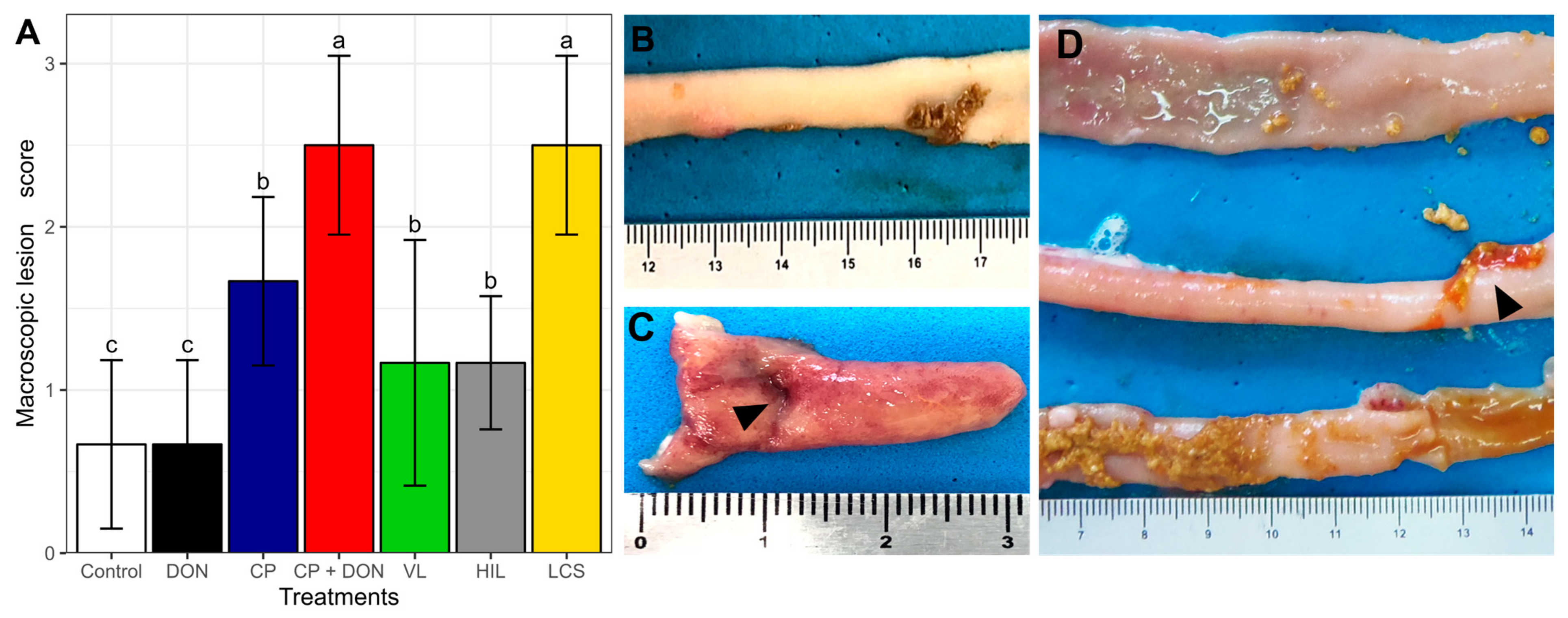
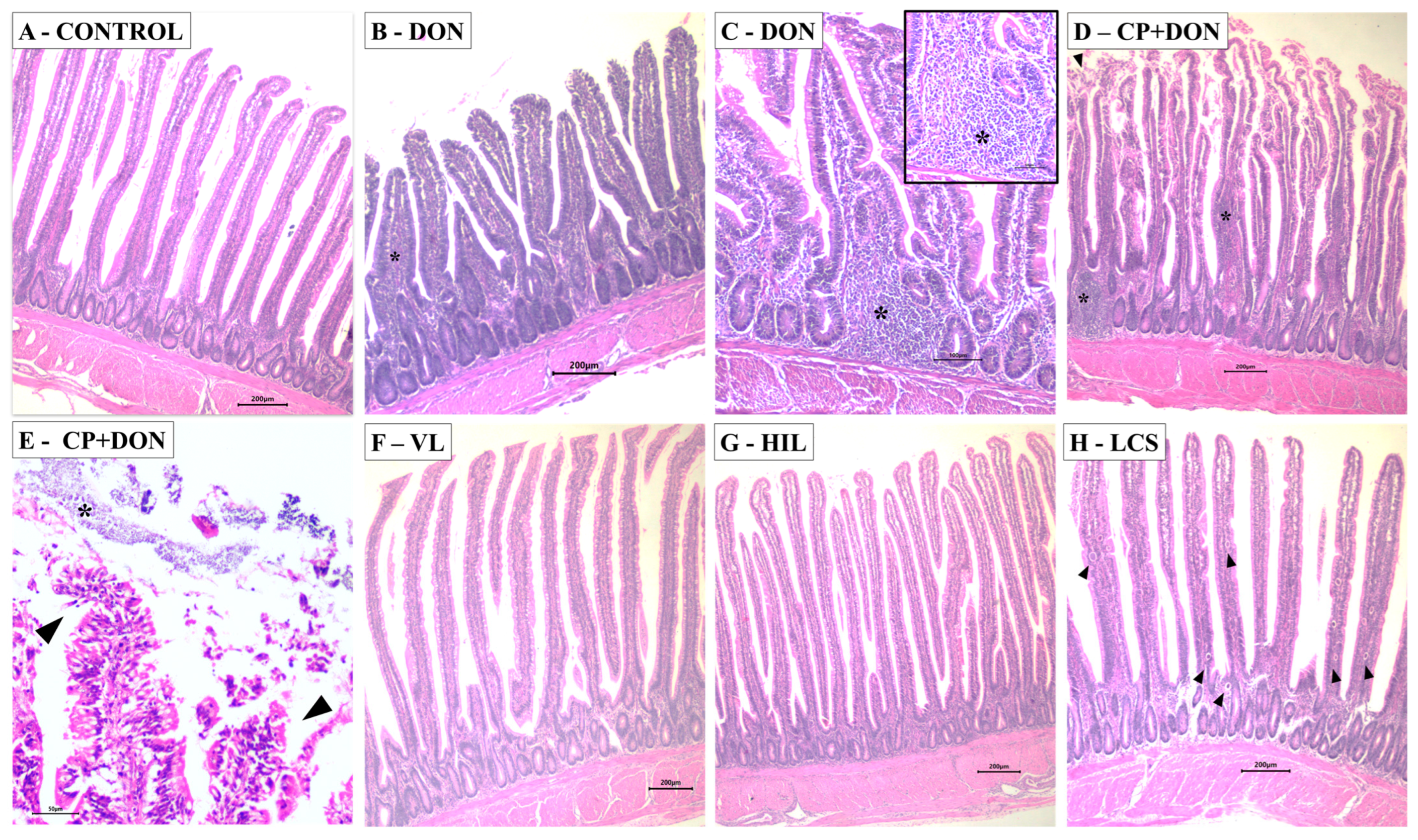
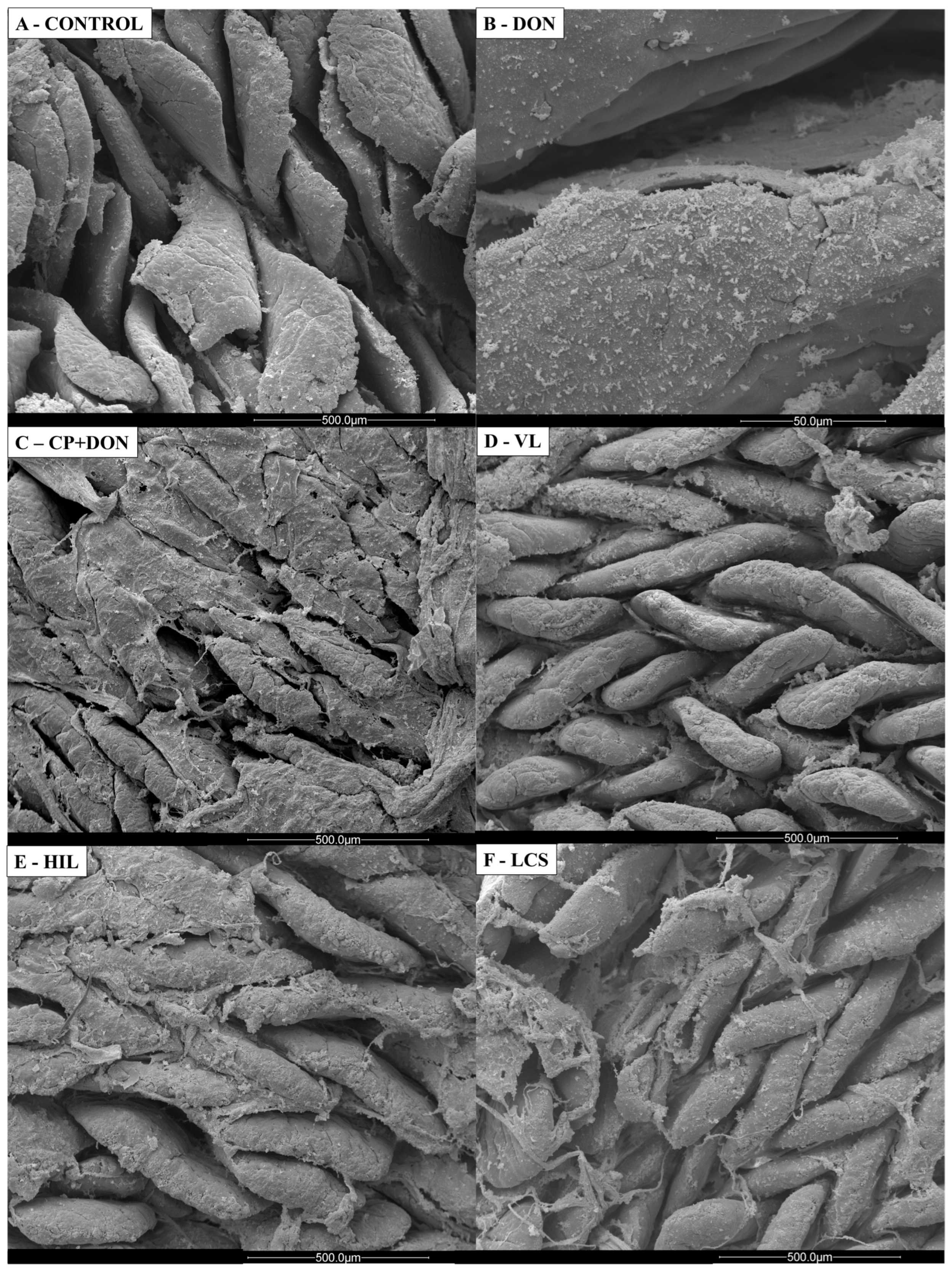
2.1. Effects of DON, C. perfringens, and Lactobacillus spp. Mixture on the Intestines

On the 23rd day, the intestines were macroscopically evaluated (Figure 1). The CP + DON group exhibited a worse intestinal gross appearance than the CP group. Among the Lactobacillus spp.-supplemented groups, VL and HIL induced the most effective protective effects. The observed changes included loss of intestinal tonus, hyperemia (Figure 1C), excessive mucus, the presence of yellow peeling content (Figure 1D), and, less frequently, a thick fibrinous mucus layer.
Intestinal morphometry is directly related to zootechnical performance [28]. In the jejunum, DON, CP, and CP + DON treatments reduced villus height and the villus: crypt ratio compared to the control and Lactobacillus spp.-supplemented groups. In the ileum, DON and CP treatments led to a reduction in villus height compared to the other groups, while no significant difference was observed in duodenal morphometry among the experimental groups (Table 1).
As sentinels of the intestinal barrier and local immune response, intraepithelial lymphocytes (IELs) were evaluated [29]. In the jejunum, only DON ingestion did not induce an increase in IELs; however, broilers receiving CP or CP + DON showed increased IEL infiltrate (≈1.5-fold on average for both groups) compared to the control group, while the VL and HIL groups were similar to the control. In the ileum, all treatments induced a higher number of IELs compared to the control (Table 1). The number of goblet cells was evaluated in the ileum, and the Lactobacillus spp.-supplemented groups showed a higher abundance compared to the control, DON, CP, and CP + DON treatments.
Morphological alterations induced by the different treatments were evaluated using a lesion score. In the duodenum, CP and CP + DON increased the lesion score by approximately 2.6-fold compared to the control, while the remaining treatments resulted in lesion scores similar to those of the control and DON groups. In the jejunum, all treatment groups showed higher scores than the control group, except for the HIL group. In the ileum, increased lesion scores were observed in all treatment groups except the VL group when compared to the control (Table 1). The most frequent changes observed in the histological evaluation included edema of the lamina propria, lymphocytic infiltrate, cell debris, apical necrosis, adhesion of bacteria on the villi surface, and cytoplasmic vacuolation of enterocytes (Figure 2).
Scanning electron microscopy (Figure 3) was performed to illustrate the effects of Lactobacillus spp. mixture, DON, and CP challenge in the jejunum. The control group showed normal villus morphology, while in the DON and CP + DON groups, a thicker mucus layer was observed. The Lactobacillus spp. mixture groups showed preserved villi morphology, similar to the control.

2.2. Effects of DON, C. perfringens, and Lactobacillus spp. Mixture on Redox Status

Oxidative stress occurs when there is an imbalance between the production of radical species and the antioxidant response in the organism [30]. Our study aimed to investigate the effects of DON and CP on the redox status. To achieve this, samples from the jejunum and ileum were used in lipid peroxidation (TBARS) and superoxide anion production (NBT) assays to assess the oxidative response. Additionally, GSH (reduced glutathione), ABTS (3-ethylbenzothiazoline-6-sulphonic acid), and FRAP (ferric reducing ability) assays were performed to evaluate antioxidant capacity.
In the jejunum, exposure to DON and CP + DON increased TBARS levels compared to the control, CP, and Lactobacillus spp.-supplemented groups. An increase in the NBT levels was detected in the CP, CP + DON, and Lactobacillus spp.-supplemented groups compared to the control and DON groups (Table 2). DON, CP, and CP + DON challenges reduced GSH levels compared to the control group, with the Lactobacillus spp.-supplemented treatments unable to restore GSH levels to those of the control. Regarding FRAP levels, the DON and control groups were similar, while the CP, CP + DON, and Lactobacillus spp.-supplemented groups showed higher levels in comparison. There were no significant differences in ABTS levels between the groups.
In the ileum, both CP and CP + DON treatments significantly increased NBT levels by approximately 5.6-fold compared to the control and DON groups. However, the Lactobacillus spp.-supplemented treatments were unable to revert superoxide anion production to control group levels. Regarding GSH levels, CP and CP + DON treatments reduced it approximately 2.85-fold compared to the control and DON groups, with the Lactobacillus spp.-supplemented treatments showing no significant differences compared to the CP and CP + DON groups. No significant differences were observed among the TBARS, ABTS, and FRAP levels between the groups.

2.3. Effects of DON, C. perfringens, and Lactobacillus spp. on Intestinal Secretory IgA Levels

Intestinal secretory IgA levels were assessed at three time points: 7, 14, and 20 days. No significant difference was observed among the treatments (p = 0.08). However, with respect to time, a lower IgA concentration was observed at 7 days (1,165,620 ± 636,766 ng/mL), compared to that at 14 (1,488,459 ± 947,397 ng/mL) and 20 (1,610,248 ± 1,050,150 ng/mL) days (p = 0.02). No interaction between time and treatment was observed (p = 0.20).

3. Discussion

Previous studies have shown that exposure to mycotoxins, such as DON alone or in combination with other mycotoxins like fumonisins, can exacerbate the occurrence of necrotic enteritis (NE) [9,12,13]. In this study, we aimed to investigate the effects of different presentations [probiotic (viable cells), paraprobiotic (heat-inactivated cells), and postbiotic (heat-inactivated cells culture supernatant)] of a Lactobacillus spp. mixture on broiler chickens challenged with DON and Clostridium perfringens (CP), as previous research has suggested that Lactobacillus spp. strains may mitigate the negative effects of single challenge with mycotoxins or CP in the intestine [5,24].
Macroscopic evaluation of the intestines revealed that the CP + DON group exhibited a higher lesion score compared to the group challenged with CP alone, consistent with previous reports indicating that a DON-contaminated diet can increase the severity of NE cases [9,12]. Among the Lactobacillus spp. groups, VL and HIL induced the lowest lesion scores. Microscopically, both DON and CP exposure induced intestinal lesions, and the combination of both factors tended to increase the lesion score, although the difference was not significant. The Lactobacillus spp. treatments appeared to mitigate the morphological changes induced by both challenges.
Zootechnical performance is closely related to increased intestinal absorptive capacity. Villus height serves as an indicator of the absorption area, and the intestinal crypts are the sites of new enterocyte multiplication [31,32]. The jejunum and ileum were the intestinal segments most affected by both DON and CP challenges, showing a reduction in villus height and the villus: crypt ratio (only in the jejunum) compared to the control and Lactobacillus spp.-supplemented groups. Since the small intestine is the primary site of DON absorption, previous studies have reported impaired intestinal morphometry as a consequence of DON ingestion [16,33]. However, in this study, the combination of DON and CP did not worsen the intestinal morphometry compared to single challenge.
Intraepithelial lymphocytes (IELs) are key components of the intestinal barrier [29]. In this study, exposure to CP increased the number of IELs in both the jejunum and ileum, regardless of DON administration. Similar findings were reported in naturally infected laying hens [34]. However, in the VL and HIL groups, the number of IELs was reduced to control levels in the jejunum but not in the ileum.
Contrary to previous reports [16,35], DON exposure did not induce an increase in goblet cell density under light microscopy evaluation in this study. However, scanning electron microscopy revealed enhanced mucus presence in the DON-exposed group compared to the control group. Lactobacillus spp. supplementation increased the number of goblet cells compared to other groups. Excess mucus can predispose to NE; however, this finding was expected, as the microbiota can influence mucus layer development, and specifically, Lactobacillus spp. are known to contribute to strengthening the intestinal mucosal barrier function [36,37,38,39].
Regarding oxidative stress in the jejunum, DON induced lipid peroxidation, and this effect was sustained after the CP challenge. However, the Lactobacillus spp. treatments decreased the lipid peroxidation/MDA levels to control levels. Previous research has also reported DON-induced lipid peroxidation [16,40,41], which is associated with mitochondrial damage [42]. The NBT assay quantifies superoxide anions indirectly through their oxidative effects on NBT and is mainly produced by inflammatory cells [43]. CP induced inflammation, as confirmed by higher NBT levels, but the Lactobacillus spp. treatments did not reverse this effect. As a consequence of lipid peroxidation and inflammation, lower GSH levels were observed in all DON- and CP-challenged groups compared to control levels. These findings align with other studies that have reported the capacity of DON and CP to reduce intestinal antioxidant defense [16,26,44,45]. The FRAP assay measures tissue ferric reducing ability, and CP-challenged groups showed higher FRAP levels, likely as a response to the inflammatory status and oxidative stress [46].
The ileum is the final segment of the small intestine, and exposure to xenobiotics such as mycotoxins is lower than that in the proximal regions [47]. In this study, the ileum showed no change in the oxidative stress response after DON exposure. It is likely that the levels of mycotoxins were reduced due to the intestinal microbiota’s detoxification activity [48]. Organisms under long-term toxicity might induce adaptations to reduce the damage [49,50,51]. On the other hand, CP induced an inflammatory status resulting in higher levels of NBT and lower levels of GSH, and Lactobacillus spp. did not exert a protective effect.
In this study, concomitant exposure to DON and CP did not worsen most of the evaluated parameters compared to single challenge. However, the Lactobacillus spp. treatments, especially LV and HIL, were effective in mitigating tissue damage. Viable or heat-inactivated cells from one strain of L. plantarum used in this study have a recognized capacity to remove DON [52]. The mechanism of action is still unclear, but based on research with similar microorganisms, it is hypothesized that viable cells can detoxify DON while heat-inactivated cells can bind to the mycotoxin, thereby reducing its toxic effects [53,54,55].
The Lactobacillus strains used in this experiment underwent in vitro evaluation, revealing their ability to antagonize the growth of C. perfringens (inhibition zone on spot on the lawn varying from 14 to 22 mm, data not shown). The protective effects of viable cells against CP-induced damage were likely a result of bacteriocin production targeting C. perfringens, whereas heat-inactivated cells might exert a prebiotic effect by modulating the gut microbiome and preventing the proliferation of pathogenic microorganisms [26,56,57,58,59].

4. Conclusions

Additional studies are required to clarify the mechanisms of action of the evaluated Lactobacillus spp. strains and establish an industrial process for producing and transforming these strains into a commercial product for animal consumption. This is especially crucial as the model employed in this trial (oral gavage) is not applicable in commercial poultry farming. Nonetheless, it can be inferred that these strains exhibited a protective effect, mitigating a significant portion of the intestinal damage induced by DON and C. perfringens.

5. Materials and Methods

5.1. Study Location and Ethical Approval

This study was conducted at the avian medicine experimental facilities at Universidade Estadual de Londrina, Londrina, Paraná, Brazil, and received approval from the institutional ethics committee for the use of animals (Comitê de Ética no Uso de Animais CEUA-UEL, protocol number 12433.2018.03, approval date 24 September 2018).

5.2. Animals and Treatments

One-day-old broiler chickens (n = 252), Ross 308 lineage, were housed in cages with water, feed, and heating provided, following lineage guidelines [60]. The animals were divided into seven treatment groups (n = 36 each), as follows: Control—uncontaminated diet; DON—diet containing DON at 19.3 mg kg−1; CP—uncontaminated diet + Clostridium perfringens challenge; CP + DON—diet containing DON at 19.3 mg kg−1 + C. perfringens challenge; VL—diet containing DON at 19.3 mg kg−1 + C. perfringens challenge + viable Lactobacillus spp. mixture; HIL—diet containing DON at 19.3 mg kg−1 + C. perfringens challenge + heat-inactivated Lactobacillus spp. mixture; LCS—diet containing DON at 19.3 mg kg−1 + C. perfringens challenge + Lactobacillus spp. mixture culture supernatant.

5.3. Diets

The experimental diets were formulated to meet the nutritional requirements of the animals (Table 3), consisting of three different diets used during the trial period: Diet 1 (0–6 days)—primarily composed of corn (48.16%) and soybean meal (43.70%), free of DON; Diet 2 (7–14 days)—composed of corn (15%), soybean meal (35.31%), and wheat (40.13%); Diet 3 (15–23 days)—composed of corn (15%), wheat (40.43%), and fish meal (35.31%). Fish meal was added to diet 3 to elevate the crude protein level and create an intestinal environment favorable for the experimental induction of necrotic enteritis [61].
On the 7th day, DON-challenged groups (excluding the control) began receiving a DON-contaminated diet containing 19.3 mg kg−1 (Figure S1). The crude DON extract used to contaminate the diets was provided by the Laboratory of Mycology, Luiz de Queiroz College of Agriculture, University of São Paulo. A blend (standard diet + DON) was prepared at the Universidade Estadual de Londrina facilities using a commercial feed mixer. The diets were sent to Lamic laboratory (Santa Maria—RS/Brazil), where the mycotoxins levels were assessed using the HPLC/MS-MS method. Three diet samples were collected throughout the experimental period: first, 0–6 days (diet 1); second, 7–23 uncontaminated diet (diets 2 and 3); and third, 7–23 DON-contaminated diet (diets 2 and 3). The results of the mycotoxin (deoxynivalenol, aflatoxins, fumonisins, and zearalenone) analysis are shown on Table 4.

5.4. Necrotic Enteritis Induction

Animals in the CP, CP + DON, VL + DON, HIL + DON, and LCS + DON groups underwent necrotic enteritis induction. They were orally challenged with 4000 oocysts of Eimeria spp. from a commercial vaccine (Livaccox®, Paulínia, Brazil) and a 10-fold dose of a commercial Gumboro disease vaccine (Bursa F®, Campinas, Brazil) [62,63] on the 14th day. The non-challenged groups received 1 mL of sterile PBS to replicate the same stress.
A C. perfringens type G, netB positive strain from the Avian Medicine UEL collection was used to challenge the birds. The strain was grown in BHI (Brain Heart Infusion, HiMedia, Sumaré, Brazil) broth at 37 °C for 18 h under anaerobic conditions using a commercial kit (GasPak®, Becton Dickinson Osasco, Brazil). From the 16th to the 22nd day, animals received 1 mL of fresh CP culture (approximately 4 × 10⁸ CFU/mL) via oral gavage twice daily (Figure 1). On each challenge day, an inoculum sample was 10-fold diluted and plated on SFP agar® (Becton Dickinson Osasco, Brazil), followed by incubation under anaerobic conditions to determine the CFU count. The non-challenged groups received 1 mL of BHI broth to simulate the same stress.

5.5. Lactobacillus spp. Mixture Administration

Animals in the VL, HIL, and LCS groups received 1 mL of a Lactobacillus spp. mixture (approximately 2.2 × 10⁹ CFU/mL) via oral gavage every other day throughout the experimental period (Figure 1). Groups not supplemented with the Lactobacillus spp. pool received 1 mL of sterile MRS (De Man, Rogosa, and Sharpe medium, HiMedia Sumaré, Brazil) broth.
The Lactobacillus spp. mixture comprised an equal quantity of three strains: two isolated from broiler chickens (L. reuteri and L. plantarum, not deposited in GenBank) and one from wheat (L. plantarum—accession number CP053912) in previous studies [52,64,65]. The strains were grown separately in MRS broth and incubated at 37 °C for 24 h under microaerophilic conditions. Samples were provided in three different forms: (i) a fresh culture of viable Lactobacillus spp. mixture; (ii) a heat-inactivated culture of Lactobacillus spp. mixture, and (iii) a supernatant culture from a heat-inactivated Lactobacillus spp. mixture. The inactivation and mixture preparation followed previously described methods [16,52]. Cell density was assessed daily through 10-fold dilution and plating.

5.6. Sample Collection

Throughout the experimental period, four samplings were conducted. On days 7, 14, and 20, ten animals per treatment group were euthanized, and on day 23, 6 animals per treatment were used for biological sample collection. The intestinal samples underwent macroscopic lesion scoring, histological examination, ELISA (enzyme-linked immunosorbent assay), and oxidative stress response assessments.
  • Macroscopic intestinal lesion score
On the 23rd day, 6 animals per treatment were euthanized, and an intestinal lesion score was determined following previously described criteria, ranging from 0 to 5 [66].
  • ELISA
Intestinal fluid was collected from 10 animals per treatment at 7, 14, and 20 days. For this purpose, 2 mL of a wash buffer (PBS pH 7.2, thimerosal 0.01%, 1% BSA, 1 mM phenylmethylsulfonyl fluoride, and 5 mM EDTA) was injected into the proximal duodenum and collected at the distal ileum. The collected samples were then centrifuged at 1200× g for 15 min at 4 °C, and the resulting supernatant was collected and stored at −20 °C. The levels of IgA were determined using the chicken IgA ELISA quantitation kit (Bethyl® Laboratories, Montgomery, TX, USA). The assay was performed in triplicate following the manufacturer’s instructions, with the plates read at 450 nm.
  • Histology and scanning electron microscopy
Morphological and morphometric evaluations were carried out on the intestines (duodenum, jejunum, and ileum) of 6 animals per treatment on the 23rd day. The Swiss roll technique [67] was used to collect and prepare the samples. The tissues were fixed in a 10% buffered formalin solution and subsequently subjected to routine histological processing. Sections of 5 µm thickness were obtained and stained with hematoxylin-eosin (HE) and Alcian Blue (AB). AB staining was utilized to determine goblet cell density.
Morphometric analysis was performed on 30 randomly selected villi and crypts per slide (180 villi and crypts per treatment) using image analysis software (Motic Image Plus 2.0, Motic Instruments, Richmond, BC, Canada). Measurements of villus height, crypt depth, and villus: crypt ratio were conducted in the duodenum, jejunum, and ileum. The morphological evaluation of the intestines followed the scoring system described by Terciolo, et al. [68], with minor modifications (including additional lesions in the score: inflammatory infiltrate, congestion, bacteria adhered to the villi, presence of Eimeria spp., and cell debris). The count of intraepithelial lymphocytes (IELs) was performed on 12 randomly chosen villi per slide (72 villi per treatment), considering IELs positioned above the enterocyte nucleus. Goblet cell density was determined exclusively in the ileum by evaluating 15 random villi per slide (90 villi per treatment).
Scanning electron microscopy was conducted specifically in the jejunum. Samples were collected on the 23rd day, fixed in a 2.5% glutaraldehyde buffered solution (sodium cacodylate solution 0.1 M, pH 7.2) for 24 h, and subsequently washed with sodium cacodylate buffer (0.1 M, pH 7.2). Treatment involved exposure to 1% osmium tetroxide in sodium cacodylate buffer (0.1 M, pH 7.2) for 1 h. Subsequent steps included gradual dehydration in different ethanol concentrations (70, 80, 90, 100%) and drying to the critical point using a CPD 030 critical point dryer (Bal-Tec Union Ltd., Vaduz, Liechtenstein). Following this, tissues were coated with gold (Sputter Coater SDC 050, Bal-Tec Union Ltd., Vaduz, Liechtenstein), and the morphology of the intestinal villi was examined using a scanning electron microscope (FEI Quanta 200, Field Electron and Ion Company, Hillsboro, USA).
  • Oxidative stress response
The oxidative stress response was evaluated in both the jejunum and ileum. On the 20th day, 4 animals per treatment were euthanized, and samples from these tissues were collected in microtubes and preserved at −80 °C until processing. The antioxidant capacity was assessed by quantification of reduced glutathione (GSH) following the method of Sedlak and Lindsay [69], ferric reducing ability (FRAP), and reduction of 2,2′-azino-bis (3-ethylbenzothiazoline-6-sulfonic acid) (ABTS), as described by Katalinic, et al. [70].
The oxidative response was evaluated using the nitroblue tetrazolium assay (NBT) [71] and the quantification of thiobarbituric acid reactive substances (TBARS) [72,73]. Tissue homogenization was carried out in buffer using a Tissue-Tearor (Bjospec, São Paulo, SP, Brazil). For the FRAP, ABTS, NBT, and TBARS protocols, the buffer consisted of KCL (1.15%) and EDTA (0.02 M) for GSH analysis.

5.7. Statistical Analysis

The experimental design was entirely randomized, except for the ELISA analysis, which followed a factorial 3 × 7 design (3 time points and 7 treatments). Each animal was considered one experimental unit. Data analysis was carried out using the free software R® version 3.4.4, and ANOVA was performed using the AgroR package with a significance level of 5%. If the means exhibited statistical significance, the data were submitted to a Scott–Knott multiple comparison test at a 5% significance level. Data that did not meet the assumption of normality of errors were subjected to logarithmic transformation before undergoing ANOVA and the Scott–Knott test.

Supplementary Materials

The following supporting information can be downloaded at: https://www.mdpi.com/article/10.3390/toxins16010046/s1, Figure S1. Experimental design. Experimental groups: Control—uncontaminated diet. DON (deoxyniva-lenol)—diet with DON 19.3 mg kg−1. CP (Clostridium perfringens)—uncontaminated diet + C. perfringens challenge. CP + DON—DON 19.3 mg kg−1 + C. perfringens challenge. LV—DON 19.3 mg kg−1 + C. perfringens challenge plus viable Lactobacillus spp. mixture. HIL—DON 19.3 mg kg−1 + C. perfringens challenge plus heat-inactivated Lactobacillus spp. mixture. LCS—DON 19.3 mg kg−1 + C. perfringens challenge plus Lactobacillus spp. mixture culture supernatant.

Author Contributions

Conceptualization, A.A.S.B. and A.P.F.R.L.B.; methodology, M.d.S., L.J., M.F.M.-C. and C.R.F.; software, G.D.S. and C.R.F.; validation, M.d.S.; formal analysis, M.d.S., L.J. and M.F.M.-C.; investigation, M.d.S. and A.A.S.B.; resources, E.M.d.G. and W.A.V.; data curation, G.D.S.; writing—original draft preparation, M.d.S.; writing—review and editing, A.P.F.R.L.B., F.V.I. and M.d.S.; visualization, A.A.S.B.; supervision, A.P.F.R.L.B.; project administration, A.P.F.R.L.B. and M.d.S.; funding acquisition, A.P.F.R.L.B. and M.d.S. All authors have read and agreed to the published version of the manuscript.

Funding

Coordenação de Aperfeiçoamento de Pessoal de Nível Superior Brasil (CAPES) and Conselho Nacional de Pesquisa e Desenvolvimento—Brasil (CNPq).

Institutional Review Board Statement

The animal study protocol was approved by the Institutional Ethics Committee for the use of animals COMITÊ DE ÉTICA NO USO DE ANIMAIS (CEUA) - UEL, protocol number 12433.2018.03, approval date 24 September 2018.

Informed Consent Statement

Not applicable.

Data Availability Statement

All data are present in the article.

Acknowledgments

The authors express sincere gratitude to Professor Admilton Gonçalves de Oliveira Junior and laboratory technician Osvaldo Capelo for their invaluable support in conducting the scanning electron microscopy (SEM) analysis. Marielen de Souza acknowledges the Coordenação de Aperfeiçoamento de Pessoal de Nível Superior Brasil (CAPES) for the sandwich doctorate fellowship (88881.624517/2021-01) and the Conselho Nacional de Pesquisa e Desenvolvimento—Brasil (CNPq) for the doctoral fellowship. Ana Paula F.R.L. Bracarense acknowledges support from CNPq (403843/2021-9) and CAPES/Cofecub (0389/2019). Camila Rodrigues Ferraz expresses gratitude for the CNPq postdoctoral fellowship. Waldiceu A. Verri acknowledges the Senior Research CNPq fellowship (#309633/2021-4).

Conflicts of Interest

The authors declare no conflicts of interest.

References

  1. Yegani, M.; Korver, D.R. Factors Affecting Intestinal Health in Poultry. Poult. Sci. 2008, 87, 2052–2063. [Google Scholar] [CrossRef]
  2. No 1831/2003; Regulation (EC) no 1831/2003 of the European Parliament and of the Council of 22 September 2003 on Additives for Use in Animal Nutrition. FAO: Rome, Italy, 2003; L 268/30, p. 15.
  3. MAPA. Instrução Normativa Nº 1, de 13 de Janeiro de 2020; Ministério da Agricultura Pecuária e Abastecimento: Brasília, Brazil, 2020. [Google Scholar]
  4. Van Immerseel, F.; Lyhs, U.; Pedersen, K.; Prescott, J.F. Recent breakthroughs have unveiled the many knowledge gaps in Clostridium perfringens-associated necrotic enteritis in chickens: The first International Conference on Necrotic Enteritis in Poultry. Avian Pathol. 2016, 45, 269–270. [Google Scholar] [CrossRef] [PubMed]
  5. Kulkarni, R.R.; Gaghan, C.; Gorrell, K.; Sharif, S.; Taha-Abdelaziz, K. Probiotics as Alternatives to Antibiotics for the Prevention and Control of Necrotic Enteritis in Chickens. Pathogens 2022, 11, 692. [Google Scholar] [CrossRef] [PubMed]
  6. Abd El-Hack, M.E.; El-Saadony, M.T.; Elbestawy, A.R.; El-Shall, N.A.; Saad, A.M.; Salem, H.M.; El-Tahan, A.M.; Khafaga, A.F.; Taha, A.E.; AbuQamar, S.F.; et al. Necrotic enteritis in broiler chickens: Disease characteristics and prevention using organic antibiotic alternatives—A comprehensive review. Poult. Sci. 2022, 101, 101590. [Google Scholar] [CrossRef] [PubMed]
  7. Mwangi, S.; Timmons, J.; Fitz-coy, S.; Parveen, S. Characterization of Clostridium perfringens recovered from broiler chicken affected by necrotic enteritis. Poult. Sci. 2019, 98, 128–135. [Google Scholar] [CrossRef] [PubMed]
  8. Dierick, E.; Hirvonen, O.P.; Haesebrouck, F.; Ducatelle, R.; Van Immerseel, F.; Goossens, E. Rapid growth predisposes broilers to necrotic enteritis. Avian Pathol. 2019, 48, 416–422. [Google Scholar] [CrossRef]
  9. Antonissen, G.; Van Immerseel, F.; Pasmans, F.; Ducatelle, R.; Haesebrouck, F.; Timbermont, L.; Verlinden, M.; Janssens, G.P.J.; Eeckhaut, V.; Eeckhout, M.; et al. The Mycotoxin Deoxynivalenol Predisposes for the Development of Clostridium perfringens-Induced Necrotic Enteritis in Broiler Chickens. PLoS ONE 2014, 9, 1–8. [Google Scholar] [CrossRef]
  10. Wade, B.; Keyburn, A. The True Cost of Necrotic Enteritis. Available online: https://www.poultryworld.net/poultry/the-true-cost-of-necrotic-enteritis/ (accessed on 30 March 2023).
  11. Paterson, R.R.M.; Lima, N. How will climate change affect mycotoxins in food? Food Res. Int. 2010, 43, 1902–1914. [Google Scholar] [CrossRef]
  12. Shanmugasundaram, R.; Adams, D.; Ramirez, S.; Murugesan, G.R.; Applegate, T.J.; Cunningham, S.; Pokoo-Aikins, A.; Glenn, A.E. Subclinical Doses of Combined Fumonisins and Deoxynivalenol Predispose Clostridium perfringens–Inoculated Broilers to Necrotic Enteritis. Front. Physiol. 2022, 13, 934660. [Google Scholar] [CrossRef]
  13. Guo, F.; Wang, F.; Ma, H.; Ren, Z.; Yang, X.; Yang, X. Study on the interactive effect of deoxynivalenol and Clostridium perfringens on the jejunal health of broiler chickens. Poult. Sci. 2021, 100, 100807. [Google Scholar] [CrossRef] [PubMed]
  14. BIOMIN. BIOMIN Mycotoxin Survey Q3 2021 Results. Available online: https://www.biomin.net/science-hub/biomin-mycotoxin-survey-q3-2021-results/ (accessed on 30 March 2023).
  15. Von Buchholz, J.S.; Ruhnau, D.; Hess, C.; Aschenbach, J.R.; Hess, M.; Awad, W.A. Paracellular intestinal permeability of chickens induced by DON and/or C. jejuni is associated with alterations in tight junction mRNA expression. Microb. Pathog. 2022, 168, 105509. [Google Scholar] [CrossRef]
  16. Souza, M.; Baptista, A.A.S.; Valdiviezo, M.J.J.; Justino, L.; Menck-Costa, M.F.; Ferraz, C.R.; da Gloria, E.M.; Verri, W.A.; Bracarense, A.P.F.R.L. Lactobacillus spp. reduces morphological changes and oxidative stress induced by deoxynivalenol on the intestine and liver of broilers. Toxicon 2020, 185, 203–212. [Google Scholar] [CrossRef] [PubMed]
  17. Osselaere, A.; Santos, R.; Hautekiet, V.; De Backer, P.; Chiers, K.; Ducatelle, R.; Croubels, S. Deoxynivalenol Impairs Hepatic and Intestinal Gene Expression of Selected Oxidative Stress, Tight Junction and Inflammation Proteins in Broiler Chickens, but Addition of an Adsorbing Agent Shifts the Effects to the Distal Parts of the Small Intestine. PLoS ONE 2013, 8, e69014. [Google Scholar] [CrossRef] [PubMed]
  18. Jia, B.; Lin, H.; Yu, S.; Liu, N.; Yu, D.; Wu, A. Mycotoxin deoxynivalenol-induced intestinal flora disorders, dysfunction and organ damage in broilers and pigs. J. Hazard. Mater. 2023, 451, 131172. [Google Scholar] [CrossRef]
  19. Awad, W.A.; Zentek, J. The feed contaminant deoxynivalenol affects the intestinal barrier permeability through inhibition of protein synthesis. Arch. Toxicol. 2015, 89, 961–965. [Google Scholar] [CrossRef]
  20. Grenier, B.; Dohnal, I.; Shanmugasundaram, R.; Eicher, S.D.; Selvaraj, R.K.; Schatzmayr, G.; Applegate, T.J. Susceptibility of broiler chickens to coccidiosis when fed subclinical doses of deoxynivalenol and fumonisins—Special emphasis on the immunological response and the mycotoxin interaction. Toxins 2016, 8, 231. [Google Scholar] [CrossRef]
  21. Cremon, C.; Barbaro, M.R.; Ventura, M.; Barbara, G. Pre- and probiotic overview. Curr. Opin. Pharm. 2018, 43, 87–92. [Google Scholar] [CrossRef]
  22. Martyniak, A.; Medyńska-Przęczek, A.; Wędrychowicz, A.; Skoczeń, S.; Tomasik, P.J. Prebiotics, Probiotics, Synbiotics, Paraprobiotics and Postbiotic Compounds in IBD. Biomolecules 2021, 11, 1903. [Google Scholar] [CrossRef]
  23. Aguilar-Toalá, J.E.; Garcia-Varela, R.; Garcia, H.S.; Mata-Haro, V.; González-Córdova, A.F.; Vallejo-Cordoba, B.; Hernández-Mendoza, A. Postbiotics: An evolving term within the functional foods field. Trends Food Sci. Technol. 2018, 75, 105–114. [Google Scholar] [CrossRef]
  24. Maidana, L.; Souza, M.; Bracarense, A.P.F.R.L. Lactobacillus plantarum and Deoxynivalenol Detoxification: A Concise Review. J. Food Prot. 2022, 85, 1815–1823. [Google Scholar] [CrossRef] [PubMed]
  25. Gong, L.; Wang, B.; Zhou, Y.; Tang, L.; Zeng, Z.; Zhang, H.; Li, W. Protective Effects of Lactobacillus plantarum 16 and Paenibacillus polymyxa 10 Against Clostridium perfringens Infection in Broilers. Front. Immunol. 2020, 11, 628374. [Google Scholar] [CrossRef]
  26. Cao, L.; Wu, X.H.; Bai, Y.L.; Wu, X.Y.; Gu, S.B. Anti-inflammatory and antioxidant activities of probiotic powder containing Lactobacillus plantarum 1.2567 in necrotic enteritis model of broiler chickens. Livest. Sci. 2019, 223, 157–163. [Google Scholar] [CrossRef]
  27. Abd El-Ghany, W.A.; Abdel-Latif, M.A.; Hosny, F.; Alatfeehy, N.M.; Noreldin, A.E.; Quesnell, R.R.; Chapman, R.; Sakai, L.; Elbestawy, A.R. Comparative efficacy of postbiotic, probiotic, and antibiotic against necrotic enteritis in broiler chickens. Poult. Sci. 2022, 101, 101988. [Google Scholar] [CrossRef] [PubMed]
  28. Schedle, K.; Pfaffl, M.W.; Plitzner, C.; Meyer, H.H.D.; Windisch, W. Effect of insoluble fibre on intestinal morphology and mRNA expression pattern of inflammatory, cell cycle and growth marker genes in a piglet model. Arch. Anim. Nutr. 2008, 62, 427–438. [Google Scholar] [CrossRef]
  29. Kaer, L.V.; Olivares-Villagomez, D. Development, homeostasis, and functions of intestinal intraepithelial lymphocytes. J. Immunol. 2018, 200, 2235–2244. [Google Scholar] [CrossRef] [PubMed]
  30. Halliwell, B. Biochemistry of oxidative stress. Biochem. Soc. Trans. 2007, 35, 1147–1150. [Google Scholar] [CrossRef]
  31. Van Loo, J. How Chicory Fructans Contribute to Zootechnical Performance and Well-Being in Livestock and Companion Animals. J. Nutr. 2007, 137, 2594S–2597S. [Google Scholar] [CrossRef]
  32. Swatson, H.K.; Gous, R.; Iji, P.A.; Zarrinkalam, R. Effect of dietary protein level, amino acid balance and feeding level on growth, gastrointestinal tract, and mucosal structure of the small intestine in broiler chickens. Anim. Res. 2002, 51, 501–515. [Google Scholar] [CrossRef]
  33. Ghareeb, K.; Awad, W.A.; Bohm, J.; Zebeli, Q. Impacts of the feed contaminant deoxynivalenol on the intestine of monogastric animals: Poultry and swine. J. Appl. Toxicol. 2015, 35, 327–337. [Google Scholar] [CrossRef]
  34. Allaart, J.G.; de Bruijn, N.D.; van Asten, A.J.A.M.; Fabri, T.H.F.; Gröne, A. NetB-producing and beta2-producing Clostridium perfringens associated with subclinical necrotic enteritis in laying hens in the Netherlands. Avian Pathol. 2012, 41, 541–546. [Google Scholar] [CrossRef] [PubMed]
  35. Wang, A.; Hogan, N.S. Performance effects of feed-borne Fusarium mycotoxins on broiler chickens: Influences of timing and duration of exposure. Anim. Nutr. 2019, 5, 32–40. [Google Scholar] [CrossRef]
  36. Nii, T.; Shinkoda, T.; Isobe, N.; Yoshimura, Y. Intravaginal injection of Lactobacillus johnsonii may modulates oviductal microbiota and mucosal barrier function of laying hens. Poult. Sci. 2023, 102, 102699. [Google Scholar] [CrossRef]
  37. Ni, Y.; Zhang, Y.; Zheng, L.; Rong, N.; Yang, Y.; Gong, P.; Yang, Y.; Siwu, X.; Zhang, C.; Zhu, L.; et al. Bifidobacterium and Lactobacillus improve inflammatory bowel disease in zebrafish of different ages by regulating the intestinal mucosal barrier and microbiota. Life Sci. 2023, 324, 121699. [Google Scholar] [CrossRef]
  38. Moore, R.J. Necrotic enteritis predisposing factors in broiler chickens. Avian Pathol. 2016, 45, 275–281. [Google Scholar] [CrossRef] [PubMed]
  39. Luis, A.S.; Hansson, G.C. Intestinal mucus and their glycans: A habitat for thriving microbiota. Cell Host Microbe 2023, 31, 1087–1100. [Google Scholar] [CrossRef] [PubMed]
  40. Yang, X.; Li, L.; Duan, Y.; Yang, X. Antioxidant activity of Lactobacillus plantarum JM113 in vitro and its protective effect on broiler chickens challenged with deoxynivalenol. J. Anim. Sci. 2017, 95, 837–846. [Google Scholar]
  41. Awad, W.A.; Ghareeb, K.; Dadak, A.; Hess, M.; Böhm, J. Single and combined effects of deoxynivalenol mycotoxin and a microbial feed additive on lymphocyte DNA damage and oxidative stress in broiler chickens. PLoS ONE 2014, 9, 1–6. [Google Scholar] [CrossRef]
  42. Silva, E.O.; Bracarense, A.P.F.L.; Oswald, I.P. Mycotoxins and oxidative stress: Where are we? World Mycotoxin J. 2018, 11, 113–134. [Google Scholar] [CrossRef]
  43. Baehner, R.L.; Boxer, L.A.; Davis, J. The biochemical basis of nitroblue tetrazolium reduction in normal human and chronic granulomatous disease polymorphonuclear leukocytes. Blood 1976, 48, 309–313. [Google Scholar] [CrossRef]
  44. Zhou, M.; Zeng, D.; Ni, X.; Tu, T.; Yin, Z.; Pan, K.; Jing, B. Effects of Bacillus licheniformis on the growth performance and expression of lipid metabolism-related genes in broiler chickens challenged with Clostridium perfringens-induced necrotic enteritis. Lipids Health Dis. 2016, 15, 48. [Google Scholar] [CrossRef] [PubMed]
  45. El-Houseiny, W.; Khalil, A.A. The effects of toxigenic Clostridium perfringens types A and D on survival, as well as innate immune, inflammatory and oxidative stress responses in Nile tilapia. Aquaculture 2020, 529, 735694. [Google Scholar] [CrossRef]
  46. Benzie, I.F.F.; Strain, J.J. The Ferric Reducing Ability of Plasma (FRAP) as a Measure of “Antioxidant Power”: The FRAP Assay. Anal. Biochem. 1996, 239, 70–76. [Google Scholar] [CrossRef] [PubMed]
  47. Maresca, M. From the gut to the brain: Journey and pathophysiological effects of the food-associated trichothecene mycotoxin deoxynivalenol. Toxins 2013, 5, 784–820. [Google Scholar] [CrossRef]
  48. Yu, H.; Zhou, T.; Gong, J.; Young, C.; Su, X.; Li, X.-Z.; Zhu, H.; Tsao, R.; Yang, R. Isolation of deoxynivalenol-transforming bacteria from the chicken intestines using the approach of PCR-DGGE guided microbial selection. BMC Microbiol. 2010, 10, 182–191. [Google Scholar] [CrossRef]
  49. Yunus, A.; Blajet-Kosicka, A.; Kosicki, R.; Khan, M.; Rehman, H.; Böhm, J. Deoxynivalenol as a contaminant of broiler feed: Intestinal development, absorptive functionality, and metabolism of the mycotoxin. Poult. Sci. 2012, 91, 852–861. [Google Scholar] [CrossRef]
  50. Dänicke, S.; Brezina, U. Kinetics and metabolism of the Fusarium toxin deoxynivalenol in farm animals: Consequences for diagnosis of exposure and intoxication and carry over. Food Chem. Toxicol. 2013, 60, 58–75. [Google Scholar] [CrossRef]
  51. Chen, S.S.; Li, Y.-H.; Lin, M.-F. Chronic Exposure to the Fusarium Mycotoxin Deoxynivalenol: Impact on Performance, Immune Organ, and Intestinal Integrity of Slow-Growing Chickens. Toxins 2017, 9, 334. [Google Scholar] [CrossRef]
  52. Franco, T.S.; Garcia, S.; Hirooka, E.Y.; Ono, Y.S.; dos Santos, J.S. Lactic acid bacteria in the inhibition of Fusarium graminearum and deoxynivalenol detoxification. J. Appl. Microbiol. 2011, 111, 739–748. [Google Scholar] [CrossRef]
  53. Zhai, Y.; Hu, S.; Zhong, L.; Lu, Z.; Bie, X.; Zhao, H.; Zhang, C.; Lu, F. Characterization of deoxynivalenol detoxification by Lactobacillus paracasei LHZ-1 isolated from yogurt. J. Food Prot. 2019, 82, 1292–1299. [Google Scholar] [CrossRef]
  54. Qu, R.; Jiang, C.; Wu, W.; Pang, B.; Lei, S.; Lian, Z.; Shao, D.; Jin, M.; Shi, J. Conversion of DON to 3-epi-DON in vitro and toxicity reduction of DON in vivo by Lactobacillus rhamnosus. Food Funct. 2019, 10, 2785–2796. [Google Scholar]
  55. El-Nezami, H.S.; Chrevatidis, A.; Auriola, S.; Salminen, S.; Mykkanen, H. Removal of common Fusarium toxins in vitro by strains of Lactobacillus and Propionibacterium. Food Addit. Contam. 2002, 19, 680–686. [Google Scholar] [CrossRef] [PubMed]
  56. Shojadoost, B.; Alizadeh, M.; Boodhoo, N.; Astill, J.; Karimi, S.H.; Shoja Doost, J.; Taha-Abdelaziz, K.; Kulkarni, R.; Sharif, S. Effects of Treatment with Lactobacilli on Necrotic Enteritis in Broiler Chickens. Probiotics Antimicrob. Proteins 2022, 14, 1110–1129. [Google Scholar] [CrossRef] [PubMed]
  57. Qing, X.; Zeng, D.; Wang, H.; Ni, X.; Liu, L.; Lai, J.; Khalique, A.; Pan, K.; Jing, B. Preventing subclinical necrotic enteritis through Lactobacillus johnsonii BS15 by ameliorating lipid metabolism and intestinal microflora in broiler chickens. AMB Express 2017, 7, 139. [Google Scholar] [CrossRef]
  58. Emami, N.K.; White, M.B.; Calik, A.; Kimminau, E.A.; Dalloul, R.A. Managing broilers gut health with antibiotic-free diets during subclinical necrotic enteritis. Poult. Sci. 2021, 100, 101055. [Google Scholar] [CrossRef]
  59. Adhikari, P.; Kiess, A.; Adhikari, R.; Jha, R. An approach to alternative strategies to control avian coccidiosis and necrotic enteritis. J. Appl. Poult. Res. 2020, 29, 515–534. [Google Scholar] [CrossRef]
  60. Aviagen. Ross Manual de Manejo de Frangos de Corte 2018; Aviagen: Huntsville, AL, USA, 2018. [Google Scholar]
  61. Drew, M.D.; Syed, N.A.; Goldade, B.G.; Laarveld, B.; Van Kessel, A.G. Effects of Dietary Protein Source and Level on Intestinal Populations of Clostridium perfringens in Broiler Chickens. Poult. Sci. 2004, 83, 414–420. [Google Scholar] [CrossRef]
  62. Sivaseelan, S.; Vijayakumar, S.; Malmarugan, S.; Balachandran, P.; Balasubramaniam, G.A. Assessment of predisposing effect of coccidiosis to necrotic enteritis in broiler chickens. Vet. Arh. 2013, 83, 653–664. [Google Scholar]
  63. Shojadoost, B.; Vince, A.R.; Prescott, J.F. The successful experimental induction of necrotic enteritis in chickens by Clostridium perfringens: a critical review. Vet. Res. 2012, 43, 74. [Google Scholar] [CrossRef]
  64. Rocha, T.S.; Baptista, A.A.S.; Donato, T.C.; Milbradt, E.L.; Okamoto, A.S.; Andreatti Filho, R.L. Identification and adhesion profile of Lactobacillus spp. strains isolated from poultry. Braz. J. Microbiol. 2014, 45, 1065–1073. [Google Scholar] [CrossRef]
  65. Maidana, L.G.; Gerez, J.; Hohmann, M.N.S.; Verri, W.A.; Bracarense, A.P.F.L. Lactobacillus plantarum metabolites reduce deoxynivalenol toxicity on jejunal explants of piglets. Toxicon 2021, 203, 12–21. [Google Scholar] [CrossRef] [PubMed]
  66. Cravens, R.L.; Goss, G.R.; Chi, F.; De Boer, E.D.; Davis, S.W.; Hendrix, S.M.; Richardson, J.A.; Johnston, S.L. The effects of necrotic enteritis, aflatoxin B1, and virginiamycin on growth performance, necrotic enteritis lesion scores, and mortality in young broilers. Poult. Sci. 2013, 92, 1997–2004. [Google Scholar] [CrossRef] [PubMed]
  67. Souza, M.d.; Cicero, C.E.; Menck-Costa, M.F.; Justino, L.; Gerez, J.R.; Baptista, A.A.S.; Bracarense, A.P.F.R.L. Histological evaluation of the intestine of broiler chickens: Comparison of three sampling methods. Semina Ciências Agrárias 2021, 42, 3247–3258. [Google Scholar] [CrossRef]
  68. Terciolo, C.; Bracarense, A.P.; Souto, P.C.M.C.; Cossalter, A.-M.; Dopavogui, L.; Loiseau, N.; Oliveira, C.A.F.; Pinton, P.; Oswald, I.P. Fumonisins at Doses below EU Regulatory Limits Induce Histological Alterations in Piglets. Toxins 2019, 11, 548. [Google Scholar] [CrossRef] [PubMed]
  69. Sedlak, J.; Lindsay, R.H. Estimation of total, protein-bound, and nonprotein sulfhydryl groups in tissue with Ellman’s reagent. Anal. Biochem. 1968, 25, 192–205. [Google Scholar] [CrossRef] [PubMed]
  70. Katalinic, V.; Modun, D.; Music, I.; Boban, M. Gender differences in antioxidant capacity of rat tissues determined by 2,2′-azinobis (3-ethylbenzothiazoline 6-sulfonate; ABTS) and ferric reducing antioxidant power (FRAP) assays. Comp. Biochem. Physiol. Part C: Toxicol. Pharmacol. 2005, 140, 47–52. [Google Scholar]
  71. Fattori, V.; Pinho-Ribeiro, F.A.; Borghi, S.M.; Alves-Filho, J.C.; Cunha, T.M.; Cunha, F.Q.; Casagrande, R.; Verri-Jr, W.A. Curcumin inhibits superoxide anion-induced pain-like behavior and leukocyte recruitment by increasing Nrf2 expression and reducing NF-κB activation. Inflammation Res. 2015, 64, 993–1003. [Google Scholar] [CrossRef]
  72. Sattler, W.; Malle, E.; Kostner, G.M. Methodological approaches for assessing lipid and protein oxidation and modification in plasma and isolated lipoproteins. Methods Mol. Biol. 1998, 110, 167–191. [Google Scholar]
  73. Manchope, M.F.; Artero, N.A.; Fattori, V.; Mizokami, S.S.; Pitol, D.L.; Issa, J.P.M.; Fukada, S.Y.; Cunha, T.M.; Alves-Filho, J.C.; Cunha, F.Q.; et al. Naringenin mitigates titanium dioxide (TiO2)-induced chronic arthritis in mice: Role of oxidative stress, cytokines, and NFκB. Inflammation Res. 2018, 67, 997–1012. [Google Scholar] [CrossRef]
Figure 1. (A) Effect of Lactobacillus spp. mixture in the macroscopic aspect of the small intestine of broilers challenged with DON and Clostridium perfringens. Values are the means ± standard deviation of the mean. ANOVA followed by Scott–Knott multiple comparison test was used to determine statistical differences among groups. a,b,c Different letters indicate a statistical difference. (B) Control—normal gross aspect of intestinal mucosa. (C) CP + DON—altered gross aspect of intestinal mucosa, moderate hyperemia, and presence of an ulcer (arrowhead). (D) LV—gross aspect of the intestine from the viable Lactobacillus spp. mixture group, discrete presence of yellow peeling content (arrowhead), and hyperemia and petechiae are observed. Control—uncontaminated diet. DON (deoxynivalenol)—diet with DON 19.3 mg kg−1. CP (Clostridium perfringens)—uncontaminated diet + C. perfringens challenge. CP + DON—DON 19.3 mg kg−1 + C. perfringens challenge. LV—DON 19.3 mg kg−1 + C. perfringens challenge plus viable Lactobacillus spp. mixture. HIL—DON 19.3 mg kg−1 + C. perfringens challenge plus heat-inactivated Lactobacillus spp. mixture. LCS—DON 19.3 mg kg−1 + C. perfringens challenge plus Lactobacillus spp. mixture culture supernatant.
Toxins 16 00046 g001
Figure 2. Morphological changes induced by DON, CP, and Lactobacillus spp. mixture supplementation on broiler jejunal tissue. (A) Control: normal villi morphology. HE, bar 200 µm. (B) DON: villi atrophy and enhanced presence of inflammatory infiltrate (*). HE, bar 200 µm. (C) DON: enhanced presence of inflammatory infiltrate (*). HE, bar 100 µm. Insert: inflammatory infiltrate predominantly composed by mononuclear cells. HE, bar 50 µm. (D) CP + DON: focal area of necrosis (*). HE, bar 200 µm. (E) CP + DON: focal area of apical necrosis with myriad of bacterial colonies attached to cell debris (*). HE, bar 50 µm. (F) Viable Lactobacillus spp. mixture: preserved villi morphology. HE, bar 200 µm. (G) Heat-inactivated Lactobacillus spp. mixture: preserved villi morphology. HE, bar 200 µm. (H) Lactobacillus spp. mixture culture supernatant: preserved villi morphology with different development stages of Eimeria spp. oocysts (▲). HE, bar 200 µm.
Toxins 16 00046 g002
Figure 3. Scanning electron microscopy images illustrating the effect of Lactobacillus mixture in the jejunum after DON and CP challenge. (A) Control—normal villi morphology. (B) DON—enhanced presence of mucus compared to the control group. (C) CP + DON—enhanced presence of mucus. (D) Viable Lactobacillus—preserved villi integrity. (E) Heat-inactivated Lactobacillus—preserved villi morphology. (F) Lactobacillus culture supernatant—preserved villi morphology.
Toxins 16 00046 g003
Table 1. Effect of Lactobacillus spp. mixture on villus height, crypt depth, villus: crypt ratio, microscopic lesion score, intraepithelial lymphocytes infiltration, and goblet cell count in the duodenum, jejunum, and ileum.
TreatmentVillus Height [µm]Crypt Depth [µm]Villi: Crypt RatioMicroscopic Lesion ScoreIELGoblet Cells
Duodenum
Control1315.02 ± 189.85145.91 ± 18.959.13 ± 1.787.50 b ± 1.22NANA
DON 1216.70 ± 182.97158.64 ± 19.967.84 ± 1.929.16 b ± 3.31NANA
CP1159.23 ± 202.50133.11 ± 15.998.71 ± 1.1911.83 a ± 5.81NANA
CP + DON 1296.70 ± 258.16148.60 ± 17.928.80 ± 1.9315.40 a ± 3.71NANA
VL1299.06 ± 135.00164.57 ± 43.968.22 ± 1.8411.40 b ± 1.52NANA
HIL1352.86 ± 99.07140.28 ± 13.899.76 ± 1.538.40 b ± 3.05NANA
LCS1180.26 ± 98.46138.52 ± 14.338.55 ± 1.199.40 b ± 2.51NANA
Jejunum
Control970.52 a ± 159.33133.87 ± 13.017.30 a ± 1.344.16 b ± 2.8620.18 b ± 2.89NA
DON 632.43 b ± 109.73129.62 ± 12.694.89 b ± 0.838.16 a ± 1.4725.68 b ± 2.66NA
CP840.82 b ± 232.02126.74 ± 11.096.59 b ± 1.4311.17 a ± 3.7128.84 a ± 2.79NA
CP + DON 789.68 b ± 77.20127.23 ± 9.136.22 b ± 0.6912.00 a ± 2.1632.70 a ± 4.94NA
VL1075.19 a ± 154.24122.39 ± 18.158.88 a ± 1.478.60 a ± 3.3624.76 b ± 3.99NA
HIL961.53 a ± 225.59131.70 ± 16.697.48 a ± 2.357.00 b ± 3.6723.41 b ± 1.90NA
LCS901.30 a ± 94.33113.33 ± 14.878.02a ± 0.969.00 a ± 3.4630.26 a ± 5.77NA
Ileum
Control750.29 a ± 49.07123.43 ± 16.566.20 ± 1.214.50 c ± 1.8718.98 b ± 1.0766.97 c ± 5.87
DON 605.68 b ± 59.83128.91 ± 25.984.86 ± 1.026.50 b ± 1.8723.52 a ± 2.2373.63 c ± 5.19
CP671.15 b ± 107.38118.87 ± 12.925.64 ± 0.628.67 a ± 3.9326.11 a ± 5.5087.34 b ± 11.32
CP + DON 713.88 a ± 86.42115.98 ± 11.156.16 ± 0.589.00 a ± 1.8726.75 a ± 2.4788.54 b ± 9.36
VL729.05 a ± 52.30137.01 ± 13.665.34 ± 0.404.20 c ± 1.7922.46 a ± 2.05108.04 a ± 9.29
HIL703.70 a ± 42.50131.71 ± 10.055.35 ± 0.269.20 a ± 2.2824.26 a ± 4.27105.21 a ± 5.83
LCS731.06 a ± 38.13131.85 ± 16.415.59 ± 0.546.80 b ± 2.4924.11 a ± 2.55104.81 a ± 10.18
Values are the mean ± standard deviation. ANOVA followed by Scott–Knott multiple comparison test was used to determine significant differences among groups. a,b,c Different letters in the same column indicate a significant difference. Control—uncontaminated diet. DON (deoxynivalenol)—diet with DON 19.3 mg kg−1. CP (Clostridium perfringens)—uncontaminated diet + C. perfringens challenge. CP + DON—DON 19.3 mg kg−1 + C. perfringens challenge. LV—DON 19.3 mg kg−1 + C. perfringens challenge, supplemented with viable Lactobacillus spp. mixture. HIL—DON 19.3 mg kg−1 + C. perfringens challenge, supplemented with heat-inactivated Lactobacillus spp. mixture. LCS—DON 19.3 mg kg−1 + C. perfringens challenge, supplemented with Lactobacillus spp. mixture culture supernatant. NA—not analyzed.
Table 2. Effect of Lactobacillus spp. mixture on the oxidative status in the small intestine. Values are the mean ± standard deviation of the mean. ANOVA followed by Scott–Knott multiple comparison test was used to determine statistical differences among groups. a,b,c Different letters in the same column indicate a statistical difference.
TreatmentTBARSNBTGSHABTSFRAP
Jejunum
Control0.04 b ± 0.036.67 b ± 3.192809.33 a ± 1215.380.70 ± 0.090.59 b ± 0.17
DON0.08 a ± 0.057.56 b ± 2.301313.11 b ± 447.711.10 ± 0.180.75 b ± 0.25
CP0.03 b ± 0.0132.46 a ± 12.51447.51 c ± 110.761.13 ± 0.441.65 a ± 0.56
CP + DON0.05 a ± 0.0124.51 a ± 13.54458.97 c ± 20.381.11 ± 0.391.21 a ± 0.23
VL0.03 b ± 0.0132.85 a ± 27.97593.57 c ± 75.591.00 ± 0.251.40 a ± 0.15
HIL0.02 b ± 0.0148.70 a ± 25.97585.41 c ± 110.410.85 ± 0.251.55 a ± 0.33
LCS0.03 b ± 0.0125.55 a ± 16.27400.20 c ± 107.881.37 ± 0.401.98 a ± 0.45
Ileum
Control0.02 ± 0.0076.81 b ± 0.641568.72 a ± 731.150.81 ± 0.171.28 ± 0.94
DON0.04 ± 0.03 6.48 b ± 3.461702.98 a ± 412.500.64 ± 0.190.96 ± 0.49
CP0.02 ± 0.00830.80 a ± 8.62588.39 b ± 120.340.89 ± 0.361.78 ± 0.57
CP + DON0.03 ± 0.0244.17 a ± 14.05556.43 b ± 111.850.75 ± 0.350.97 ± 0.21
VL0.02 ± 0.0139.59 a ± 14.33639.80 b ± 250.440.88 ± 0.221.18 ± 0.30
HIL0.03 ± 0.00536.29 a ± 13.26606.61 b ± 87.300.57 ± 0.341.41 ± 0.52
LCS0.03 ± 0.00926.03 a ± 17.26663.84 b ± 137.500.59 ± 0.340.93 ± 0.23
Control—uncontaminated diet. DON (deoxynivalenol)—diet with DON 19.3 mg kg−1. CP (Clostridium perfringens)—uncontaminated diet + C. perfringens challenge. CP + DON—DON 19.3 mg kg−1 + C. perfringens challenge. LV—DON 19.3 mg kg−1 + C. perfringens challenge plus viable Lactobacillus spp. mixture. HIL—DON 19.3 mg kg−1 + C. perfringens challenge plus heat-inactivated Lactobacillus spp. mixture. LCS—DON 19.3 mg kg−1 + C. perfringens challenge plus Lactobacillus spp. mixture culture supernatant. Results are expressed as: TBARS (thiobarbituric acid reactive substances)—ΔOD A535−A532/mg of tissue; NBT (nitroblue tetrazolium)—OD/mg of protein; GSH (reduced glutathione)—nmol/mg of protein; ABTS (2,2′-azino-bis (3-ethylbenzothiazoline-6-sulphonic acid)—nmol Trolox Eq/mg of tissue; FRAP (ferric reducing antioxidant power)—nmol Trolox Eq/mg of tissue.
Table 3. Composition of the experimental diets.
Ingredients (g/kg)Diet 1
0–6 Days
Diet 2
7–14 Days
Diet 3
15–23 Days
Corn481.6150150
Soya bean meal (46% CP)437353.14-
Soybean oil325454
Wheat-404.33404.33
Fishmeal--353.14
Dicalcium phosphate259.289.29
Limestone1.513.1713.16
Sodium chloride65.035.04
Premix555
L-lysine HCL0.9682.12.08
DL-methionine3.3632.99
L-threonine0.380.980.98
Nutritional Levels
Energy mcal/kg297530503010
Protein (%)24.2723.3125.61
Linoleic acid (%)3.355--
Calcium (%)0.9710.8782.910
Phosphorus available (%)0.4630.3101.563
Lysine dig (%)1.3071.2561.418
Methionine dig (%)0.6460.6000.869
Methionine + cistine dig (%)0.9670.9291.176
Threonine dig (%)0.8630.8290.931
Tryptophan dig (%)0.2770.2710.190
Sodium (%)0.2250.2180.451
Premix—Iron 8400.00 mg kg−1; Copper 3200.00 mg kg−1; Manganese 13.60 g kg−1; Zinc 10.80 g kg−1; Iodine 146.00 mg kg−1; Selenium 52.00 mg kg−1; Vitamin A 2,500,000.00 UI/kg; Vitamin D3 420,000.00 UI/kg; Vitamin E 6000.00 UI/kg; Vitamin K3 500.00 mg kg−1; Vitamin B1 500.00 mg kg−1; Vitamin B2 1600.00 mg kg−1; Niacin 7000.00 mg kg−1; Vitamin B6 900.00 mg kg−1; Folic acid 200.00 mg kg−1; Biotin 36.00 mg kg−1; Vitamin B12 16,000.00 mcg kg−1; Colin 80.00 g kg−1; Methionine 178.20 g kg−1.
Table 4. Mycotoxin contamination levels of the experimental diets used in the trial, as determined by HPLC/MS-MS.
Contamination Level [µg kg−1]
Mycotoxin1–6 Days
Uncontaminated Diet
7–23 Days
Uncontaminated Diet
7–23 Days
Contaminated Diet
DON<LOQ20019,309.4
AFB1<LOQ<LOQ<LOQ
AFB2<LOQ<LOQ<LOQ
AFG1<LOQ<LOQ<LOQ
AFG2<LOQ<LOQ<LOQ
FB1252.9<LOQ<LOQ
FB2<LOQ<LOQ<LOQ
ZEA31.4<LOQ4878.7
LOQ: limit of quantification. DON: deoxynivalenol; AFB1: aflatoxin B1; AFB2: aflatoxin B2; AFG1: aflatoxin G1; AFG2: aflatoxin G2; FB1: fumonisin B1; FB2: fumonisin B2; ZEA: zearalenole. LOQ: DON, 200 µg kg−1; AFB1, AFB2, AFG1, AFG2, 1 µg kg−1; FB1, FB2, 125 µg kg−1; ZEA, 20 µg kg−1.
Disclaimer/Publisher’s Note: The statements, opinions and data contained in all publications are solely those of the individual author(s) and contributor(s) and not of MDPI and/or the editor(s). MDPI and/or the editor(s) disclaim responsibility for any injury to people or property resulting from any ideas, methods, instructions or products referred to in the content.

