Article

Biotransformation of Deoxynivalenol by a Dual-Member Bacterial Consortium Isolated from Tenebrio molitor Larval Feces

1
Institute of Grain and Oil Quality and Safety, Academy of National Food and Strategic Reserves Administration, No.11 Baiwanzhuang Street, Xicheng District, Beijing 100037, China
2
School of Health Science and Engineering, University of Shanghai for Science and Technology, Shanghai 200093, China
*
Author to whom correspondence should be addressed.
Toxins 2023, 15(8), 492; https://doi.org/10.3390/toxins15080492
Submission received: 27 June 2023 / Revised: 29 July 2023 / Accepted: 2 August 2023 / Published: 4 August 2023

Abstract

:
In this study, a dual-member bacterial consortium with the ability to oxidize deoxynivalenol (DON) to 3-keto-DON, designated SD, was first screened from the feces of Tenebrio molitor larvae. This consortium consisted of Pseudomonas sp. SD17-1 and Devosia sp. SD17-2, as determined by 16S rRNA-based phylogenetic analysis. A temperature of 30 °C, a pH of 8.0–9.0, and an initial inoculum concentration ratio of Devosia to Pseudomonas of 0.1 were optimal single-factor parameters for the DON oxidation activity of the bacterial consortium SD. Genome-based bioinformatics analysis revealed the presence of an intact PQQ biosynthesis operon (pqqFABCDEG) and four putative pyrroloquinoline quinone (PQQ)-dependent alcohol dehydrogenase (ADH) genes in the genomes of Pseudomonas strain SD17-1 and Devosia strain SD17-2, respectively. Biochemical analyses further confirmed the PQQ-producing phenotype of Pseudomonas and the DON-oxidizing enzymatic activities of two of four PQQ-dependent ADHs in Devosia. The addition of PQQ-containing a cell-free fermentation supernatant from Pseudomonas activated DON-oxidizing activity of Devosia. In summary, as members of the bacterial consortium SD, Pseudomonas and Devosia play indispensable and complementary roles in SD’s oxidation of DON. Specifically, Pseudomonas is responsible for producing the necessary PQQ cofactor, whereas Devosia expresses the PQQ-dependent DON dehydrogenase, together facilitating the oxidation of DON.
Key Contribution: The DON-oxidizing bacterial consortium, consisting of Pseudomonas sp. SD17-1 and Devosia sp. SD17-2, was first isolated from yellow mealworm feces. This consortium utilizes a synergistic mechanism for oxidizing DON, with Devosia producing two DON dehydrogenases, and Pseudomonas producing its cofactor PQQ.

Graphical Abstract

1. Introduction

Mycotoxins, a class of toxic secondary metabolites produced by some fungi, pose a significant threat to food and feed safety [1]. Deoxynivalenol (DON) is a prevalent mycotoxin found in cereals, especially maize, wheat, and barley [2], with the characteristics of high incidence and high contamination level. Due to its high thermal stability, DON is difficult to remove during food and feed processing. Consumption of DON can trigger a range of pathological reactions in humans and animals, mainly including gastrointestinal disturbances and immunotoxicity [3,4]. Therefore, there is an urgent need to develop an efficient and practical approach to eliminate DON contamination in food and feed. Numerous physical and chemical methods have been proposed to mitigate DON contamination, such as ultraviolet radiation [5], heat treatment [6], adsorption treatment [7,8], and ozone treatment [9]. However, these physicochemical approaches tend to be expensive and inefficient while also prone to secondary contamination and nutrient loss. In contrast, biological detoxification utilizes the selected microorganism and biocatalyst to mediate highly specific detoxification, thereby avoiding the drawbacks of secondary contamination and nutrient loss caused by physical and chemical treatments. As a result, the biological detoxification strategy provides a more sustainable and targeted solution to mitigate the risks associated with mycotoxin contamination in food and feed.
Research on DON biotransformation began in the 1980s. Since then, many different species of microorganisms with the ability to transform DON have been successfully isolated and identified. The majority of these microbes are pure cultures, including Devosia spp. [10,11], Nocardioides spp. [12,13], Paradevosia shaoguanensis DDB001 [14], Agrobacterium-Rhizobium E3-39 [15], Pelagibacterium halotolerans ANSP101 [16], Sphingomonas spp. [17,18], Bacillus spp. [19,20], Marmoricola sp. MIM116 [21], Lactobacillus rhamnosus SHA113 [22], Eubacterium sp. BBSH 797 [23], Slackia sp. D-G6 [24], and Desulfitobacterium sp. PGC-3-9 [25]. The minority are microbial consortia, such as DX100 [26], PGC3 [27], a blended culture of Pseudomonas sp. Y1 and Lysobacter sp. S1 [28], an artificial bacterial consortium comprising of Devosia sp. A8 and Paracoccus yeei A9 [29], and a mixed culture of Pseudomonas sp. B6-24 and Devosia sp. A6-243 [30]. Whether as pure or mixed cultures, these microbes transform DON primarily through oxidation or epimerization at the C3-OH group, hydroxylation at C16, de-epoxidation of the C12/C13 epoxy group, isomerization, and mineralization, thereby achieving biological detoxification [12,15,17,20,21,23]. Of all these pathways, the transformation mechanisms for the oxidation of DON’s C3-OH group and the subsequent epimerization are the most common and well-studied, especially in pure culture. Two oxidoreductases, namely, the NADP-dependent aldo-keto reductase AKR18A1 from Sphingomonas strain S3-4 [18] and a dehydrogenase DepA from Devosia mutans 17-2-E-8 [31], can catalyze the oxidative conversion of DON into 3-keto-DON. It is worth noting that DepA is dependent on the PQQ cofactor, and Ca2+ assists in assembling the active enzyme form. Additionally, a NADPH-dependent aldo-keto reductase DepB [32] from strain 17-2-E-8 further stereoselectively reduces 3-keto-DON to the epimer of DON, 3-epi-DON. Despite the successful isolation of several mixed cultures that transform DON, elucidating the biotransformation mechanisms of DON by mixed cultures is a challenging task due to the complexity of interactions between microorganisms and the diversity of their metabolism. As a result, the biotransformation mechanisms of DON by mixed cultures still remain unclear.
Tenebrio molitor larva, also known as the yellow mealworm, is a species of darkling beetle that has been studied for various applications, including as a model for toxicity and pharmacokinetic studies, as an alternative protein source, and even for the degradation of polystyrene [33,34,35]. However, there is limited information available on mycotoxin transformation by T. molitor. The available studies mainly focus on the high tolerance of yellow mealworms to mycotoxins, the effects of mycotoxins on the growth performance of yellow mealworms [36,37], and the analysis of mycotoxin metabolites [38]. Long-term feeding of cereal bran diets potentially contaminated with mycotoxins may promote the acclimation of mycotoxin-transforming bacterial strains in the gut of yellow mealworm larvae. Based on this assumption, Wang et al. [39] isolated a novel DON-transforming bacterial strain, Ketogulonicigenium vulgare D3_3, from a sample of Tenebrio molitor larval feces, suggesting that insect feces may be a novel resource for screening mycotoxin-transforming microorganisms. DON and its metabolites are generally not retained in insect larvae fed mycotoxin-contaminated diets but are detectable in larval feces, suggesting yellow mealworms and/or their gut microbiota may metabolize DON. However, it is unclear if T. molitor itself or its intestinal microorganisms drive this metabolism. Isolating DON-degrading microorganisms from T. molitor larval feces could help reveal if intestinal microorganisms contribute to DON metabolism in these insects. In conclusion, whereas T. molitor larvae show potential for mycotoxin transformation, current research on this subject is limited. Further studies are needed to isolate and identify specific mycotoxin-transforming bacteria from the larvae’s guts and feces, as well as to gain a deeper insight into the transformation mechanism and explore potential applications.
In this study, a dual-member bacterial consortium capable of transforming DON was isolated from the feces of T. molitor larvae. The effects of various factors on its transformation activity were investigated. An intact PQQ biosynthesis gene cluster and two genes encoding PQQ-dependent DON dehydrogenases were identified in Pseudomonas strain SD17-1 and Devosia strain SD17-2, respectively. The PQQ production phenotype of SD17-1 and DON-transforming enzymatic activity of the two dehydrogenases from SD17-2 were also confirmed. A cooperative interaction between Pseudomonas and Devosia in DON transformation was proposed: Pseudomonas sp. SD17-1 acts as a source of PQQ necessary for the dehydrogenase enzymes. Meanwhile, Devosia sp. SD17-2, despite its inability to synthesize PQQ, possesses DON dehydrogenase and functions as the key strain responsible for catalyzing the oxidation of DON.

2. Results

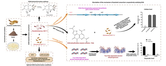
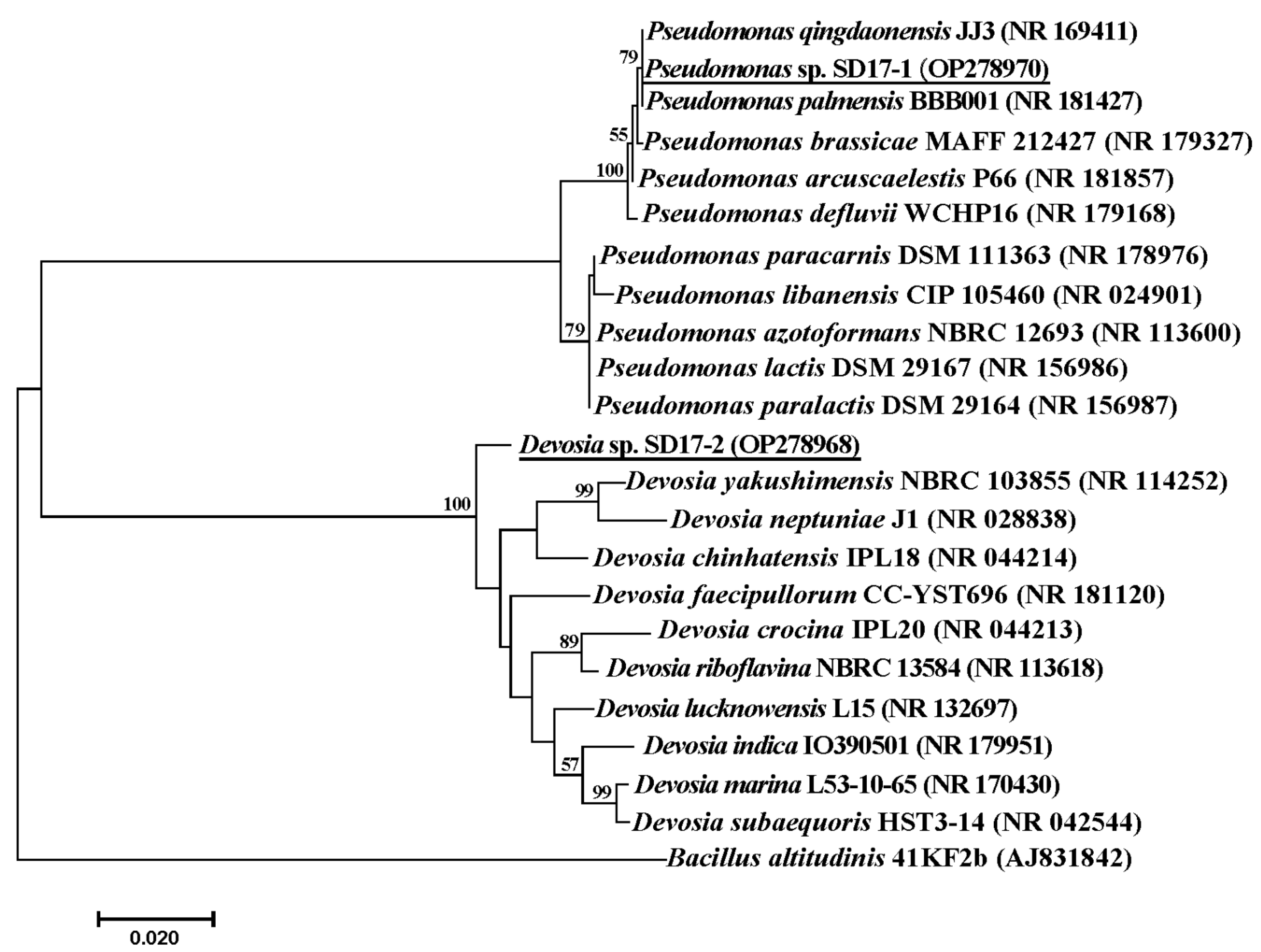
2.1. Isolation and Identification of the DON-Transforming Bacterial Community

Employing the enrichment culture technique, a fecal insect sample from Shandong Province, China, was enriched in MMF medium and demonstrated a remarkable ability to transform DON after four consecutive subcultures. Intriguingly, none of the individual colonies picked from TSA plate during the isolation procedure displayed DON transformation activity, necessitating the use of a mixed culture of pairwise colony combinations to obtain a microbial consortium with the desired ability. Consequently, the mixed community SD containing the two strains SD17-1 and SD17-2 was acquired. The 16S rRNA gene sequence of SD17-1 (Accession number: OP278970) displayed 98.0–100% similarity with those of the closely related type strains of the genus Pseudomonas, sharing the highest similarity of 100% with those of both P. palmensis BBB001T and P. qingdaonensis JJ3T. Additionally, the 16S rRNA gene sequence of SD17-2 (Accession number: OP278968) exhibited 94.8–98.0% similarity with those of the closely related type strains of the genus Devosia, with the highest similarity of 98.0% with that of D. lucknowensis L15T. Phylogenetic tree analysis, shown as Figure 1, revealed that SD17-1 formed a clade with P. palmensis BBB001T and P. qingdaonensis JJ3T, whereas SD17-2 constituted a taxon that was independent of other known species. Regarding the genomic ANIm (average nucleotide identity calculation based on MUMmer) analysis result, Devosia sp. SD17-1 showed ANIm values ranging from 83.52% to 84.52% when compared to the most related top 25 type strains in the genus Devosia, as shown in Table S1. An ANIm value below 95% indicated classification as a novel species, so SD17-1 appeared to be a novel Devosia species. Pseudomonas sp. SD17-2 showed ANIm values from 85.59% to 99.33% when compared to the top 25 Pseudomonas type strains. However, its ANIm values of 99.29% and 99.33% with P. palmensis and P. qingdaoensis, respectively, as shown in Table S2, exceeded the 95% threshold for species classification. Therefore, the exact species of SD17-2 could not be determined. These conclusions were consistent with the phylogenetic analysis based on 16S rRNA sequences.

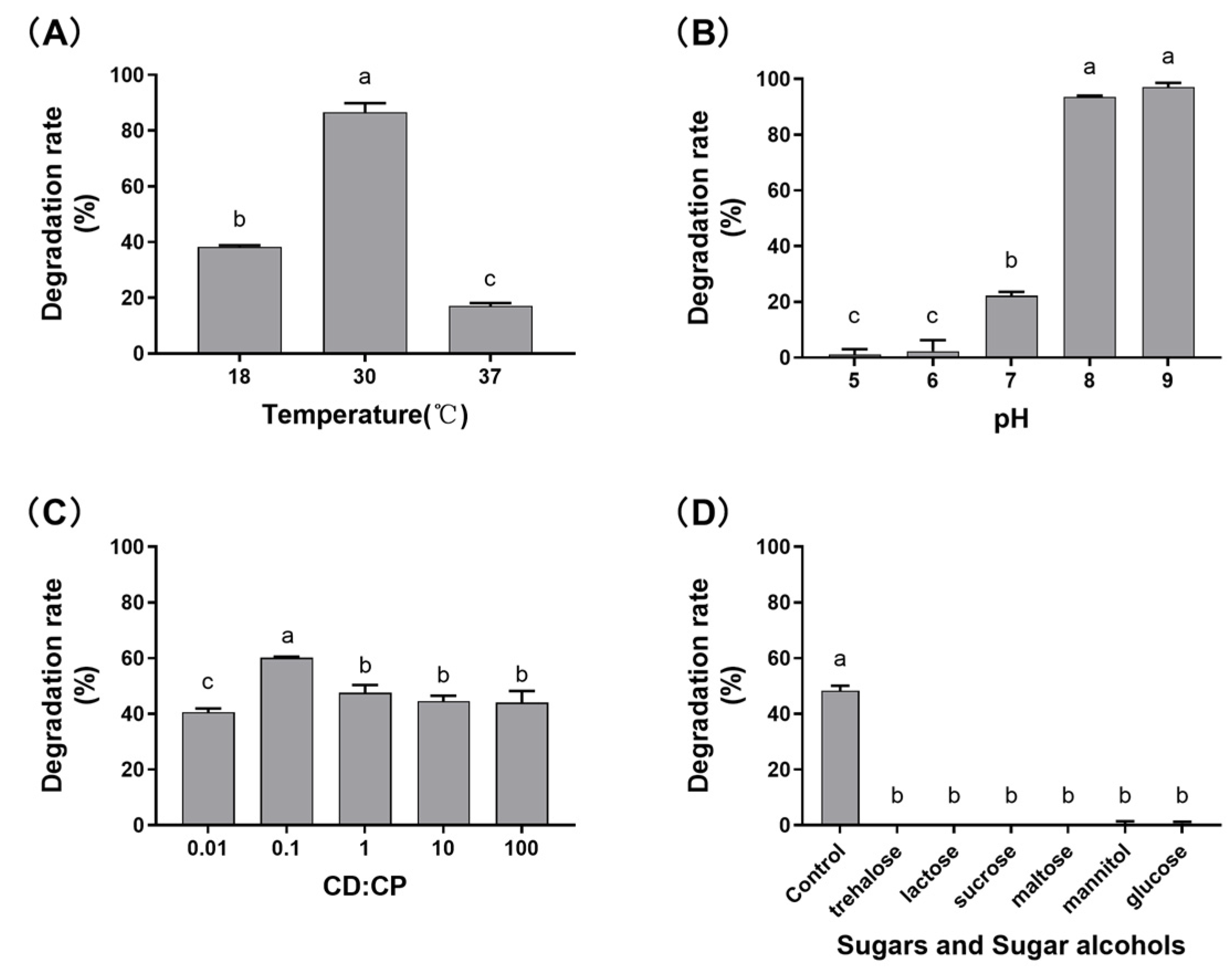
2.2. Characterization of the Effects of Different Factors on DON Transformation Activity

The impacts of temperature, initial pH, inoculation ratio of two isolates, as well as sugars and sugar alcohols on DON transformation activity of the mixed culture SD in MMF medium were investigated. The results from Figure 2A indicated that the mixed culture SD exhibited the highest transformation rate at 30 °C, with a rate of 86.6 ± 3.3%. Optimal pH values for DON transformation activity were found to be 8 and 9; however, acidic conditions (pH 5 and 6) resulted in virtually no activity (Figure 2B). Under a CFU ratios of Devosia to Pseudomonas (0.1), the mixed culture SD exhibited the highest DON transformation activity (Figure 2C). Furthermore, the addition of various sugars and sugar alcohols, such as trehalose, lactose, sucrose, maltose, mannitol, and glucose, strongly inhibited the DON transformation activity (Figure 2D).

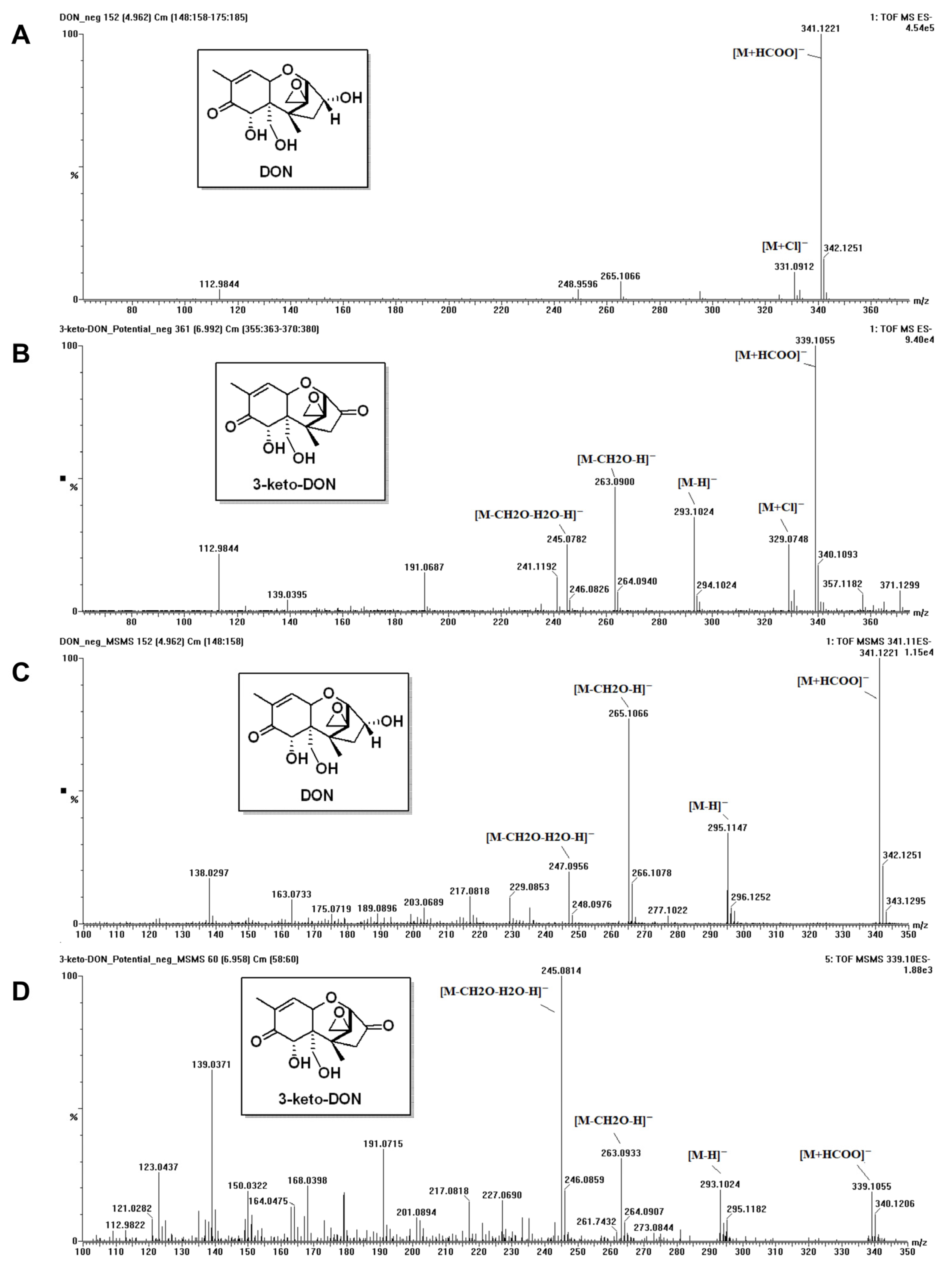
2.3. Determination of DON Metabolite’ Chemical Structure

To identify the DON’s metabolite generated by the microbial consortium SD, we performed UPLC-MS analysis on the DON standard and the supernatant that contained the metabolite. In the negative ion mode, on full-scan spectrum, DON afforded pseudomolecular ions at m/z 341.1221, which belonged to the formate adduct ([M+HCOO], calc. 341.1242), and 331.0912 corresponded to the chlorine anion adduct ([M+Cl], calc. 331.0954) due to its distinctive isotopic pattern. For the metabolite, similar adducts were observed at m/z 339.1055 ([M+HCOO], calc. 339.1085) and 329.0785 ([M+Cl], calc. 329.0797) on its full-scan spectrum (Figure 3A,B). The exact differences in masses of formate adducts and chlorine adducts between DON and its metabolite were both two Da, which indicated that the metabolite was the oxidative product of DON with the loss of two hydrogen atoms. For further identification, an MS/MS experiment was performed to analyze the fragmentation of the formate adducts of DON and its metabolite (C, D in Figure 3). The pseudomolecular ion of DON’s formate adduct (m/z 341.1221) yielded characteristic MS2 spectrum with product ions at m/z 295.1147 ([M–H]), 265.1066 ([M–CH2O–H]), and 247.0956 ([M–CH2O–H2O–H]). The main fragmented ions of formate adducts of DON and metabolites have been proposed and included in the Supplementary Materials (Figure S1 and Figure S2). In comparison, the fragmentation of m/z 339.1055 of the metabolite led to product ions at m/z 293.1024 ([M–H]), 263.0933 ([M–CH2O–H]), and 245.0814 ([M–CH2O–H2O–H]). The mass differences of the characteristic product ions between DON and its metabolite were all about two Da, in accordance with the atomic mass of two hydrogens. Both MS1 and MS2 results indicated that a hydroxyl group on DON was transformed into a carboxyl group, which was in line with that in the literature [16]. Due to the stability of the possible products (15-aldehyde-DON, 3-keto-DON), the transformation mostly occurred on the 3-position of DON molecule to the 3-keto-DON. Moreover, in our MS operating condition, the metabolite of DON could readily afford a characteristic ions profile in the full-scan spectrum similar to that in its MS/MS experiment due to the in-source fragmentation.

2.4. Elucidation the Underlying Mechanisms of DON Biotransformation by the Microbial Consortium SD

2.4.1. Sequencing, Assembly, and Annotation of the Genomes of Strains SD17-1 and SD17-2

To elucidate the potential mechanisms underlying the ability of the mixed culture SD, consisting of Pseudomonas and Devosia, to oxidize DON, we performed genome sequencing, assembly, and annotation of the two strains, providing a biological information basis for subsequent research. As shown as Table 1, the genome of Pseudomonas sp. SD17-1 had a size of 5.7 Mb with a GC content of 64.2% and contained 5170 protein-coding sequences, whereas Devosia sp. SD17-2 had a size of 4.2 Mb with a GC content of 62.1% and contained 4004 protein-coding sequences.

2.4.2. Homology Search for PQQ Biosynthetic Gene Cluster in Pseudomonas sp. 17-1 and PQQ-Dependent ADH Genes in Devosia sp. 17-2

Based on the BLASTp search results, an intact PQQ biosynthesis gene cluster was identified in the genome of the Pseudomonoas sp. SD17-1. The arrangement of the PQQ synthesis gene cluster in the genomic sequence of Pseudomonas sp. SD17-1, which contained seven open reading frames, pqqF (GenBank accession number of the corresponding encoding protein: WEJ21288.1), pqqA (WEJ24285.1 and WEJ24188.1), pqqB (WEJ21289.1), pqqC (WEJ21290.1), pqqD (WEJ21291.1), pqqE (WEJ21292.1), and pqqG (WEJ21293.1), was depicted in Figure 4. In particular, the gene pqqA encoding the PQQ precursor peptide contained two copies, pqqA1 and pqqA2, and these multiple copies of the pqqA gene have been shown to play an important role in PQQ synthesis and to contribute differently in different growth environments [40]. Like other Pseudomonas, the two S9 family peptidase encoding genes pqqF and pqqG in the genome of SD17-1 were located on either side of the typical pqqA-E biosynthetic operon. The results suggested that strain 17-1 had the potential to produce PQQ and might play a crucial role as a PQQ provider in the DON oxidation process carried out by the microbial consortium SD. On the other hand, several essential enzymes involved in PQQ biosynthesis, namely, PqqA, PqqB, PqqC, PqqD, and PqqE, were not identified in the genome of Devosia sp. SD17-2, suggesting that this strain lacked the ability to produce PQQ.
To identify potential genes that encoded PQQ-dependent DON dehydrogenase, a BLASTp search was performed using DepA as the query sequence against the genomes of Pseudomonas sp. SD17-1 and Devosia sp. SD17-2. Several PQQ-dependent ADHs were found in the genomic sequence of Pseudomonas strain SD17-1, as presented in Table S3. However, due to their relatively low similarity and coverage with DepA, coupled with the absence of DON oxidizing capability in Pseudomonas cultures alone, further cloning, expression, and DON oxidation activity assays for these genes were deemed unnecessary. In contrast, the genomic sequence of Devosia sp. SD17-2 contained four candidate PQQ-dependent ADH genes, namely, 17dh1 (GenBank accession number of the corresponding encoding protein: WEJ32717.1), 17dh2 (WEJ32716.1), 17dh3 (WEJ31544.1), and 17dh4 (WEJ31519.1), with sizes of 1770, 1770, 1761, and 1725 bp, respectively. The corresponding proenzymes encoded by these regions comprised 589, 589, 586, and 574 amino acids, respectively. SignalP 6.0 analysis revealed that the N-terminal regions of these four enzymes contained signal peptides consisting of 1–24, 1–24, 1–25, and 1–25 amino acids, respectively. Additionally, all four enzymes belonged to the quinoprotein alcohol dehydrogenase superfamily and shared amino acid sequence homology with the DepA, with similarities of 36.9%, 36.7%, 35.9%, and 34.2%, respectively.

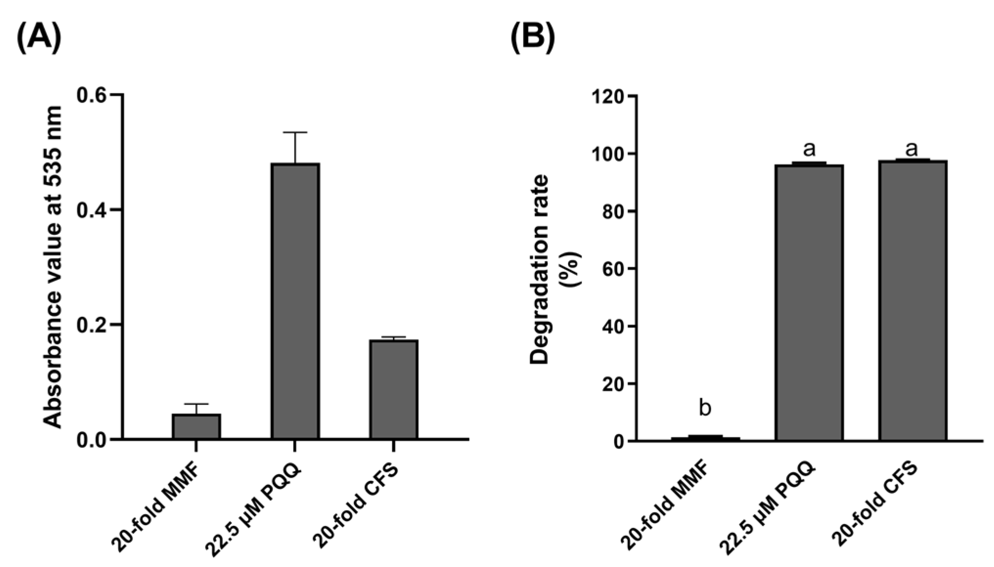
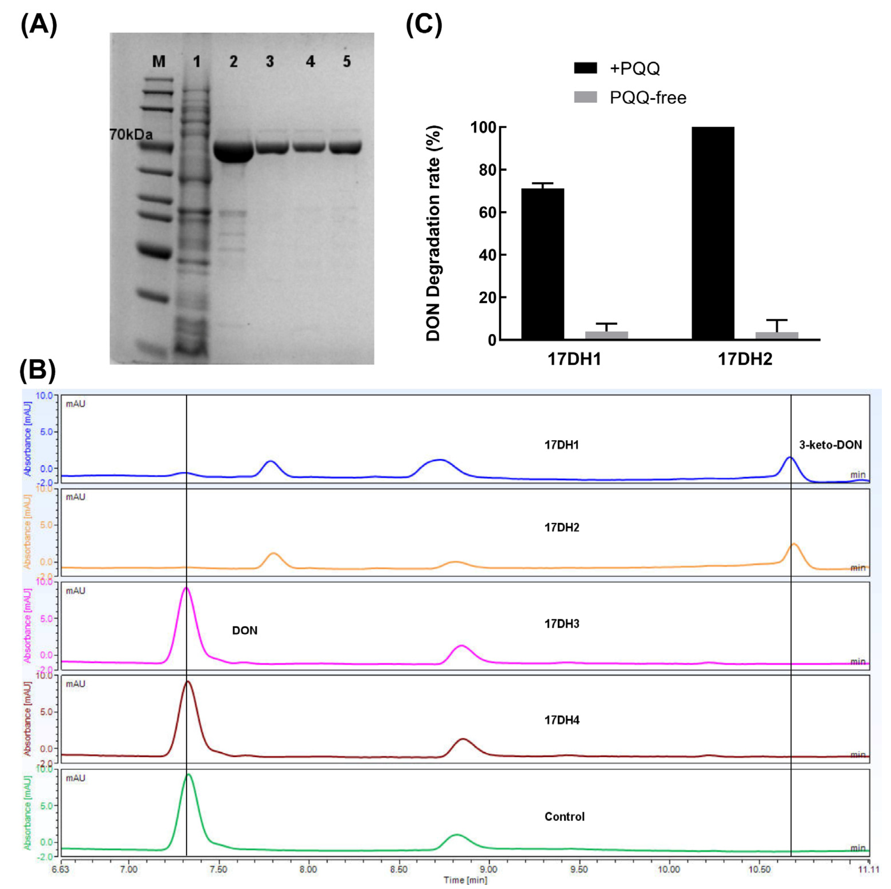
2.4.3. Biochemical Evidence for Pesudomonas and Devosia as Producer of PQQ and DON Dehydrogenase

The PQQ content in the 20-fold concentrated cell-free supernatant of Pseudomonas’ MMF culture was determined to be approximately 8.8 μmol/L using the NBT method (Figure 5A), demonstrating Pseudomonas’ ability to produce PQQ at the cellular level. When the supernatant was added to Devosia cell pellets suspension, the Devosia cells showed DON oxidative activity similar to Devosia cells treated with 22.5 μM PQQ standard; whereas Devosia cells supplied with 20-fold concentrated MMF medium as a control did not exhibit such activity (Figure 5B).
The four recombinant ADHs were heterologously expressed and then analyzed by Coomassie brilliant blue-stained SDS-PAGE. As shown in Figure 6, the approximate molecular weights of purified recombinant 17DH1, 17DH 2, 17DH 3, and 17DH 4 determined by SDS-PAGE were consistent with their respective theoretical molecular weights of 63.4, 63.4, 62.1, and 61.4 kDa. The UPLC analysis demonstrated the ability of recombinant 17DH1 and 17DH2 to transform DON, as evidenced by the complete disappearance of the DON peak and the appearance of the 3-keto-DON peak after incubation with PQQ and Ca2+, as well as an electron acceptor, PMS. Conversely, 17DH3 and 17DH4 did not exhibit any such activity (Figure 6). Furthermore, according to the findings of the PQQ-dependency assay, it was observed that 17DH1 and 17DH2 exhibited remarkable DON oxidation activity only in the presence of PQQ. However, in the absence of PQQ, their activity was almost disappeared, indicating that these two enzymes were strictly dependent on PQQ and functioned as DON dehydrogenases. Two PQQ-dependent DON dehydrogenases with DON oxidation activities, 17DH1 and 17DH2, exhibited amino acid sequence similarities of 36.9% and 36.7%, respectively, with DepA, whereas they shared 84.8% similarity between each other. This demonstrated the sequence diversity of PQQ-dependent ADHs with DON oxidation activity. Such sequence diversity may have accumulated through evolutionary pressures for the enzymes to adapt to the recognition and action of different substrates.
Conclusively, a cooperative mechanism can be proposed for the biotransformation of DON between Pseudomonas and Devosia bacteria based on these experimental findings. Specifically, Pseudomonas, which produces PQQ as a cofactor for Devosia’s DON dehydrogenase, acts as the PQQ supplier. Devosia, equipped with DON dehydrogenase, acts as the effector strain that carries out the oxidation of DON.

3. Discussion

Microbial pure cultures and consortia capable of transforming DON have been isolated from various environmental sources, including soil [11,13,14,15,18,20,25,26,27,28,30,41], aquatic systems [16,17], animal intestines [23,24,42], and wheat [20,21]. This suggests that microorganisms able to metabolize DON are widespread in nature. In the current study, we isolated for the first time a DON-transforming microbial consortium from yellow mealworm feces, further confirming the ubiquity of DON-transforming microorganisms. Furthermore, a novel DON-transforming bacterial strain D3_3, recently isolated from a single sample of yellow mealworm feces [39], together with the DON-transforming microbial consortia SD isolated from three additional mealworm fecal samples in the current study, suggest that yellow mealworm feces represent an unconventional yet highly efficient source for isolating mycotoxin-transforming microbes. Long-term feeding of grain feed contaminated with mycotoxins may promote the domestication of microorganisms with DON transformation capabilities in the mealworm gut, thus providing a rational explanation for the efficiency of mealworm feces as a source for screening DON transformation microbial strains.
Microbial communities with the same DON transformation mean may have different microbial compositions and vice versa. On one hand, the microbial consortia DX100 and PGC3 both have the ability to de-epoxide DON, though they differ in composition. DX100 consists mainly of Stenotrophomonas (80.03%), followed by Blautia (11.19%) and unknown microbes (3.38%), whereas PGC3 consists of 10 different genera of microbes, among which Desulfitobacterium may be the primary contributor to DON de-epoxidation [26,27]. On the other hand, a microbial consortium consisting of Pseudomonas sp. B6-24 and Devosia sp. A6-243 [30], which has a similar genus-level composition to the microbial community SD in this study, can transform DON into 3-epi-DON. In contrast, the microbial community SD in this study can oxidize DON to 3-keto-DON. Furthermore, we tried to analyze whether the two members of the microbial community SD with DON oxidation in this study, Pseudomonas sp. SD17-1 and Devosia sp. SD17-2, differ from Pseudomonas sp. B6-24 and Devosia sp. A6-243 at the microbial species level. Regrettably, as the 16S rRNA sequences of strains B6-24 and A6-243 have no available data in the NCBI database and related published articles, it is impossible to determine the phylogenetic relation between the Pseudomonas and Devosia bacteria identified in this study and those identified in Gao’s study [30]. Additionally, based on the ANIb analysis, Pseudomonas sp. SD17-1 shares 99.05% similarity with the two validly published type strains P. palmensis BBB001T and P. qingdaonensis JJ3T, exceeding the 95% threshold for species classification. However, the determination of its precise classification status requires further study using the polyphasic taxonomy approach. Devosia sp. SD17-2 exhibits the highest similarity of 77.82% to D. elaeis S37T, suggesting it may represent a novel Devosia species.
The mixed culture that consisted of Pseudomonas sp. SD17-1 and Devosia sp. SD17-2 in this study, as well as the reported microbial consortia comprising of Devosia sp. A8 and Paracoccus yeei sp. A9 [29], could both oxidize DON to the final product 3-keto-DON. However, the two microbial communities differ in their DON catalytic mechanisms. Strain A8 can oxidize DON to 3-keto-DON when cultured alone, but this capability is enhanced when it is co-cultured with electricigens such as P. yeei A9 or when NADPH and riboflavin are added. Researchers believed that Devosia sp. A8might have an NADPH-dependent aldo-keto reductase similar to AKR18A1, which catalyzes the conversion of DON to 3-keto-DON via oxidation [29]. On the other hand, Devosia sp. SD17-2 has two PQQ-dependent DON dehydrogenases that have the same catalytic function as DepA, but not AKR18A1-like enzymes, which are responsible for the oxidation. It is noteworthy that neither of the two microbial communities could continue to stereoselectively reduce 3-keto-DON to 3-epi-DON. This is because the absence of 3-keto-DON aldo-keto reductase similar to DepB was the reason why 3-epi-DON was not detected in the DON conversion products of the two microbial communities.
Regarding the toxicity of 3-keto-DON, some research concludes that 3-keto-DON shows reduced toxicity compared to DON when tested on various cell lines and animal models [15,39,43]. However, in plant-based models, 3-keto-DON retains its ability to function as a potent plant toxin and has the power to inhibit plant growth [39,44], suggesting that while 3-keto-DON may be less harmful to animals, it still poses a significant threat to plants. Collectively, the microbial consortium SD harboring the capacity to enzymatically convert DON into 3-keto-DON exhibits potential for deployment in the reduction in DON in animal feed. Nevertheless, it would not be wise to pursue the development of transgenic crops via the incorporation of DON oxidizing enzyme genes from SD for resistance.
PQQ-dependent ADHs are a class of enzyme mainly present in Gram-negative bacteria, with a small number also found in eukaryotes and archaea, which catalyze the oxidation of alcohols and aldose sugars [45]. In this study, two PQQ-dependent DON dehydrogenases, 17DH1 and 17DH2, were identified from Devosia sp. SD17-2. The amino acid sequence similarities between 17DH1/17DH2 and DepA, a known PQQ-dependent DON dehydrogenase, were found to be 36.9% and 36.7%, respectively, indicating significant sequence diversity among PQQ-dependent ADHs with DON oxidation activities. In additional, considering the finding that the presence of sugars and sugar alcohols strongly inhibited the DON oxidation activity of the microbial community SD, and sugars and sugar alcohols may be the natural substrates of the PQQ-dependent alcohol dehydrogenase rather than DON, which is chemically a sesquiterpene derivative. Therefore, we deduce that the reason for the strong inhibitory effect of sugar alcohols on DON oxidation activity is the high concentration of sugar alcohols effectively inhibits the DON oxidation activities of the two PQQ-dependent ADHs in the effective strain SD17-2 within the microbial community through substrate inhibition effect. Although 17DH3 and 17DH4 also showed relatively high amino acid similarities of 35.9% and 34.2% with DepA, respectively, no detectable DON oxidation activity was observed for these enzymes. This suggests that factors in addition to amino acid sequence similarity may determine DON oxidation activity in PQQ-dependent ADHs. Further studies are needed to elucidate the mechanisms underlying these observations and the role of other factors in conferring DON catalytic activity.
Although the Pseudomonas sp. SD17-1 and Devosia sp. SD17-2 strains cultured together that could synergistically oxidize DON were isolated from the yellow mealworm gut, it is still unclear whether they are indigenous or transient bacteria of the yellow mealworm gut. Additional research is necessary to determine the potential pathogenicity of Pseudomonas strain SD17-1 towards yellow mealworms and to assess the colonization capability of both strains in the gut of the yellow mealworm. Successful colonization would pave the way for developing a novel biological agent for detoxifying DON. Using the colonized mealworms as a biological agent could offer a simple, cost-effective, sustainable and nutritious solution for detoxifying DON-contaminated feed. These yellow mealworms colonized with DON-degrading bacteria would be likely to exhibit better DON tolerance and thus could be fed with highly DON contaminated feed, converting low-quality mycotoxin-contaminated plant protein into high-quality animal protein, thereby realizing an improvement in the utilization efficiency of mycotoxin-contaminated feed. In addition, these two PPQ-dependent alcohol dehydrogenases could potentially be modified through protein engineering to improve their activity, specificity, stability, and other properties and further developed into detoxified enzyme agents for application in feed and food industries.

4. Conclusions

In the present study, a microbial consortium SD with DON-transforming ability was isolated from the larval frass of yellow mealworm. The consortium consisted of two members, Pseudomonas sp. SD17-1 and Devosia sp. SD17-2, which could effectively oxidize 50 μg/mL DON into the low-toxicity metabolite 3-keto-DON within 72 h under optimal conditions. The isolation of SD from yellow mealworm frass suggested that the frass microbiome may be a source of mycotoxin-transforming microbes. The mechanism by which the bacterial consortium SD co-cultivates the oxidation of DON was elucidated based on genomic information analysis and biochemical testing. Specifically, SD17-1, a PQQ producer, provided the cofactor PQQ to SD17-2, which lacked the ability to produce PQQ but produced two PQQ-dependent DON dehydrogenases. Together, these two bacteria collaborated to achieve the oxidation of DON. Understanding the mechanism of DON oxidation by the microbial community allows for the development of strategies to optimize the biodetoxification process and reduce DON contamination.

5. Materials and Methods

5.1. Reagent and Culture Medium

A sterile stock solution of 10,000 μg/mL concentration was produced from DON standard procured from Pribolab of Qingdao, China. The solution was then preserved at −20 °C until its use. PQQ, phenazine methosulfate (PMS), and nitroblue tetrazolium (NBT) were purchased from Yuanye Bio-Technology (Shanghai, China). Methanol and acetonitrile of chromatography grade were purchased from Shanghai Titan Scientific Co., Ltd. (Shanghai, China). All other reagents and chemicals unless otherwise stated were from Sangon Biotech (Shanghai, China).
MMF medium was used for the enrichment and isolation of DON-transforming microorganism. It contained the following ingredients per liter of distilled water: 4.03 g of Na2HPO4·12H2O, 1.0 g of KH2PO4, 0.59 g of KNO3, 0.5 g of (NH4)2SO4, 0.5 g of MgSO4·7H2O, 0.019 g of CaCl2, 200 μL of trace element solution [46], and 100 mL of aseptic yellow mealworm feces supernatant. To prepare the aseptic insect feces supernatant, 10 g of insect feces were dissolved in 200 mL of distilled water, centrifuged, and filtered through a sterile filter to obtain a 5% (w/v) sterile solution. The other components of the MMF medium were subjected to moist heat sterilization, after which the sterile supernatant was added to the medium.

5.2. Screening and Identification of the DON-Transforming Bacterial Strains

Microbial enrichment and isolation technique was used to screen for DON-transforming microorganisms from three fecal samples of yellow mealworm collected from different regions (Beijing city, Shandong province and Fujian province) in China. For the enrichment procedure, 3 g of each fecal sample was added to 45 mL of sterile water. This mixture was vortexed for 30 min followed by 5 min of settling time. Then, 500 μL of the slurry was inoculated into 5 mL of MMF medium containing 50 μg/mL DON for cultivation. Unless otherwise stated, the cultivation conditions were at 30 °C temperature, 220 rpm rotation speed, and 3 d cultivation time. In total, 500 μL of culture was taken for sub-culturing in fresh MMF + 50 μg/mL DON medium. This process was repeated four times to obtain a stable mixed culture with DON transformation activity. For the isolation procedure, the positive mixed culture determined by UPLC analysis was diluted and spread on TSA plates, and single colonies were randomly selected for their DON transformation activities. Unfortunately, all single colonies did not show DON transformation activities. Therefore, the pure cultures were further paired and co-cultured to assess their transformation activity.
Genomic DNA of the two isolates was obtained using a bacterial genomic DNA kit (Sangon Biotech, Shanghai, China) following the manufacturer’s protocol. The 16S rRNA sequence was amplified with universal primers 27F (5′-AGAGTTTGATCCTGGCTCAG-3′) and 1492R (5′-GTTACCTTGTTACGACT-3′) on a Biometra GmbH Thermocycler (Göttingen, Germany). The PCR product was sequenced by Bomaide Gene Technology Co., Ltd. (Beijing, China). The resulting 16S rRNA sequence was analyzed by BLAST search in the NCBI database (http://www.ncbi.nlm.nih.gov/, accessed on 21 April 2023), and a phylogenetic tree was constructed by MEGA 7.0 (MEGA, Auckland, New Zealand) using the Maximum Likelihood method with 1000 bootstrap replicates.
To determine the taxonomic status of the two isolates at the species level, we conducted a genome-based taxonomic analysis using average nucleotide identity (ANI) calculations. The genomes of isolates SD17-1 and SD17-2 obtained from Section 5.5.1 of the materials and methods were individually uploaded to the JSpeciesWS web service at https://jspecies.ribohost.com/jspeciesws/#home, accessed on 30 July 2023. Through tetra correlation search (TCS), the top 25 most closely related congeneric genome sequences were retrieved from the genomic database for each query genome. Average nucleotide identity calculations based on MUMmer (ANIm) were then performed between the query genomes and the retrieved genomes. The resulting ANIm values were used to determine the taxonomic status of isolates SD17-1 and SD17-2 according to established phylogenetic thresholds on the JSpeciesWS website.

5.3. Analysis of DON and Its Metabolite

5.3.1. Ultra-Performance Liquid Chromatography (UPLC) Analysis for DON

Residual DON analysis was performed by a Thermo UltiMate3000 UPLC-DAD system (Waltham, MA, USA). Briefly, 2 μL of sample was injected onto an ACQUITY UPLC® BEH C18 column (2.1 × 100 mm, 1.7 μm; Waters), and DON was detected using the following parameters: 220 nm wavelength, 40 °C temperature, 0.2 min/L flow rate, and a elution gradient program: 0–6 min, gradient 5–25% acetonitrile in water (v/v); 6–12 min, isocratic 25% acetonitrile; 12–13 min, gradient 25–5% acetonitrile; 13–18 min, and isocratic 5% acetonitrile. All analyses were performed in triplicate. The DON transformation rate was calculated utilizing the subsequent equation:
Dr = (1 − Dt/Dc) × 100%
In this equation, Dr represents the DON conversion rate, and Dt and Dc represent the integrated area of the absorption peak of residual DON at a wavelength of 220 nm for the treated group and the control group, respectively.

5.3.2. MS (Mass Spectrometry) Analysis of DON Metabolite

The analysis of DON and its metabolite generated by the microbial community SD was conducted utilizing an Acquity UPLC combined with an Xevo G2-S quadrupole time-of-flight (Q-TOF) mass spectrometer that was furnished with an electrospray ionization source (Waters, Medford, MA, USA). The liquid chromatographic separation was executed on an ACQUITY UPLC BEH C18 column (100 mm × 2.1 mm, 1.7 μm) at 40 °C. The injection volume was 1 μL. The mobile phase consisted of two solutions: A, an aqueous solution containing 5 mmol/L ammonium formate and 0.1% (v/v) formic acid, and B, methanol; the flow rate was 0.2 mL/min, and the gradient elution program was 10% B, 0–1 min; 10–90% B, 1–19 min; 90% B, 19–24 min; 90–10% B 24–25 min; and 10% B, 25–30 min. The Q-TOF-MS was performed in the negative mode under following the optimum parameters: capillary voltage, 2.0 kV; desolvation gas (N2), 800 L/h; desolvation temperature, 450 °C; source temperature, 120 °C; and cone gas (N2), 50 L/h. Mass spectra were acquired at full-scan mode with a m/z range of 100–800. Argon was used as the collision gas and the collision energy range was 10–40 eV. Data were analyzed using MassLynx version 4.1 (Waters, Milford, MA, USA).

5.4. Effects of Various Factors on DON Transformation Rate

In a transformation system, 2 mL MMF medium containing 50 μg/mL DON, the effects of different temperatures (18 °C, 30 °C, and 37 °C), various initial pH values (5.0, 6.0, 7.0, 8.0, and 9.0), the addition of different types of sugars and sugar alcohols (2% w/v of D-trehalose, lactose, sucrose, maltose, D-mannitol, and glucose), and varying CFU ratios of the dual-member microbial consortium inoculum (Devosia: Pseudomonas = 1:100, 1:10, 1:1, 10:1, and 100:1) on the DON transformation activity of the microbial consortium were investigated. Residual DON and DON transformation rates were measured and calculated, respectively, according to the Materials and Methods described in Section 5.3.1. The experiments were conducted in triplicate, and the presented transformation rate values represented the mean of three replicates, with the error bars indicating the standard deviations. To compare between multiple groups, an analysis of variance (ANOVA) was carried out, followed by pairwise comparisons using Tukey’s test (p < 0.05).

5.5. Elucidation of the Mechanism of DON Degradation by Microbial Community SD

5.5.1. Genomic Sequencing and Analysis

In Biomaker Technology Inc., located in Beijing, China, the genomes of Devosia strain SD 17-2 and Pseudomonas strain SD 17-1 underwent sequencing through a combination of the Nanopore PromethION 48 system (Oxford, Oxon, UK) and the Illumina NovaSeq 6000 platform (San Diego, CA, USA). Further technical information on the sequencing, assembly, and annotation processes can be found in a previous report by Wang and Wang [47]. Subsequently, BLASTp was used with an E value of 10−5 by TBtools version 1.098769 [48] to search for candidate DON-oxidizing encoding genes in the genomic sequences of Devosia strain SD17-2 using DepA (known PQQ-dependent DON dehydrogenase, GenBank accession: KFL25551.1) from D. mutans 17-2-E-8 as a query sequence. Similarly, the known PQQ biosynthesis enzymes PqqA, PqqB, PqqC, PqqD, and PqqE from Pseudomonas were used as queries to search for an intact PQQ biosynthesis gene cluster in the genomes of Pseudomonas strain SD17-1, with the same method and parameter set applied as for the DON-oxidizing enzyme search. Diagram of PQQ biosynthesis gene cluster arrangement was drawn by IBS 2.0 [49].

5.5.2. Identification of Two PQQ-Dependent DON Dehydrogenases from Devosia sp. SD17-2

Using Q5 High-Fidelity DNA Polymerase (NEB, America), we amplified four possible genes for DON oxidation and linearized pET28a from the genomic DNA of strain SD17-2 and expression vector pET28a, respectively. The PCR conditions and primers used are listed in Table S4. The resulting amplicons for the candidate gene and the linearized pET28a were individually ligated using the T5 exonuclease-dependent DNA assembly (TEDA) method [50] and then transformed into E. coli BL21(DE3) for expression as N-terminal His6-fusion protein. The expression and purification of recombinant proteins was performed using a previously reported method [51]. Subsequently, the purified proteins were concentrated, analyzed by Coomassie blue-stained SDS-PAGE, and stored at −80 °C until required.
To test the DON transformation activity of the four purified recombinant enzymes, reaction mixtures contained 50 μg/mL of DON, 40 μM of PMS, 1 mM of CaCl2, 100 μM of PQQ, and 50 nM of purified recombinant enzyme in 25 mM Tris-HCl buffer (pH 8.0). These samples, including a control without enzyme, were incubated for 2 h at 30 °C and then subjected to UPLC analysis for DON.

5.5.3. NBT Assay for PQQ Produced by Pseudomonas sp. SD17-1

The cell-free supernatant of a 10 mL culture of Pseudomonas sp. strain SD7-1 grown in MMF medium for 3 d was prepared by centrifugation and filtration. The supernatant was freeze-dried to a powder, reconstituted in 500 μL of sterile water, and centrifuged. The resulting supernatant (20-fold CFS) was analyzed for PQQ content using NBT assay [30]. Briefly, 45 μL of the concentrated sample was added to a reaction mixture consisting of 25 μL of 3.6 mM NBT solution, 40 μL of 20 mM phosphate-buffered saline (pH 7.0) and, 90 μL of 20 mM glycine–potassium hydroxide buffer (pH 10.0) and incubated in the dark at 30 °C for 1 h. The absorbance at 535 nm was then measured. Positive and negative controls were 22.5 μM PQQ standard and 20-fold concentrated MMF medium, respectively.

5.5.4. Activation of DON Oxidation Activity of Devosia sp. SD17-2 by PQQ Supplement

An aliquot of 100 μL of the aforementioned 20-fold CFS was added to a 400 μL reaction mixture containing a working concentration of 50 μg/mL DON, 1 mM CaCl2, 6.1 × 10⁷ CFU/mL of Devosia sp. SD17-2, and 25 mM Tris-HCl buffer (pH 8.0). Reaction mixture supplemented with 20-fold MMF served as negative control, whereas that supplemented with 22.5 μM PQQ standard served as positive control. These mixtures were incubated at 30 °C for 48 h and then subjected to UPLC assay for DON.

Supplementary Materials

The following supporting information can be downloaded at: https://www.mdpi.com/article/10.3390/toxins15080492/s1, Table S1: The genomic ANIm analysis of Devosia sp. SD17-2 against the top 25 most closely related type strains in the genus of Devosia. Table S2: The genomic ANIm analysis of Pseudomonas sp. SD17-1 against the top 25 most closely related type strains in the genus of Pseudomonas. Table S3: Several putative PQQ-dependent dehydrogenases in Pseudomonas sp. SD17-1. Table S4: Primer sequence and PCR reaction program. Figure S1: The proposed product ions in the fragmentation of m/z 341.1221 ([M+HCOO]) for DON. Figure S2: The proposed product ions in the fragmentation of m/z 339.1055 ([M+HCOO]) for DON metabolite.

Author Contributions

Conceptualization, Y.W. and B.G.; methodology, Y.W. and D.Z.; formal analysis, D.Z. and W.Z.; investigation, Y.W.; resources, D.Z.; writing—original draft preparation, Y.W.; writing—review and editing, Y.W. and B.G.; supervision, S.W. and K.H.; project administration, B.G.; funding acquisition, Y.W. and B.G. All authors have read and agreed to the published version of the manuscript.

Funding

This research was funded by the National Natural Science Foundation of China (Grant No. 31972605) and the Key Research and Development Program of Ningxia Hui Autonomous Region, China (Grant No. 2022BBF03031).

Institutional Review Board Statement

Not applicable.

Informed Consent Statement

Not applicable.

Data Availability Statement

The complete genomic sequences of Pseudomonas sp. SD17-1 and Devosia sp. SD17-2 have been deposited in NCBI/GenBank. Each strain has its own unique BioProject number, GenBank accession number, and BioSample number: PRJNA875557, GCA_029201585.1, and SAMN30622627 for strain SD17-1 and PRJNA874206, GCA_029201565.1, and SAMN30543117 for strain SD17-2, respectively. The 16S rRNA genes of strain SD17-1 and SD17-2 have been deposited under GenBank accession numbers OP278970 and OP278968, respectively. Additionally, data supporting the findings of this work are available within the paper and its Supplementary Information.

Conflicts of Interest

The authors declare no conflict of interest.

References

  1. Mishra, S.; Srivastava, S.; Dewangan, J.; Divakar, A.; Kumar Rath, S. Global occurrence of deoxynivalenol in food commodities and exposure risk assessment in humans in the last decade: A survey. Crit. Rev. Food Sci. Nutr. 2020, 60, 1346–1374. [Google Scholar] [CrossRef]
  2. Tima, H.; Berkics, A.; Hannig, Z.; Ittzés, A.; Kecskésné Nagy, E.; Mohácsi-Farkas, C.; Kiskó, G. Deoxynivalenol in wheat, maize, wheat flour and pasta: Surveys in Hungary in 2008–2015. Food Addit. Contam. Part B Surveill. 2018, 11, 37–42. [Google Scholar] [CrossRef]
  3. Sobrova, P.; Adam, V.; Vasatkova, A.; Beklova, M.; Zeman, L.; Kizek, R. Deoxynivalenol and its toxicity. Interdiscip. Toxicol. 2010, 3, 94–99. [Google Scholar] [CrossRef]
  4. Liao, Y.; Peng, Z.; Chen, L.; Nüssler, A.K.; Liu, L.; Yang, W. Deoxynivalenol, gut microbiota and immunotoxicity: A potential approach? Food Chem. Toxicol. 2018, 112, 342–354. [Google Scholar] [CrossRef]
  5. Murata, H.; Yamaguchi, D.; Nagai, A.; Shimada, N. Reduction of deoxynivalenol contaminating corn silage by short-term ultraviolet irradiation: A pilot study. J. Vet. Med. Sci. 2011, 73, 1059–1060. [Google Scholar] [CrossRef] [Green Version]
  6. Bretz, M.; Beyer, M.; Cramer, B.; Knecht, A.; Humpf, H.-U. Thermal Degradation of the Fusarium Mycotoxin Deoxynivalenol. J. Agric. Food Chem. 2006, 54, 6445–6451. [Google Scholar] [CrossRef]
  7. Sabater-Vilar, M.; Malekinejad, H.; Selman, M.H.J.; van der Doelen, M.A.M.; Fink-Gremmels, J. In vitro assessment of adsorbents aiming to prevent deoxynivalenol and zearalenone mycotoxicoses. Mycopathologia 2007, 163, 81–90. [Google Scholar] [CrossRef] [Green Version]
  8. Zhang, Q.; Zhang, Y.; Liu, S.; Wu, Y.; Zhou, Q.; Zhang, Y.; Zheng, X.; Han, Y.; Xie, C.; Liu, N. Adsorption of deoxynivalenol by pillared montmorillonite. Food Chem. 2021, 343, 128391. [Google Scholar] [CrossRef]
  9. Wang, L.; Shao, H.; Luo, X.; Wang, R.; Li, Y.; Li, Y.; Luo, Y.; Chen, Z. Effect of Ozone Treatment on Deoxynivalenol and Wheat Quality. PLoS ONE 2016, 11, e0147613. [Google Scholar] [CrossRef]
  10. Hassan, Y.I.; He, J.W.; Perilla, N.; Tang, K.; Karlovsky, P.; Zhou, T. The enzymatic epimerization of deoxynivalenol by Devosia mutans proceeds through the formation of 3-keto-DON intermediate. Sci. Rep. 2017, 7, 6929. [Google Scholar] [CrossRef] [Green Version]
  11. Wang, G.; Wang, Y.; Ji, F.; Xu, L.; Yu, M.; Shi, J.; Xu, J. Biodegradation of deoxynivalenol and its derivatives by Devosia insulae A16. Food Chem. 2019, 276, 436–442. [Google Scholar] [CrossRef]
  12. Ikunaga, Y.; Sato, I.; Grond, S.; Numaziri, N.; Yoshida, S.; Yamaya, H.; Hiradate, S.; Hasegawa, M.; Toshima, H.; Koitabashi, M.; et al. Nocardioides sp. strain WSN05-2, isolated from a wheat field, degrades deoxynivalenol, producing the novel intermediate 3-epi-deoxynivalenol. Appl. Microbiol. Biotechnol. 2011, 89, 419–427. [Google Scholar] [CrossRef] [Green Version]
  13. Zhang, H.; Zhang, H.; Qin, X.; Wang, X.; Wang, Y.; Bin, Y.; Xie, X.; Zheng, F.; Luo, H. Biodegradation of Deoxynivalenol by Nocardioides sp. ZHH-013: 3-keto-Deoxynivalenol and 3-epi-Deoxynivalenol as Intermediate Products. Front. Microbiol. 2021, 12, 658421. [Google Scholar] [CrossRef]
  14. Wang, Y.; Zhang, H.H.; Zhao, C.; Han, Y.T.; Liu, Y.C.; Zhang, X.L. Isolation and characterization of a novel deoxynivalenol-transforming strain Paradevosia shaoguanensis DDB001 from wheat field soil. Lett. Appl. Microbiol. 2017, 65, 414–422. [Google Scholar] [CrossRef]
  15. Shima, J.; Takase, S.; Takahashi, Y.; Iwai, Y.; Fujimoto, H.; Yamazaki, M.; Ochi, K. Novel detoxification of the trichothecene mycotoxin deoxynivalenol by a soil bacterium isolated by enrichment culture. Appl. Environ. Microbiol. 1997, 63, 3825–3830. [Google Scholar] [CrossRef]
  16. Zhang, J.; Qin, X.; Guo, Y.; Zhang, Q.; Ma, Q.; Ji, C.; Zhao, L. Enzymatic degradation of deoxynivalenol by a novel bacterium, Pelagibacterium halotolerans ANSP101. Food Chem. Toxicol. 2020, 140, 111276. [Google Scholar] [CrossRef]
  17. Ito, M.; Sato, I.; Ishizaka, M.; Yoshida, S.; Koitabashi, M.; Yoshida, S.; Tsushima, S. Bacterial Cytochrome P450 System Catabolizing the Fusarium Toxin Deoxynivalenol. Appl. Environ. Microbiol. 2013, 79, 1619–1628. [Google Scholar] [CrossRef] [Green Version]
  18. He, W.-J.; Zhang, L.; Yi, S.-Y.; Tang, X.-L.; Yuan, Q.-S.; Guo, M.-W.; Wu, A.-B.; Qu, B.; Li, H.-P.; Liao, Y.-C. An aldo-keto reductase is responsible for Fusarium toxin-degrading activity in a soil Sphingomonas strain. Sci. Rep. 2017, 7, 9549. [Google Scholar] [CrossRef] [Green Version]
  19. Wang, S.; Hou, Q.; Guo, Q.-Q.; Zhang, J.; Sun, Y.; Wei, H.; Shen, L. Isolation and Characterization of a Deoxynivalenol-Degrading Bacterium Bacillus licheniformis YB9 with the Capability of Modulating Intestinal Microbial Flora of Mice. Toxins 2020, 12, 184. [Google Scholar] [CrossRef] [Green Version]
  20. Li, B.; Duan, J.; Ren, J.; Francis, F.; Li, G. Isolation and Characterization of Two New Deoxynivalenol-Degrading Strains, Bacillus sp. HN117 and Bacillus sp. N22. Toxins 2022, 14, 781. [Google Scholar] [CrossRef]
  21. Ito, M.; Sato, I.; Koitabashi, M.; Yoshida, S.; Imai, M.; Tsushima, S. A novel actinomycete derived from wheat heads degrades deoxynivalenol in the grain of wheat and barley affected by Fusarium head blight. Appl. Microbiol. Biotechnol. 2012, 96, 1059–1070. [Google Scholar] [CrossRef]
  22. Qu, R.; Jiang, C.; Wu, W.; Pang, B.; Lei, S.; Lian, Z.; Shao, D.; Jin, M.; Shi, J. Conversion of DON to 3-epi-DON in vitro and toxicity reduction of DON in vivo by Lactobacillus rhamnosus. Food Funct. 2019, 10, 2785–2796. [Google Scholar] [CrossRef]
  23. Fuchs, E.; Binder, E.M.; Heidler, D.; Krska, R. Structural characterization of metabolites after the microbial degradation of type A trichothecenes by the bacterial strain BBSH 797. Food Addit. Contam. 2002, 19, 379–386. [Google Scholar] [CrossRef]
  24. Gao, X.; Mu, P.; Zhu, X.; Chen, X.; Tang, S.; Wu, Y.; Miao, X.; Wang, X.; Wen, J.; Deng, Y. Dual Function of a Novel Bacterium, Slackia sp. D-G6: Detoxifying Deoxynivalenol and Producing the Natural Estrogen Analogue, Equol. Toxins 2020, 12, 85. [Google Scholar] [CrossRef] [Green Version]
  25. He, W.-J.; Shi, M.-M.; Yang, P.; Huang, T.; Yuan, Q.-S.; Yi, S.-Y.; Wu, A.-B.; Li, H.-P.; Gao, C.-B.; Zhang, J.-B.; et al. Novel Soil Bacterium Strain Desulfitobacterium sp. PGC-3-9 Detoxifies Trichothecene Mycotoxins in Wheat via De-Epoxidation under Aerobic and Anaerobic Conditions. Toxins 2020, 12, 363. [Google Scholar] [CrossRef]
  26. Ahad, R.; Zhou, T.; Lepp, D.; Pauls, K.P. Microbial detoxification of eleven food and feed contaminating trichothecene mycotoxins. BMC Biotechnol. 2017, 17, 30. [Google Scholar] [CrossRef] [Green Version]
  27. He, W.; Yuan, Q.-S.; Zhang, Y.; Guo, M.; Gong, A.; Zhang, J.; Wu, A.; Huang, T.; Qu, B.; Li, H.; et al. Aerobic De-Epoxydation of Trichothecene Mycotoxins by a Soil Bacterial Consortium Isolated Using In Situ Soil Enrichment. Toxins 2016, 8, 277. [Google Scholar] [CrossRef] [Green Version]
  28. Zhai, Y.; Zhong, L.; Gao, H.; Lu, Z.; Bie, X.; Zhao, H.; Zhang, C.; Lu, F. Detoxification of Deoxynivalenol by a Mixed Culture of Soil Bacteria With 3-epi-Deoxynivalenol as the Main Intermediate. Front Microbiol. 2019, 10, 2172. [Google Scholar] [CrossRef] [Green Version]
  29. Wang, Y.; Hu, J.; Dai, Y.; Wang, Y.; Shi, J.; Wang, G.; Xu, J.; De Saeger, S. Design and characterization of an artificial two-strain bacterial consortium for the efficient biodegradation of deoxynivalenol. Biol. Control. 2023, 179, 105172. [Google Scholar] [CrossRef]
  30. Gao, H.; Niu, J.; Yang, H.; Lu, Z.; Zhou, L.; Meng, F.; Lu, F.; Chen, M. Epimerization of Deoxynivalenol by the Devosia Strain A6-243 Assisted by Pyrroloquinoline Quinone. Toxins 2022, 14, 16. [Google Scholar] [CrossRef]
  31. Carere, J.; Hassan, Y.I.; Lepp, D.; Zhou, T. The enzymatic detoxification of the mycotoxin deoxynivalenol: Identification of DepA from the DON epimerization pathway. Microb. Biotechnol. 2018, 11, 1106–1111. [Google Scholar] [CrossRef]
  32. Carere, J.; Hassan, Y.I.; Lepp, D.; Zhou, T. The Identification of DepB: An Enzyme Responsible for the Final Detoxification Step in the Deoxynivalenol Epimerization Pathway in Devosia mutans 17-2-E-8. Front. Microbiol. 2018, 9, 1573. [Google Scholar] [CrossRef]
  33. Yang, S.; Wu, W.; Brandon, A.M.; Fan, H.; Receveur, J.P.; Li, Y.; Wang, Z.; Fan, R.; McClellan, R.L.; Gao, S.-H.; et al. Ubiquity of polystyrene digestion and biodegradation within yellow mealworms, larvae of Tenebrio molitor Linnaeus (Coleoptera: Tenebrionidae). Chemosphere 2018, 212, 262–271. [Google Scholar] [CrossRef] [Green Version]
  34. Brai, A.; Poggialini, F.; Trivisani, C.I.; Vagaggini, C.; Tarchi, F.; Francardi, V.; Dreassi, E. Efficient use of agricultural waste to naturally fortify Tenebrio molitor mealworms and evaluation of their nutraceutical properties. J. Insects Food Feed. 2022, 9, 599–610. [Google Scholar] [CrossRef]
  35. Brai, A.; Poggialini, F.; Vagaggini, C.; Pasqualini, C.; Simoni, S.; Francardi, V.; Dreassi, E. Tenebrio molitor as a Simple and Cheap Preclinical Pharmacokinetic and Toxicity Model. Int. J. Mol. Sci. 2023, 24, 2296. [Google Scholar] [CrossRef]
  36. Niermans, K.; Woyzichovski, J.; Kröncke, N.; Benning, R.; Maul, R. Feeding study for the mycotoxin zearalenone in yellow mealworm (Tenebrio molitor) larvae—Investigation of biological impact and metabolic conversion. Mycotoxin Res. 2019, 35, 231–242. [Google Scholar] [CrossRef] [Green Version]
  37. Dilshaan, D. Yellow Mealworm Larvae (Tenebrio molitor) Grown on Deoxynivalenol-Contaminated Wheat as a Feed Ingredient for Broiler Chickens. PhD Thesis, University of Saskatchewan, Saskatoon, SK, Canada, 2021. [Google Scholar]
  38. Zhao, D.; Xie, H.; Gao, L.; Zhang, J.; Li, Y.; Mao, G.; Zhang, H.; Wang, F.; Lam, S.S.; Song, A. Detoxication and bioconversion of aflatoxin B1 by yellow mealworms (Tenebrio molitor): A sustainable approach for valuable larval protein production from contaminated grain. Ecotoxicol. Environ. Saf. 2022, 242, 113935. [Google Scholar] [CrossRef]
  39. Wang, Y.; Zhao, D.; Zhang, W.; Wang, S.; Wu, Y.; Wang, S.; Yang, Y.; Guo, B. Four PQQ-Dependent Alcohol Dehydrogenases Responsible for the Oxidative Detoxification of Deoxynivalenol in a Novel Bacterium Ketogulonicigenium vulgare D3_3 Originated from the Feces of Tenebrio molitor Larvae. Toxins 2023, 15, 367. [Google Scholar] [CrossRef]
  40. Ge, X.; Wang, W.; Du, B.; Wang, J.; Xiong, X.; Zhang, W. Multiple pqqA genes respond differently to environment and one contributes dominantly to pyrroloquinoline quinone synthesis. J. Basic Microbiol. 2015, 55, 312–323. [Google Scholar] [CrossRef]
  41. He, W.-J.; Shi, M.-M.; Yang, P.; Huang, T.; Zhao, Y.; Wu, A.-B.; Dong, W.-B.; Li, H.-P.; Zhang, J.-B.; Liao, Y.-C. A quinone-dependent dehydrogenase and two NADPH-dependent aldo/keto reductases detoxify deoxynivalenol in wheat via epimerization in a Devosia strain. Food Chem. 2020, 321, 126703. [Google Scholar] [CrossRef]
  42. Yu, H.; Zhou, T.; Gong, J.; Young, C.; Su, X.; Li, X.-Z.; Zhu, H.; Tsao, R.; Yang, R. Isolation of deoxynivalenol-transforming bacteria from the chicken intestines using the approach of PCR-DGGE guided microbial selection. BMC Microbiol. 2010, 10, 182. [Google Scholar] [CrossRef] [Green Version]
  43. He, J.W. Detoxification of Deoxynivalenol by a Soil Bacterium Devosia mutans 17-2-E-8. PhD Thesis, University of Guelph, Guelph, ON, Canada, 2015. [Google Scholar]
  44. Li, X.-Z.; Hassan, Y.I.; Lepp, D.; Zhu, Y.; Zhou, T. 3-keto-DON, but Not 3-epi-DON, Retains the in Planta Toxicological Potential after the Enzymatic Biotransformation of Deoxynivalenol. Int. J. Mol. Sci. 2022, 23, 7230. [Google Scholar] [CrossRef]
  45. Sarmiento-Pavía, P.D.; Sosa-Torres, M.E. Bioinorganic insights of the PQQ-dependent alcohol dehydrogenases. J. Biol. Inorg. Chem. 2021, 26, 177–203. [Google Scholar] [CrossRef]
  46. Studier, F.W. Protein production by auto-induction in high-density shaking cultures. Protein Expr. Purif. 2005, 41, 207–234. [Google Scholar] [CrossRef]
  47. Wang, Y.; Wang, S. Complete Genome Sequence of Paradevosia shaoguanensis Type Strain J5-3, Obtained Using Nanopore and Illumina Sequencing Technologies. Microbiol. Resour. Announc. 2021, 10, e0009921. [Google Scholar] [CrossRef]
  48. Chen, C.; Chen, H.; Zhang, Y.; Thomas, H.R.; Frank, M.H.; He, Y.; Xia, R. TBtools: An Integrative Toolkit Developed for Interactive Analyses of Big Biological Data. Mol. Plant 2020, 13, 1194–1202. [Google Scholar] [CrossRef]
  49. Xie, Y.; Li, H.; Luo, X.; Li, H.; Gao, Q.; Zhang, L.; Teng, Y.; Zhao, Q.; Zuo, Z.; Ren, J. IBS 2.0: An upgraded illustrator for the visualization of biological sequences. Nucleic Acids Res. 2022, 50, W420–W426. [Google Scholar] [CrossRef]
  50. Xia, Y.; Li, K.; Li, J.; Wang, T.; Gu, L.; Xun, L. T5 exonuclease-dependent assembly offers a low-cost method for efficient cloning and site-directed mutagenesis. Nucleic Acids Res. 2019, 47, e15. [Google Scholar] [CrossRef] [Green Version]
  51. Yang, H.; Yan, R.; Li, Y.; Lu, Z.; Bie, X.; Zhao, H.; Lu, F.; Chen, M. Structure-Function Analysis of a Quinone-Dependent Dehydrogenase Capable of Deoxynivalenol Detoxification. J. Agric. Food Chem. 2022, 70, 6764–6774. [Google Scholar] [CrossRef]
Figure 1. Maximum Likelihood phylogenetic tree based on 16S rRNA gene sequences indicating the taxonomic positions of strain SD17-1 and SD17-2. Sequence accession numbers are indicated in parenthesis. The two isolates obtained in this study are underlined. Numbers nearby nodes are bootstrap values (%) based on 1000 replicates. Bootstrap values greater than 50% are shown at the branching points.
Toxins 15 00492 g001
Figure 2. Effects of different factors on the transformation activity of DON by the bacterial consortium SD. The effects of culture temperature (A), pH (B), and inoculation ratio of Devosia to Pseudomonas (C), as well as sugars and sugar alcohols (D) on the transformation rate of DON by bacterial consortium SD. In (C), CD: CP indicates the initial inoculum concentration ratio of Devosia to Pseudomonas. The transformation rates’ values are indicative of the means derived from triplicate measurements, whereas the error bars serve to illustrate the standard deviations. The utilization of distinct lowercase letters signifies a significant difference (p < 0.05).
Toxins 15 00492 g002
Figure 3. MS profiles for DON standard and its metabolite produced by the bacterial consortium SD. (A) Full-scan spectrum of DON; (B) Full-scan spectrum of DON’s metabolite; (C) MS/MS fragmentation profile of m/z 341.1221 of DON; (D) MS/MS fragmentation profile of m/z 339.1055 of the metabolite.
Toxins 15 00492 g003
Figure 4. PQQ biosynthesis gene cluster structure in Pseudomonas sp. SD17-1.
Toxins 15 00492 g004
Figure 5. Determination of PQQ in the cell-free MMF culture supernatant of Pseudomonas sp. SD17-1 and its activation effect of the DON-oxidizing activity of Devosia sp. SD17-2. (A) Determination of PQQ in the cell-free MMF culture supernatant of Pseudomonas sp. SD17-1 using NBT method. (B) The activating effect of PQQ on the DON-oxidizing activity of Devosia sp. SD17-2. 20-fold CFS: 20-fold concentrated cell-free supernatant of Pseudomonas’ MMF culture. 20-fold MMF: 20-fold concentrated MMF medium. The distinct lowercase letters show a significant difference (p < 0.05).
Toxins 15 00492 g005
Figure 6. SDS-PAGE analysis, DON oxidation activity assay, and PQQ-dependence determination (C) of four PQQ-dependent ADHs revealed that two PQQ-dependent ADHs were responsible for DON-oxidizing activity in Devosia sp. SD17-2. (A) SDS-PAGE analysis for four PQQ-dependent ADHs. M: protein maker, 1: crude extract of E. coli BL21(DE3)/pET28a, 2–5: purified recombinant 17DH1, 17DH2, 17DH3, and 17DH4. (B) LC profiles for DON oxidation activity assay of four purified PQQ-dependent ADHs. (C) PQQ-dependence determination for two DON-oxidizing enzymes 17DH1 and 17DH2.
Toxins 15 00492 g006
Table 1. Genome summary table of Pseudomonas sp. SD17-1 and Devosia sp. SD17-2.
Pseudomonas sp. SD17-1Devosia sp. SD17-2
Genome coverage180.0×261.0×
Contig Length (bp)5,727,0874,231,113
Typechromosomechromosome
Topologycircularcircular
Genes52494152
Protein-coding51704004
GC content (%)64.2562.11
rRNA number1912
tRNA number7358
Assembly accession numberGCA_029201585.1GCF_029201565.1
Disclaimer/Publisher’s Note: The statements, opinions and data contained in all publications are solely those of the individual author(s) and contributor(s) and not of MDPI and/or the editor(s). MDPI and/or the editor(s) disclaim responsibility for any injury to people or property resulting from any ideas, methods, instructions or products referred to in the content.

