Article

Comparative Analysis of Alpha-1 Orthosteric-Site Binding by a Clade of Central American Pit Vipers (Genera Atropoides, Cerrophidion, Metlapilcoatlus, and Porthidium)

1
Venom Evolution Laboratory, School of the Environment, University of Queensland, St Lucia, Queensland 4072, Australia
2
Facultad de Ciencias Biológicas, Universidad Juárez del Estado de Durango, Av. Universidad s/n. Fracc. Filadelfia, Gómez Palacio 35010, Dgo., Mexico
3
Instituto de Biotecnología, Universidad Nacional Autónoma de México, Avenida Universidad 2001, Chamilpa, Cuernavaca 62210, Mor., Mexico
*
Authors to whom correspondence should be addressed.
Toxins 2023, 15(8), 487; https://doi.org/10.3390/toxins15080487
Submission received: 22 May 2023 / Revised: 18 July 2023 / Accepted: 19 July 2023 / Published: 2 August 2023
(This article belongs to the Special Issue Snakebite and Clinical Toxinology)

Abstract

:
The distribution and relative potency of post-synaptic neurotoxic activity within Crotalinae venoms has been the subject of less investigation in comparison with Elapidae snake venoms. No previous studies have investigated post-synaptic neurotoxic activity within the Atropoides, Metlapilcoatlus, Cerrophidion, and Porthidium clade. Given the specificity of neurotoxins to relevant prey types, we aimed to uncover any activity present within this clade of snakes that may have been overlooked due to lower potency upon humans and thus not appearing as a clinical feature. Using biolayer interferometry, we assessed the relative binding of crude venoms to amphibian, lizard, bird, rodent and human α-1 nAChR orthosteric sites. We report potent alpha-1 orthosteric site binding in venoms from Atropoides picadoi, Metlapilcoatlus occiduus, M. olmec, M. mexicanus, M. nummifer. Lower levels of binding, but still notable, were evident for Cerrophidion godmani, C. tzotzilorum and C. wilsoni venoms. No activity was observed for Porthidium venoms, which is consistent with significant alpha-1 orthosteric site neurotoxicity being a trait that was amplified in the last common ancestor of Atropoides/Cerrophidion/Metlapilcoatlus subsequent to the split by Porthidium. We also observed potent taxon-selective activity, with strong selection for non-mammalian targets (amphibian, lizard, and bird). As these are poorly studied snakes, much of what is known about them is from clinical reports. The lack of affinity towards mammalian targets may explain the knowledge gap in neurotoxic activity within these species, since symptoms would not appear in bite reports. This study reports novel venom activity, which was previously unreported, indicating toxins that bind to post-synaptic receptors may be more widespread in pit vipers than previously considered. While these effects appear to not be clinically significant due to lineage-specific effects, they are of significant evolutionary novelty and of biodiscovery interest. This work sets the stage for future research directions, such as the use of in vitro and in vivo models to determine whether the alpha-1 orthosteric site binding observed within this study confers neurotoxic venom activity.
Key Contribution: We report novel neurotoxic effects for the Atropoides, Metlapilcoatlus and Cerrophidion clade of pit vipers.

1. Introduction

Snake venom toxins have evolved to disrupt a range of pathophysiological targets to immobilise prey [1]. One way in which snake venoms can incapacitate prey is targeting the nervous system, causing flaccid or spastic paralysis. While post-synaptic neurotoxicity is typically found within Elapidae and Colubridae, there is growing evidence for widespread post-synaptic neurotoxic activity within viperid snakes [2,3,4,5].
Snake venom neurotoxins can cause flaccid paralysis via the inhibition of the orthosteric site of post-synaptic muscle-type α-1 nicotinic acetylcholine receptors (nAChRs). Snake toxins targeting the α-1 nAChR have evolved independently on three separate occasions: 3FTx (three-finger toxins) which are abundant in many elapid and colubrid snake venoms [6,7,8,9]; phospholipase A2 (PLA2) toxins such as those from Bitis [10,11,12]; de novo evolution of neurotoxic peptides within the propeptide region of the gene encoding for natriuretic peptides, which are present in linear forms, including azemiopsins from Azemiops [10] and waglerins from Tropidolaemus venoms [12,13]. While these toxins are responsible for lethal effects in some venoms, as seen in elapid snakes, they may be highly specific for non-mammalian lineages [4,7,8,14]. Consequently, as many snake venom effects are known only from clinical reports, the non-mammalian selective neurotoxic activity may not be noted in case studies. Such selectivity may be present within a higher order lineage; this can be seen in the venom of the snake-specialist Ophiophagus hannah, which is not only selective for reptiles, but possesses even higher selectivity towards snake nAChRs relative to lizard [15].
Coagulotoxicity dominates the clinical landscape for viper venoms, and consequently this aspect of venom biochemistry has been the focus of extensive research [16,17,18,19,20,21,22,23,24,25,26,27,28,29,30]. Of the neurotoxins, the effect of presynaptic neurotoxins is significant in human envenomations [31,32]; as such, the focus of viper venom neurotoxicity research has been on presynaptic toxins. Notably, crotoxin isolated from Crotalus dissurus terrificus, and subsequent isoforms found in other species, cause flaccid paralysis by preventing the release of acetylcholine [33,34,35,36,37,38,39,40,41]. While post-synaptic neurotoxicity has long been known from Tropidolaemus [5] and Azemiops [10], it has only been described recently from few pit vipers such as Bothriechis, Calloselasma, and Tropidolaemus [3,5,36].
Venom from the Atropoides, Cerrophidion, Metlapilcoatlus, Porthidium clade have been subject to limited research, with mainly proteomics and few coagulotoxic investigations performed [28,42,43,44,45,46,47,48]. Proteomic analysis shows that venom composition remains similar between species, with metalloproteinases (SVMP), serine proteases (SVSP) and PLA2s comprising the majority of toxins present. Remarkably, Angulo’s analysis of M. mexicanus venom revealed the presence of a 3FTx in the venom proteome [47]. However, the extent of the functional biological activity of this toxin within M. mexicanus’s venom remains unknown.
Functional coagulotoxic research has revealed that, despite proteomic similarity, venoms are active on a plethora of pathophysiological targets within the coagulation cascade. For instance, Porthidium volcanicum was uniquely procoagulant within the clade, activating FXII to form strong blood clots in vitro [28]. While no neurotoxic symptoms have been reported clinically, humans are of course not a natural prey item to these species, and many of the species take a large proportion of non-mammalian prey in their diet. Given that diet is a main driver of venom evolution, as well as the specificity of α-neurotoxins and the diversity of venom effects seen within this clade, the absence of symptoms in bite victims does not exclude the presence of neurotoxins [49]. Therefore, the functional testing of neurotoxicity against relevant prey types is necessary to understand not only the evolutionary history of a venom, but also the ecological role it plays.
To understand the potential unique neurotoxic effects of venoms, as well as species-specific activity, we used biolayer interferometry to measure binding to the alpha-1 orthosteric site of a variety of relevant prey mimotopes. Venoms from these genera have never been assessed for neurotoxicity, providing an opportunity to describe new venom phenotypes.

2. Results

Of the 14 species tested, we observed alpha-1 orthosteric site binding by eight species (Figure 1). All Metlapilcoatlus species included within this study showed potent binding similar to the positive control, T. wagleri. Metlapilcoatlus occiduus was the most potent, displaying strong binding across all taxon receptors, but with non-mammalian targets bound much stronger than rodent or human. Within Cerrophidion, lower levels of binding were seen for C. godmani, C. wilsoni, and C. tzotzilorum. Whereas C. petlalcalensis showed no significant binding. None of the Porthidium species tested presented significant binding to any mimotope tested.
AUC (Area under curve) values indicate taxon-specific activity where binding was observed in relation to snake phylogeny (Figure 2). The highest relative binding was observed with the bird mimotope, while lower binding is reported for mammalian mimotopes. Metlapilcoatlus mexicanus showed the greatest increase in binding (318%) from the rodent to the bird receptors.

3. Discussion

This study reports that venoms from all Metlapilcoatlus species tested show high affinity for alpha-1 orthosteric sites, with M. occiduus, M. olmec, and M. mexicanus being equipotent to the results obtained for the Asian pit viper Tropidolaemus wagleri (Figure 1), which is well-documented as being neurotoxic [5,12,14]. In addition to the overall levels of binding, Metlapilcoatlus and Atropoides species also had a faster onset in comparison to that of T. wagleri. This is consistent with Atropoides and Metlapilcoatlus being sit-and-wait ambush-predators. As they are morphologically stout, and consequently slow-moving (but fast-striking), they have limited abilities to pursue prey. Therefore, prey escape is a strong selection pressure acting upon these venoms, which is consistent with our results of fast-acting neurotoxins. This is congruent with other species of venomous animal, in which venoms are selected for very fast-onsetting neurotoxins due to the potential for prey escape (such as cone snails) [51,52] or prey retaliation (seen in the blue long-glanded coral snake Calliophis bivirgatus which feeds on other venomous snakes) [53].
Consistent with the hypothesis that prey escape potential is a strong selection pressure for these venoms, all Atropoides and Metlapilcoatlus venoms displayed the strongest affinity toward the bird receptor (Figure 1 and Figure 2). Based on few limited diet studies, Metlapilcoatlus are thought to be generalists that opportunistically feed upon diverse prey, with amphibians, lizards, small passerines, orthopterans, and mammals recovered from dietary studies [54]. Venom from these species reflects Calloselasma rhodostoma, an Asian pit viper species with similar morphology, and inhabits the same niche as Atropoides, Metlapilcoatlus and Cerrophidion, who have also been shown to possess post-synaptic neurotoxic activity [55]. This research further emphasises the link between diet and venom activity, highlighted by the specificity of α- neurotoxins to relevant prey receptors. In particular, the greatest increase in binding was seen between the rodent and the bird receptors, indicating these toxins are specific for non-mammalian targets (Figure 2). Furthermore, the low binding observed in the human receptor may explain the absence of neurotoxic symptoms described within clinical envenomation reports [56,57]. As humans are not prey items for these species, there is low selection pressure for neurotoxins to act upon human α-1 binding regions.
The toxin types responsible for the alpha-1 orthosteric site binding observed in this study are unknown, just as they are for Central American pit vipers within the Bothriechis genus [3] or the Asian pit viper Calloselasma rhodostoma [5]. While 3FTx has been shown in very small quantities within the venom proteome of M. mexicanus, the levels present are far too low to explain the level of activity observed in this study. As post-synaptic PLA2 have previously been isolated from Bitis venoms [58] and post-synaptic neurotoxicity is broadly present within that genus [2], as well as other viperine genera [4], it is hypothesised that similar PLA2 are responsible for the alpha-1 orthosteric site binding activity in pit vipers such as the species in this study. This may also be the case for the aforementioned activity in other pit vipers, such as the American Bothriechis [3] and the Asian Calloselasma [5].
In contrast to the fast and potent activity of Atropoides and Metlapilcoatlus, only comparatively low binding was observed for Cerrophidion species, with C. petlalcalensis devoid of any discernible significant activity. Similarly, all Porthidium species lacked notable alpha-1 orthosteric site binding. This pattern is congruent with that shown for relative anticoagulant actions through the inhibition of clotting factors or destruction of fibrinogen. The clades in this study that were the least neurotoxic, were shown to be the most potently anticoagulant through enzyme-inhibition or fibrinogen-destruction [28]. Reciprocally, the species shown to be the most potently neurotoxic in this study, were those in the clade previously shown to be anticoagulant through the pseudo-procoagulant mechanism whereby fibrinogen is converted into abnormal, weakly structured fibrin clots that break down much more rapidly than endogenously produced fibrin clots, and therefore ultimately contribute to a net anticoagulant state by depleting fibrinogen levels [28].
Phylogenetic reconstruction of the evolutionary history and identification of the toxins responsible for the alpha-1 orthosteric site binding activity is necessary to ascertain if these toxins are homologous to those responsible for post-synaptic neurotoxicity in other vipers (e.g., azemiopsin/waglerin peptides and PLA2s). Regardless, as both Atropoides and Metlapilcoatlus show significant neurotoxicity, but are not sister genera, this suggests that post-synaptic neurotoxicity was present in the last common ancestor of this clade, and was independently amplified as a trait in Atropoides and Metlapilcoatlus. In light of this, it is hypothesised that the toxins responsible will be shared with Bothriechis, the other genus from the Americas shown to have significant levels of post-synaptic neurotoxicity [3]. Future studies should be conducted to provide a more definitive answer of neurotoxic activity, such as utilizing both in vitro and in vivo models to determine if the binding observed within this assay confers post-synaptic neurotoxicity.

4. Conclusions

We report widespread alpha-1 orthosteric site binding activity for Atropoides and Metlapilcoatlus and lower levels in Cerrophidion species. In addition, we have shown the neurotoxic toxins present are more selective towards non-mammalian prey. Further ecological and natural history studies are required to link the functional testing of venom to life history in order to understand the evolutionary history of these genera. The comparatively low levels on human receptors are congruent with these effects not being usual clinical features.

5. Materials and Methods

5.1. Venoms

All venom work was performed under the University of Queensland Animal Ethics Approval 2021/AE000075 and UQ Biosafety Committee Approval # IBC/134B/SBS/2015.
Venoms were sources from fourteen captive snakes used within this study: Atropoides picadoi (Costa Rica), Cerrophidion godmani (Mexico), Cerrophidion petlalcalensis (San Andres Tenejapan, Veracrus, Mexico), Cerrophidion tzotzilorum (San Cristobal, Chiapas, Mexico), Cerrophidion wilsoni (Honduras), Metlapilcoatlus mexicanus (Chiapas, Mexico), Metlapilcoatlus nummifer (Veracruz, Mexico), Metlapilcoatlus occiduus (Mapastepec, Chiapas, Mexico), Metlapilcoatlus olmec (Los Tuxtlas, Veracruz, Mexico), Porthidium dunni (Oaxaca, Mexico), Porthidium nasutum (Mexico), Porthidium ophryomegas (Costa Rica), Porthidium volcanicum (Costa Rica), and Porthidium yucatanicum (Solidaridad, Quintana Roo, Mexico). Lyophilized venoms were reconstituted in double-deionised water (DDH2O) and centrifuged (4 °C, 10 min, 14,000 RCF). Working stock solutions were made to a concentration of 1 mg/mL 50% DDH2O and 50% glycerol. Concentrations of the working stock were determined in triplicate using a NanoDrop 2000 (Thermofisher, Sydney, Australia) at 280 nm wavelength.

5.2. Mimotope Preparation

Following previously validated methods [15], a thirteen–fourteen amino acid chain mimotope of the vertebrate α-1 nAChR orthosteric site was designed using publicly available sequences of cholinergic receptors from amphibian, gecko, anole, bird, rodent, and human receptors. Receptor sequences were sourced from Genbank and UniProt, and developed by GeneticBio (Shanghai, China). Mimotope dry stocks were reconstituted in 100% dimethyl sulfoxide (DMSO) and diluted 1:10 to produce a 50 µg/mL working stock in 10% DMSO.

5.3. Biolayer Interferometry

In order to test the neurotoxic activity, biolayer interferometry (BLI) was used on the Octet HTX system (ForteBio, Fremont, CA, USA). BLI is an optical technique that measures the thickness of biomolecules accumulated on an optical-fibre-coated biosensor. Bound molecules cause a spectral shift in the wavelength of light being reflected through the fibre-optic biosensor, allowing a quantified measure of binding. Full details of the assay used within this study can be found in the validated protocol [15]. Prior to experimentation, Streptavidin biosensors were hydrated in a running buffer (Dulbecco’s phosphate-buffered saline (DPBS) with 0.1% BSA and 0.05% Tween-20) for 30–60 min while shaken at 2.0 revolutions per minute (RPM). Venom (analyte) samples were diluted to make an experimental concentration of 50 µg/mL per well and mimotope aliquots were diluted to a final concentration of 1 µg/mL per well. A standard acidic glycine buffer solution (10 mM glycine (pH 1.5–1.7) in DDH2O) was used to cause the dissociation of analytes.

5.4. Data Analysis

Data processing was performed in accordance with the validated assay. Data were extracted directly from the Octet HTX and imported into Prism 9.5.0 software (GraphPad Software Inc., La Jolla, CA, USA) for data analysis. This study utilized area under the curve (AUC) for ease of data viewing in a heatmap format. All raw data are available in supplementary material.

Supplementary Materials

The following supporting information can be downloaded at https://www.mdpi.com/article/10.3390/toxins15080487/s1. Data can be viewed within Supplementary File S1.

Author Contributions

Conceptualization, L.J. and B.G.F.; data curation, L.J. and C.W.; formal analysis, L.J. and B.G.F.; investigation, L.J.; methodology, L.J. and B.G.F.; visualization, L.J., C.W. and B.G.F.; resources, E.N.-C. and B.G.F.; writing—original draft preparation, L.J.; writing—review and editing, L.J., C.W., E.N.-C. and B.G.F.; supervision, B.G.F.; project administration and funding acquisition, B.G.F. All authors have read and agreed to the published version of the manuscript.

Funding

B.G.F. was funded by the Australian Research Council Discovery Project DP190100304 and L.J. was the recipient of a Ph.D. Scholarship from the University of Queensland.

Institutional Review Board Statement

Research was undertaken with the following University of Queensland institutional review board approvals: Biosafety Committee #IBC134BSBS2015, and the Animal Ethics Committee 2021/AE000075 (15 March 2021).

Informed Consent Statement

Not applicable.

Data Availability Statement

All raw data are available in the Supplementary Materials.

Acknowledgments

We thank Alid Guadarrama for his help in obtaining Porthidium venom samples.

Conflicts of Interest

The authors declare no conflict of interest.

References

  1. Fry, B.G.; Casewell, N.R.; Wüster, W.; Vidal, N.; Young, B.; Jackson, T.N. The structural and functional diversification of the Toxicofera reptile venom system. Toxicon 2012, 60, 434–448. [Google Scholar] [CrossRef] [PubMed]
  2. Youngman, N.J.; Harris, R.J.; Huynh, T.M.; Coster, K.; Sundman, E.; Braun, R.; Naude, A.; Hodgson, W.C.; Fry, B.G. Widespread and differential neurotoxicity in venoms from the Bitis genus of viperid snakes. Neurotox. Res. 2021, 39, 697–704. [Google Scholar] [CrossRef] [PubMed]
  3. Youngman, N.J.; Peng, Y.-H.; Harris, R.J.; Jones, L.; Llinas, J.; Haworth, M.; Gillett, A.; Fry, B.G. Differential coagulotoxic and neurotoxic venom activity from species of the arboreal viperid snake genus Bothriechis (palm-pitvipers). Comp. Biochem. Physiol. Part C Toxicol. Pharmacol. 2022, 256, 109326. [Google Scholar] [CrossRef] [PubMed]
  4. Chowdhury, A.; Zdenek, C.N.; Fry, B.G. Diverse and Dynamic Alpha-Neurotoxicity Within Venoms from the Palearctic Viperid Snake Clade of Daboia, Macrovipera, Montivipera, and Vipera. Neurotox. Res. 2022, 40, 1793–1801. [Google Scholar] [CrossRef]
  5. Harris, R.J.; Zdenek, C.N.; Debono, J.; Harrich, D.; Fry, B.G. Evolutionary interpretations of nicotinic acetylcholine receptor targeting venom effects by a clade of Asian Viperidae snakes. Neurotox. Res. 2020, 38, 312–318. [Google Scholar] [CrossRef]
  6. Fry, B.G.; Lumsden, N.G.; Wüster, W.; Wickramaratna, J.C.; Hodgson, W.C.; Manjunatha Kini, R. Isolation of a neurotoxin (α-colubritoxin) from a nonvenomous colubrid: Evidence for early origin of venom in snakes. J. Mol. Evol. 2003, 57, 446–452. [Google Scholar] [CrossRef]
  7. Pawlak, J.; Mackessy, S.P.; Sixberry, N.M.; Stura, E.A.; Le Du, M.H.; Ménez, R.; Foo, C.S.; Ménez, A.; Nirthanan, S.; Kini, R.M. Irditoxin, a novel covalently linked heterodimeric three-finger toxin with high taxon-specific neurotoxicity. FASEB J. 2009, 23, 534–545. [Google Scholar] [CrossRef] [Green Version]
  8. Heyborne, W.H.; Mackessy, S.P. Identification and characterization of a taxon-specific three-finger toxin from the venom of the Green Vinesnake (Oxybelis fulgidus; family Colubridae). Biochimie 2013, 95, 1923–1932. [Google Scholar] [CrossRef]
  9. Fry, B.G.; Wüster, W.; Kini, R.M.; Brusic, V.; Khan, A.; Venkataraman, D.; Rooney, A. Molecular evolution and phylogeny of elapid snake venom three-finger toxins. J. Mol. Evol. 2003, 57, 110–129. [Google Scholar] [CrossRef] [Green Version]
  10. Utkin, Y.N.; Weise, C.; Kasheverov, I.E.; Andreeva, T.V.; Kryukova, E.V.; Zhmak, M.N.; Starkov, V.G.; Hoang, N.A.; Bertrand, D.; Ramerstorfer, J. Azemiopsin from Azemiops feae viper venom, a novel polypeptide ligand of nicotinic acetylcholine receptor. J. Biol. Chem. 2012, 287, 27079–27086. [Google Scholar] [CrossRef] [Green Version]
  11. Vulfius, C.A.; Spirova, E.N.; Serebryakova, M.V.; Shelukhina, I.V.; Kudryavtsev, D.S.; Kryukova, E.V.; Starkov, V.G.; Kopylova, N.V.; Zhmak, M.N.; Ivanov, I.A. Peptides from puff adder Bitis arietans venom, novel inhibitors of nicotinic acetylcholine receptors. Toxicon 2016, 121, 70–76. [Google Scholar] [CrossRef] [PubMed]
  12. Debono, J.; Xie, B.; Violette, A.; Fourmy, R.; Jaeger, M.; Fry, B.G. Viper venom botox: The molecular origin and evolution of the waglerin peptides used in anti-wrinkle skin cream. J. Mol. Evol. 2017, 84, 8–11. [Google Scholar] [CrossRef] [PubMed]
  13. Weinstein, S.A.; Schmidt, J.J.; Bernheimer, A.W.; Smith, L.A. Characterization and amino acid sequences of two lethal peptides isolated from venom of Wagler’s pit viper, Trimeresurus wagleri. Toxicon 1991, 29, 227–236. [Google Scholar] [CrossRef] [PubMed]
  14. Harris, R.J.; Zdenek, C.N.; Harrich, D.; Frank, N.; Fry, B.G. An appetite for destruction: Detecting prey-selective binding of α-neurotoxins in the venom of Afro-Asian elapids. Toxins 2020, 12, 205. [Google Scholar] [CrossRef] [PubMed] [Green Version]
  15. Zdenek, C.N.; Harris, R.J.; Kuruppu, S.; Youngman, N.J.; Dobson, J.S.; Debono, J.; Khan, M.; Smith, I.; Yarski, M.; Harrich, D. A taxon-specific and high-throughput method for measuring ligand binding to nicotinic acetylcholine receptors. Toxins 2019, 11, 600. [Google Scholar] [CrossRef] [Green Version]
  16. Serrano, S.M.; Shannon, J.D.; Wang, D.; Camargo, A.C.; Fox, J.W. A multifaceted analysis of viperid snake venoms by two-dimensional gel electrophoresis: An approach to understanding venom proteomics. Proteomics 2005, 5, 501–510. [Google Scholar] [CrossRef]
  17. Chowdhury, A.; Lewin, M.R.; Carter, R.; Soria, R.; Aldridge, M.; Fry, B.G. Extreme Procoagulant Potency in Human Plasma of Venoms from the African Viperid Genera Atheris, Cerastes, and Proatheris and the Relative Efficacy of Antivenoms and Synthetic Enzyme-Inhibitors. Toxins 2022, 14, 836. [Google Scholar] [CrossRef]
  18. Chowdhury, A.; Zdenek, C.N.; Dobson, J.S.; Bourke, L.A.; Soria, R.; Fry, B.G. Clinical implications of differential procoagulant toxicity of the Palearctic viperid genus Macrovipera, and the relative neutralization efficacy of antivenoms and enzyme inhibitors. Toxicol. Lett. 2021, 340, 77–88. [Google Scholar] [CrossRef]
  19. Gené, J.; Roy, A.; Rojas, G.; Gutiérrez, J.; Cerdas, L. Comparative study on coagulant, defibrinating, fibrinolytic and fibrinogenolytic activities of Costa Rican crotaline snake venoms and their neutralization by a polyvalent antivenom. Toxicon 1989, 27, 841–848. [Google Scholar] [CrossRef]
  20. Isbister, G.K. Procoagulant snake toxins: Laboratory studies, diagnosis, and understanding snakebite coagulopathy. Semin. Thromb. Hemost. 2009, 35, 93–103. [Google Scholar] [CrossRef]
  21. Kini, R.M. Anticoagulant proteins from snake venoms: Structure, function and mechanism. Biochem. J. 2006, 397, 377–387. [Google Scholar] [CrossRef] [PubMed] [Green Version]
  22. Kini, R.M.; Rao, V.S.; Joseph, J.S. Procoagulant proteins from snake venoms. Pathophysiol. Haemost. Thromb. 2001, 31, 218–224. [Google Scholar] [CrossRef]
  23. Nielsen, V.G.; Frank, N.; Afshar, S. De novo assessment and review of pan-american pit viper anticoagulant and procoagulant venom activities via kinetomic analyses. Toxins 2019, 11, 94. [Google Scholar] [CrossRef] [PubMed] [Green Version]
  24. Youngman, N.J.; Debono, J.; Dobson, J.S.; Zdenek, C.N.; Harris, R.J.; Coimbra, F.C.; Naude, A.; Coster, K.; Sundman, E.; Braun, R. Venomous landmines: Clinical implications of extreme coagulotoxic diversification and differential neutralization by antivenom of venoms within the viperid snake genus Bitis. Toxins 2019, 11, 422. [Google Scholar] [CrossRef] [PubMed] [Green Version]
  25. Bourke, L.A.; Zdenek, C.N.; Neri-Castro, E.; Bénard-Valle, M.; Alagón, A.; Gutiérrez, J.M.; Sanchez, E.F.; Aldridge, M.; Fry, B.G. Pan-American lancehead pit-vipers: Coagulotoxic venom effects and antivenom neutralisation of Bothrops asper and B. atrox geographical variants. Toxins 2021, 13, 78. [Google Scholar] [CrossRef]
  26. Debono, J.; Bos, M.H.; Do, M.S.; Fry, B.G. Clinical implications of coagulotoxic variations in Mamushi (Viperidae: Gloydius) snake venoms. Comp. Biochem. Physiol. Part C Toxicol. Pharmacol. 2019, 225, 108567. [Google Scholar] [CrossRef]
  27. Debono, J.; Bos, M.H.; Nouwens, A.; Ge, L.; Frank, N.; Kwok, H.F.; Fry, B.G. Habu coagulotoxicity: Clinical implications of the functional diversification of Protobothrops snake venoms upon blood clotting factors. Toxicol. Vitr. 2019, 55, 62–74. [Google Scholar] [CrossRef] [Green Version]
  28. Jones, L.; Youngman, N.J.; Neri-Castro, E.; Guadarrama-Martínez, A.; Lewin, M.R.; Carter, R.; Frank, N.; Fry, B.G. Differential Antivenom and Small-Molecule Inhibition of Novel Coagulotoxic Variations in Atropoides, Cerrophidion, Metlapilcoatlus, and Porthidium American Viperid Snake Venoms. Toxins 2022, 14, 511. [Google Scholar] [CrossRef]
  29. Seneci, L.; Zdenek, C.N.; Rodrigues, C.F.; Chowdhury, A.; Neri-Castro, E.; Bénard-Valle, M.; Alagón, A.; Fry, B. A Clot Twist: Potent Coagulotoxicity Found in Mexican Neotropical Rattlesnakes. Front. Immunol. 2021, 12, 552. [Google Scholar] [CrossRef]
  30. Sousa, L.F.; Zdenek, C.N.; Dobson, J.S.; Op den Brouw, B.; Coimbra, F.C.; Gillett, A.; Del-Rei, T.H.; Chalkidis, H.d.M.; Sant’Anna, S.; Teixeira-da-Rocha, M.M. Coagulotoxicity of Bothrops (lancehead pit-vipers) venoms from Brazil: Differential biochemistry and antivenom efficacy resulting from prey-driven venom variation. Toxins 2018, 10, 411. [Google Scholar] [CrossRef] [Green Version]
  31. Bush, S.P.; Siedenburg, E. Neurotoxicity associated with suspected southern Pacific rattlesnake (Crotalus viridis helleri) envenomation. Wilderness Environ. Med. 1999, 10, 247–249. [Google Scholar] [CrossRef] [PubMed]
  32. Calvete, J.J.; Sanz, L.; Cid, P.; de la Torre, P.; Flores-Diaz, M.; Dos Santos, M.C.; Borges, A.; Bremo, A.; Angulo, Y.; Lomonte, B. Snake venomics of the Central American rattlesnake Crotalus simus and the South American Crotalus durissus complex points to neurotoxicity as an adaptive paedomorphic trend along Crotalus dispersal in South America. J. Proteome Res. 2010, 9, 528–544. [Google Scholar] [CrossRef] [PubMed]
  33. Neri-Castro, E.; Hernández-Dávila, A.; Olvera-Rodríguez, A.; Cardoso-Torres, H.; Bénard-Valle, M.; Bastiaans, E.; López-Gutierrez, O.; Alagón, A. Detection and quantification of a β-neurotoxin (crotoxin homologs) in the venom of the rattlesnakes Crotalus simus, C. culminatus and C. tzabcan from Mexico. Toxicon X 2019, 2, 100007. [Google Scholar] [CrossRef]
  34. Segura, Á.; Herrera, M.; Mares, F.R.; Jaime, C.; Sánchez, A.; Vargas, M.; Villalta, M.; Gómez, A.; Gutiérrez, J.M.; León, G. Proteomic, toxicological and immunogenic characterization of Mexican west-coast rattlesnake (Crotalus basiliscus) venom and its immunological relatedness with the venom of Central American rattlesnake (Crotalus simus). J. Proteom. 2017, 158, 62–72. [Google Scholar] [CrossRef] [PubMed]
  35. Chen, Y.-H.; Wang, Y.-M.; Hseu, M.-J.; Tsai, I.-H. Molecular evolution and structure–function relationships of crotoxin-like and asparagine-6-containing phospholipases A2 in pit viper venoms. Biochem. J. 2004, 381, 25–34. [Google Scholar] [CrossRef] [Green Version]
  36. Lomonte, B.; Mora-Obando, D.; Fernández, J.; Sanz, L.; Pla, D.; Gutiérrez, J.M.; Calvete, J.J. First crotoxin-like phospholipase A2 complex from a New World non-rattlesnake species: Nigroviriditoxin, from the arboreal Neotropical snake Bothriechis nigroviridis. Toxicon 2015, 93, 144–154. [Google Scholar] [CrossRef]
  37. Neri-Castro, E.; Lomonte, B.; Valdés, M.; Ponce-López, R.; Bénard-Valle, M.; Borja, M.; Strickland, J.L.; Jones, J.M.; Grünwald, C.; Zamudio, F. Venom characterization of the three species of Ophryacus and proteomic profiling of O. sphenophrys unveils Sphenotoxin, a novel Crotoxin-like heterodimeric β-neurotoxin. J. Proteom. 2019, 192, 196–207. [Google Scholar] [CrossRef]
  38. Yang, Z.-M.; Guo, Q.; Ma, Z.-R.; Chen, Y.; Wang, Z.-Z.; Wang, X.-M.; Wang, Y.-M.; Tsai, I.-H. Structures and functions of crotoxin-like heterodimers and acidic phospholipases A2 from Gloydius intermedius venom: Insights into the origin of neurotoxic-type rattlesnakes. J. Proteom. 2015, 112, 210–223. [Google Scholar] [CrossRef]
  39. Yang, Z.-M.; Yang, Y.-E.; Chen, Y.; Cao, J.; Zhang, C.; Liu, L.-L.; Wang, Z.-Z.; Wang, X.-M.; Wang, Y.-M.; Tsai, I.-H. Transcriptome and proteome of the highly neurotoxic venom of Gloydius intermedius. Toxicon 2015, 107, 175–186. [Google Scholar] [CrossRef]
  40. Chang, C.C.; Lee, J.D. Crotoxin, the neurotoxin of South American rattlesnake venom, is a presynaptic toxin acting like β-bungarotoxin. Naunyn-Schmiedeberg’s Arch. Pharmacol. 1977, 296, 159–168. [Google Scholar] [CrossRef]
  41. Neri-Castro, E.; Sanz, L.; Olvera-Rodríguez, A.; Bénard-Valle, M.; Alagón, A.; Calvete, J.J. Venomics and biochemical analysis of the black-tailed horned pitviper, Mixcoatlus melanurus, and characterization of Melanurutoxin, a novel crotoxin homolog. J. Proteom. 2020, 225, 103865. [Google Scholar]
  42. Jiménez-Charris, E.; Montealegre-Sanchez, L.; Solano-Redondo, L.; Mora-Obando, D.; Camacho, E.; Castro-Herrera, F.; Fierro-Pérez, L.; Lomonte, B. Proteomic and functional analyses of the venom of Porthidium lansbergii lansbergii (Lansberg’s hognose viper) from the Atlantic Department of Colombia. J. Proteom. 2015, 114, 287–299. [Google Scholar] [CrossRef] [PubMed]
  43. Lomonte, B.; Fernández, J.; Sanz, L.; Angulo, Y.; Sasa, M.; Gutiérrez, J.M.; Calvete, J.J. Venomous snakes of Costa Rica: Biological and medical implications of their venom proteomic profiles analyzed through the strategy of snake venomics. J. Proteom. 2014, 105, 323–339. [Google Scholar]
  44. Lomonte, B.; Rey-Suárez, P.; Tsai, W.-C.; Angulo, Y.; Sasa, M.; Gutiérrez, J.M.; Calvete, J.J. Snake venomics of the pit vipers Porthidium nasutum, Porthidium ophryomegas, and Cerrophidion godmani from Costa Rica: Toxicological and taxonomical insights. J. Proteom. 2012, 75, 1675–1689. [Google Scholar] [CrossRef]
  45. Méndez, R.; Bonilla, F.; Sasa, M.; Dwyer, Q.; Fernández, J.; Lomonte, B. Proteomic profiling, functional characterization, and immunoneutralization of the venom of Porthidium porrasi, a pitviper endemic to Costa Rica. Acta Trop. 2019, 193, 113–123. [Google Scholar] [CrossRef]
  46. Ruiz-Campos, M.; Sanz, L.; Bonilla, F.; Sasa, M.; Lomonte, B.; Zaruma-Torres, F.; Terán, M.; Fernández, J.; Calvete, J.J.; Caldeira, C.A. Venomics of the poorly studied hognosed pitvipers Porthidium arcosae and Porthidium volcanicum. J. Proteom. 2021, 249, 104379. [Google Scholar] [CrossRef]
  47. Angulo, Y.; Escolano, J.; Lomonte, B.; Gutiérrez, J.M.; Sanz, L.; Calvete, J.J. Snake venomics of Central American pitvipers: Clues for rationalizing the distinct envenomation profiles of Atropoides nummifer and Atropoides picadoi. J. Proteome Res. 2008, 7, 708–719. [Google Scholar] [PubMed]
  48. García-Osorio, B.; Lomonte, B.; Bénard-Valle, M.; de León, J.L.; Román-Domínguez, L.; Mejía-Domínguez, N.R.; Lara-Hernández, F.; Alagón, A.; Neri-Castro, E. Ontogenetic changes in the venom of Metlapilcoatlus nummifer, the mexican jumping viper. Toxicon 2020, 184, 204–214. [Google Scholar] [CrossRef]
  49. Daltry, J.C.; Wüster, W.; Thorpe, R.S. Diet and snake venom evolution. Nature 1996, 379, 537–540. [Google Scholar] [CrossRef]
  50. Alencar, L.R.; Quental, T.B.; Grazziotin, F.G.; Alfaro, M.L.; Martins, M.; Venzon, M.; Zaher, H. Diversification in vipers: Phylogenetic relationships, time of divergence and shifts in speciation rates. Mol. Phylogenetics Evol. 2016, 105, 50–62. [Google Scholar]
  51. Jin, A.-H.; Israel, M.R.; Inserra, M.C.; Smith, J.J.; Lewis, R.J.; Alewood, P.F.; Vetter, I.; Dutertre, S. δ-Conotoxin SuVIA suggests an evolutionary link between ancestral predator defence and the origin of fish-hunting behaviour in carnivorous cone snails. Proc. R. Soc. B Biol. Sci. 2015, 282, 20150817. [Google Scholar] [CrossRef] [PubMed]
  52. Aman, J.W.; Imperial, J.S.; Ueberheide, B.; Zhang, M.-M.; Aguilar, M.; Taylor, D.; Watkins, M.; Yoshikami, D.; Showers-Corneli, P.; Safavi-Hemami, H. Insights into the origins of fish hunting in venomous cone snails from studies of Conus tessulatus. Proc. Natl. Acad. Sci. USA 2015, 112, 5087–5092. [Google Scholar] [CrossRef]
  53. Yang, D.C.; Deuis, J.R.; Dashevsky, D.; Dobson, J.; Jackson, T.N.; Brust, A.; Xie, B.; Koludarov, I.; Debono, J.; Hendrikx, I. The snake with the scorpion’s sting: Novel three-finger toxin sodium channel activators from the venom of the long-glanded blue coral snake (Calliophis bivirgatus). Toxins 2016, 8, 303. [Google Scholar] [CrossRef] [Green Version]
  54. Campbell, J.A.; Brodie, E.D. Biology of the Pitvipers; Selva: Tyler, TX, USA, 1992. [Google Scholar]
  55. Daltry, J.C.; Wüster, W.; Thorpe, R.S. Intraspecific variation in the feeding ecology of the crotaline snake Calloselasma rhodostoma in Southeast Asia. J. Herpetol. 1998, 32, 198–205. [Google Scholar] [CrossRef]
  56. Otero, R.; Gutiérrez, J.; Mesa, M.a.B.; Duque, E.; Rodŕguez, O.; Arango, J.L.; Gómez, F.; Toro, A.; Cano, F.; Rodŕguez, L.M.a. Complications of Bothrops, Porthidium, and Bothriechis snakebites in Colombia. A clinical and epidemiological study of 39 cases attended in a university hospital. Toxicon 2002, 40, 1107–1114. [Google Scholar] [CrossRef]
  57. Warrell, D. Snakebites in Central and South America: Epidemiology, clinical features, and clinical management. In the Venomous Reptiles of the Western Hemisphere; Campbell, J.A., Lamar, W.W., Eds.; Cornell University Press: New York, NY, USA, 2004. [Google Scholar]
  58. Vulfius, C.A.; Gorbacheva, E.V.; Starkov, V.G.; Osipov, A.V.; Kasheverov, I.E.; Andreeva, T.V.; Astashev, M.E.; Tsetlin, V.I.; Utkin, Y.N. An unusual phospholipase A2 from puff adder Bitis arietans venom–a novel blocker of nicotinic acetylcholine receptors. Toxicon 2011, 57, 787–793. [Google Scholar] [CrossRef] [PubMed]
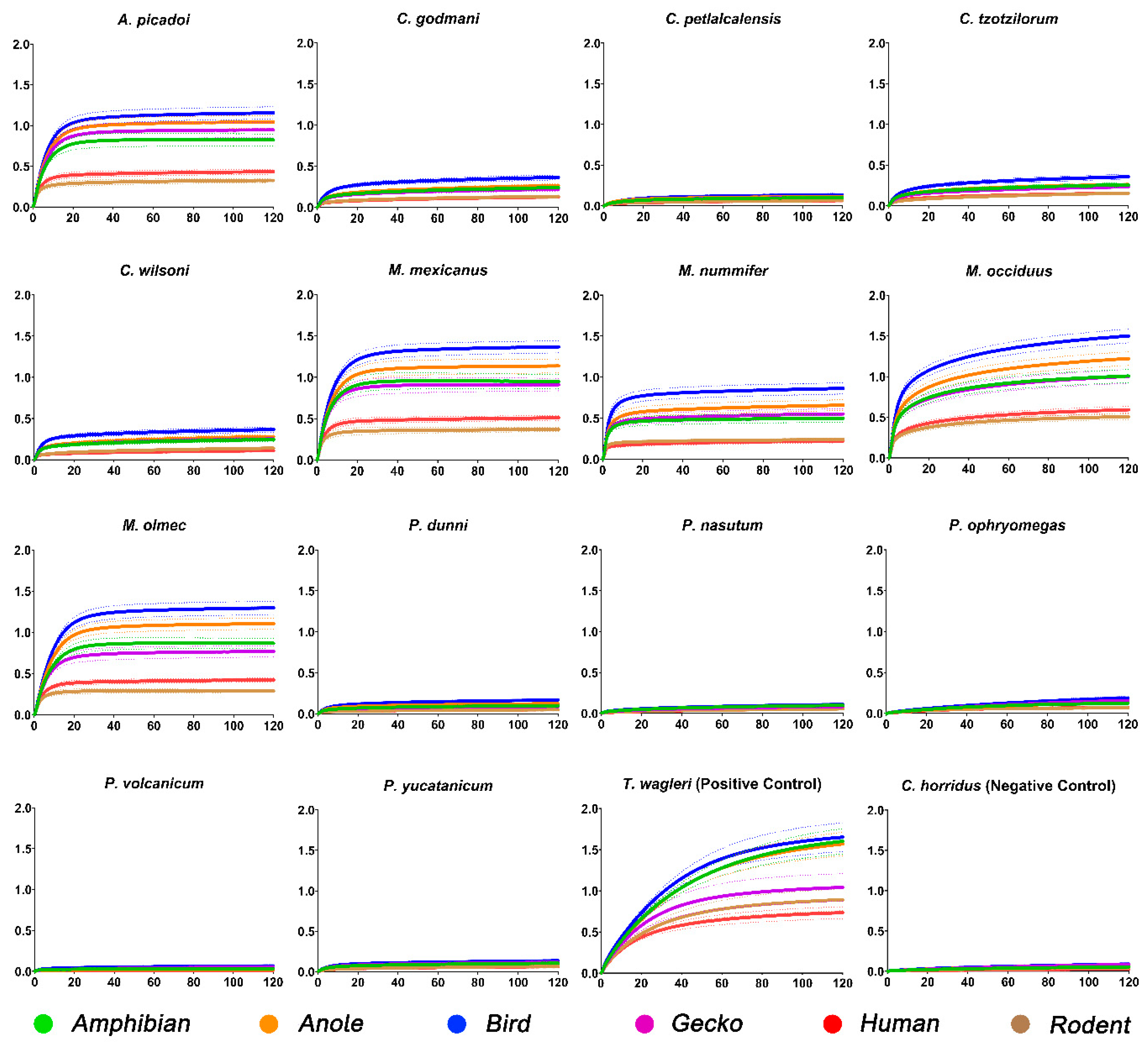
Figure 1. Comparison of wavelength (nm) curves of Atropoides/Cerrophidion/Metlapilcoatlus/Porthidium venom over a 120 s period. To keep controls consistent to Crotalinae, T. wagleri was used as the positive control and C. horridus as the negative. All venoms were tested against amphibian (green), anole (orange), bird (blue), gecko (purple), human (red), and rodent (brown) mimotopes. Dots surrounding the mean lines represent the standard error of the mean (SEM). Each venom was tested in triplicate (n = 3).
Toxins 15 00487 g001
Figure 2. Heat map comparison of Atropoides/Cerrophidion/Metlapilcoatlus/Porthidium species. Values are AUC ± SEM and are derived from binding-rate curves; warmer colours indicate stronger binding whereas cooler colours indicate weaker/no binding. Snake phylogeny is based on Alencar et al. [50]. Species phylogeny was obtained by entering the taxa into www.timetree.org (accessed 1 May 2023).
Toxins 15 00487 g002
Disclaimer/Publisher’s Note: The statements, opinions and data contained in all publications are solely those of the individual author(s) and contributor(s) and not of MDPI and/or the editor(s). MDPI and/or the editor(s) disclaim responsibility for any injury to people or property resulting from any ideas, methods, instructions or products referred to in the content.