Article

Biocontrol Capabilities of Bacillus subtilis E11 against Aspergillus flavus In Vitro and for Dried Red Chili (Capsicum annuum L.)

Key Laboratory of Plant Resource Conservation and Germplasm Innovation in Mountainous Region (Ministry of Education), College of Life Sciences/Institute of Agro-Bioengineering, Guizhou University, Guiyang 550025, China
*
Author to whom correspondence should be addressed.
Toxins 2023, 15(5), 308; https://doi.org/10.3390/toxins15050308
Submission received: 20 March 2023 / Revised: 22 April 2023 / Accepted: 23 April 2023 / Published: 26 April 2023
(This article belongs to the Section Mycotoxins)

Abstract

:
As a condiment with extensive nutritional value, chili is easy to be contaminated by Aspergillus flavus (A. flavus) during field, transportation, and storage. This study aimed to solve the contamination of dried red chili caused by A. flavus by inhibiting the growth of A. flavus and detoxifying aflatoxin B1 (AFB1). In this study, Bacillus subtilis E11 (B. subtilis) screened from 63 candidate antagonistic bacteria exhibited the strongest antifungal ability, which could not only inhibit 64.27% of A. flavus but could also remove 81.34% of AFB1 at 24 h. Notably, scanning electron microscopy (SEM) showed that B. subtilis E11 cells could resist a higher concentration of AFB1, and the fermentation supernatant of B. subtilis E11 could deform the mycelia of A. flavus. After 10 days of coculture with B. subtilis E11 on dried red chili inoculated with A. flavus, the mycelia of A. flavus were almost completely inhibited, and the yield of AFB1 was significantly reduced. Our study first concentrated on the use of B. subtilis as a biocontrol agent for dried red chili, which could not only enrich the resources of microbial strains for controlling A. flavus but also could provide theoretical guidance to prolong the shelf life of dried red chili.
Key Contribution: Bacillus subtilis E11, a strain that can effectively control the growth of A. flavus and AFB1 pollution in vitro and on dried red chili.

1. Introduction

China has the largest production of chili worldwide [1], in which dried red chili (Capsicum annuum L.) is one of the common condiments. However, it is vulnerable to pollution by aflatoxins (AFTs) as other crops [2]. The contamination and growth of Aspergillus flavus (A. flavus) may occur anywhere and anytime during the pre-harvest or post-harvest of chili [3]. After contamination with A. flavus, chili may not only cause serious economic losses but may also endanger human health. For instance, Rajendran et al. measured the AFTs in 42 samples of chili grown and marketed in Tamil Nadu. The results showed that approximately 66.7% of the samples collected from retail stores exceeded the European Commission standard limit (10 μg/kg). The content of AFTs in the samples collected across the value chain ranged from 3.83 to 37.80 μg/kg [4,5]. Therefore, it is urgent to globally solve the challenges of A. flavus pollution and the production of AFTs.
A. flavus is a common saprophytic fungus that widely exists in nature. AFTs are secondary toxic metabolites that are mainly produced by A. flavus and Aspergillus parasiticus [6]. To date, more than 18 types of AFTs have been found [7], mainly including aflatoxin B1 (AFB1), aflatoxin B2 (AFB2), aflatoxin G1 (AFG1), and aflatoxin G2 (AFG2) [8,9]. AFB1 has been classified as a group I carcinogen by the International Agency for Research on Cancer (IARC) because of its strong toxicity [10]. The toxicity of AFB1 has been demonstrated to act on most animals [11], which is carcinogenic, teratogenic, and mutagenic [12]. AFB1 can also cause irreversible effects on the kidneys, pancreas, bladder, bones, viscera, and central nervous system. Among them, damage to the liver is the most well-known concern. Besides, it may cause a series of chronic and acute diseases [13]. For crops, AFB1 pollution exists widely, which usually occurs during pre and post-harvesting of regional crops and food commodities [14].
The AFTs synthesis gene cluster is located on chromosome 3, with a DNA region of about 70 kb [15]. Based on the current research results, the biosynthesis of AFTs is regulated by at least 53 genes, containing 39 positive regulatory genes, 12 negative regulatory genes, and two other regulatory genes [16]. It is noteworthy that aflR and aflS are two regulatory genes in the biochemical synthesis pathway of AFB1. The aflR gene is a transcriptional activator of the AFTs gene cluster, which is necessary to activate the expression of other AFTs genes. The aflR gene is responsible for encoding the zinc-finger transcription factor. The aflS gene is located in the attachment of the aflR gene and is a transcriptional co-activator of the AFTs gene cluster. The ketoreductase encoded by the aflD gene could convert norsolorinic acid (NOR) into averantin (AVN), belonging to structural genes [17,18,19]. These three genes play a crucial role in the synthesis of AFTs.
At present, the control strategies of AFB1 mainly include physical (adsorption, irradiation, thermal treatment, and mechanical sorting), chemical (curcumin, salts, acids, and alkaline), and biological methods [20,21,22,23,24]. The control of AFB1 based on physical and chemical methods cannot be widely used because of the nutrient loss, chemical residue, sensory quality reduction, and high cost. In contrast, biodegradation is more moderate and environmentally safe, with less impact on food quality [25,26]. Thus, biological methods to inhibit the growth of A. flavus and the production of AFTs have noticeably attracted scholars’ attention. It was reported that some species of bacteria, fungi, and algae could well prevent the growth of A. flavus and the production of toxins [27]. The reported biological control methods of AFB1 have mainly used bacteria or fungi and antagonistic substances produced by their metabolism to adsorb AFB1, degrade it enzymatically or convert AFB1 into other substances with lower toxicity [28]. For instance, Cai et al. [29] screened a Stenotrophomonas acidoaminiphila strain that could reduce AFB1 concentration, and they demonstrated that this strain could degrade AFB1 through the combination of enzymes and micro-molecular oxides. A recent study showed that the supernatant of Trichoderma reesei could effectively degrade AFB1 with a concentration of 500 ng/kg and with a degradation rate of 91.8%, while the degradation rates of intracellular components and mycelial adsorption were 19.5% and 8.9%, respectively. It was also proved that the degradation products of this strain were nontoxic [30]. In another study, Zhao et al. [31] purified an extracellular enzyme called myxobacteria aflatoxin degradation enzyme (MADE) from Myxococcus fulvus ANSM068, which could simultaneously degrade AFB1 (71.89%), AFG1 (68.13%), and aflatoxin M1 (AFM1) (63.82%).
Bacillus subtilis (B. subtilis) and Lactic acid bacteria (LAB) are commonly known as generally recognized as safe (GRAS) [32,33], which are recognized as biological control agents against a variety of fungi [34,35]. Ren et al. [36] demonstrated that Bacillus was found in 21% of the research articles on controlling aflatoxigenic fungi and AFB1. Because of its fast growth rate, and its ability to produce a wide range of antifungal compounds, Bacillus has been widely used in the control of aflatoxigenic fungi and the production of AFTs. For instance, Zhao et al. [37] screened a strain from 22 strains of Lactobacillus plantarum that could completely inhibit the growth of A. flavus at a concentration of 5 × 10⁵ CFU/mL and reduced the yield of AFTs by 93%. The cell-free supernatant of another Lactobacillus plantarum UM55 inhibited the production of AFTs by 91% [38]. According to Suresh et al.’s study [28], B. subtilis could degrade 60% of AFB1. Another study showed that the treatment of pistachios with B. subtilis UTBSP1 could reduce the growth of A. flavus R5 and the production of AFB1 on pistachios [39].
In conclusion, although biological control is one of the most promising methods to solve the pollution of A. flavus and AFB1, it may be inapplicable to some types of food. Some scholars have shown that the biological detoxication of mycotoxins and their ability to alter the chemical structures deserve further attention [40]. On the other hand, as antagonistic bacteria that inhibit the growth of A. flavus and detoxify AFB1, they could not promptly and effectively inhibit the growth of A. flavus, which would ultimately lead to the accumulation of AFB1 [41]. At present, the control of the production of AFTs mainly depends on two key points, one is to prevent the growth of AFTs-producing fungi, and the other is to remove toxic compounds through various methods to achieve the purpose of detoxification in case of pollution [42]. Therefore, the present study aimed to screen bacteria with strong antifungal activity and detoxification capability of AFB1 in order to provide a new strategy to solve the pollution of A. flavus and AFB1 on dried red chili.
In the present study, we screened a strain of B. subtilis E11 that exhibited significant biological control capabilities against A. flavus in vitro and on dried red chili (Denglongjiao, one of the main varieties of chili cultured in the Guizhou province of China). Therefore, the results of the present study may provide an effective theoretical basis for the development of biological preservatives that can effectively control the growth and toxicity of A. flavus and broaden the biological control pathway of A. flavus and AFB1 in dried red chili storage.

2. Results

2.1. Screening of Strains with Biological Antagonistic Ability to A. flavus

Preliminary screening and rescreening results of antagonistic bacteria are shown in Table S1 and Figure S1. Among 63 strains of LAB and B. subtilis, three strains of B. subtilis, including E11, V1J1, and 9932, were finally selected as antagonistic bacteria for the next study according to their inhibitory abilities to A. flavus (Figure 1). According to the results of confronting incubation, the inhibitory rates of B. subtilis E11, V1J1, and 99,332 on the growth of A. flavus mycelia were 64.27 ± 2.06%, 62.57 ± 1.85%, and 69.31 ± 5.45%, respectively. Considering that B. subtilis E11, V1J1, and 9932 had strong inhibitory effects on the mycelia of A. flavus, we believe that these three strains have potential in the biological control of A. flavus and should be further studied.

2.2. The Abilities of B. subtilis E11, B. subtilis V1J1, and B. subtilis 9932 to Remove AFB1

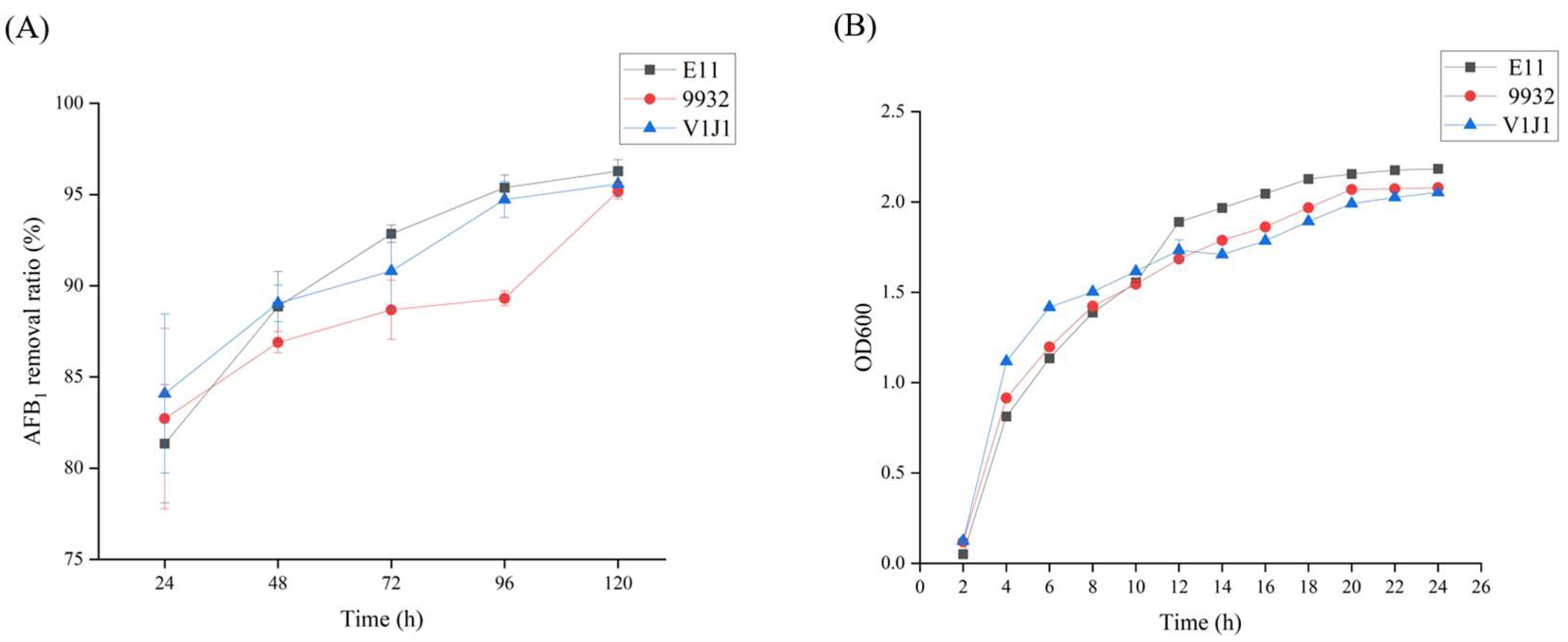
The abilities of B. subtilis E11, B. subtilis 9932, and B. subtilis V1J1 to remove AFB1 were further detected by an ELISA kit at different time points. It can be seen from Figure 2A and Table S2 that the ability of B. subtilis E11 to remove AFB1 was slightly higher than that of B. subtilis 9932 and B. subtilis V1J1. Afterward, according to the growth curves of the three strains, it was found that the number of cells in the platform stage of B. subtilis E11 was higher than that in the other two strains, which could lead to more secondary accumulated metabolites (Figure 2B). Therefore, B. subtilis E11 was selected for further study.

2.3. The Biodegradation Ability of AFB1 by Culture Supernatant, Cells, and Cell Lysates

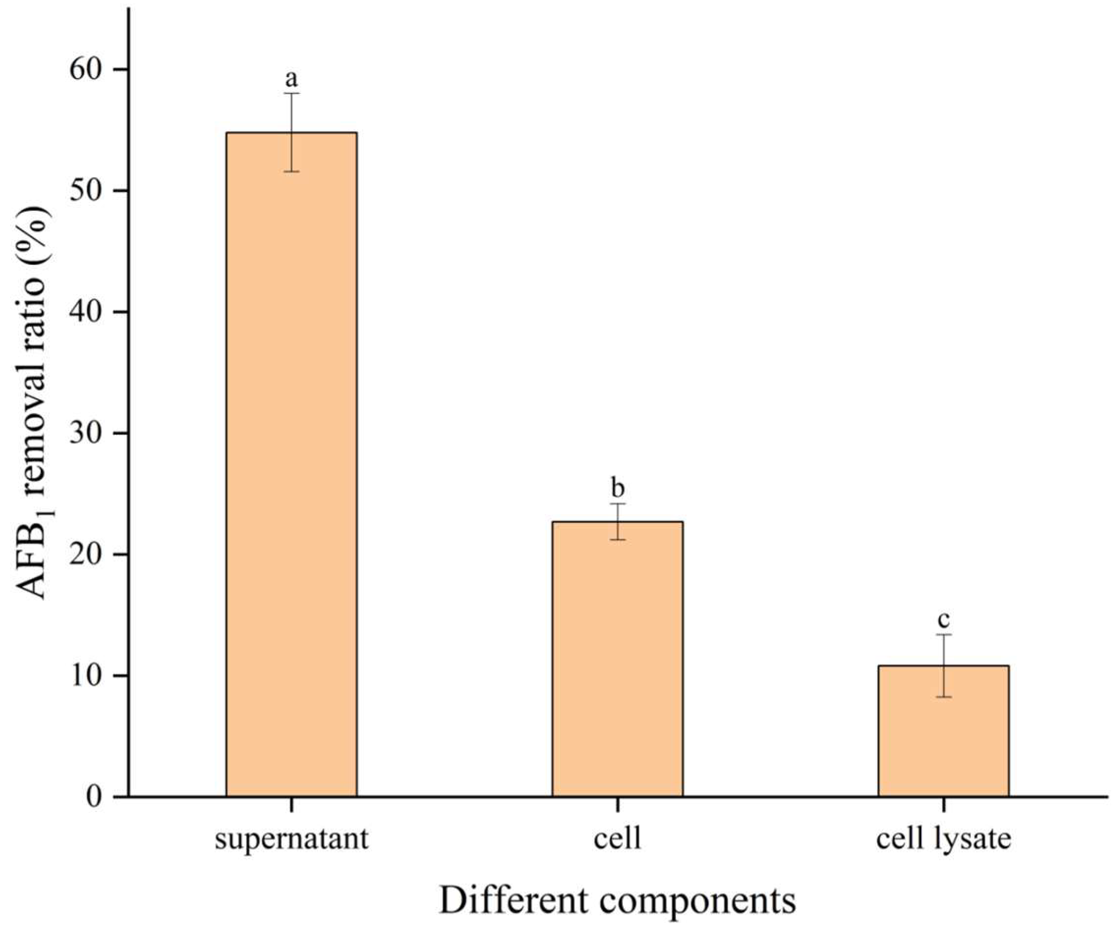
AFB1 was added to the fermentation supernatant, cells, and cell lysates of B. subtilis E11 and cultured for 24 h, in which the results showed that the degradation rate of AFB1 in the culture supernatant of B. subtilis E11 was the highest (54.78%) (Figure 3). Moreover, degradation rates of 22.68% and 10.81% were obtained when AFB1 was treated with cells and cell lysates, respectively. It was revealed that the fermentation supernatant of B. subtilis E11 had the greatest contribution to the degradation of AFB1.

2.4. The Effects of B. subtilis E11 on aflR, aflS, and aflD Key Genes for AFB1 Production

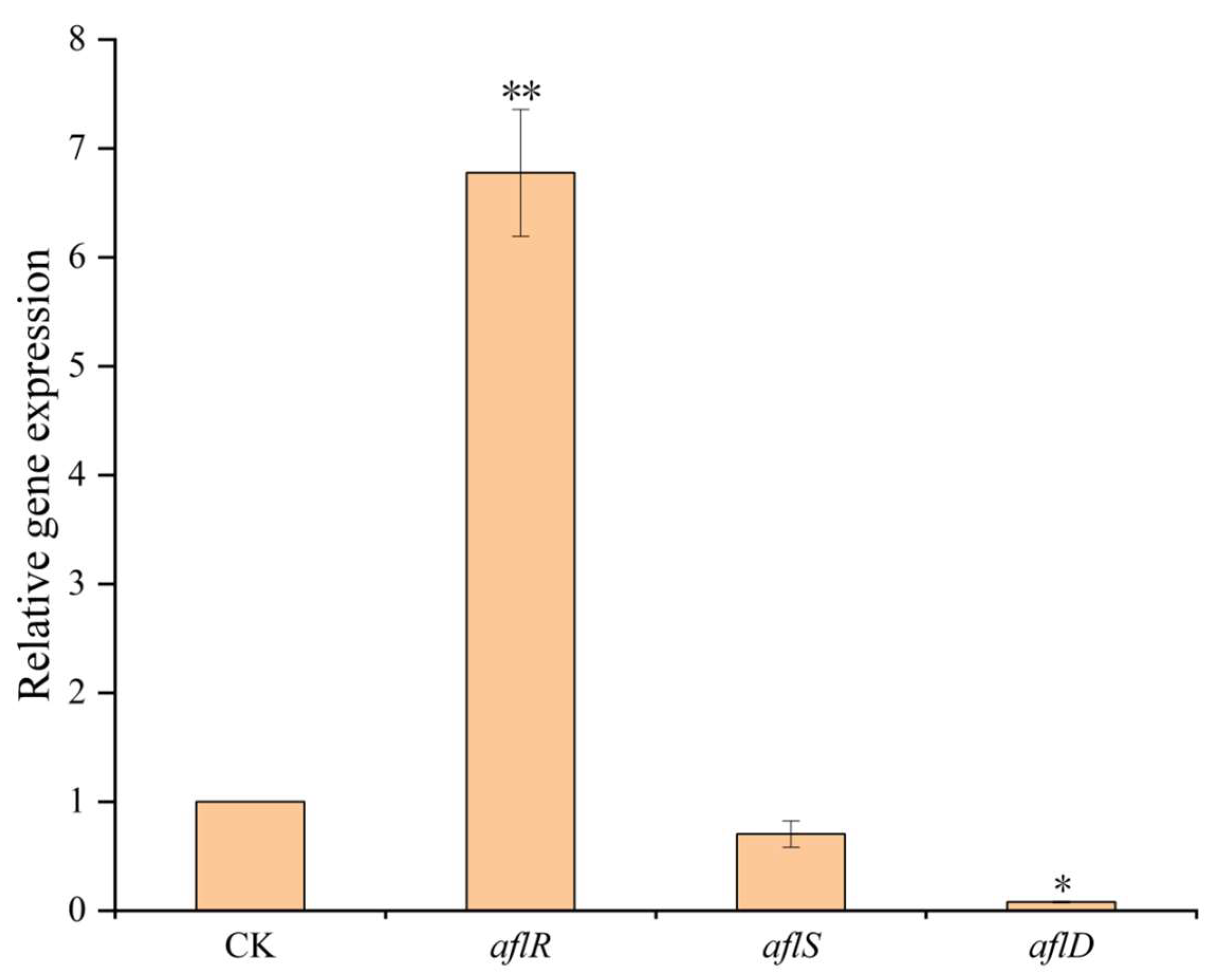
Based on the results of the expression of three key genes in the AFB1 synthesis pathway, the fermentation supernatant of B. subtilis E11 could significantly increase the aflR expression level and significantly repress the aflD expression level, while the aflS expression level slightly decreased compared with the control group (Figure 4).

2.5. The Ultrastructural Changes of B. subtilis E11 and A. flavus

The ultrastructural changes of B. subtilis E11 treated with AFB1 and A. flavus treated with B. subtilis E11 fermentation supernatant were observed by scanning electron microscopy (SEM) (Figure 5). Figure S2 shows the effect of the fermentation supernatant of B. subtilis E11 on A. flavus mycelia. It was revealed that A. flavus in the control group could form a mycelia ball after 48 h of culture, while the mycelia of A. flavus treated with the supernatant of B. subtilis E11 presented a loose flocculent structure and could not form mycelia ball. The purpose of using SEM to analyze the ultrastructural changes of B. subtilis E11 was to indicate whether cells of B. subtilis E11 could resist a high concentration of AFB1. The ultrastructural changes of B. subtilis E11 after coculture with AFB1 showed that the B. subtilis cells of E11 became irregular, and the surface became rough with slight wrinkles after 24 h (Figure 5A). The cells of B. subtilis E11 without AFB1 treatment were regularly rod-shaped, and the cell surface was smooth (Figure 5B). The mycelia morphology of A. flavus cocultured with fermentation supernatants of B. subtilis E11 had obvious folds and deformation (Figure 5C). However, the mycelia of A. flavus in the control group had a smooth appearance without wrinkles (Figure 5D). Therefore, B. subtilis E11 cells not only have the ability to tolerate a high concentration of AFB1, but also, their fermentation supernatant can disrupt the mycelia of A. flavus, which may affect the spore production and toxin synthesis of A. flavus.

2.6. The Potential of B. subtilis E11 as a Natural Food Preservative for Dried Red Chili

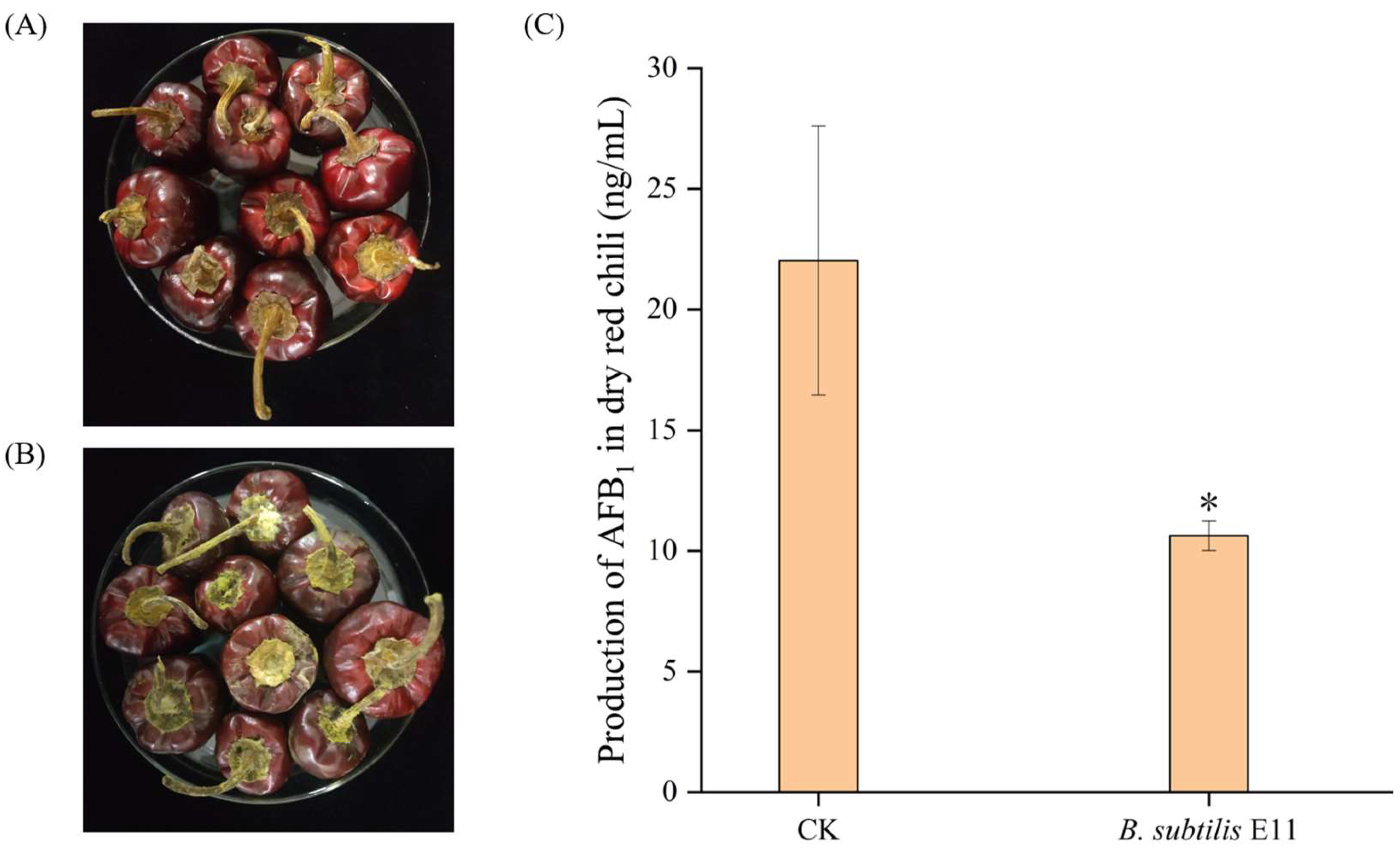
The biological control ability of B. subtilis E11 was confirmed on dried red chili (Figure 6). After coculture for 10 d, the dried red chili in the control group was covered with A. flavus mycelia and spores on the carpopodium. However, only a very small number of mycelia appeared on the surface of chili treated with a culture solution of B. subtilis E11 (Figure 6A,B). Figure 6C illustrates the content of AFB1 in dried red chili after 10 days of coculture with A. flavus and B. subtilis E11 (water was used as a control). It was indicated that the yield of AFB1 in dried red chili could be significantly reduced after B. subtilis E11 treatment of chili samples. This showed that B. subtilis E11 has the potential to be used as a biological control agent in the storage of dried red chili. A. flavus is one of the common harmful fungi in dried red chili. The results of the present study may provide theoretical support for extending the storage period of dried red chili.

3. Discussion

According to the report of the Rapid Alert System for Food and Feed (RASFF) [43], contamination caused by AFTs accounted for about 36% of the export safety warning of dried pepper in China. Consequently, it is urgent to find a green environmental protection method to control the contamination of dried red chili by A. flavus. At present, scholars are committed to controlling the production of mycotoxins through biological methods, which can not only protect crops from fungal infection but also reduce damage to the ecological environment [44].
In our study, the B. subtilis E11 we screened was not only environmentally friendly but also, as an antagonistic bacterium, it could firstly inhibit the growth of A. flavus and secondly remove the AFB1 when A. flavus grew and produced it. Our results indicated that the three strains of B. subtilis have a stronger ability to resist A. flavus than some previously reported strains. For instance, a total of 10 bacterial strains were isolated from the rhizosphere soil of Camellia sinensis by Wu et al. [45] in the plate confrontation test, and the results showed that only Bacillus tequilensis and Bacillus velezensis had significant inhibitory effects on the growth of A. flavus, with inhibition rates of 34.22% and 35.72%, respectively. Another study achieved the inhibition rates of 4–90% and 5–92% for 250–2500 μL Bacillus amyloliquefaciens (UTB2) and B. subtilis (UTB3) cell culture filtrate on the growth of Aspergillus parasiticus, respectively [46]. Other strains, such as Candida nivariensis DMKU-CE18, could inhibit the growth of 64.9 ± 7.0% of A. flavus A39 mycelia [47]. It was revealed that the three strains of B. subtilis could effectively inhibit the growth of more than 60% of A. flavus hyphae by measuring the diameter of A. flavus in the control group and the experimental group.
Studies have pointed out that Bacillus can produce various secondary metabolites and lytic enzymes in the metabolic process, which play an important role in the antagonism of Bacillus [48]. The ability of B. subtilis E11 screened in this experiment to reduce AFB1 was higher than that of some reported strains. For example, Shu et al. [49] isolated a strain from soil samples named Bacillus velezensis DY3108, which had a strong AFB1 degradation activity (91.5%). In Fang et al.’s study [50], the rate of AFB1 degradation at 72 h was 88.59% by Aspergillus niger RAF106. Petchkongkaew et al. [51] found that one strain of Bacillus licheniformis could simultaneously inhibit the growth of A. flavus and Aspergillus westerdijkiae, and it could remove 74% of AFB1 and 92.5% of ochratoxin A (OTA), while another strain of B. subtilis could inhibit the growth of A. flavus and degrade AFB1 (85%). In another study, when 2 μg/mL AFB1 was added to the lysogeny broth (LB) medium, Bacillus amylolyticus WE2020 could reduce 70.22% of AFB1 at 48 h, degraded more than 84% of AFB1 at 72 h, and reached the maximum value [52]. A strain of Bacillus cereus XSWW9 was isolated from Daqu of Baijiu, which could degrade 86.7% of AFB1 (1 mg/L) after incubation at 37 °C for 72 h [53]. A strain of Microbacterium proteolyticum B204 was isolated from bovine feces, which could degrade 77% of AFB1 at 24 h [54]. The reported antifungal compounds produced by B. subtilis mainly include lipopeptides, such as surfactin, iturins, and fengycin [39,55]. B. subtilis is one of the most commonly used microorganisms in the industry for producing lipopeptides, which have wide applications in biology (anti-bacterial, anti-fungal, anti-viral, and anti-tumor), chemical, agricultural, pharmaceuticals, and food industries [56]. Bertuzzi et al. [48] analyzed the lipopeptides in raw and cell-free B. subtilis QST 713 broth by LC-MS/MS, and found that the contents of surfactants, iturins, and fengycin family in the broth were 292 mg/L, 3034 mg/L, and 1033 mg/L, respectively. However, the content of all three lipopeptides in cell-free broth was lower than that in raw broth. In addition, they also found that raw B. subtilis broth can completely inhibit the production of AFB1. As B. subtilis E11 exhibited to have a strong ability to degrade AFB1 that might be a potentially detoxification strain, the characteristics of detoxification of AFB1 by B. subtilis E11 need to be further explored. In the present study, it was only found that B. subtilis E11 could detoxify AFB1, while the antagonistic substances of B. subtilis E11 were not determined. Therefore, the main active substances of B. subtilis E11 for detoxifying AFB1 should be identified in the future research.
Our study found that the ability of different components of B. subtilis E11 to remove AFB1 was in the order of fermentation supernatant, cells, and cell contents. Similar results have been previously reported. For instance, the cell-free extract of B. subtilis fermentation could reduce AFB1 by 60% [28]. A strain of Bacillus albus YUN5 was isolated from fermented soybean paste in Korea. Through the degradation experiment of AFB1, it was found that the cell-free fermentation supernatant of Bacillus albus YUN5 had the highest degradation ability, reaching 76.28 ± 0.15%. However, the degradation ability of AFB1 by cells and intracellular components was negligible [57].
Next, we analyzed the expression of three key genes in the AFB1 synthesis pathway. Our results were similar to some published articles. Similarly, 20 strains of Aspergillus niger isolated from peanuts showed that the culture filtrate of Aspergillus niger could upregulate the expression level of the aflR gene while downregulating the expression levels of aflS and aflD genes [58]. Kong et al. [59] pointed out that AflR and AflS could exert a transcriptional regulatory effect on the synthetic gene cluster as a protein complex, including four AflS with one AflR. When aflS is expressed normally, there are sufficient AflS proteins to bind to AflR to activate the complex to regulate aflatoxin synthesis. Transcription of toxin synthetic clustered structural genes is also downregulated when the aflS expression level is insufficient for complex formation. In the present study, the fermentation supernatants of B. subtilis E11 might reduce the AFB1 production by upregulating the aflR expression level and downregulating the aflS expression level, thereby changing the aflR/aflS ratio. Although an aflR/aflS ratio above 1 may lead to the activation of AFB1 biosynthesis, it was found in some studies that AFB1 accumulation would not occur when the ratio was greater than 1 [60,61].
A. flavus is an opportunistic plant pathogen and a human pathogen [62], negatively influencing crop consumers’ health [63]. Chili may be contaminated by A. flavus at any stage, from field to storage. However, AFB1 is very difficult to remove once present in the chili production chain [2]. So, we investigated the ability of B. subtilis E11 as a biocontrol agent on dried red chili. The results showed that E11 not only inhibited the growth of A. flavus on dried chili but also reduced the content of AFB1. Currently, there are many successful cases of using microorganisms as biological control agents in food. For instance, it was demonstrated that Bacillus velezensis HC6 could simultaneously inhibit the growth of A. flavus, Aspergillus ochraceus, Fusarium oxysporum, Fusarium graminearum, Aspergillus sulphureus, and Aspergillus parasiticus in maize, and Bacillus velezensis HC6 could significantly reduce the contents of AFB1 and OTA [64]. Shakeel et al. evaluated the potential of Streptomyces yangliansis 3-10 as an antifungal agent on peanuts. It was found that different concentrations of culture filtrate and crude extract of Streptomyces yanglinensis 3-10 could inhibit the growth of A. flavus and reduce the production of AFB1. Moreover, it could significantly inhibit the expression levels of aflR and aflS during AFB1 synthesis. The ultrastructural changes of A. flavus treated with Streptomyces yangliansis 3-10 culture filtrate were observed by SEM and transmission electron microscopy (TEM). It was revealed that the hyphae grew abnormally and atrophied, the organelles degenerated and collapsed, and large vacuoles appeared [65]. Furthermore, 28 microorganisms isolated from red grapes to test their antifungal and anti-mycotoxigenic abilities showed that when strain UTA6 was used as a biological preservative in grapes contaminated by A. flavus and Botrytis cinerea, the spores of A. flavus and Botrytis cinerea in grapes were reduced by 0.4 and 0.6 log10 spores/g in grapes, while those of AFB1 and fumonisin B1 (FB1) decreased by 28–100% [66]. In the present study, the culture of B. subtilis E11 reduced the yield of AFB1 by 51.79%. In the subsequent study, the fermentation conditions of B. subtilis E11 will be optimized to enhance its ability to reduce the production of AFB1. On the other hand, Shifa et al.’s findings [67] will be valuable, and it will be attempted to consider applying B. subtilis E11 to the growth of chili in the field and actual warehouse storage.
Overall, the B. subtilis E11 we have screened has great potential for application in the storage of dried red chili. Firstly, B. subtilis E11 is a GRAS bacterium. Secondly, B. subtilis E11 has a fast growth rate and low nutritional requirements, which can reduce costs in practical applications. Finally, B. subtilis E11 can effectively inhibit the growth of A. flavus and reduce the production of AFB1 in vitro and on dried red chili. Hence, the results of this study can provide a theoretical basis for extending the shelf life of dried red chili.

4. Materials and Methods

4.1. Fungal and Bacterial Strains

The A. flavus was purchased from China General Microbiology Culture Center (CGMCC; Shanghai, China), and the strain number was CGMCC 3.4408. In total, 63 bacterial strains, including 25 LAB and 35 B. subtilis, were isolated from fermented food and preserved in our laboratory. The other 3 LAB strains were purchased from CGMCC (CGMCC 1.2427) and China Center of Industrial Culture Collection (CICC 6239 and CICC 24,450).

4.2. Preparation of Spore Suspension

A spore suspension was prepared according to the method described by Natarajan et al. [68] with some modifications. A. flavus was transferred to the Potato Dextrose Agar (PDA) slants and incubated at 28 °C for 3–7 days. Then, sterile saline (10 mL) was utilized to wash the spores from the slants by rubbing the surface with a sterile glass rod and filtered through 4 layers of sterile gauze to obtain spore suspension. At last, the spore suspension was adjusted to 107 CFU/mL using a hemocytometer under a microscope (Olympus, Tokyo, Japan) with sterile saline and used in all experiments.

4.3. Screening of Strains with Resistance to A. flavus

First, LAB and B. subtilis were cultured overnight at 37 °C on Man Rogosa and Sharpe (MRS) and LB plates, respectively. Then, 100 μL spore suspension was evenly plated on the PDA, and the single visible colony of 63 bacteria was picked up on the PDA. Each plate was equally spaced with 8 strains. The plates were incubated at 28 °C for 3–5 days. Bacteria with antifungal capacity were selected for the following experiments.
The rescreening of the antifungal ability of the strains was carried out using the method described by Zhang et al. [69] with minor variations. A 10 mm filter paper disc incubated with 30 μL of the A. flavus spore suspension was placed on the center of the PDA. Afterward, 30 μL of bacterial culture and sterile water aliquots were independently inoculated onto sterile filter paper discs that were placed at 4 equal distance positions, 25 mm away from the center of the PDA. The colony diameter of A. flavus was measured by a vernier caliper at 7 days after incubation at 28 °C. All the experiments were performed in triplicate. The inhibition rate (%) was calculated as follows: Inhibition rate (%) = (d − d’)/d × 100%, where d (mm) represents the diameter of the A. flavus colony in the control group, and d’ represents the A. flavus colony of the tested bacteria.

4.4. Ability of the Selected Strains to Degrade AFB1

The stock solution of AFB1 (MACKLIN, Shanghai, China) at 10 mg/L concentration was prepared in acetonitrile and kept at −20 °C. The ability of each strain to remove AFB1 was based on the method presented by Zhu et al. [41] with some variations. Each strain was inoculated at 37 °C for 12 h in the LB medium, followed by inoculation (5% vol/vol) of each strain into 50 mL LB medium at 37 °C for 48 h. When optical density (OD) at a wavelength of 600 nm for each strain was adjusted to make consistency, 29.55 mL inoculum (an equal volume of LB medium was used as control) was transferred to a 100 mL sterile conical flask and mixed with 0.45 mL of AFB1 (10 mg/L). The final concentration of AFB1 was 150 μg/L. Subsequently, the mixture was incubated at 28 °C with agitation at 150 rpm. The inoculums were taken at 24, 48, 72, 96 and 120 h to investigate the ability of selected strains to decrease AFB1 at different time points. According to the manufacturer’s instructions, a commercial enzyme-linked immunosorbent assay kit (ELISA kit, MM-092501, Jiangsu Meimian, Industrial Co., Ltd., Jiangsu, China) was utilized to determine the AFB1 removal capacity of the selected strains. A microplate reader (Thermo Fisher Scientific, Waltham, MA, USA) was used to measure the OD at a wavelength of 450 nm. The ability of the selected strains to degrade AFB1 was calculated using the following formula: (concentration of AFB1 in the control group-concentration of AFB1 in the treatment group)/concentration of AFB1 in the control group ×100%.

4.5. Determination of Growth Curves of B. subtilis E11, B. subtilis 9932, and B. subtilis V1J1

The OD 600 of 3 B. subtilis strains was measured using a UV-Visible spectrophotometer (UV-2100; Guangzhou, China). According to the method proposed by Ju et al. [70], B. subtilis E11, B. subtilis 9932, and B. subtilis V1J1 were inoculated in 5 mL liquid LB medium for activation for 2 generations, and then, the 3 B. subtilis strains were respectively inoculated into a 50 mL liquid LB medium (5% vol/vol) at 37 °C, 180 rpm, and OD600 was measured every 2 h.

4.6. AFB1 Biodegradation by Culture Supernatant, Cells, and Cell Lysates

The culture supernatant, cells, and cell lysates were prepared according to the method presented by Wang et al. [71] with minor modifications. First, B. subtilis E11 was cultured in an LB medium at 37 °C for 24 h and was then cultured (5% vol/vol) in a 50 mL LB medium at 37 °C for 48 h. The supernatant and cells were collected after centrifugation at 8000× g for 10 min at 4 °C, and a 0.22 µm filter was then utilized to filter the supernatant for subsequent experiments. The cell pellet was thrice washed with phosphate-buffered saline (PBS, pH = 7) and then resuspended in PBS. The cell suspension was divided into 2 parts, 1 without any treatment served as cells for AFB1 degradation, and the other was broken by ultrasonicator. The cell lysates were obtained after centrifugation at 10,000× g for 30 min at 4 °C and filtered through a 0.22 μm filter. To explore the AFB1 degradation, the culture supernatant, cells, and cell lysates were treated with AFB1 (final concentration was 150 μg/L) and were then incubated at 37 °C for 24 h with agitation at 150 rpm. The biodegradation of AFB1 was investigated by ELISA, according to Section 4.4.

4.7. The Effects of B. subtilis E11 Culture Supernant on the Expression Levels of Genes Associated with AFB1 Production of A. flavus

4.7.1. Preparation of Mycelia Samples

Samples were prepared according to Zhu et al.’s method [41] with minor modifications. In short, 15 mL of E11 fermentation supernatant (the fermentation supernatant was collected as described in Section 4.6, and 15 mL of LB medium was used as a control) and 1.5 mL of A. flavus spore suspension were added to 15 mL of Potato Dextrose Broth (PDB), respectively, and the mixture was then incubated at 28 °C for 72 h with agitation at 150 rpm.

4.7.2. Extraction of Total RNA

The mycelia, after 72 h of culture, were ground with liquid nitrogen. The extraction of total RNA was performed according to the instructions of the Fungal RNA kit (Omega, New York, NY, USA). The concentration and quality were subsequently measured using a NanoDrop spectrophotometer (Thermo Fisher Scientific), and the qualified samples were stored at −80 °C for subsequent experiments.

4.7.3. Synthesis of cDNA

The cDNA synthesis was performed using the StarScript II RT Mix with gDNA Remover kit (GenStar, Beijing, China). The first step was to remove residues of genomic DNA from the RNA. Then, 2 μL of RNA, 2 μL 5 × gDNA remover buffer, 1 μL gDNA remover, and 5 μL Nuclease-free water (DEPC-treated) were added to the reaction tube. The mixture was incubated at 37 °C for 5 min after being mixed and centrifuged.
Synthesis of the first strand of the cDNA was immediately carried out. Additional 4 μL of 5 × StarScript II Buffer (The Buffer was pre-mixed with Random Primer and Oligo18 (dT) Primer.), 1 μL StarScript Enzyme Mix, and 5 μL Nuclease-free water (DEPC-treated) were added to the reaction tube after incubation. The components of the reactant were evenly mixed and centrifuged. The mixture was first incubated at 42 °C for 15 min and was then heated at 85 °C for 5 min to inactivate the StarScript Enzyme Mix. The cDNA at the end of the reaction was kept at −20 °C for subsequent experiments.

4.7.4. Real-Time Quantitative Polymerase Chain Reaction (RT-qPCR)

The RT-qPCR instrument equipped with Bio-Rad CFX Manager 3.1 (Bio-Rad Laboratories Inc., Hercules, CA, USA) was used to measure the expression levels of 3 genes (aflR, aflS, and aflD). The housekeeping gene (β-tubulin) was used as an endogenous control. The primers involved in this study are presented in Table 1. Each amplification reaction comprised cDNA (2 μL), SYBR Green Supermix (GenStar, Beijing, China; 10 μL), primers (forward/reverse, 10 μM; 0.4 μL), and nuclease-free water (7.2 μL). The RT-qPCR amplification conditions included initial denaturation at 94 °C for 5 min, followed by 40 amplification cycles (at 95 °C for 15 s and at 55 °C for 30 s).

4.8. SEM Analysis

The SEM analysis included 2 parts. One part was to observe the ultrastructural changes of B. subtilis E11 after coculture with AFB1 at 24 h, and the other part was to observe the ultrastructural changes of A. flavus after coculture with A. flavus 3.4408 and B. subtilis E11 supernatant for 72 h. The ultrastructural changes of B. subtilis E11 and A. flavus were observed using the SEM. This method was partially modified by referring to Zhao et al.’s research [61]. Cultures described in Section 4.6 and Section 4.7.1 were collected and centrifuged at 8000× g for 10 min at 4 °C. The precipitate was then washed 4 times with sterile water for 10 min. The precipitate was subsequently fixed with 2.5% glutaraldehyde overnight at 4 °C. Next, the samples were dehydrated using gradient concentrations of ethanol (10%, 30%, 50%, 70%, 95%, 100%) for 10 min. The samples were then dried in a vacuum dryer (LABCONCO Inc., New York, NY USA), which was coated with a gold ion sputter (E-1010; Hitachi, Tokyo, Japan) and examined using the SEM (S-3400N; Hitachi).

4.9. The Biocontrol of B. subtilis E11 on Dried Red Chili

The biocontrol of B. subtilis E11 on dried red chili was conducted by the method proposed by Einloft et al. [72] with some modifications. Denglongjiao was irradiated with ultraviolet radiation for 3 h to remove microorganisms on the surface of the chilis. Then, 20 g Denglongjiao was mixed with 1 mL of spore suspension and 1 mL of B. subtilis E11 culture suspension (sterile water of the same volume was used as control) in sterile PE self-sealing bags. After incubation at 25 °C for 10 days, the content of AFB1 in each sample was determined by ELISA as described in Section 4.4.

4.10. Statistical Analysis

All experiments were performed in triplicate. Analysis of variance (ANOVA) of SPSS 26 software (IBM, Armonk, NY, USA) was used to perform statistical analysis. The results were expressed as the mean ± SD. p < 0.05 was considered statistically significant.

Supplementary Materials

The following supporting information can be downloaded at: https://www.mdpi.com/article/10.3390/toxins15050308/s1. Table S1: Preliminary screening of bacteria against A. flavus. Table S2: The abilities of B. subtilis E11, B. subtilis V1J1, and B. subtilis 9932 to degrade AFB1 at different time points. Figure S1: Rescreening of antagonistic bacteria against A. flavus. Figure S2: The effect of the fermentation supernatant of B. subtilis E11 on A. flavus mycelia.

Author Contributions

Conceptualization, S.Y. and Y.W.; methodology, S.Y., Y.W. and S.T.; software, S.Y.; validation, S.Y.; investigation, S.Y. and Y.C.; resources, Y.W; data curation, S.Y.; writing—original draft, S.Y.; writing—review & editing, Y.W. and J.J.; project administration, Y.W.; funding acquisition, Y.W., S.T. and L.Z. All authors have read and agreed to the published version of the manuscript.

Funding

This research was funded by the Guizhou Pepper Industry Technology System [GZSLJCYTX-2022], Guizhou Engineering Technology Research Center for the Processing of Pepper Products [Qianjiaohe KV (2021) 006], Guizhou Pepper Fermented Products Engineering and Technology Research Center [Qiankehe Platform Talent (2020) 2102], Guizhou Provincial Science and Technology Projects [Qian Ke He Zhi Cheng-ZK (2021) General 262], Guizhou Provincial Science and Technology Projects [Qian Ke He Zhi Cheng-ZK (2022) General 172], Guizhou Provincial Science and Technology Projects [NO. Qiankehe (2022) 156].

Institutional Review Board Statement

Not applicable.

Informed Consent Statement

Not applicable.

Data Availability Statement

Not applicable.

Conflicts of Interest

All authors declare that there is no conflict of interest associated with the content of this article.

References

  1. Hernandez-Perez, T.; Gomez-Garcia, M.d.R.; Valverde, M.E.; Paredes-Lopez, O. Capsicum annuum (hot pepper): An ancient Latin-American crop with outstanding bioactive compounds and nutraceutical potential. A review. Compr. Rev. Food Sci. Food Saf. 2020, 19, 2972–2993. [Google Scholar] [CrossRef]
  2. Costa, J.; Rodriguez, R.; Garcia-Cela, E.; Medina, A.; Magan, N.; Lima, N.; Battilani, P.; Santos, C. Overview of Fungi and Mycotoxin Contamination in Capsicum Pepper and in Its Derivatives. Toxins 2019, 11, 27. [Google Scholar] [CrossRef]
  3. Rushing, B.R.; Selim, M.I. Aflatoxin B1: A review on metabolism, toxicity, occurrence in food, occupational exposure, and detoxification methods. Food Chem. Toxicol. 2019, 124, 81–100. [Google Scholar] [CrossRef]
  4. Rajendran, S.; Shunmugam, G.; Mallikarjunan, K.; Paranidharan, V.; Venugopal, A.P. Prevalence of aflatoxin contamination in red chilli pepper (Capsicum annuum L.) from India. Int. J. Food Sci. Technol. 2022, 57, 2185–2194. [Google Scholar] [CrossRef]
  5. European Commission. Commission Regulation (EC) No 1881/2006 Setting Maximum Levels for Certain Contaminants in Foodstuffs. Off. J. Eur. Union 2006, 364, 5–24. [Google Scholar]
  6. Watanakij, N.; Visessanguan, W.; Petchkongkaew, A. Aflatoxin B-1-degrading activity fromBacillus subtilisBCC 42005 isolated from fermented cereal products. Food Addit. Contam. Part A-Chem. Anal. Control Expo. Risk Assess. 2020, 37, 1579–1589. [Google Scholar] [CrossRef]
  7. Benkerroum, N. Aflatoxins: Producing-Molds, Structure, Health Issues and Incidence in Southeast Asian and Sub-Saharan African Countries. Int. J. Environ. Res. Public Health 2020, 17, 1215. [Google Scholar] [CrossRef]
  8. Chen, T.; Liu, J.; Li, Y.; Wei, S. Burden of Disease Associated with Dietary Exposure to Aflatoxins in China in 2020. Nutrients 2022, 14, 1027. [Google Scholar] [CrossRef]
  9. Xuan, Z.; Ye, J.; Zhang, B.; Li, L.; Wu, Y.; Wang, S. An Automated and High-Throughput Immunoaffinity Magnetic Bead-Based Sample Clean-Up Platform for the Determination of Aflatoxins in Grains and Oils Using UPLC-FLD. Toxins 2019, 11, 583. [Google Scholar] [CrossRef]
  10. Chitarrini, G.; Nobili, C.; Pinzari, F.; Antonini, A.; De Rossi, P.; Del Fiore, A.; Procacci, S.; Tolaini, V.; Scala, V.; Scarpari, M.; et al. Buckwheat achenes antioxidant profile modulates Aspergillus flavus growth and aflatoxin production. Int. J. Food Microbiol. 2014, 189, 1–10. [Google Scholar] [CrossRef]
  11. Marchese, S.; Polo, A.; Ariano, A.; Velotto, S.; Costantini, S.; Severino, L. Aflatoxin B1 and M1: Biological Properties and Their Involvement in Cancer Development. Toxins 2018, 10, 214. [Google Scholar] [CrossRef] [PubMed]
  12. Limaye, A.; Yu, R.-C.; Chou, C.-C.; Liu, J.-R.; Cheng, K.-C. Protective and Detoxifying Effects Conferred by Dietary Selenium and Curcumin against AFB1-Mediated Toxicity in Livestock: A Review. Toxins 2018, 10, 25. [Google Scholar] [CrossRef]
  13. Benkerroum, N. Chronic and Acute Toxicities of Aflatoxins: Mechanisms of Action. Int. J. Environ. Res. Public Health 2020, 17, 423. [Google Scholar] [CrossRef]
  14. Kumar, A.; Pathak, H.; Bhadauria, S.; Sudan, J. Aflatoxin contamination in food crops: Causes, detection, and management: A review. Food Prod. Process. Nutr. 2021, 3, 1–9. [Google Scholar] [CrossRef]
  15. Tumukunde, E.; Xie, R.; Wang, S.H. Updates on the Functions and Molecular Mechanisms of the Genes Involved in Aspergillus flavus Development and Biosynthesis of Aflatoxins. J. Fungi. 2021, 7, 666. [Google Scholar] [CrossRef]
  16. Liao, J.; He, Z.; Xia, Y.; Lei, Y.; Liao, B. A review on biosynthesis and genetic regulation of aflatoxin production by major Aspergillus fungi. Oil Crop Sci. 2020, 5, 166–173. [Google Scholar] [CrossRef]
  17. Caceres, I.; Al Khoury, A.; El Khoury, R.; Lorber, S.; Oswald, I.; El Khoury, A.; Atoui, A.; Puel, O.; Bailly, J.-D. Aflatoxin Biosynthesis and Genetic Regulation: A Review. Toxins 2020, 12, 150. [Google Scholar] [CrossRef] [PubMed]
  18. Pandey, M.K.; Kumar, R.; Pandey, A.K.; Soni, P.; Gangurde, S.S.; Sudini, H.K.; Fountain, J.C.; Liao, B.S.; Desmae, H.; Okori, P.; et al. Mitigating Aflatoxin Contamination in Groundnut through A Combination of Genetic Resistance and Post-Harvest Management Practices. Toxins 2019, 11, 315. [Google Scholar] [CrossRef]
  19. Gil-Serna, J.; Vazquez, C.; Patino, B. Genetic regulation of aflatoxin, ochratoxin A, trichothecene, and fumonisin biosynthesis: A review. Int. Microbiol. 2020, 23, 89–96. [Google Scholar] [CrossRef]
  20. Peles, F.; Sipos, P.; Kovacs, S.; Gyori, Z.; Pocsi, I.; Pusztahelyi, T. Biological Control and Mitigation of Aflatoxin Contamination in Commodities. Toxins 2021, 13, 104. [Google Scholar] [CrossRef]
  21. Zabiulla, I.; Malathi, V.; Swamy, H.V.L.N.; Naik, J.; Pineda, L.; Han, Y. The Efficacy of a Smectite-Based Mycotoxin Binder in Reducing Aflatoxin B-1 Toxicity on Performance, Health and Histopathology of Broiler Chickens. Toxins 2021, 13, 856. [Google Scholar] [CrossRef] [PubMed]
  22. Ayob, O.; Hussain, P.R.; Naqash, F.; Riyaz, L.; Kausar, T.; Joshi, S.; Azad, Z.R.A.A. Aflatoxins: Occurrence in red chilli and control by gamma irradiation. Int. J. Food Sci. Technol. 2022, 57, 2149–2158. [Google Scholar] [CrossRef]
  23. Li, S.; Muhammad, I.; Yu, H.; Sun, X.; Zhang, X. Detection of Aflatoxin adducts as potential markers and the role of curcumin in alleviating AFB1-induced liver damage in chickens. Ecotoxicol. Environ. Saf. 2019, 176, 137–145. [Google Scholar] [CrossRef] [PubMed]
  24. Shabeer, S.; Asad, S.; Jamal, A.; Ali, A. Aflatoxin Contamination, Its Impact and Management Strategies: An Updated Review. Toxins 2022, 14, 307. [Google Scholar] [CrossRef]
  25. Eshelli, M.; Harvey, L.; Edrada-Ebel, R.; McNeil, B. Metabolomics of the Bio-Degradation Process of Aflatoxin B1 by Actinomycetes at an Initial pH of 6.0. Toxins 2015, 7, 439–456. [Google Scholar] [CrossRef] [PubMed]
  26. Ali, S.; Hassan, M.; Essam, T.; Ibrahim, M.A.; Al-Amry, K. Biodegradation of aflatoxin by bacterial species isolated from poultry farms. Toxicon 2021, 195, 7–16. [Google Scholar] [CrossRef]
  27. Verheecke, C.; Liboz, T.; Mathieu, F. Microbial degradation of aflatoxin B1: Current status and future advances. Int. J. Food Microbiol. 2016, 237, 1–9. [Google Scholar] [CrossRef]
  28. Suresh, G.; Cabezudo, I.; Pulicharla, R.; Cuprys, A.; Rouissi, T.; Brar, S.K. Biodegradation of aflatoxin B-1 with cell-free extracts of Trametes versicolor and Bacillus subtilis. Res. Vet. Sci. 2020, 133, 85–91. [Google Scholar] [CrossRef]
  29. Cai, M.; Qian, Y.; Chen, N.; Ling, T.; Wang, J.; Jiang, H.; Wang, X.; Qi, K.; Zhou, Y. Detoxification of aflatoxin B1 by Stenotrophomonas sp. CW117 and characterization the thermophilic degradation process. Environ. Pollut. 2020, 261, 114178. [Google Scholar] [CrossRef]
  30. Yue, X.; Ren, X.; Fu, J.; Wei, N.; Altomare, C.; Haidukowski, M.; Logrieco, A.F.; Zhang, Q.; Li, P. Characterization and mechanism of aflatoxin degradation by a novel strain of Trichoderma reesei CGMCC3.5218. Front. Microbiol. 2022, 13, 1003039. [Google Scholar] [CrossRef]
  31. Zhao, L.H.; Guan, S.; Gao, X.; Ma, Q.G.; Lei, Y.P.; Bai, X.M.; Ji, C. Preparation, purification and characteristics of an aflatoxin degradation enzyme from Myxococcus fulvus ANSM068. J. Appl. Microbiol. 2011, 110, 147–155. [Google Scholar] [CrossRef] [PubMed]
  32. Zhang, X.; Al-Dossary, A.; Hussain, M.; Setlow, P.; Li, J. Applications of Bacillus subtilis Spores in Biotechnology and Advanced Materials. Appl. Environ. Microbiol. 2020, 86, e01096-20. [Google Scholar] [CrossRef] [PubMed]
  33. Raman, J.; Kim, J.-S.; Choi, K.R.; Eun, H.; Yang, D.; Ko, Y.-J.; Kim, S.-J. Application of Lactic Acid Bacteria (LAB) in Sustainable Agriculture: Advantages and Limitations. Int. J. Mol. Sci. 2022, 23, 7784. [Google Scholar] [CrossRef] [PubMed]
  34. Chen, Y.-H.; Lee, P.-C.; Huang, T.-P. Biological Control of Collar Rot on Passion Fruits Via Induction of Apoptosis in the Collar Rot Pathogen by Bacillus subtilis. Phytopathology 2021, 111, 627–638. [Google Scholar] [CrossRef] [PubMed]
  35. De Simone, N.; Capozzi, V.; de Chiara, M.L.V.; Amodio, M.L.; Brahimi, S.; Colelli, G.; Drider, D.; Spano, G.; Russo, P. Screening of Lactic Acid Bacteria for the Bio-Control of Botrytis cinerea and the Potential of Lactiplantibacillus plantarum for Eco-Friendly Preservation of Fresh-Cut Kiwifruit. Microorganisms 2021, 9, 773. [Google Scholar] [CrossRef] [PubMed]
  36. Ren, X.; Zhang, Q.; Zhang, W.; Mao, J.; Li, P. Control of Aflatoxigenic Molds by Antagonistic Microorganisms: Inhibitory Behaviors, Bioactive Compounds, Related Mechanisms, and Influencing Factors. Toxins 2020, 12, 24. [Google Scholar] [CrossRef]
  37. Zhao, Y.; Zhang, C.; Folly, Y.M.E.; Chang, J.; Wang, Y.; Zhou, L.; Zhang, H.; Liu, Y. Morphological and Transcriptomic Analysis of the Inhibitory Effects of Lactobacillus plantarum on Aspergillus flavus Growth and Aflatoxin Production. Toxins 2019, 11, 636. [Google Scholar] [CrossRef]
  38. Guimaraes, A.; Santiago, A.; Teixeira, J.A.; Venancio, A.; Abrunhosa, L. Anti-aflatoxigenic effect of organic acids produced by &ITL actobacillus plantarum &IT. Int. J. Food Microbiol. 2018, 264, 31–38. [Google Scholar] [CrossRef]
  39. Farzaneh, M.; Shi, Z.-Q.; Ahmadzadeh, M.; Hu, L.-B.; Ghassempour, A. Inhibition of the Aspergillus flavus Growth and Aflatoxin B1 Contamination on Pistachio Nut by Fengycin and Surfactin-Producing Bacillus subtilis UTBSP1. Plant Pathol. J. 2016, 32, 209–215. [Google Scholar] [CrossRef]
  40. Awuchi, C.G.; Ondari, E.N.; Ogbonna, C.U.; Upadhyay, A.K.; Baran, K.; Okpala, C.O.R.; Korzeniowska, M.; Guine, R.P.F. Mycotoxins Affecting Animals, Foods, Humans, and Plants: Types, Occurrence, Toxicities, Action Mechanisms, Prevention, and Detoxification Strategies—A Revisit. Foods 2021, 10, 1279. [Google Scholar] [CrossRef] [PubMed]
  41. Zhu, Y.; Xu, Y.; Yang, Q. Antifungal properties and AFB (1) detoxification activity of a new strain of Lactobacillus plantarum. J. Hazard. Mater. 2021, 414, 125569. [Google Scholar] [CrossRef] [PubMed]
  42. Boukaew, S.; Prasertsan, P.; Mahasawat, P.; Sriyatep, T.; Petlamul, W. Efficacy of the antifungal metabolites of Streptomyces philanthi RL-1-178 on aflatoxin degradation with its application to prevent aflatoxigenic fungi in stored maize grains and identification of the bioactive compound. World J. Microbiol. Biotechnol. 2023, 39, 1–12. [Google Scholar] [CrossRef] [PubMed]
  43. Li, B.; Wang, Z.; Yang, G.; Huang, S.; Liao, S.; Chen, K.; Du, M.; Zalan, Z.; Hegyi, F.; Kan, J. Biocontrol potential of 1-pentanal emitted from lactic acid bacteria strains against Aspergillus flavus in red pepper (Capsicum annuum L.). Food Control 2022, 142, 109261. [Google Scholar] [CrossRef]
  44. Habschied, K.; Krstanovic, V.; Zdunic, Z.; Babic, J.; Mastanjevic, K.; Saric, G.K. Mycotoxins Biocontrol Methods for Healthier Crops and Stored Products. J. Fungi. 2021, 7, 348. [Google Scholar] [CrossRef] [PubMed]
  45. Wu, Q.; Li, H.; Wang, S.; Zhang, Z.; Zhang, Z.; Jin, T.; Hu, X.; Zeng, G. Differential Expression of Genes Related to Growth and Aflatoxin Synthesis in Aspergillus flavus When Inhibited by Bacillus velezensis Strain B2. Foods 2022, 11, 3620. [Google Scholar] [CrossRef]
  46. Siahmoshteh, F.; Hamidi-Esfahani, Z.; Spadaro, D.; Shams-Ghahfarokhi, M.; Razzaghi-Abyaneh, M. Unraveling the mode of antifungal action of Bacillus subtilis and Bacillus amyloliquefaciens as potential biocontrol agents against aflatoxigenic Aspergillus parasiticus. Food Control 2018, 89, 300–307. [Google Scholar] [CrossRef]
  47. Jaibangyang, S.; Nasanit, R.; Limtong, S. Biological control of aflatoxin-producing Aspergillus flavus by volatile organic compound-producing antagonistic yeasts. Biocontrol 2020, 65, 377–386. [Google Scholar] [CrossRef]
  48. Bertuzzi, T.; Leni, G.; Bulla, G.; Giorni, P. Reduction of Mycotoxigenic Fungi Growth and Their Mycotoxin Production by Bacillus subtilis QST 713. Toxins 2022, 14, 797. [Google Scholar] [CrossRef]
  49. Shu, X.; Wang, Y.; Zhou, Q.; Li, M.; Hu, H.; Ma, Y.; Chen, X.; Ni, J.; Zhao, W.; Huang, S.; et al. Biological Degradation of Aflatoxin B-1 by Cell-Free Extracts of Bacillus velezensis DY3108 with Broad PH Stability and Excellent Thermostability. Toxins 2018, 10, 330. [Google Scholar] [CrossRef]
  50. Fang, Q.a.; Du, M.; Chen, J.; Liu, T.; Zheng, Y.; Liao, Z.; Zhong, Q.; Wang, L.; Fang, X.; Wang, J. Degradation and Detoxification of Aflatoxin B1 by Tea-Derived Aspergillus niger RAF106. Toxins 2020, 12, 777. [Google Scholar] [CrossRef]
  51. Petchkongkaew, A.; Taillandier, P.; Gasaluck, P.; Lebrihi, A. Isolation of Bacillus spp. from Thai fermented soybean (Thua-nao): Screening for aflatoxin B-1 and ochratoxin A detoxification. J. Appl. Microbiol. 2008, 104, 1495–1502. [Google Scholar] [CrossRef] [PubMed]
  52. Chen, G.; Fang, Q.a.; Liao, Z.; Xu, C.; Liang, Z.; Liu, T.; Zhong, Q.; Wang, L.; Fang, X.; Wang, J. Detoxification of Aflatoxin B1 by a Potential Probiotic Bacillus amyloliquefaciens WF2020. Front. Microbiol. 2022, 13, 891091. [Google Scholar] [CrossRef] [PubMed]
  53. Xue, G.; Qu, Y.; Wu, D.; Huang, S.; Che, Y.; Yu, J.; Song, P. Biodegradation of Aflatoxin B-1 in the Baijiu Brewing Process by Bacillus cereus. Toxins 2023, 15, 65. [Google Scholar] [CrossRef] [PubMed]
  54. Yan, Y.; Zhang, X.; Chen, H.; Huang, W.; Jiang, H.; Wang, C.; Xiao, Z.; Zhang, Y.; Xu, J. Isolation and Aflatoxin B1-Degradation Characteristics of a Microbacterium proteolyticum B204 Strain from Bovine Faeces. Toxins 2022, 14, 525. [Google Scholar] [CrossRef]
  55. Li, S.; Xu, X.; Zhao, T.; Ma, J.; Zhao, L.; Song, Q.; Sun, W. Screening of Bacillus velezensis E2 and the Inhibitory Effect of Its Antifungal Substances on Aspergillus flavus. Foods 2022, 11, 140. [Google Scholar] [CrossRef]
  56. Zhao, H.B.; Shao, D.Y.; Jiang, C.M.; Shi, J.L.; Li, Q.; Huang, Q.S.; Rajoka, M.S.R.; Yang, H.; Jin, M.L. Biological activity of lipopeptides from Bacillus. Appl. Microbiol. Biotechnol. 2017, 101, 5951–5960. [Google Scholar] [CrossRef]
  57. Kumar, V.; Bahuguna, A.; Lee, J.S.; Sood, A.; Han, S.S.; Chun, H.S.; Kim, M. Degradation mechanism of aflatoxin B1 and aflatoxin G1 by salt tolerant Bacillus albus YUN5 isolated from ‘doenjang’, a traditional Korean food. Food Res. Int. 2023, 165, 112479. [Google Scholar] [CrossRef]
  58. Xing, F.; Wang, L.; Liu, X.; Selvaraj, J.N.; Wang, Y.; Zhao, Y.; Liu, Y. Aflatoxin B-1 inhibition in Aspergillus flavus by Aspergillus niger through down-regulating expression of major biosynthetic genes and AFB (1) degradation by atoxigenic A. flavus. Int. J. Food Microbiol. 2017, 256, 1–10. [Google Scholar] [CrossRef]
  59. Kong, Q.; Chi, C.; Yu, J.; Shan, S.; Li, Q.; Li, Q.; Guan, B.; Nierman, W.C.; Bennett, J.W. The inhibitory effect of Bacillus megaterium on aflatoxin and cyclopiazonic acid biosynthetic pathway gene expression in Aspergillus flavus. Appl. Microbiol. Biotechnol. 2014, 98, 5161–5172. [Google Scholar] [CrossRef] [PubMed]
  60. Verheecke, C.; Liboz, T.; Anson, P.; Diaz, R.; Mathieu, F. Reduction of aflatoxin production by Aspergillus flavus and Aspergillus parasiticus in interaction with Streptomyces. Microbiol.-Sgm 2015, 161, 967–972. [Google Scholar] [CrossRef]
  61. Zhao, Q.; Qiu, Y.; Wang, X.; Gu, Y.; Zhao, Y.; Wang, Y.; Yue, T.; Yuan, Y. Inhibitory Effects of Eurotium cristatum on Growth and Aflatoxin B-1 Biosynthesis in Aspergillus flavus. Front. Microbiol. 2020, 11, 921. [Google Scholar] [CrossRef] [PubMed]
  62. Skerker, J.M.; Pianalto, K.M.; Mondo, S.J.; Yang, K.; Arkin, A.P.; Keller, N.P.; Grigoriev, I.V.; Glass, N.L.L. Chromosome assembled and annotated genome sequence of Aspergillus flavus NRRL 3357. G3-Genes Genomes Genet. 2021, 11, jkab213. [Google Scholar] [CrossRef]
  63. Gilbert, M.K.; Mack, B.M.; Lebar, M.D.; Chang, P.-K.; Gross, S.R.; Sweany, R.R.; Cary, J.W.; Rajasekaran, K. Putative Core Transcription Factors Affecting Virulence in Aspergillus flavus during Infection of Maize. J. Fungi 2023, 9, 118. [Google Scholar] [CrossRef] [PubMed]
  64. Liu, Y.; Teng, K.; Wang, T.; Dong, E.; Zhang, M.; Tao, Y.; Zhong, J. Antimicrobial Bacillus velezensis HC6: Production of three kinds of lipopeptides and biocontrol potential in maize. J. Appl. Microbiol. 2020, 128, 242–254. [Google Scholar] [CrossRef]
  65. Shakeel, Q.; Lyu, A.; Zhang, J.; Wu, M.; Li, G.; Hsiang, T.; Yang, L. Biocontrol of Aspergillus flavus on Peanut Kernels Using Streptomyces yansingensis 3–10. Front. Microbiol. 2018, 9, 1049. [Google Scholar] [CrossRef] [PubMed]
  66. Dopazo, V.; Luz, C.; Manes, J.; Quiles, J.M.; Carbonell, R.; Calpe, J.; Meca, G. Bio-Preservative Potential of Microorganisms Isolated from Red Grape against Food Contaminant Fungi. Toxins 2021, 13, 412. [Google Scholar] [CrossRef]
  67. Shifa, H.; Tasneem, S.; Gopalakrishnan, C.; Velazhahan, R. Biological control of pre-harvest aflatoxin contamination in groundnut (Arachis hypogaea L.) with Bacillus subtilis G1. Arch. Phytopathol. Plant Prot. 2016, 49, 137–148. [Google Scholar] [CrossRef]
  68. Natarajan, S.; Balachandar, D.; Senthil, N.; Paranidharan, V. Interaction of water activity and temperature on growth, gene expression, and aflatoxin B-1 production in Aspergillus flavus on Indian senna (Cassia angustifolia Vahl.). Int. J. Food Microbiol. 2022, 361, 109457. [Google Scholar] [CrossRef]
  69. Zhang, Y.; Li, Z.; Lu, Y.; Zhang, J.; Sun, Y.; Zhou, J.; Tu, T.; Gong, W.; Sun, W.; Wang, Y. Characterization of Bacillus velezensis E2 with abilities to degrade ochratoxin A and biocontrol against Aspergillus westerdijkiae fc-1. Toxicon 2022, 216, 125–131. [Google Scholar] [CrossRef]
  70. Ju, S.; Cao, Z.; Wong, C.; Liu, Y.; Foda, M.F.; Zhang, Z.; Li, J. Isolation and Optimal Fermentation Condition of the Bacillus subtilis Subsp. natto Strain WTC016 for Nattokinase Production. Ferment. -Basel 2019, 5, 92. [Google Scholar] [CrossRef]
  71. Wang, L.; Huang, W.; Sha, Y.; Yin, H.; Liang, Y.; Wang, X.; Shen, Y.; Wu, X.; Wu, D.; Wang, J. Co-Cultivation of Two Bacillus Strains for Improved Cell Growth and Enzyme Production to Enhance the Degradation of Aflatoxin B-1. Toxins 2021, 13, 435. [Google Scholar] [CrossRef] [PubMed]
  72. Einloft, T.C.; de Oliveira, P.B.; Radunz, L.L.; Dionello, R.G. Biocontrol capabilities of three Bacillus isolates towards aflatoxin B1 producer A. flavus in vitro and on maize grains. Food Control 2021, 125, 107978. [Google Scholar] [CrossRef]
Figure 1. Inhibitory effects of Bacillus subtilis E11 (B. subtilis), B. subtilis V1J1, and B. subtilis 9932 on Aspergillus flavus (A. flavus). (AC) represented the inhibition rates of B. subtilis E11, B. subtilis V1J1, and B. subtilis 9932 on A. flavus mycelia, respectively. Each Potato Dextrose Agar (PDA) plate had five filter paper discs. The central filter paper disc was inoculated with A. flavus spore suspension, the two vertical filter paper discs were inoculated with water as the control group, and the two horizontal filter paper discs were inoculated with a bacterial solution as the experimental group.
Toxins 15 00308 g001
Figure 2. Determination of aflatoxin B1 (AFB1) removal ability and growth curve of B. subtilis E11, B. subtilis V1J1 and B. subtilis 9932. (A) The abilities of B. subtilis E11, B. subtilis V1J1, and B. subtilis 9932 to degrade AFB1 at different time points. (B) The growth curves of B. subtilis E11, B. subtilis V1J1, and B. subtilis 9932 at different time points. Bars were expressed as means ± standard deviation (SD) from three biological repeats.
Toxins 15 00308 g002
Figure 3. The abilities of different components of B. subtilis E11 to degrade AFB1. Values were expressed as means ± SD; a, b, and c indicated significant differences among different components (p < 0.05).
Toxins 15 00308 g003
Figure 4. Relative expression levels of aflR, aflS, and aflD genes. CK indicated the control group. Values were expressed as means ± SD. Significantly differential gene expression was indicated as * p-value < 0.05, ** p-value < 0.01.
Toxins 15 00308 g004
Figure 5. Ultrastructural changes of B. subtilis E11 and A. flavus. (A) Morphology of B. subtilis treated with AFB1; (B) Morphology of untreated B. subtilis E11 was used as a control group; (C) Mycelia morphology of A. flavus cocultured with fermentation supernatants of B. subtilis E11; (D) Mycelia morphology of untreated A. flavus was used as a control group. The red arrows in Figure 5A,C represented the microstructural changes of B. subtilis E11 and A. flavus, respectively.
Toxins 15 00308 g005
Figure 6. Biocontrol of B. subtilis E11 on dried red chili. (A) Dried red chili inoculated with A. flavus and B. subtilis E11; (B) Dried red chili inoculated with A. flavus without B. subtilis E11 as the control group; (C) Effect of B. subtilis E11 on reducing the yield of AFB1 in dried red chili. CK indicated the control group. Values were expressed as means ± SD; Values in the column followed by one asterisk were significantly different from those in the control group (p < 0.05).
Toxins 15 00308 g006
Table 1. The primers used for real-time quantitative polymerase chain reaction (RT-qPCR).
GeneSequence (5′–3′)Reference
β-tubulinF: TCTTCATGGTTGGCTTCGCT[61]
R: CTTGGGGTCGAACATCTGCT
aflRF: GGCATCTCCCGGACCGAT
R: TTGACAGGCAAGACCACGTTCGAT
aflSF: TGGTGCGACCATATTTACA
R: GGTTGGGTCACGAACTGTTT
aflDF: ATGCTCCCGTCCTACTGTTT
R: ATGTTGGTGATGGTGCTGAT
Disclaimer/Publisher’s Note: The statements, opinions and data contained in all publications are solely those of the individual author(s) and contributor(s) and not of MDPI and/or the editor(s). MDPI and/or the editor(s) disclaim responsibility for any injury to people or property resulting from any ideas, methods, instructions or products referred to in the content.