Review

Challenges and Opportunities in Clinical Diagnostic Routine of Envenomation Using Blood Plasma Proteomics

by
Joeliton dos Santos Cavalcante
1,
Denis Emanuel Garcia de Almeida
2,
Micael Saggion Moraes
2,
Sophia Ribeiro Santos
2,
Pedro Moriel Pincinato
2,
Pedro Marques Riciopo
2,
Laís Lacerda B. de Oliveira
3,
Wuelton Marcelo Monteiro
4 and
Rui Seabra Ferreira-Junior
1,5,*
1
Graduate Program in Tropical Diseases, Botucatu Medical School (FMB), São Paulo State University (UNESP—Universidade Estadual Paulista), Botucatu 18618-687, São Paulo, Brazil
2
Department of Bioprocess and Biotechnology, School of Agriculture, Agronomic Sciences, São Paulo State University (UNESP—Universidade Estadual Paulista), Botucatu 18610-034, São Paulo, Brazil
3
Graduate Program in Translational Medicine, Drug Research and Development Center (NPDM), Federal University of Ceará (UFC), Fortaleza 60020-181, Ceará, Brazil
4
Graduate Program in Tropical Medicine, Department of Research at Fundação de Medicina Tropical Dr. Heitor Vieira Dourado, Amazonas State University, Manaus 69850-000, Amazonas, Brazil
5
Center for the Study of Venoms and Venomous Animals (CEVAP), São Paulo State University (UNESP—Universidade Estadual Paulista), Botucatu 18619-002, São Paulo, Brazil
*
Author to whom correspondence should be addressed.
Toxins 2023, 15(3), 180; https://doi.org/10.3390/toxins15030180
Submission received: 13 December 2022 / Revised: 3 February 2023 / Accepted: 9 February 2023 / Published: 27 February 2023
(This article belongs to the Special Issue Biotechnological Potential of Animal Venom and Toxins)

Abstract

:
Specific and sensitive tools for the diagnosis and monitoring of accidents by venomous animals are urgently needed. Several diagnostic and monitoring assays have been developed; however, they have not yet reached the clinic. This has resulted in late diagnoses, which represents one of the main causes of progression from mild to severe disease. Human blood is a protein-rich biological fluid that is routinely collected in hospital settings for diagnostic purposes, which can translate research progress from the laboratory to the clinic. Although it is a limited view, blood plasma proteins provide information about the clinical picture of envenomation. Proteome disturbances in response to envenomation by venomous animals have been identified, allowing mass spectrometry (MS)-based plasma proteomics to emerge as a tool in a range of clinical diagnostics and disease management that can be applied to cases of venomous animal envenomation. Here, we provide a review of the state of the art on routine laboratory diagnoses of envenomation by snakes, scorpions, bees, and spiders, as well as a review of the diagnostic methods and the challenges encountered. We present the state of the art on clinical proteomics as the standardization of procedures to be performed within and between research laboratories, favoring a more excellent peptide coverage of candidate proteins for biomarkers. Therefore, the selection of a sample type and method of preparation should be very specific and based on the discovery of biomarkers in specific approaches. However, the sample collection protocol (e.g., collection tube type) and the processing procedure of the sample (e.g., clotting temperature, time allowed for clotting, and anticoagulant used) are equally important to eliminate any bias.
Key Contribution: We present methods and challenges for discovering and validating protein markers in blood plasma samples for envenomation by venomous animals.

Graphical Abstract

1. Introduction

Human envenoming by venomous animals represents the main neglected health problem in tropical and subtropical countries [1,2,3,4]. Due to its climatic heterogeneity and different biomes, Brazil harbors a great variety of venomous animals with harmful potential for humans [5,6,7]. The therapeutic procedures for envenoming by venomous animals consist of the administration of antivenoms [8]. An antivenom is a product that contains antibodies, or fragments thereof, that act on the active toxins that are circulating in the victim’s plasma [9].
However, the application of an antivenom is not a trivial task due to uncertainties about the identity of the species responsible for the accident [10], the identification of dry bites, the late appearance of symptoms and presence of asymptomatic windows [11], the amount of venom injected, and the dose of antivenom to be used [12]. The lack of knowledge/preparation of the medical team, associated with the lack of diagnostic tools, contributes to these uncertainties as well. Diagnosis, including identifying the animal/genus responsible and the severity of the envenoming, is challenging but crucial in deciding when and how much antivenom to use. Thus far, specific and sensitive diagnostic tools have not yet reached the clinic, resulting in late diagnoses based on clinical manifestations and laboratory tests [1]. This leaves room for errors regarding the administration (or lack thereof) of antivenoms and the amount of antivenom to be administered. The waiting time for decision making based on the observation of signs and symptoms may represent a factor in the progression of the clinical condition from mild to severe illness.
Considering that animal venoms use plasma as a means of biodistribution, their effects on this fluid, as well as the activation of biochemical pathways, modulate the organism’s proteome, providing abundant information and biomarkers for investigation [13]. Serum and/or plasma have attracted substantial attention due to their ease of sample collection and preparation. The dynamics in the abundance of proteins and [14] metabolites, in association with the correlation between disease progression and severity [15,16], can be used for diagnosis, differentiation between pathologies with very similar etiology and clinical manifestations, and monitoring disease progression [17].
Previous studies have addressed the importance and advancements in the field of diagnosis of envenomation by venomous animals, but the use of mass spectrometry, particularly proteomics, has not yet been fully explored [14]. Establishing predictive blood biomarkers for the diagnosis, severity, and complications of envenomations would be highly valuable for prognosis, monitoring disease progression and responses to therapy, and predicting outcomes [14]. Research on plasma biomarkers has increased exponentially in an effort to elucidate the complex pathogenesis of human diseases, leading to the creation of plasma and serum marker panels [13].
However, caution must be taken from the sample collection phase to the interpretation of results in order to fill in gaps and minimize errors and variables during the clinical sample collection and laboratory workbench. The need for new tools in the diagnostic and prognostic field of clinical toxinology and the scarcity of proteomics studies with clinical samples justify the importance of this publication, since it will initiate a discussion on the relevance of the proposed strategy and methodology. Data analysis, including open-source frameworks, software libraries, and tools, will not be considered here, since this is an extensive topic that should be addressed in a future manuscript.

2. Diagnostic and Monitoring Tools for Envenomation: An Urgent Need

2.1. Snakebite

The diagnosis of snakebite envenomation is hampered by uncertainties regarding the identity of the species that caused the envenomation, and it is often restricted to clinical manifestations and case reports [10,18,19]. The anamnesis of snakebite victims has been based on five basic questions: (i) where were you bitten?; (ii) what time/when were you bitten?; (iii) what were you doing when you were bitten?; (iv) where was the snake and what did it look like?; and, finally, (v) how are you feeling now? [1]. In addition, laboratory tests (such as clotting time) have been used to evaluate hemostasis due to the higher incidence of snake envenomations that induce coagulopathy. These tests include the 20-min whole blood clotting test (20WBCT), bleeding time (TS), prothrombin time (PT), and activated partial thromboplastin time (APTT) [20,21].
Hematological analysis based on cell counts has been used to diagnose snakebite envenomation. Clinically, cases of thrombotic microangiopathy (TMA) with accompanying kidney injury and thrombocytopenia after Bothrops envenomations have been reported in Brazil [22,23,24]. However, markers of microvascular hemolysis and anemia after snake envenomation are not specific yet [25]. The platelet count used to identify thrombocytopenia does not show specificity for the genus and type of venom due to the variability of the action of toxins on platelets, which can cause platelet aggregation or inhibition. Neutrophilia and severe thrombocytopenia have been reported in cases of snakebites that had tissue loss and/or limb amputation [26,27,28], showing a low predictive potential for clinical outcome. This reinforces the need to discover and develop new tools for monitoring snakebites.
Biochemical parameters are used to monitor envenoming, but they have low specificity and sensitivity and may not reflect the actual clinical condition of the patient. For example, creatine kinase (CK) quantification assays are used to identify and monitor myonecrosis caused by venoms. CK has a relatively short half-life, and its activity returns to normal quickly after the cessation of myodegeneration or local necrosis [29]. Although it has been considered a gold standard for assessing muscle damage, evidence has shown that this biomarker does not reflect the amount of tissue damage [29,30]. CK activity depends not only on tissue damage but also on the number of CK molecules present in the plasma/serum and the glutathione concentrations, which tend to decrease during rhabdomyolysis [30].
In addition, asymptomatic cases have been reported in epidemiological studies worldwide [31,32,33,34,35,36]. The absence of clinical manifestations in these cases could be associated with the lack of venom injection, but their diagnosis to decide on the need (or lack thereof) to apply antivenoms and the lack of methods to detect the presence of venom in the patient’s blood may lead to an erroneous interpretation of the case, leading to a misapplication of antivenom that can result in early or late adverse reactions to it [9,11,37].
In this context, Enzyme-Linked Immunosorbent Assay (ELISA) and lateral flow strip assays for the detection of venom in blood samples from patients have been developed [38,39,40]. However, numerous variables come into play with this form of production. One of the problems to be listed regarding the development of these diagnostic kits refers to the cross-reactivity observed between the venoms. Some classes of proteins in each venom overlap, and detection devices are not species/genus-specific and detect a variety of species when using immunological techniques, which may direct the diagnosis to a false-positive or false-negative result [38,39,40]. Another critical factor to be considered is the amount of injected and free venom in the patient’s plasma in relation to the sensitivity of the kit, since the toxins can be highly diluted in the plasma samples and, consequently, outside the detection range [40]. Additionally, there are several toxins that are quickly absorbed into deep tissue, while others act locally and are never absorbed, remaining in the bite/sting site [41].

2.2. Scorpion Envenomation

In scorpion envenomation, the pain caused by the venom of these animals, as well as the morphological characteristics described by patients who take the specimen to the hospital, help in the diagnosis [42]. Although little used in the clinic, enzyme-linked immunosorbent assays of the ELISA type have allowed for the determination of the concentration of venom in patients envenomated by scorpions [43,44,45]. Furthermore, in cases where the victim does not see the animal, the diagnosis can still be made based on the set of elements that involve the occurrence (place of occurrence, pain, clinical signs, and others) [46].
In addition, the use of antivenoms for treatment is based on the clinical condition presented by the victims [2,42], which can lead to errors regarding the administration (or lack thereof) of antivenoms and, in cases where the application is necessary, errors as to the amount of antivenom to be administered. Late systemic manifestations and clinical complications can be detected using electrocardiography to identify cardiac changes [47] and radiography and echocardiography to identify changes in the cardiac area, as well as signs of acute pulmonary edema and other complications [46,48].

2.3. Honey Bee Stings

Human envenomation by honey bees can result in a complex pathophysiological picture that includes inflammatory reactions, allergic manifestations, anaphylactic shock, and systemic toxic reactions, depending mainly on the number of stings that the victim received, the age, weight, comorbidities, and medical conditions of the patient [5,49]. However, there is no specific diagnosis or protocol for monitoring the clinical condition of patients. During the multicenter phase I/II clinical trial of the first apilic antivenom for the treatment of Africanized bee stings, several clinical and biochemical parameters were taken into account to monitor treatment and therapeutic success [49,50]. The laboratory findings observed included the presence of stinging at the bite sites, hemodynamic changes, respiratory disorders, elevated levels of CK, C-reactive protein (CRP), fibrinogen, alanine transaminase (ALT), and leukocytosis. In addition, for the first time, plasma samples from patients were analyzed by mass spectrometry, through which it was possible to identify the presence of melittin in bee venom, suggesting that this tool can be used to identify biomarkers for envenomation [50].

2.4. Spider Envenomation

The diagnosis of spider envenomation becomes challenging when the spider is not felt or seen by the patient, being completely dependent on clinical and laboratory analysis as well as knowledge of the distribution of the species in the region [51]. The diagnosis based on systemic and/or local symptoms may lead to confusion with other medical conditions that have been or may be diagnosed as recluse bites [52]. Despite being useful as a diagnostic tool in some contexts, this method is primitive and can provide false information, which highlights the need for better diagnoses.
The diagnosis is mainly clinical and focused on the skin wound; however, the clinical team also uses laboratory tests, although nonspecific, to obtain a possible differential diagnosis [52,53,54]. The laboratory diagnosis relies on the presence of several hematological tests (analysis of the red series and WBC) to identify hemolysis and leukocytosis, hemostatic tests (fibrinogen, APTT, PT, and D-dimer assay) to assess the presence of disseminated intravascular coagulopathy, and biochemical tests (ALT, AST, total and direct bilirubin, urea, creatinine, CRP, lactate, lactate dehydrogenase, CK, sodium, potassium, glucose, and venous blood gases) to diagnose and monitor kidney injury [55].
Although several spiders cause medically important envenoming, most studies involving the development of tools to identify and quantify toxins are developed for Loxosceles sp. and Latrodectus sp. Thus, skin exudate samples (passive hemagglutination inhibition test and ELISA) [56,57,58], biopsy and hair samples (competitive ELISA) [56,58,59], and serum [60] have already been used to detect Loxosceles venom. Although the possibility of detecting Loxosceles venoms has been reported for a long time, their use in the clinic is not yet common knowledge. In addition, assays with serum samples are under development for the diagnosis of envenomation by Phoneutria, Atrax, and Hadronyche spiders. In the case of Phoneutria venom, its detection was possible 8 h after experimental envenoming [61], and there are already reports of its detection in human samples [62,63]. The detection of Atrax and Hadronyche venom in human samples has also been reported [63]. Thus, there is a need for studies that demonstrate the effectiveness of these kits in the laboratory-hospital routine.

3. Biomarkers for Envenomation by Venomous Animals: What to Look For?

During the transition from the discovery phase to the verification and validation phases, a different set of quality assessments is required to ensure the analytical validity of the biomarkers. The discovery of new biomarker candidates by MS has evolved, thanks to new applications of MS methods in omics research (lipidomics, metabolomics, and proteomics), allowing for the analysis of complex samples, including blood, urine, and biopsy tissues, and the obtaining of a profile of molecules that indicate a certain pathological scenario [64,65]. Within this context, proteomics has played a distinguished role due to the wide variety of proteins being explored as biomarkers for different human pathogenic conditions [13].
Considering that animal venoms use plasma as a means of biodistribution, their effects on this fluid, as well as the activation of biochemical pathways, modulate the organism’s proteome, providing an abundance of information and biomarkers for investigation [13]. Serum and/or plasma have attracted substantial attention due to their ease of sample collection and preparation, as well as the dynamics in the abundance of proteins and metabolites in association with the correlation between disease progression and severity [15,16,66]. In addition, many of these proteins can be used for diagnosis, differentiation between pathologies with similar etiology and clinical manifestations, and monitoring disease progression [17]
Human envenomation by venomous animals causes changes in plasma protein abundance that are currently monitored by assays that primarily assess changes in hemostasis [20,21]. Additionally, other proteins that are or are not involved in hemostasis have already been identified as potential biomarker candidates but require validation for monitoring clinical complications associated with envenomation [67,68,69,70]. Furthermore, the possibility of identifying circulating animal toxins in plasma samples from patients after multiple bee stings [50] and snake bites [41,71] using proteomics tools has already been reported. Several toxins show a high degree of homology with human blood proteins, which makes accurate identification using MS difficult. Thus, previous experiments can be performed based on the immunodetection of the toxins present in the plasma using Western blot assays [71], and the bands that correspond to the toxins can be easily identified by MS. Therefore, it is also necessary to overcome another challenge with regard to the immunodetection of toxins: the use of specific antibodies that have thus far been obtained from commercial antivenoms [40].
Studies involving mass spectrometry in toxinology have focused on the study of proteins present in animal venoms, converging on (i) the identification of isolated and purified proteins from venoms [72,73,74], (ii) the description of the composition of venoms [75,76,77,78,79,80,81], and (iii) the identification of altered biological processes in tissues after tissue damage caused by snake venom [41,67,68,69,70,82,83]. MS can assist in the development and tracking of target toxins and improve the sensitivity and specificity of ELISA and Western blot assays by identifying toxins circulating in the blood of the victims. This information can be used for the production of antibodies against specific toxins, aiding in the elimination of heterophilic antibodies and reducing the probability of a cross-reaction in the diagnosis. When used in combination, proteomics tools (ELISA, Western blot, and mass spectrometry) have the ability to identify and validate biomarkers for tracking early tissue damage, predicting clinical outcomes, and evaluating therapeutic response (Figure 1).
The use of specific antibodies in the development of diagnostic tools (ELISA and lateral flow strip assays) for envenoming by venomous animals can accurately determine the type of snakebite incident and classify the severity of the case based on the amount of circulating venom, thus avoiding unjustified administration and unnecessary antivenom [11,40,50,60,61,84]. Consequently, the specificity of the antibodies present in antivenoms is associated with the venoms of species selected for hyperimmunization, and variations in intra/interspecific toxins can result in reduced recognition of toxins from different venoms [85,86,87].
Different studies report the use of polyclonal antibodies with low specificity and saturation in high concentrations of snake venom, for example, the polyclonal diagnostic antibodies used to classify the cases of envenomation by hematotoxic and neurotoxic snakes and the polyclonal diagnostic antibodies used for individual species and bees [40,50]. The purification of antibodies from these preparations can compromise the reliability of the number and type of toxins detected, since detection will be dependent on the presence of antibodies that recognize a particular toxin. Therefore, we believe that the study of the production of genus-specific antibodies for venomous animals is indispensable and can be obtained through various techniques of immunization of animals [88,89].

4. Biomarker’s Development Phase

Biomarkers are indicator biomolecules that help in early diagnosis, discriminate among different diseases, and provide valuable tools for monitoring the progression/severity of diseases [16,17,90,91,92,93]. The development of biomarkers for human diseases is divided into three phases: discovery, verification, and validation, with the latter being further divided into two stages: analytical validation and clinical validation. As the study progresses through these phases, the number of candidate biomarkers (peptides and proteins) decreases, and they are measured in more samples and subjects [13]. During the transition from the discovery phase to the verification and validation phases, a different set of quality assessments is required to ensure the analytical validity of the biomarkers.
In the discovery phase, a large number of biomarker candidates are identified through an in-depth and untargeted analysis of the proteome of biological samples, which is considered the analyte source that aims to identify as many candidates as possible. However, due to the complexity of obtaining clinical samples, economic cost, and other factors, this phase is carried out with a very small number of patients [13,16,17,90,92,93,94,95]. Peptides, and therefore proteins, are identified by matching experimental tandem mass spectra (MS/MS) to computationally predicted MS/MS spectra.
In the verification phase, assays are performed to validate the abundance of target peptides/proteins in patient samples relative to the control group. One of the most commonly performed tests for this purpose is the addition of synthetic peptides labeled with stable isotopes to the samples. This facilitates the reliable identification and quantification of targeted peptides using mass spectrometry techniques such as selected/multiple reaction monitoring (SRM/MRM) [96]. In the verification phase, the number of samples analyzed depends on the disease, its complexity, previous research, and the analytical testing platform. The number of patients should be based on a power analysis. In addition, the number of subjects ranges from tens to hundreds to confirm the abundance of biomarker candidates [66].
Analytical validity includes the analysis of several parameters such as precision, specificity, sensitivity, recovery, and stability. Precision analysis includes a measure of repeatability, which focuses on investigating within-day variability, and reproducibility, which refers to day-to-day variability [97]. To define the coefficient of variation, repeated analyses under different conditions and at different concentrations are used, and the robustness of a coefficient of variation must be interpreted within the context of what is considered a clinically significant change in the analyte. In addition, verifying that assays yield similar results when performed by different individuals and in different laboratories is part of validating reproducibility [98].
Afterwards, the analytical validation phase is performed to confirm the usefulness of biomarker candidates and their respective assays. To provide a measure of robustness, an expanded cohort of patients used in the earlier stages, and even a cohort of individuals with the same disease/condition not previously analyzed, is considered for analysis. The validation phase also requires a number of patients defined through power analysis, and the number of biomarkers to be tested must also be considered. This can range from tens to thousands of patient samples, which are analyzed by immunological assays such as ELISA, immunohistochemistry, dot blot, Western blot, and others [13,15,66,99,100].

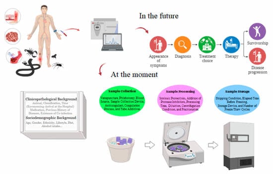
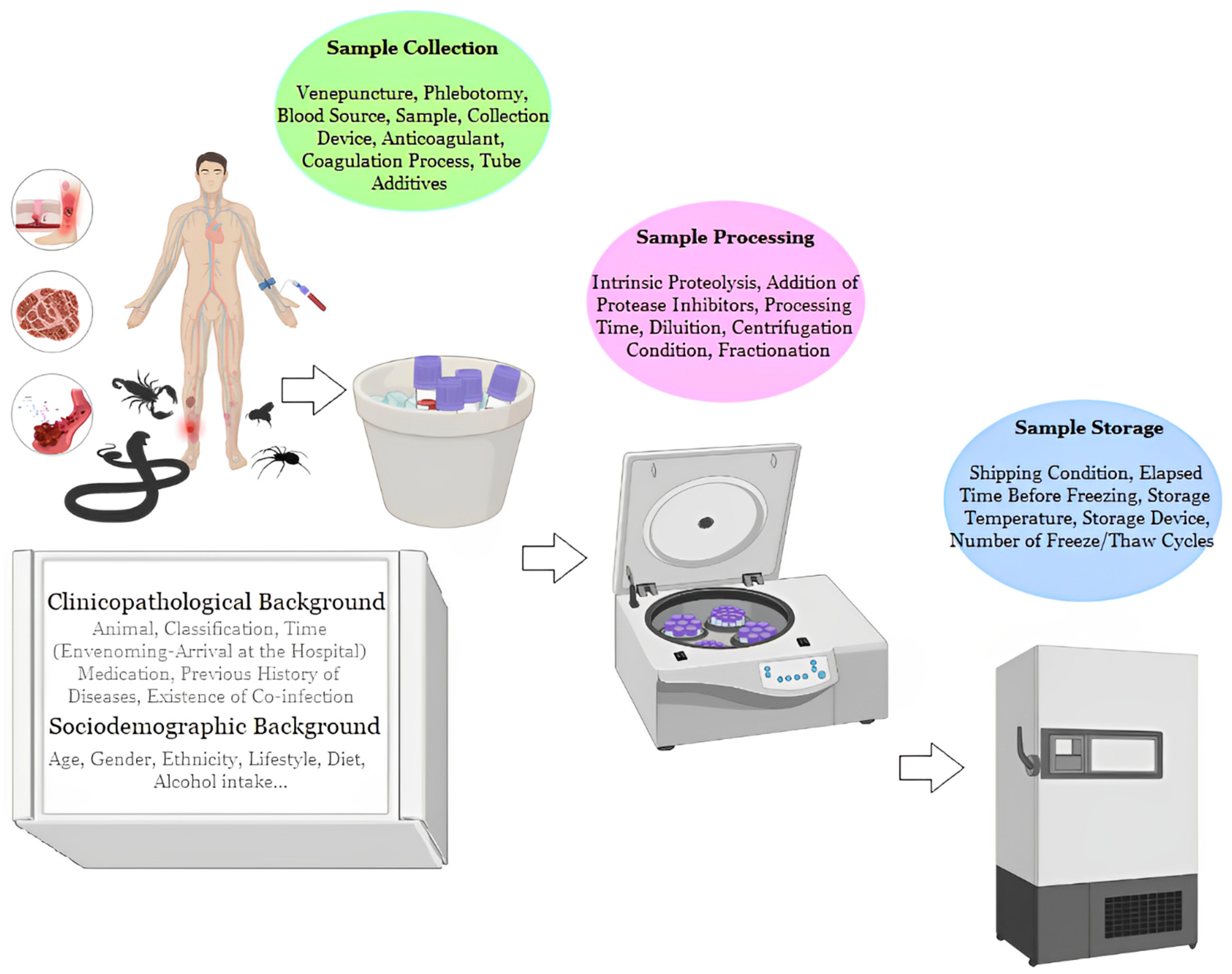
5. Preanalytical Variables: Sample Preparation Challenges

Blood is the most commonly used medium in clinical analysis and an important biofluid for researching diagnostic and prognostic biomarkers of human diseases [13,15,16,17,66,90,92,93,95,97,100,101]. An innumerable number of variables can have an immeasurable impact on the results of the analyses (Figure 2). Therefore, they must be minimized by adopting strict criteria and carrying out the collections through the development and implementation of standard operating procedures (SOPs). SOPs must be strictly followed and include detailed criteria and information to be followed from sample collection to processing [102]. This will help to validate and interpret blood-based biomarker results across all studies and will facilitate the implementation of these biomarkers in diagnostic and assay settings.
The use of blood has its own advantages, such as ease of collection and obtaining, in addition to yield, since more plasma is obtained from an equal amount of whole blood compared to serum. The use of plasma with EDTA or citrate anticoagulant without the addition of protease inhibitors, following the guidelines of the Human Proteome Organization, favors greater reproducibility of the results due to a lower degree of ex vivo degradation [103,104,105,106].
Processing time is also an essential issue for sample preparation and experimental design in research [107]. For example, the processing time alters the integrity of human peripheral blood mononuclear cells (PBMC), leading to contamination and activation of granulocytes that can alter the proteome in a biased manner and should be considered during the performance of the studies [108,109]. On the other hand, samples submitted to freeze/thaw cycles do not show degradation, although hemolysis is a known issue associated with delayed sample processing [110]. However, the greatest pre-analytical variation is reported in the centrifugation time and the time from centrifugation to storage, during which tubes are kept at room temperature or cold [111]. Other pre-analytical variables, such as centrifugation conditions, delay time for the first centrifugation, and blood and anticoagulant storage temperatures, contribute significantly to plasma proteomic variation and may result in increased intracellular plasma proteins [107,112].
The removal of major proteins to improve proteomic analysis has been widely studied, and it is a variable of great relevance for the study of biomarkers. Removing major proteins such as albumin allows the detection of other proteins of lower abundance, and as more proteins are removed, additional proteins can be identified [113]. A wide variety of columns for the depletion of major proteins other than albumin (such as IgG, antitrypsin, IgA, transferrin, and haptoglobin) can also be used [114]. However, the investigator must pay attention to the percentage of target-protein capture efficiency, which is related to the manufacturer, workflow, and amount of target proteins from the column selected for depletion. This influences the number of proteins detected and increases the degree of variability in the effectiveness of depletion among individual isoforms of certain proteins [115]. A potential problem with plasma protein depletion is that some non-target proteins may be removed along with the target proteins, either due to their association with the target proteins or because they interact non-specifically with the column [114,116]. Furthermore, the use of depletion columns involves inserting additional sample manipulation steps, leading to losses and introducing more variability into the data [113].
The efficiency, reproducibility, and non-specific binding of different depletion products have been investigated [117,118,119]; however, most studies have focused on removing human serum albumin (HSA) and IgG [119,120,121,122,123]. Different columns for the depletion of these proteins are commercially available, such as those capable of retaining 7 (MARS Hu-7 from Agilent Technologies), 14 (Seppro IgY14 from Sigma Aldrich or the MARS Hu-14 kit from Agilent Technologies), or 20 high-abundant proteins (HAPs) (ProteoPrep20 from Sigma). A topic that requires attention is the ability to immunocapture the largest number of proteins, which should be considered the most efficient depletion system currently available (such as the ProteoMiner approach). Furthermore, the efficiency in obtaining the data must also be considered. For example, the ProteoMiner column induces a reduction in terms of the total number of identified proteins and the total number of peptides, resulting in a high number of proteins (30%) being identified with only one significant peptide [124].

6. Sociodemographic Background

Human envenomations by venomous animals are known to have a higher incidence in countries with limited resources, as well as those with lower income and other indicators of poverty [125]. This alerts us to a series of patient and environmental characteristics that are factors that can interfere with proteomic analysis. Factors such as the diversity of socio-epidemiological background, the exposure to various environmental risk factors and infectious agents, ethnicity, lifestyle, diet, alcohol intake, cigarette use, and hormonal variables can significantly alter serum/plasma components [13,126,127].
In patients with neglected diseases, it is also possible to observe a reflection of these sociodemographic factors, since they are endemic diseases in low-income populations. Proteomic studies point to variations in plasma proteins in patients with tuberculosis [128], Chagas disease [129], schistosomiasis [130], and leprosy [131], among others.
Regarding the ethnicity and diet of the groups studied, Brenner et al. (2011) evaluated 54 high-abundance plasma proteins in a multiethnic population of healthy young adults and found significant associations with previously identified dietary patterns. Most of these proteins have functions related to processes such as inflammation and lipid metabolism. Another example is the study by García-Bailo and co-authors [91], who observed that Caucasians had higher mean concentrations of adiponectin than East Asians and South Asians.
Specifically, in patients with chronic alcohol abuse, an alteration in the microheterogeneity of serum glycoproteins has already been observed, with abnormal isoforms of transferrin and alpha-1 antitrypsin in the serum [132]. In passive smokers, it was possible to identify nine proteins differentially expressed in plasma. Of these, ceruloplasmin and Inter-alpha-trypsin heavy chain H4 inhibitor (ITIH4), which are two acute-phase inflammatory proteins, showed a high number of isoforms and exhibited an increase in their abundance associated with tobacco exposure. This may have been due to a specific proteolytic cleavage or increased instability due to oxidative modifications [133].
Plasma hormonal variables are closely related to sex, as pointed out by the study by Ramsey et al. (2016), which showed that the serum concentrations of 117 of the 171 (68%) molecules studied were associated with sex and/or female hormonal status. The study took into consideration variables such as age, Body Mass Index, medication use, lifestyle, health, and other relevant demographic variables. The biomarker studies produced up to 40% of false leads when the patient and control groups were not matched for sex, and up to 41% of false results when premenopausal women were not matched for oral contraceptive pill use. Failure to account for sex and female hormonal status as important sources of variability in serum molecule concentrations can result in confounding these variables with disease status, reducing the power to detect differences, and contributing to poor analysis performance [126].
Consequently, the selection of research subjects should follow comparable sex ratios, age groups, demographic characteristics, and dietary considerations. The choice and careful recording of these factors are essential to minimize pre-analytical variation, since the lack of detailed information can lead to misinterpretations of the results obtained. Therefore, meticulous monitoring of all variables is extremely important for data reproducibility and for correct analysis/interpretation [132].

7. Clinicopathological Background

Clinicopathological details, mainly information about the time between envenomation and arrival at the hospital, local and systemic manifestations, clinical complications, the amount of antivenom administered, and use of other drugs, are crucial during the analysis of the plasma proteome of patients. The lack of detailed information can lead to erroneous interpretations of the results obtained. Therefore, meticulous screening of all clinical variables is extremely important to obtain reproducible results and for further analysis/interpretation of the results. To this end, clinical evaluation forms must be completed as clearly and completely as possible and accompany the samples to be entered in the clinical study. Obtaining well-annotated samples is very challenging, but assembling stable biobanks/biorepositories from collected and stored samples from a large number of samples with comprehensive clinical-pathological and socio-epidemiological information can speed up the studies [94,134].

8. Other Directions for Studies of Biomarkers in Envenomations

In recent years, there has been a tremendous advance in the use of clinical proteomics in the search for new biomarkers. Critically, the development and refinement of techniques that allow identifying and validating the use of biomarkers has paved the way for preliminary studies that investigate their usefulness in clinical settings [13,16,17,90,93]. However, in clinical toxinology, the process of searching for new molecules for use in diagnosis, monitoring, and prognosis is still in its early stages. Several significant obstacles begin to emerge from the design of studies for this purpose, but the efforts will bring immeasurable gains in terms of the implementation and usefulness of these molecules in clinical care for envenomation by venomous animals.
The number of species within the same genus can be a critical challenge for studies of biomarkers in envenomation by venomous animals. As observed in experimental studies, different biomarker candidates may be associated with the species that caused the envenomation, requiring the search for candidates that overlap at the genus level [67,68,69]. There is an opportunity for high specificity if the biomarker target is not endogenous, for example, specific toxins with markers [41,50,71]. This highlights the need for large-scale studies and clinical trials involving the recruitment of participants in different regions affected by different species and genera of venomous animals for discovery, validation, and standardization purposes.
Furthermore, the ability to actually target and separate the biomarkers associated with clinical complications from those that are not could be a potential challenge. One of the limitations encountered so far in studies is that they display a map of the proteome differentially expressed at a single moment during disease progression compared to healthy individuals or individuals who did not present a specific event [37,67,68,69,70]. Single-point analysis (case versus control) in the case of envenomations by venomous animals, which involves obtaining an overview of biomarkers at the moment the patient enters the hospital service, will help to identify possible diagnostic markers.
However, longitudinal studies involving repeated observations of the same individuals over different time periods, such as different intervals after antivenom treatment until hospital discharge, may provide valuable additional information about changes in biomarkers and their usefulness as monitoring or prognostic markers [135,136]. In addition, the longitudinal analysis of molecules associated with clinical complications can be identified in this way, with the change in the abundance of these biomarkers being monitored and validated as a potential predictor [83].
While blood-based proteomic analyses have been the most common avenue of investigation, other samples such as exudate and the contents present in blisters offer a unique opportunity to further refine biomarkers and increase their sensitivity, specificity, and overall reliability [41,137]. Indeed, studies comparing the efficacy of plasma and other fluid-based biomarkers (or their combinations) will be of great interest.

9. Conclusions

Human envenomation by venomous animals is of great epidemiological importance in the world due to the high annual number of cases and deaths, which is aggravated by the lack of more precise and specific diagnostic tools for monitoring associated complications. The development of simple, accurate, low-cost, and stable diagnostic, monitoring, and predictive tools for clinical complications and their implementation in developing countries are crucial due to the high incidence of complications and deaths resulting from envenomation by venomous animals, mainly due to the lack of early and predictive diagnosis and timely treatments. Therefore, without a doubt, the use of proteomics in the clinic will be one of the most promising applications for the identification of diagnostic and prognostic biomarkers and the development of predictive tools for complications and envenomation outcomes, which can effectively improve diagnosis and therapy.

Author Contributions

Conceptualization, J.d.S.C.; writing—original draft preparation, J.d.S.C., D.E.G.d.A., M.S.M., S.R.S., P.M.P., P.M.R. and L.L.B.d.O.; writing—review and editing, W.M.M. and R.S.F.-J.; visualization, J.d.S.C.; supervision, R.S.F.-J.; project administration, R.S.F.-J.; funding acquisition, R.S.F.-J. All authors have read and agreed to the published version of the manuscript.

Funding

This research was funded by FAPESP Proc 2021/11936-3 [R.S.F.-J.] and the APC was funded by FAPESP Proc 2021/11936-3 [R.S.F.-J.].

Institutional Review Board Statement

Not applicable.

Informed Consent Statement

Not applicable.

Data Availability Statement

Not applicable.

Acknowledgments

We are thankful to the Coordination of Superior Level Staff Improvement (CAPES)-n° 88887.674376/2022-00 (JSC) and FAPESP Proc 2021/11936-3 [R.S.F.-J.]. R.S.F.-J. is a CNPq PQ1C fellow researcher [303224/2018-5].

Conflicts of Interest

The authors declare no conflict of interest.

References

  1. Williams, H.F.; Layfield, H.J.; Vallance, T.; Patel, K.; Bicknell, A.B.; Trim, S.A.; Vaiyapuri, S. The Urgent Need to Develop Novel Strategies for the Diagnosis and Treatment of Snakebites. Toxins 2019, 11, 363. [Google Scholar] [CrossRef] [PubMed] [Green Version]
  2. Abroug, F.; Ouanes-Besbes, L.; Tilouche, N.; Elatrous, S. Scorpion Envenomation: State of the Art. Intensive Care Med. 2020, 46, 401–410. [Google Scholar] [CrossRef] [PubMed]
  3. Kono, I.S.; Freire, R.L.; Caldart, E.T.; de Souza Rodrigues, F.; Santos, J.A.; Freire, L.G.D.; Faccin, T.C. Bee Stings in Brazil: Epidemiological Aspects in Humans. Toxicon 2021, 201, 59–65. [Google Scholar] [CrossRef] [PubMed]
  4. Schneider, M.C.; Min, K.; Hamrick, P.N.; Montebello, L.R.; Ranieri, T.M.; Mardini, L.; Camara, V.M.; Raggio Luiz, R.; Liese, B.; Vuckovic, M.; et al. Overview of Snakebite in Brazil: Possible Drivers and a Tool for Risk Mapping. PLoS Negl. Trop. Dis. 2021, 15, e0009044. [Google Scholar] [CrossRef] [PubMed]
  5. Pucca, M.B.; Cerni, F.A.; Oliveira, I.S.; Jenkins, T.P.; Argemí, L.; Sørensen, C.V.; Ahmadi, S.; Barbosa, J.E.; Laustsen, A.H. Bee Updated: Current Knowledge on Bee Venom and Bee Envenoming Therapy. Front. Immunol. 2019, 10, 2090. [Google Scholar] [CrossRef] [Green Version]
  6. Teixeira, C.; Fernandes, C.M.; Leiguez, E.; Chudzinski-Tavassi, A.M. Inflammation Induced by Platelet-Activating Viperid Snake Venoms: Perspectives on Thromboinflammation. Front. Immunol. 2019, 10, 2082. [Google Scholar] [CrossRef] [PubMed] [Green Version]
  7. dos Santos Cavalcante, J.; Nogueira Júnior, F.A.; Bezerra Jorge, R.J.; Almeida, C. Pain Modulated by Bothrops Snake Venoms: Mechanisms of Nociceptive Signaling and Therapeutic Perspectives. Toxicon 2021, 201, 105–114. [Google Scholar] [CrossRef]
  8. Pucca, M.B.; Cerni, F.A.; Janke, R.; Bermúdez-Méndez, E.; Ledsgaard, L.; Barbosa, J.E.; Laustsen, A.H. History of Envenoming Therapy and Current Perspectives. Front. Immunol. 2019, 10, 1598. [Google Scholar] [CrossRef] [Green Version]
  9. Laustsen, A.H.; María Gutiérrez, J.; Knudsen, C.; Johansen, K.H.; Bermúdez-Méndez, E.; Cerni, F.A.; Jürgensen, J.A.; Ledsgaard, L.; Martos-Esteban, A.; Øhlenschlæger, M.; et al. Pros and Cons of Different Therapeutic Antibody Formats for Recombinant Antivenom Development. Toxicon 2018, 146, 151–175. [Google Scholar] [CrossRef]
  10. de Castañeda, R.R.; Durso, A.M.; Ray, N.; Fernández, J.L.; Williams, D.J.; Alcoba, G.; Chappuis, F.; Salathé, M.; Bolon, I. Snakebite and Snake Identification: Empowering Neglected Communities and Health-Care Providers with AI. Lancet Digit. Health 2019, 1, e202–e203. [Google Scholar] [CrossRef] [Green Version]
  11. Pucca, M.B.; Knudsen, C.; Oliveira, I.S.; Rimbault, C.; Cerni, F.A.; Wen, F.H.; Sachett, J.; Sartim, M.A.; Laustsen, A.H.; Monteiro, W.M. Current Knowledge on Snake Dry Bites. Toxins 2020, 12, 668. [Google Scholar] [CrossRef] [PubMed]
  12. Daswani, B. Comparison of Different Dosing Protocols of Anti-Snake Venom (ASV) in Snake Bite Cases. J. Clin. Diagn. Res. 2017, 11, FC17–FC21. [Google Scholar] [CrossRef] [PubMed]
  13. Ray, S.; Patel, S.K.; Kumar, V.; Damahe, J.; Srivastava, S. Differential Expression of Serum/Plasma Proteins in Various Infectious Diseases: Specific or Nonspecific Signatures. Proteom. Clin. Appl. 2014, 8, 53–72. [Google Scholar] [CrossRef] [PubMed]
  14. Knudsen, C.; Jürgensen, J.A.; Føns, S.; Haack, A.M.; Friis, R.U.W.; Dam, S.H.; Bush, S.P.; White, J.; Laustsen, A.H. Snakebite Envenoming Diagnosis and Diagnostics. Front. Immunol. 2021, 12, 661457. [Google Scholar] [CrossRef]
  15. Huang, Z.; Ma, L.; Huang, C.; Li, Q.; Nice, E.C. Proteomic Profiling of Human Plasma for Cancer Biomarker Discovery. Proteomics 2017, 17, 1600240. [Google Scholar] [CrossRef]
  16. Suvarna, K.; Biswas, D.; Pai, M.G.J.; Acharjee, A.; Bankar, R.; Palanivel, V.; Salkar, A.; Verma, A.; Mukherjee, A.; Choudhury, M.; et al. Proteomics and Machine Learning Approaches Reveal a Set of Prognostic Markers for COVID-19 Severity With Drug Repurposing Potential. Front. Physiol. 2021, 12, 652799. [Google Scholar] [CrossRef]
  17. Kumar, V.; Ray, S.; Aggarwal, S.; Biswas, D.; Jadhav, M.; Yadav, R.; Sabnis, S.V.; Banerjee, S.; Talukdar, A.; Kochar, S.K.; et al. Multiplexed Quantitative Proteomics Provides Mechanistic Cues for Malaria Severity and Complexity. Commun. Biol. 2020, 3, 683. [Google Scholar] [CrossRef]
  18. Theakston, R.; Laing, G. Diagnosis of Snakebite and the Importance of Immunological Tests in Venom Research. Toxins 2014, 6, 1667–1695. [Google Scholar] [CrossRef] [Green Version]
  19. Hifumi, T.; Sakai, A.; Kondo, Y.; Yamamoto, A.; Morine, N.; Ato, M.; Shibayama, K.; Umezawa, K.; Kiriu, N.; Kato, H.; et al. Venomous Snake Bites: Clinical Diagnosis and Treatment. J. Intensive Care 2015, 3, 16. [Google Scholar] [CrossRef] [Green Version]
  20. Lamb, T.; Abouyannis, M.; de Oliveira, S.S.; Shenoy, K.R.; Geevar, T.; Zachariah, A.; Sharma, S.K.; Bhatt, N.; Mukaka, M.; Harriss, E.; et al. The 20-Minute Whole Blood Clotting Test (20WBCT) for Snakebite Coagulopathy—A Systematic Review and Meta-Analysis of Diagnostic Test Accuracy. PLoS Negl. Trop. Dis. 2021, 15, e0009657. [Google Scholar] [CrossRef]
  21. Hamza, M.; Knudsen, C.; Gnanathasan, C.A.; Monteiro, W.; Lewin, M.R.; Laustsen, A.H.; Habib, A.G. Clinical Management of Snakebite Envenoming: Future Perspectives. Toxicon X 2021, 11, 100079. [Google Scholar] [CrossRef] [PubMed]
  22. Bucaretchi, F.; Pimenta, M.M.B.; Borrasca-Fernandes, C.F.; Prado, C.C.; de Capitani, E.M.; Hyslop, S. Thrombotic Microangiopathy Following Bothrops Jararaca Snakebite: Case Report. Clin. Toxicol. 2019, 57, 294–299. [Google Scholar] [CrossRef] [PubMed]
  23. Malaque, C.M.S.; Duayer, I.F.; Santoro, M.L. Acute Kidney Injury Induced by Thrombotic Microangiopathy in Two Cases of Bothrops Envenomation. Clin. Toxicol. 2019, 57, 213–216. [Google Scholar] [CrossRef]
  24. Mota, S.M.B.; Albuquerque, P.L.M.M.; da Silva Júnior, G.B.; Daher, E.D.F. Thrombotic Microangiopathy Due to Bothrops Erythromelas: A Case Report in Northeast Brazil. Rev. Inst. Med. Trop. Sao Paulo 2020, 62, e53. [Google Scholar] [CrossRef]
  25. Noutsos, T.; Currie, B.J.; Isbister, G.K. Snakebite Associated Thrombotic Microangiopathy: A Protocol for the Systematic Review of Clinical Features, Outcomes, and Role of Interventions. Syst. Rev. 2019, 8, 212. [Google Scholar] [CrossRef] [Green Version]
  26. Luciano, P.M.; Silva, G.E.B.; Azevedo-Marques, M.M. Acidente Botrópico Fatal. Medicina 2009, 42, 61–65. [Google Scholar] [CrossRef] [Green Version]
  27. Valle, L.A.; Silva, D.D.F.R.; Magalhães, P.H.; Mattos, P.A.; Leal, J.A. Bilateral amputation of inferior extremities due to serious Bothrops accident: A case report. Arq. Med. Hosp. Fac. Cienc. Med. Santa Casa São Paulo 2008, 53, 81–84. [Google Scholar]
  28. Graciano, A.R.; De Carvalho, K.C.N. Compartimental syndrome associated to snake bite of the Bothrops gender: Case report. Rev Pesq Saúde 2017, 18, 54–56. [Google Scholar]
  29. Lippi, G.; Schena, F.; Ceriotti, F. Diagnostic Biomarkers of Muscle Injury and Exertional Rhabdomyolysis. Clin. Chem. Lab. Med. 2018, 57, 175–182. [Google Scholar] [CrossRef]
  30. Delanghe, J.R.; Speeckaert, M.M.; de Buyzere, M.L. Is Creatine Kinase an Ideal Biomarker in Rhabdomyolysis? Reply to Lippi et al.: Diagnostic Biomarkers of Muscle Injury and Exertional Rhabdomyolysis (https://doi.org/10.1515/cclm-2018-0656). Clin. Chem. Lab. Med. 2019, 57, e75–e76. [Google Scholar] [CrossRef] [Green Version]
  31. Kularatne, S.A.M.; Budagoda, B.D.S.S.; Gawarammana, I.B.; Kularatne, W.K.S. Epidemiology, Clinical Profile and Management Issues of Cobra (Naja Naja) Bites in Sri Lanka: First Authenticated Case Series. Trans. R. Soc. Trop. Med. Hyg. 2009, 103, 924–930. [Google Scholar] [CrossRef] [PubMed]
  32. Nicoleti, A.F.; de Medeiros, C.R.; Duarte, M.R.; de Siqueira França, F.O. Comparison of Bothropoides Jararaca Bites with and without Envenoming Treated at the Vital Brazil Hospital of the Butantan Institute, State of São Paulo, Brazil. Rev. Soc. Bras. Med. Trop. 2010, 43, 657–661. [Google Scholar] [CrossRef] [Green Version]
  33. Spano, S.; Macias, F.; Snowden, B.; Vohra, R. Snakebite Survivors Club: Retrospective Review of Rattlesnake Bites in Central California. Toxicon 2013, 69, 38–41. [Google Scholar] [CrossRef]
  34. Valenta, J.; Stach, Z.; Stříteský, M.; Michálek, P. Common Viper Bites in the Czech Republic—Epidemiological and Clinical Aspects during 15 Year Period (1999–2013). Prague Med. Rep. 2014, 115, 120–127. [Google Scholar] [CrossRef] [Green Version]
  35. Roth, B.; Sharma, K.; Onisko, N.; Chen, T. Prospective Evaluation of Pain, Swelling, and Disability from Copperhead Envenomation. Clin. Toxicol. 2016, 54, 271–276. [Google Scholar] [CrossRef]
  36. Bawaskar, H.; Bawaskar, P. Diagnosis of Envenomation by Russell’s and Echis Carinatus Viper: A Clinical Study at Rural Maharashtra State of India. J. Family Med. Prim. Care 2019, 8, 1386. [Google Scholar] [CrossRef]
  37. Soares, F.G.S.; Ibiapina, H.N.; Sartim, M.A.; Mendonça-da-Silva, I.; Nascimento, E.F.; Ferreira, L.C.L.; Cerni, F.A.; Malheiro, A.; Pucca, M.B.; Wen, F.H.; et al. Lower Levels of CXCL-8 and IL-2 on Admission as Predictors of Early Adverse Reactions to Bothrops Antivenom in the Brazilian Amazon. Cytokine 2022, 152, 155825. [Google Scholar] [CrossRef]
  38. Dong, L. Immunogenicity of Venoms from Four Common Snakes in the South of Vietnam and Development of ELISA Kit for Venom Detection. J. Immunol. Methods 2003, 282, 13–31. [Google Scholar] [CrossRef]
  39. Kulawickrama, S.; O’Leary, M.A.; Hodgson, W.C.; Brown, S.G.A.; Jacoby, T.; Davern, K.; Isbister, G.K. Development of a Sensitive Enzyme Immunoassay for Measuring Taipan Venom in Serum. Toxicon 2010, 55, 1510–1518. [Google Scholar] [CrossRef]
  40. Liu, C.-C.; Yu, J.-S.; Wang, P.-J.; Hsiao, Y.-C.; Liu, C.-H.; Chen, Y.-C.; Lai, P.-F.; Hsu, C.-P.; Fann, W.-C.; Lin, C.-C. Development of Sandwich ELISA and Lateral Flow Strip Assays for Diagnosing Clinically Significant Snakebite in Taiwan. PLoS Negl. Trop. Dis. 2018, 12, e0007014. [Google Scholar] [CrossRef] [Green Version]
  41. Macêdo, J.K.A.; Joseph, J.K.; Menon, J.; Escalante, T.; Rucavado, A.; Gutiérrez, J.M.; Fox, J.W. Proteomic Analysis of Human Blister Fluids Following Envenomation by Three Snake Species in India: Differential Markers for Venom Mechanisms of Action. Toxins 2019, 11, 246. [Google Scholar] [CrossRef] [PubMed] [Green Version]
  42. Monteiro, W.M.; de Oliveira, S.S.; Pivoto, G.; Alves, E.C.; de Almeida Gonçalves Sachett, J.; Alexandre, C.N.; Fé, N.F.; das Graças Vale Barbosa Guerra, M.; da Silva, I.M.; Tavares, A.M.; et al. Scorpion Envenoming Caused by Tityus Cf. Silvestris Evolving with Severe Muscle Spasms in the Brazilian Amazon. Toxicon 2016, 119, 266–269. [Google Scholar] [CrossRef] [PubMed]
  43. Krifi, M.N.; Kharrat, H.; Zghal, K.; Abdouli, M.; Abroug, F.; Bouchoucha, S.; Dellagi, K.; el Ayeb, M. Development of an ELISA for the Detection of Scorpion Venoms in Sera of Humans Envenomed by Androctonus Australis Garzonii (Aag) and Buthus Occitanus Tunetanus (Bot): Correlation with Clinical Severity of Envenoming in Tunisia. Toxicon 1998, 36, 887–900. [Google Scholar] [CrossRef] [PubMed]
  44. Chávez-Olórtegui, C.; Fonseca, S.C.G.; Campolina, D.; Amaral, C.F.S.; Diniz, C.R. ELISA for the Detection of Toxic Antigens in Experimental and Clinical Envenoming by Tityus Serrulatus Scorpion Venom. Toxicon 1994, 32, 1649–1656. [Google Scholar] [CrossRef] [PubMed]
  45. de Rezende, N.A.; Dias, M.B.; Campolina, D.; Chavéz-Olortegui, C.; Amaral, C.F.S. Standardization of an Enzyme Linked Immunosorbent Assay (ELISA) for Detecting Circulating Toxic Venom Antigens in Patients Stung by the Scorpion Tityus Serrulatus. Rev. Inst. Med. Trop. Sao Paulo 1995, 37, 71–74. [Google Scholar] [CrossRef] [PubMed] [Green Version]
  46. Bahloul, M.; Chaari, A.; Dammak, H.; Samet, M.; Chtara, K.; Chelly, H.; ben Hamida, C.; Kallel, H.; Bouaziz, M. Pulmonary Edema Following Scorpion Envenomation: Mechanisms, Clinical Manifestations, Diagnosis and Treatment. Int. J. Cardiol. 2013, 162, 86–91. [Google Scholar] [CrossRef] [PubMed]
  47. Abdi, A.; Farshidi, H.; Rahimi, S.; Amini, A.; Tasnim Eftekhari, S.F. Electrocardiologic and Echocardiographic Findings in Patients with Scorpion Sting. Iran. Red. Crescent Med. J. 2013, 15, 446–447. [Google Scholar] [CrossRef] [Green Version]
  48. Kumar, C.M.; Naveen Prasad, S. v Echocardiologic Evaluation and Follow-up of Cardiovascular Complications in Children with Scorpion Sting in Coastal South India. Indian J. Crit. Care Med. 2015, 19, 42–46. [Google Scholar] [CrossRef] [PubMed]
  49. Barbosa, A.N.; Boyer, L.; Chippaux, J.-P.; Medolago, N.B.; Caramori, C.A.; Paixão, A.G.; Poli, J.P.V.; Mendes, M.B.; dos Santos, L.D.; Ferreira, R.S.; et al. A Clinical Trial Protocol to Treat Massive Africanized Honeybee (Apis mellifera) Attack with a New Apilic Antivenom. J. Venom. Anim. Toxins Incl. Trop. Dis. 2017, 23, 14. [Google Scholar] [CrossRef] [Green Version]
  50. Barbosa, A.N.; Ferreira, R.S.; de Carvalho, F.C.T.; Schuelter-Trevisol, F.; Mendes, M.B.; Mendonça, B.C.; Batista, J.N.; Trevisol, D.J.; Boyer, L.; Chippaux, J.-P.; et al. Single-Arm, Multicenter Phase I/II Clinical Trial for the Treatment of Envenomings by Massive Africanized Honey Bee Stings Using the Unique Apilic Antivenom. Front. Immunol. 2021, 12, 653151. [Google Scholar] [CrossRef]
  51. Vetter, R.S.; Isbister, G.K. Medical Aspects of Spider Bites. Annu. Rev. Entomol. 2008, 53, 409–429. [Google Scholar] [CrossRef]
  52. Dunbar, J.P.; Vitkauskaite, A.; O’Keeffe, D.T.; Fort, A.; Sulpice, R.; Dugon, M.M. Bites by the Noble False Widow Spider Steatoda Nobilis Can Induce Latrodectus -like Symptoms and Vector-Borne Bacterial Infections with Implications for Public Health: A Case Series. Clin. Toxicol. 2022, 60, 59–70. [Google Scholar] [CrossRef]
  53. Jerusalem, K.; Salavert Lletí, M. Probable Cutaneous Loxoscelism with Mild Systemic Symptoms: A Case Report from Spain. Toxicon 2018, 156, 7–12. [Google Scholar] [CrossRef]
  54. Langner, T.R.; Ganatra, H.A.; Schwerdtfager, J.; Stoecker, W.; Thornton, S. Viscerocutaneous Loxoscelism Manifesting with Myocarditis: A Case Report. Am. J. Case Rep. 2021, 22, e932378. [Google Scholar] [CrossRef]
  55. Lopes, P.H.; Squaiella-Baptistão, C.C.; Marques, M.O.T.; Tambourgi, D.V. Clinical Aspects, Diagnosis and Management of Loxosceles Spider Envenomation: Literature and Case Review. Arch. Toxicol. 2020, 94, 1461–1477. [Google Scholar] [CrossRef]
  56. Stoecker, W.V.; Green, J.A.; Gomez, H.F. Diagnosis of Loxoscelism in a Child Confirmed with an Enzyme-Linked Immunosorbent Assay and Noninvasive Tissue Sampling. J. Am. Acad. Dermatol. 2006, 55, 888–890. [Google Scholar] [CrossRef] [Green Version]
  57. Keklikci, U.; Akdeniz, S.; Sakalar, Y.B.; Cakmak, S.S.; Unlu, K. Loxosceles Reclusa Bite to the Eyelid. Eur. J. Ophthalmol. 2008, 18, 633–635. [Google Scholar] [CrossRef]
  58. Krywko, D.M.; Gomez, H.F. Detection of Loxosceles Species Venom in Dermal Lesions: A Comparison of 4 Venom Recovery Methods. Ann. Emerg. Med. 2002, 39, 475–480. [Google Scholar] [CrossRef]
  59. Miller, M.J.; Gomez, H.F.; Snider, R.J.; Stephens, E.L.; Czop, R.M.; Warren, J.S. Detection of Loxosceles Venom in Lesional Hair Shafts and Skin: Application of a Specific Immunoassay to Identify Dermonecrotic Arachnidism. Am. J. Emerg. Med. 2000, 18, 626–628. [Google Scholar] [CrossRef]
  60. Chávez-Olórtegui, C.; Zanetti, V.C.; Ferreira, A.P.; Minozzo, J.C.; Mangili, O.C.; Gubert, I.C. ELISA for the Detection of Venom Antigens in Experimental and Clinical Envenoming by Loxosceles Intermedia Spiders. Toxicon 1998, 36, 563–569. [Google Scholar] [CrossRef]
  61. Chávez-Olórtegui, C.; Bohórquez, K.; Alvarenga, L.M.; Kalapothakis, E.; Campolina, D.; Maria, W.S.; Diniz, C.R. Sandwich-ELISA Detection of Venom Antigens in Envenoming by Phoneutria Nigriventer Spider. Toxicon 2001, 39, 909–911. [Google Scholar] [CrossRef] [PubMed]
  62. Bucaretchi, F.; Mello, S.M.; Vieira, R.J.; Mamoni, R.L.; Blotta, M.H.S.L.; Antunes, E.; Hyslop, S. Systemic Envenomation Caused by the Wandering Spider Phoneutria Nigriventer, with Quantification of Circulating Venom. Clin. Toxicol. 2008, 46, 885–889. [Google Scholar] [CrossRef] [Green Version]
  63. Miller, M.; O’Leary, M.A.; Isbister, G.K. Towards Rationalisation of Antivenom Use in Funnel-Web Spider Envenoming: Enzyme Immunoassays for Venom Concentrations. Clin. Toxicol. 2016, 54, 245–251. [Google Scholar] [CrossRef] [PubMed]
  64. Feng, K.; Lan, Y.; Zhu, X.; Li, J.; Chen, T.; Huang, Q.; Ho, C.-T.; Chen, Y.; Cao, Y. Hepatic Lipidomics Analysis Reveals the Antiobesity and Cholesterol-Lowering Effects of Tangeretin in High-Fat Diet-Fed Rats. J. Agric. Food Chem. 2020, 68, 6142–6153. [Google Scholar] [CrossRef] [PubMed]
  65. Aggarwal, H.; Pathak, P.; Kumar, Y.; Jagavelu, K.; Dikshit, M. Modulation of Insulin Resistance, Dyslipidemia and Serum Metabolome in INOS Knockout Mice Following Treatment with Nitrite, Metformin, Pioglitazone, and a Combination of Ampicillin and Neomycin. Int. J. Mol. Sci. 2021, 23, 195. [Google Scholar] [CrossRef]
  66. Shu, T.; Ning, W.; Wu, D.; Xu, J.; Han, Q.; Huang, M.; Zou, X.; Yang, Q.; Yuan, Y.; Bie, Y.; et al. Plasma Proteomics Identify Biomarkers and Pathogenesis of COVID-19. Immunity 2020, 53, 1108–1122.e5. [Google Scholar] [CrossRef]
  67. Cavalcante, J.S.; Brito, I.M.d.C.; de Oliveira, L.A.; de Barros, L.C.; Almeida, C.; Rossini, B.C.; Sousa, D.L.; Alves, R.S.; Jorge, R.J.B.; Santos, L.D. dos Experimental Bothrops Atrox Envenomation: Blood Plasma Proteome Effects after Local Tissue Damage and Perspectives on Thromboinflammation. Toxins 2022, 14, 613. [Google Scholar] [CrossRef]
  68. Cavalcante, J.S.; da Silva, W.R.G.B.; de Oliveira, L.A.; Brito, I.M.C.; Muller, K.S.; Vidal, I.S.J.; dos Santos, L.D.; Jorge, R.J.B.; Almeida, C.; Bicho, C.D.L. Blood Plasma Proteome Alteration after Local Tissue Damage Induced by Bothrops Erythromelas Snake Venom in Mice. J. Proteom. 2022, 269, 104742. [Google Scholar] [CrossRef]
  69. Cavalcante, J.d.S.; de Almeida, C.A.S.; Clasen, M.A.; da Silva, E.L.; de Barros, L.C.; Marinho, A.D.; Rossini, B.C.; Marino, C.L.; Carvalho, P.C.; Jorge, R.J.B.; et al. A Fingerprint of Plasma Proteome Alteration after Local Tissue Damage Induced by Bothrops Leucurus Snake Venom in Mice. J. Proteom. 2022, 253, 104464. [Google Scholar] [CrossRef]
  70. Dong, D.; Deng, Z.; Yan, Z.; Mao, W.; Yi, J.; Song, M.; Li, Q.; Chen, J.; Chen, Q.; Liu, L.; et al. Oxidative Stress and Antioxidant Defense in Detoxification Systems of Snake Venom-Induced Toxicity. J. Venom. Anim. Toxins Incl. Trop. Dis. 2020, 26, e20200053. [Google Scholar] [CrossRef]
  71. Luna, K.P.O.; Xavier, E.M.; Pascoal, V.P.M.; Martins-Filho, O.A.; Pereira, V.R.A. Humoral Immune Response of Patients Bitten by the Snake Bothrops Erythromelas. Rev. Soc. Bras. Med. Trop. 2010, 43, 731–732. [Google Scholar] [CrossRef] [Green Version]
  72. Fu, Y.; Zhang, Y.; Ju, S.; Ma, B.; Huang, W.; Luo, S. Isolation and Characterization of Five Novel Disulfide-Poor Conopeptides from Conus Marmoreus Venom. J. Venom. Anim. Toxins Incl. Trop. Dis. 2022, 28, e20210116. [Google Scholar] [CrossRef] [PubMed]
  73. Bao, N.; Lecaer, J.-P.; Nghia, N.D.; Vinh, P.T.K. Isolation and Structural Identification of a New T1-Conotoxin with Unique Disulfide Connectivities Derived from Conus Bandanus. J. Venom. Anim. Toxins Incl. Trop. Dis. 2020, 26, e20190095. [Google Scholar] [CrossRef] [PubMed]
  74. dos Santos, R.T.F.; Silva, M.F.P.; Porto, R.M.; Lebrun, I.; de Camargo Gonçalves, L.R.; de Fátima Correia Batista, I.; Sandoval, M.R.L.; Abdalla, F.M.F. Effects of Mlx-8, a Phospholipase A2 from Brazilian Coralsnake Micrurus Lemniscatus Venom, on Muscarinic Acetylcholine Receptors in Rat Hippocampus. J. Venom. Anim. Toxins Incl. Trop. Dis. 2020, 26, e20190041. [Google Scholar] [CrossRef] [PubMed]
  75. de Melo-Braga, M.N.; da Silva Moreira, R.; Gervásio, J.H.D.B.; Felicori, L.F. Overview of Protein Posttranslational Modifications in Arthropoda Venoms. J. Venom. Anim. Toxins Incl. Trop. Dis. 2022, 28, e20210047. [Google Scholar] [CrossRef]
  76. Marchi, F.C.; Mendes-Silva, E.; Rodrigues-Ribeiro, L.; Bolais-Ramos, L.G.; Verano-Braga, T. Toxinology in the Proteomics Era: A Review on Arachnid Venom Proteomics. J. Venom. Anim. Toxins Incl. Trop. Dis. 2022, 28, 20210034. [Google Scholar] [CrossRef]
  77. Anita, S.; Sadjuri, A.R.; Rahmah, L.; Nugroho, H.A.; Mulyadi; Trilaksono, W.; Ridhani, W.; Safira, N.; Bahtiar, H.; Maharani; et al. Venom Composition of Trimeresurus slbolabris, T. insularis, T. puniceus and T. purpureomaculatus from Indonesia. J. Venom. Anim. Toxins Incl. Trop. Dis. 2022, 28, e20210103. [Google Scholar] [CrossRef]
  78. Chanhome, L.; Khow, O.; Reamtong, O.; Vasaruchapong, T.; Laoungbua, P.; Tawan, T.; Suntrarachun, S.; Sitprija, S.; Kumkate, S.; Chaiyabutr, N. Biochemical and Proteomic Analyses of Venom from a New Pit Viper, Protobothrops Kelomohy. J. Venom. Anim. Toxins Incl. Trop. Dis. 2022, 28, e20210080. [Google Scholar] [CrossRef]
  79. Nie, X.; He, Q.; Zhou, B.; Huang, D.; Chen, J.; Chen, Q.; Yang, S.; Yu, X. Exploring the Five-Paced Viper (Deinagkistrodon acutus) Venom Proteome by Integrating a Combinatorial Peptide Ligand Library Approach with Shotgun LC-MS/MS. J. Venom. Anim. Toxins Incl. Trop. Dis. 2021, 27, e20200196. [Google Scholar] [CrossRef]
  80. Faisal, T.; Tan, N.H.; Sim, S.M.; Gnanathasan, C.A.; Tan, C.H. Proteomics, Toxicity and Antivenom Neutralization of Sri Lankan and Indian Russell’s Viper (Daboia russelii) Venoms. J. Venom. Anim. Toxins Incl. Trop. Dis. 2021, 27, e20200177. [Google Scholar] [CrossRef]
  81. Sanz, L.; Pérez, A.; Quesada-Bernat, S.; Diniz-Sousa, R.; Calderón, L.A.; Soares, A.M.; Calvete, J.J.; Caldeira, C.A.S. Venomics and Antivenomics of the Poorly Studied Brazil’s Lancehead, Bothrops Brazili (Hoge, 1954), from the Brazilian State of Pará. J. Venom. Anim. Toxins Incl. Trop. Dis. 2020, 26, e20190103. [Google Scholar] [CrossRef] [PubMed]
  82. Tasoulis, T.; Isbister, G. A Review and Database of Snake Venom Proteomes. Toxins 2017, 9, 290. [Google Scholar] [CrossRef] [PubMed] [Green Version]
  83. Herrera, C.; Macêdo, J.K.A.; Feoli, A.; Escalante, T.; Rucavado, A.; Gutiérrez, J.M.; Fox, J.W. Muscle Tissue Damage Induced by the Venom of Bothrops Asper: Identification of Early and Late Pathological Events through Proteomic Analysis. PLoS Negl. Trop. Dis. 2016, 10, e0004599. [Google Scholar] [CrossRef] [PubMed] [Green Version]
  84. Brasileiro-Martins, L.M.; Nascimento, T.P.; Silva-Neto, A.V.; Martins, F.; Cavalcante, S.A.; Martins, R.B.; Marques, H.; Colombini, M.; Martins, M.; Sartim, M.A.; et al. The Severity of Acute Kidney Injury Correlates with Plasma Venom Levels in Bothrops Atrox Envenomings. Toxicon 2022, 219, 106924. [Google Scholar] [CrossRef]
  85. Casewell, N.R.; Jackson, T.N.W.; Laustsen, A.H.; Sunagar, K. Causes and Consequences of Snake Venom Variation. Trends Pharmacol. Sci. 2020, 41, 570–581. [Google Scholar] [CrossRef]
  86. Rashmi, U.; Khochare, S.; Attarde, S.; Laxme, R.R.S.; Suranse, V.; Martin, G.; Sunagar, K. Remarkable Intrapopulation Venom Variability in the Monocellate Cobra (Naja kaouthia) Unveils Neglected Aspects of India’s Snakebite Problem. J. Proteom. 2021, 242, 104256. [Google Scholar] [CrossRef]
  87. Bourke, L.A.; Zdenek, C.N.; Neri-Castro, E.; Bénard-Valle, M.; Alagón, A.; Gutiérrez, J.M.; Sanchez, E.F.; Aldridge, M.; Fry, B.G. Pan-American Lancehead Pit-Vipers: Coagulotoxic Venom Effects and Antivenom Neutralisation of Bothrops Asper and B. Atrox Geographical Variants. Toxins 2021, 13, 78. [Google Scholar] [CrossRef]
  88. Laustsen, A.H.; Ainsworth, S.; Lomonte, B.; Kini, R.M.; Chávez-Olórtegui, C. Editorial: Novel Immunotherapies Against Envenomings by Snakes and Other Venomous Animals. Front. Immunol. 2020, 11, 1004. [Google Scholar] [CrossRef]
  89. Long, C.; Wu, F.; Lu, Q.; Xie, B.; Shen, C.; Li, J.; Deng, Y.; Liang, P.; Yu, Y.; Lai, R. A Strategy for Efficient Preparation of Genus-Specific Diagnostic Antibodies for Snakebites. Front. Immunol. 2021, 12, 775678. [Google Scholar] [CrossRef]
  90. García-Gutiérrez, M.S.; Navarrete, F.; Sala, F.; Gasparyan, A.; Austrich-Olivares, A.; Manzanares, J. Biomarkers in Psychiatry: Concept, Definition, Types and Relevance to the Clinical Reality. Front. Psychiatry 2020, 11, 432. [Google Scholar] [CrossRef]
  91. García-Bailo, B.; Brenner, D.R.; Nielsen, D.; Lee, H.-J.; Domanski, D.; Kuzyk, M.; Borchers, C.H.; Badawi, A.; Karmali, M.A.; El-Sohemy, A. Dietary Patterns and Ethnicity Are Associated with Distinct Plasma Proteomic Groups. Am. J. Clin. Nutr. 2012, 95, 352–361. [Google Scholar] [CrossRef] [PubMed] [Green Version]
  92. Ray, S.; Kumar, V.; Bhave, A.; Singh, V.; Gogtay, N.J.; Thatte, U.M.; Talukdar, A.; Kochar, S.K.; Patankar, S.; Srivastava, S. Proteomic Analysis of Plasmodium Falciparum Induced Alterations in Humans from Different Endemic Regions of India to Decipher Malaria Pathogenesis and Identify Surrogate Markers of Severity. J. Proteom. 2015, 127, 103–113. [Google Scholar] [CrossRef] [PubMed]
  93. Moussa, E.M.; Huang, H.; Thézénas, M.L.; Fischer, R.; Ramaprasad, A.; Sisay-Joof, F.; Jallow, M.; Pain, A.; Kwiatkowski, D.; Kessler, B.M.; et al. Proteomic Profiling of the Plasma of Gambian Children with Cerebral Malaria. Malar J. 2018, 17, 337. [Google Scholar] [CrossRef] [PubMed]
  94. Ray, S.; Moiyadi, A.; Srivastava, S. Biorepositories for Cancer Research in Developing Countries. Nat. Rev. Clin. Oncol. 2013, 10, 434–436. [Google Scholar] [CrossRef]
  95. Ray, S.; Patel, S.K.; Venkatesh, A.; Chatterjee, G.; Ansari, N.N.; Gogtay, N.J.; Thatte, U.M.; Gandhe, P.; Varma, S.G.; Patankar, S.; et al. Quantitative Proteomics Analysis of Plasmodium Vivax Induced Alterations in Human Serum during the Acute and Convalescent Phases of Infection. Sci. Rep. 2017, 7, 4400. [Google Scholar] [CrossRef] [Green Version]
  96. Vidova, V.; Spacil, Z. A Review on Mass Spectrometry-Based Quantitative Proteomics: Targeted and Data Independent Acquisition. Anal. Chim. Acta 2017, 964, 7–23. [Google Scholar] [CrossRef]
  97. Masucci, G.V.; Cesano, A.; Hawtin, R.; Janetzki, S.; Zhang, J.; Kirsch, I.; Dobbin, K.K.; Alvarez, J.; Robbins, P.B.; Selvan, S.R.; et al. Validation of Biomarkers to Predict Response to Immunotherapy in Cancer: Volume I—Pre-Analytical and Analytical Validation. J. Immunother. Cancer 2016, 4, 76. [Google Scholar] [CrossRef] [Green Version]
  98. Santos, F.L.N.; Celedon, P.A.F.; Zanchin, N.I.T.; de Souza, W.V.; da Silva, E.D.; Foti, L.; Krieger, M.A.; de Miranda Gomes, Y. Accuracy of Chimeric Proteins in the Serological Diagnosis of Chronic Chagas Disease—A Phase II Study. PLoS Negl. Trop. Dis. 2017, 11, e0005433. [Google Scholar] [CrossRef] [Green Version]
  99. Manole, E.; Bastian, A.E.D.; Popescu, I.; Constantin, C.; Mihai, S.; Gaina, G.F.; Codrici, E.; Neagu, M.T. Immunoassay Techniques Highlighting Biomarkers in Immunogenetic Diseases. In Immunogenetics; IntechOpen: London, UK, 2019; Volume 6. [Google Scholar] [CrossRef] [Green Version]
  100. Manjunath, D.; Kumaraswamy, S.B.; Venkatakrishniah, S.A.; Appaiah, H.N.; Thomas, A.; Banerjee, S.D. Validation and Evaluation of a Common Biomarker in Human Cancers Sera Protein Detected by a Monoclonal Antibody UNIVmAb. BMC Res. Notes 2019, 12, 744. [Google Scholar] [CrossRef] [Green Version]
  101. Huang, C.; Wang, Y.; Liu, S.; Ding, G.; Liu, W.; Zhou, J.; Kuang, M.; Ji, Y.; Kondo, T.; Fan, J. Quantitative Proteomic Analysis Identified Paraoxonase 1 as a Novel Serum Biomarker for Microvascular Invasion in Hepatocellular Carcinoma. J. Proteome Res. 2013, 12, 1838–1846. [Google Scholar] [CrossRef]
  102. Stewart, T.; Shi, M.; Mehrotra, A.; Aro, P.; Soltys, D.; Kerr, K.F.; Zabetian, C.P.; Peskind, E.R.; Taylor, P.; Shaw, L.M.; et al. Impact of Pre-Analytical Differences on Biomarkers in the ADNI and PPMI Studies: Implications in the Era of Classifying Disease Based on Biomarkers. J. Alzheimer’s Dis. 2019, 69, 263–276. [Google Scholar] [CrossRef] [PubMed]
  103. Omenn, G.S. Exploring the Human Plasma Proteome. Proteomics 2005, 5, 3223–3225. [Google Scholar] [CrossRef] [PubMed] [Green Version]
  104. Omenn, G.S.; States, D.J.; Adamski, M.; Blackwell, T.W.; Menon, R.; Hermjakob, H.; Apweiler, R.; Haab, B.B.; Simpson, R.J.; Eddes, J.S.; et al. Overview of the HUPO Plasma Proteome Project: Results from the Pilot Phase with 35 Collaborating Laboratories and Multiple Analytical Groups, Generating a Core Dataset of 3020 Proteins and a Publicly-Available Database. Proteomics 2005, 5, 3226–3245. [Google Scholar] [CrossRef]
  105. Mahboob, S.; Ahn, S.B.; Mohamedali, A.; Amirkhani, A.; Tan, S.; Ranganathan, S.; Nice, E.; Baker, M.S. Addition of Protease Inhibitors to EDTA Containing Blood Collection Tubes Does Not Deliver Significant Advantage for Preservation of Plasma for Down-Stream Analysis. Clin. Proteom. Bioinform. 2016, 1, 1–9. [Google Scholar] [CrossRef]
  106. Lan, J.; Núñez Galindo, A.; Doecke, J.; Fowler, C.; Martins, R.N.; Rainey-Smith, S.R.; Cominetti, O.; Dayon, L. Systematic Evaluation of the Use of Human Plasma and Serum for Mass-Spectrometry-Based Shotgun Proteomics. J. Proteome Res. 2018, 17, 1426–1435. [Google Scholar] [CrossRef]
  107. Hassis, M.E.; Niles, R.K.; Braten, M.N.; Albertolle, M.E.; Ewa Witkowska, H.; Hubel, C.A.; Fisher, S.J.; Williams, K.E. Evaluating the Effects of Preanalytical Variables on the Stability of the Human Plasma Proteome. Anal. Biochem. 2015, 478, 14–22. [Google Scholar] [CrossRef] [PubMed] [Green Version]
  108. Agashe, C.; Chiang, D.; Grishin, A.; Masilamani, M.; Jones, S.M.; Wood, R.A.; Sicherer, S.H.; Burks, A.W.; Leung, D.Y.M.; Dawson, P.; et al. Impact of Granulocyte Contamination on PBMC Integrity of Shipped Blood Samples: Implications for Multi-Center Studies Monitoring Regulatory T Cells. J. Immunol. Methods 2017, 449, 23–27. [Google Scholar] [CrossRef]
  109. Bonilauri, B.; Santos, M.D.M.; Camillo-Andrade, A.C.; Bispo, S.; Nogueira, F.C.S.; Carvalho, P.C.; Zanchin, N.I.T.; de S. da G. Fischer, J. The Impact of Blood-Processing Time on the Proteome of Human Peripheral Blood Mononuclear Cells. Biochim. et Biophys. Acta (BBA)—Proteins Proteom. 2021, 1869, 140581. [Google Scholar] [CrossRef]
  110. Mateos, J.; Carneiro, I.; Corrales, F.; Elortza, F.; Paradela, A.; del Pino, M.S.; Iloro, I.; Marcilla, M.; Mora, M.I.; Valero, L.; et al. Multicentric Study of the Effect of Pre-Analytical Variables in the Quality of Plasma Samples Stored in Biobanks Using Different Complementary Proteomic Methods. J. Proteom. 2017, 150, 109–120. [Google Scholar] [CrossRef] [Green Version]
  111. Verberk, I.M.W.; Misdorp, E.O.; Koelewijn, J.; Ball, A.J.; Blennow, K.; Dage, J.L.; Fandos, N.; Hansson, O.; Hirtz, C.; Janelidze, S.; et al. Characterization of Pre-analytical Sample Handling Effects on a Panel of Alzheimer’s Disease–Related Blood-based Biomarkers: Results from the Standardization of Alzheimer’s Blood Biomarkers (SABB) Working Group. Alzheimer’s Dement. 2022, 18, 1484–1497. [Google Scholar] [CrossRef]
  112. Halvey, P.; Farutin, V.; Koppes, L.; Gunay, N.S.; Pappas, D.A.; Manning, A.M.; Capila, I. Variable Blood Processing Procedures Contribute to Plasma Proteomic Variability. Clin. Proteom. 2021, 18, 5. [Google Scholar] [CrossRef] [PubMed]
  113. Roche, S.; Tiers, L.; Provansal, M.; Seveno, M.; Piva, M.-T.; Jouin, P.; Lehmann, S. Depletion of One, Six, Twelve or Twenty Major Blood Proteins before Proteomic Analysis: The More the Better? J. Proteom. 2009, 72, 945–951. [Google Scholar] [CrossRef] [PubMed]
  114. Tu, C.; Rudnick, P.A.; Martinez, M.Y.; Cheek, K.L.; Stein, S.E.; Slebos, R.J.C.; Liebler, D.C. Depletion of Abundant Plasma Proteins and Limitations of Plasma Proteomics. J. Proteome Res. 2010, 9, 4982–4991. [Google Scholar] [CrossRef] [PubMed] [Green Version]
  115. Cao, X.; Sandberg, A.; Araújo, J.E.; Cvetkovski, F.; Berglund, E.; Eriksson, L.E.; Pernemalm, M. Evaluation of Spin Columns for Human Plasma Depletion to Facilitate MS-Based Proteomics Analysis of Plasma. J. Proteome Res. 2021, 20, 4610–4620. [Google Scholar] [CrossRef] [PubMed]
  116. Zolotarjova, N.; Mrozinski, P.; Chen, H.; Martosella, J. Combination of Affinity Depletion of Abundant Proteins and Reversed-Phase Fractionation in Proteomic Analysis of Human Plasma/Serum. J. Chromatogr. A 2008, 1189, 332–338. [Google Scholar] [CrossRef]
  117. Qian, W.-J.; Kaleta, D.T.; Petritis, B.O.; Jiang, H.; Liu, T.; Zhang, X.; Mottaz, H.M.; Varnum, S.M.; Camp, D.G.; Huang, L.; et al. Enhanced Detection of Low Abundance Human Plasma Proteins Using a Tandem IgY12-SuperMix Immunoaffinity Separation Strategy. Mol. Cell. Proteom. 2008, 7, 1963–1973. [Google Scholar] [CrossRef] [PubMed] [Green Version]
  118. Polaskova, V.; Kapur, A.; Khan, A.; Molloy, M.P.; Baker, M.S. High-Abundance Protein Depletion: Comparison of Methods for Human Plasma Biomarker Discovery. Electrophoresis 2010, 31, 471–482. [Google Scholar] [CrossRef]
  119. Ahmed, N.; Barker, G.; Oliva, K.; Garfin, D.; Talmadge, K.; Georgiou, H.; Quinn, M.; Rice, G. An Approach to Remove Albumin for the Proteomic Analysis of Low Abundance Biomarkers in Human Serum. Proteomics 2003, 3, 1980–1987. [Google Scholar] [CrossRef]
  120. Colantonio, D.A.; Dunkinson, C.; Bovenkamp, D.E.; van Eyk, J.E. Effective Removal of Albumin from Serum. Proteomics 2005, 5, 3831–3835. [Google Scholar] [CrossRef]
  121. Chen, Y.-Y.; Lin, S.-Y.; Yeh, Y.-Y.; Hsiao, H.-H.; Wu, C.-Y.; Chen, S.-T.; Wang, A.H.-J. A Modified Protein Precipitation Procedure for Efficient Removal of Albumin from Serum. Electrophoresis 2005, 26, 2117–2127. [Google Scholar] [CrossRef]
  122. Govorukhina, N.I.; Keizer-Gunnink, A.; van der Zee, A.G.J.; de Jong, S.; de Bruijn, H.W.A.; Bischoff, R. Sample Preparation of Human Serum for the Analysis of Tumor Markers. J. Chromatogr. A 2003, 1009, 171–178. [Google Scholar] [CrossRef] [PubMed]
  123. Quero, C.; Colomé, N.; Prieto, M.R.; Carrascal, M.; Posada, M.; Gelpí, E.; Abian, J. Determination of Protein Markers in Human Serum: Analysis of Protein Expression in Toxic Oil Syndrome Studies. Proteomics 2004, 4, 303–315. [Google Scholar] [CrossRef]
  124. Millioni, R.; Tolin, S.; Puricelli, L.; Sbrignadello, S.; Fadini, G.P.; Tessari, P.; Arrigoni, G. High Abundance Proteins Depletion vs Low Abundance Proteins Enrichment: Comparison of Methods to Reduce the Plasma Proteome Complexity. PLoS ONE 2011, 6, e19603. [Google Scholar] [CrossRef] [PubMed] [Green Version]
  125. Harrison, R.A.; Hargreaves, A.; Wagstaff, S.C.; Faragher, B.; Lalloo, D.G. Snake Envenoming: A Disease of Poverty. PLoS Negl. Trop. Dis. 2009, 3, e569. [Google Scholar] [CrossRef] [Green Version]
  126. Ramsey, J.M.; Cooper, J.D.; Penninx, B.W.J.H.; Bahn, S. Variation in Serum Biomarkers with Sex and Female Hormonal Status: Implications for Clinical Tests. Sci. Rep. 2016, 6, 26947. [Google Scholar] [CrossRef] [PubMed] [Green Version]
  127. Rai, A.J.; Vitzthum, F. Effects of Preanalytical Variables on Peptide and Protein Measurements in Human Serum and Plasma: Implications for Clinical Proteomics. Expert Rev. Proteom. 2006, 3, 409–426. [Google Scholar] [CrossRef]
  128. Bahk, Y.Y.; Kim, S.A.; Kim, J.-S.; Euh, H.-J.; Bai, G.-H.; Cho, S.-N.; Kim, Y.S. Antigens Secreted FromMycobacterium Tuberculosis: Identification by Proteomics Approach and Test for Diagnostic Marker. Proteomics 2004, 4, 3299–3307. [Google Scholar] [CrossRef]
  129. Garg, N.J.; Soman, K.V.; Zago, M.P.; Koo, S.-J.; Spratt, H.; Stafford, S.; Blell, Z.N.; Gupta, S.; Nuñez Burgos, J.; Barrientos, N.; et al. Changes in Proteome Profile of Peripheral Blood Mononuclear Cells in Chronic Chagas Disease. PLoS Negl. Trop. Dis. 2016, 10, e0004490. [Google Scholar] [CrossRef]
  130. Bexkens, M.L.; van Gestel, R.A.; van Breukelen, B.; Urbanus, R.T.; Brouwers, J.F.; Nieuwland, R.; Tielens, A.G.M.; van Hellemond, J.J. Schistosoma Mansoni Infection Affects the Proteome and Lipidome of Circulating Extracellular Vesicles in the Host. Mol. Biochem. Parasitol. 2020, 238, 111296. [Google Scholar] [CrossRef]
  131. Marques, M.A.M.; Neves-Ferreira, A.G.C.; da Silveira, E.K.X.; Valente, R.H.; Chapeaurouge, A.; Perales, J.; da Silva Bernardes, R.; Dobos, K.M.; Spencer, J.S.; Brennan, P.J.; et al. Deciphering the Proteomic Profile of Mycobacterium Leprae Cell Envelope. Proteomics 2008, 8, 2477–2491. [Google Scholar] [CrossRef]
  132. Thadikkaran, L.; Siegenthaler, M.A.; Crettaz, D.; Queloz, P.-A.; Schneider, P.; Tissot, J.-D. Recent Advances in Blood-Related Proteomics. Proteomics 2005, 5, 3019–3034. [Google Scholar] [CrossRef] [PubMed]
  133. Pacheco, S.A.; Torres, V.M.; Louro, H.; Gomes, F.; Lopes, C.; Marçal, N.; Fragoso, E.; Martins, C.; Oliveira, C.L.; Hagenfeldt, M.; et al. Effects of Occupational Exposure to Tobacco Smoke: Is There a Link Between Environmental Exposure and Disease? J. Toxicol. Environ. Health A 2013, 76, 311–327. [Google Scholar] [CrossRef]
  134. Sgaier, S.K.; Jha, P.; Mony, P.; Kurpad, A.; Lakshmi, V.; Kumar, R.; Ganguly, N.K. Biobanks in Developing Countries: Needs and Feasibility. Science 2007, 318, 1074–1075. [Google Scholar] [CrossRef] [Green Version]
  135. Ibiapina, H.N.S.; Costa, A.G.; Sachett, J.A.G.; Silva, I.M.; Tarragô, A.M.; Neves, J.C.F.; Kerr, M.W.A.; Santana, M.F.; Martins-Filho, O.A.; Lacerda, M.V.G.; et al. An Immunological Stairway to Severe Tissue Complication Assembly in Bothrops Atrox Snakebites. Front. Immunol. 2019, 10, 1882. [Google Scholar] [CrossRef] [PubMed] [Green Version]
  136. Neves, J.C.F.; Ibiapina, H.N.S.; Magalhães-Gama, F.; Sachett, J.A.G.; Silva, I.M.; Coelho, K.F.; Alves, E.C.; Tarragô, A.M.; de Lima Ferreira, L.C.; Malheiro, A.; et al. CCL-2 and CXCL-8: Potential Prognostic Biomarkers of Acute Kidney Injury after a Bothrops Atrox Snakebite. Mediat. Inflamm. 2022, 2022, 8285084. [Google Scholar] [CrossRef] [PubMed]
  137. Gimenes, S.N.C.; Sachett, J.A.G.; Colombini, M.; Freitas-de-Sousa, L.A.; Ibiapina, H.N.S.; Costa, A.G.; Santana, M.F.; Park, J.-J.; Sherman, N.E.; Ferreira, L.C.L.; et al. Observation of Bothrops Atrox Snake Envenoming Blister Formation from Five Patients: Pathophysiological Insights. Toxins 2021, 13, 800. [Google Scholar] [CrossRef]
Figure 1. Suggested workflow for discovery of biomarkers (toxins and toxin fragments) in victims of venomous animals. (a,b) After collecting plasma samples from patients, the concentration of venom in the plasma should be explored using ELISA assays. (c) Then, plasma proteins must be fractionated using SDS-PAGE and analyzed using Western blot with antibodies specific to the target venoms. This will provide the catalog of toxins and their fragments present in the patients’ plasma. (d) Bands and spots corresponding to toxins and their fragments identified using Western blot must be excised, digested, and analyzed using LC–MS/MS. (e) Peptides should be identified and quantified using bioinformatics tools. Created with BioRender.com (accessed on 8 September 2022) by Joeliton S. Cavalcante.
Toxins 15 00180 g001
Figure 2. Different sources of pre-analytical variability for proteomics biomarker discovery. Created with BioRender.com (accessed on 8 September 2022) by Joeliton S. Cavalcante.
Toxins 15 00180 g002
Disclaimer/Publisher’s Note: The statements, opinions and data contained in all publications are solely those of the individual author(s) and contributor(s) and not of MDPI and/or the editor(s). MDPI and/or the editor(s) disclaim responsibility for any injury to people or property resulting from any ideas, methods, instructions or products referred to in the content.

