Article

Contextual Constraints: Dynamic Evolution of Snake Venom Phospholipase A2

by
Vivek Suranse
1,
Timothy N. W. Jackson
2 and
Kartik Sunagar
1,*
1
Evolutionary Venomics Laboratory, Centre for Ecological Sciences, Indian Institute of Science, Bangalore 560012, India
2
Australian Venom Research Unit, Department of Pharmacology and Therapeutics, The University of Melbourne, Parkville, VIC 3010, Australia
*
Author to whom correspondence should be addressed.
Toxins 2022, 14(6), 420; https://doi.org/10.3390/toxins14060420
Submission received: 3 May 2022 / Revised: 30 May 2022 / Accepted: 31 May 2022 / Published: 20 June 2022
(This article belongs to the Special Issue Evolution of Animal Toxins)

Abstract

:
Venom is a dynamic trait that has contributed to the success of numerous organismal lineages. Predominantly composed of proteins, these complex cocktails are deployed for predation and/or self-defence. Many non-toxic physiological proteins have been convergently and recurrently recruited by venomous animals into their toxin arsenal. Phospholipase A2 (PLA2) is one such protein and features in the venoms of many organisms across the animal kingdom, including snakes of the families Elapidae and Viperidae. Understanding the evolutionary history of this superfamily would therefore provide insight into the origin and diversification of venom toxins and the evolution of novelty more broadly. The literature is replete with studies that have identified diversifying selection as the sole influence on PLA2 evolution. However, these studies have largely neglected the structural/functional constraints on PLA2s, and the ecology and evolutionary histories of the diverse snake lineages that produce them. By considering these crucial factors and employing evolutionary analyses integrated with a schema for the classification of PLA2s, we uncovered lineage-specific differences in selection regimes. Thus, our work provides novel insights into the evolution of this major snake venom toxin superfamily and underscores the importance of considering the influence of evolutionary and ecological contexts on molecular evolution.
Key Contribution: In this study we highlight the role of ecological factors, evolutionary histories of diverse snake lineages, and structural–functional constraints on snake venom PLA2s in underpinning their evolutionary diversification.

1. Introduction

Venoms are functional traits used by one organism to subdue or deter another—their function is thus intrinsically ecological, as it mediates interactions between two organisms [1]. Venoms are secretory cocktails composed of a variety of ‘exophysiological’ proteins and peptides (collectively ‘toxins’) that interfere with the functioning of target molecules in key regulatory pathways [1,2,3]. Snake venoms, for example, are constituted by several toxin superfamilies, including phospholipase A2 (PLA2), three-finger toxins (3FTx), snake venom metalloproteinases (SVMP), snake venom serine proteases (SVSP), lectins, L-amino acid oxidase (LAAO), hyaluronidase, and Kunitz-type serine protease inhibitors [4]. While some of the major toxin superfamilies are especially exploited by specific lineages of snakes (e.g., 3FTx by elapids and SVMPs by viperids), others, such as PLA2s, are ubiquitously found in advanced snakes. Understanding the evolutionary origin and diversification of toxin superfamilies and their role in underpinning the evolutionary success of snakes is therefore intriguing.
PLA2s belong to a family of proteins that catalyse the hydrolysis of phospholipids by cleaving the 2-acyl ester linkage of 3-sn-phospholipids in a calcium-dependent manner [5,6]. In mammals, PLA2s play a pivotal role in crucial physiological processes, including fertilisation [7], cell proliferation [8], lipid metabolism [9] and signal transduction [10]. PLA2s are also amongst the major components of snake venoms, where they exhibit diverse biochemical activities including cytotoxicity, myotoxicity, neurotoxicity, inflammation and anticoagulatory effects [11].
Evidence has generally been consistent with the hypothesis that genes encoding snake venom toxins evolve under the influence of positive selection [12,13,14,15,16]. Venom PLA2s are no exception [15,17,18,19]. Unfortunately, however, studies investigating the nature of the selection pressures sculpting the evolution of PLA2s have failed to incorporate the unique evolutionary histories of snakes, including the ecological constraints experienced by them, as well as the structural and functional constraints on PLA2 toxins. These influences have been previously shown to constrain the evolutionary rates of other prominent snake venom toxin superfamilies, such as three-finger toxins (3FTx) of Elapidae snakes [14].
Snake venom PLA2s are classified into group I (Elapidae snakes) and group II (Viperidae snakes) based on their structural similarities with mammalian pancreatic and synovial PLA2s, respectively [20,21,22]. Group I PLA2s are further subclassified into pancreatic and non-pancreatic sequences based on the presence or absence of the pancreatic loop, a five amino acid motif. Given their medical relevance, snake venom PLA2s have been extensively studied over the past few decades. The two groups of PLA2 were independently ‘recruited’ as snake venom toxins. Recruitment of group II as a toxin appears to have occurred in the viperid snake lineage as a result of the co-option of a gene copy that was previously constitutively expressed in the venom gland [23]. Subsequent to recruitment, the family has expanded and contracted in various viperid lineages [24]. The evolutionary history of group I has not been reconstructed with the same level of detail; however, current literature is in broad agreement that snake venom PLA2s in general have diversified via a ‘birth-and-death’ model of evolution under the influence of positive selection [15,17,18,19,25,26]. However, both structural–functional constraints on the molecular evolution of PLA2s and the ecology and evolutionary history of snakes that produce them likely contribute to shaping the evolution of this toxin superfamily. Consideration of these factors has been largely neglected. Failure to incorporate these considerations hampers our understanding of the evolution of snake venom and could potentially lead to erroneous conclusions.
To address these shortcomings, we assembled a dataset of 912 sequences and classified them into various structurally, ecologically, and evolutionarily appropriate subgroups (Table 1; Figures S1 and S2). We then quantified the influence of selection on the evolution of this snake venom superfamily, utilising a range of bioinformatic analyses. Our findings highlight the dynamic evolution of snake venom PLA2s and clearly demonstrate the need to investigate the evolutionary histories of venom proteins in the light of their structural and functional constraints and the ecological and evolutionary contexts of the organisms that produce them.

2. Results and Discussion

2.1. Structural and Functional Underpinnings of Snake Venom PLA2s

Partitioning of datasets in this study is based on a variety of considerations. For example, β-bungarotoxins, which are found in the venoms of kraits (Elapidae: Bungarus), provide a vivid example of the way in which the structure–function relationships of molecules can constrain their evolution. β-bungarotoxins are chimeric toxins formed by the dimerisation of phospholipase A2 and Kunitz peptide subunits [27,28]. For the chains to dimerise and produce an active toxin, it is imperative to conserve contact residues that are involved in dimerisation. Hence, due to this unique constraint, these toxins were considered a distinct dataset. On the other hand, certain group I PLA2s are known to possess a characteristic five-amino-acid-long pancreatic loop [29]. Based on the presence or absence of this structural feature, we divided these PLA2s into pancreatic and non-pancreatic PLA2s, respectively ([20]; Supplementary Files S1 and S2).
In group II PLA2s of Viperidae snakes, the 49th amino acid residue of the mature toxin chain is responsible for the catalytic activity [30,31]. The presence of aspartic acid (D) at this position is known to impart catalytic activity, and the substitution of this amino acid for lysine (K) results in a complete loss of phospholipase activity. Several other substitutions have also been reported at this position [32,33,34]. Based on the amino acid substitution observed at this position, viperid PLA2s (a total of 494 sequences) were classified as D-, K-, N- or S-type datasets (Supplementary file S3). We were also able to retrieve H-, R- and T-type sequences. However, since these sequences were in limited numbers, these datasets were excluded from analyses.

2.2. The Influence of Diverse Ecologies on the Evolution of Snake Venom PLA2s

We considered the diverse ecological contexts in which snakes are embedded while delineating PLA2 datasets. For example, the venom system of the marbled sea snake (Aipysurus eydouxii)—a lineage that has experienced a significant shift in feeding ecology from actively hunting fish to feeding on their eggs (oophagy)—has undergone considerable degeneration [35]. When toxin encoding genes were investigated in this species, it was found that the only 3FTx genes still expressed had undergone a frameshift mutation, rendering them dysfunctional [36]. A previous study, when investigating the evolution of the PLA2 genes in this species, reported a decelerated rate of evolution [37]. However, the methodology that was implemented to assess the influence of selection may have led to misleading results. Hence, we partitioned A. eydouxii PLA2s as a distinct dataset and reinvestigated the evolution of this toxin in this fascinating species. Similarly, coral snakes are a lineage that exhibits unique feeding ecology. Many species of coral snakes are known for their dietary specialisation, as they may chiefly feed on other snakes [38,39,40,41,42], lizards [38,43], or amphibians [38,43].
Sea kraits represent a fascinating case of colonisation of marine habitats by terrestrial snakes [44,45,46]. In this novel habitat, these descendants of landlubbers would encounter prey and predatory animals with markedly different physiologies. Consequently, such ecological transitions result in distinct evolutionary selection pressures [47]. Similarly, Australian elapids are also an extremely speciose group of snakes that epitomise the influence of drastic ecological transitions on venom evolution, especially given that the entire lineage may have descended from a semi-aquatic ancestor [48]. Subsequent to the colonisation of the Australian continent by semi-aquatic elapid snakes, they have undergone adaptive radiation, giving rise to more than 160 extant species of snakes, including a lineage (the ‘true sea snakes’) that has colonised the marine environment [49]. This adaptive radiation of snake species was also accompanied by a rapid expansion of their venom arsenal [50,51]. Therefore, we classified and analysed sea krait and Australian elapid PLA2s as distinct groups.

2.3. Distinct Phylogenetic Histories of Elapid and Viperid Venom PLA2s

Finally, we incorporated the unique phylogenetic histories of PLA2s while partitioning datasets. Nucleotide datasets were assembled by retrieving 418 and 494 group I and II sequences, respectively, and phylogenetic analyses were performed to reconstruct the evolutionary histories of snake venom PLA2s. Bayesian phylogenetic reconstructions of group I non-pancreatic PLA2s retrieved two distinct lineages (Figure 1 and Figure S3). One of the lineages was primarily composed of PLA2 sequences from Australian elapids, including orphan clades I-III, and was paraphyletic with sea krait (Laticauda spp.) and sea snake (A. eydouxii) PLA2s. The second lineage contained coral snake (Micrurus spp.), certain Australian elapid (orphan clade IV) and Afro-Asian and Middle Eastern elapid PLA2s (Figure 1 and Figure S3). The group I pancreatic PLA2, on the other hand, was analysed separately (Figure S4). In comparison to previously reported trees [17,26], our phylogenetic reconstructions provide robust relationships for PLA2s across a broader taxonomic distribution with well-supported nodes. Interestingly several deeply nested groups included closely related taxa and formed reciprocally monophyletic clades. For instance, PLA2 toxins from the genera Naja, Bungarus, Micrurus and Laticauda occupied distinct monophyletic clades (Figure 1, Figures S3 and S5–S9). These clustering patterns suggest that there has been a genus-specific diversification of group I PLA2s under the influence of organismal ecology and evolution. It should also be noted that all the aforementioned structural–functional classes of group I PLA2s, as well as those that were partitioned based on ecological considerations, formed distinct monophyletic clades.
Interestingly, in contrast to elapid group I PLA2s, group II in Viperidae snakes did not form diverse phylogenetic groups (Figure 2 and Figure S10). The phylogeny of group II PLA2 was also marked by limited node support, despite numerous attempts to resolve their relationships. We speculate that this is primarily because of the ancestral recruitment of group II PLA2s in viperids, which was followed by the convergent evolution of diverse substitutions at the catalytic site. Convergent evolution of functional residues has previously been shown to confound phylogenetic analyses of this toxin family when transcriptomic sources of data are exclusively relied upon [52]. Extensive sequence divergence and recurrent evolution of the catalytic site perhaps make it difficult to robustly determine their phylogenetic relationships. In future, when high-quality genomic data are more available, microsyntenic analyses of gene arrangements could resolve this issue (see, e.g., [53]). However, considering our inability to precisely resolve evolutionary relationships, we retained our classification of group II PLA2 sets based on the amino acid substitution at the catalytic site and investigated the regimes of selection pressures acting on them. A caveat of this strategy is that it may not represent the precise phylogenetic history of these snake venom PLA2s. However, its advantage is that it enables us to determine whether or not convergently recruited catalytic sites influence the regime of selection pressure under which these toxins evolve (see below).

2.4. The Impact of Structure–Function, Ecology and Evolution on the Diversification of Snake Venom PLA2s

To determine the effects of structural–functional constraints on PLA2s and the unique ecological and evolutionary histories of snakes that produce them, we employed maximum-likelihood approaches and estimated the omega (ω) ratio for each toxin group. ω represents the ratio of non-synonymous (substitutions that change the coded protein) to synonymous substitutions (changes that do not alter the coded protein) and thereby sheds light on the nature and strength of the selection regime experienced by the genes [54,55,56,57]. An omega ratio of greater, lesser, or equal to 1 is indicative of positive selection, negative selection, and neutral evolution, respectively.
Site models implemented in PAML identified several lineages experiencing the influence of positive selection (Table 2). However, the nature and strength of selection varied across snake lineages. The strongest effect of positive selection was recorded for sea kraits and A. eydouxii, while the lowest was estimated for the Naja group [ω: 1.19; positively selected (PS): 15; Figure 3; Table 2]. As previously outlined, sea kraits exemplify a drastic shift in habitat, and the observed rapid rate of evolution (ω: 2.4; PS: 26) could be a consequence of colonising a novel niche. Similarly, the stark shift in feeding ecology recorded in the marbled sea snake could potentially explain the accelerated evolution (ω: 2.34; PS: 2) of this venom protein, either as a result of the complete relaxation of constraint or the derivation of novel forms with a primarily digestive function. These findings contradict an earlier report of decelerated evolution for A. eydouxii PLA2 [37]. The limited number of positively selected sites identified in these may further corroborate the suggestion that the accelerated rate of non-synonymous mutations in these sequences may result from a relaxation of selection, rather than the role of positive selection fixing site-specific substitutions.
Similar to the sea kraits, Australian elapids are a lineage that has successfully colonised novel ecological niches. The strong effects of positive selection observed in this group (ω: 1.5 to 1.7; PS: between 18 and 35) could be a consequence of such ecological adaptations, which have occurred rapidly within this relatively young lineage [Figure 3; Table 2; [58]]. The Bayes Empirical Bayes (BEB) approach implemented in model 8 of PAML confidently identified 86 sites in Australian elapid PLA2s that are experiencing a strong influence of positive selection.
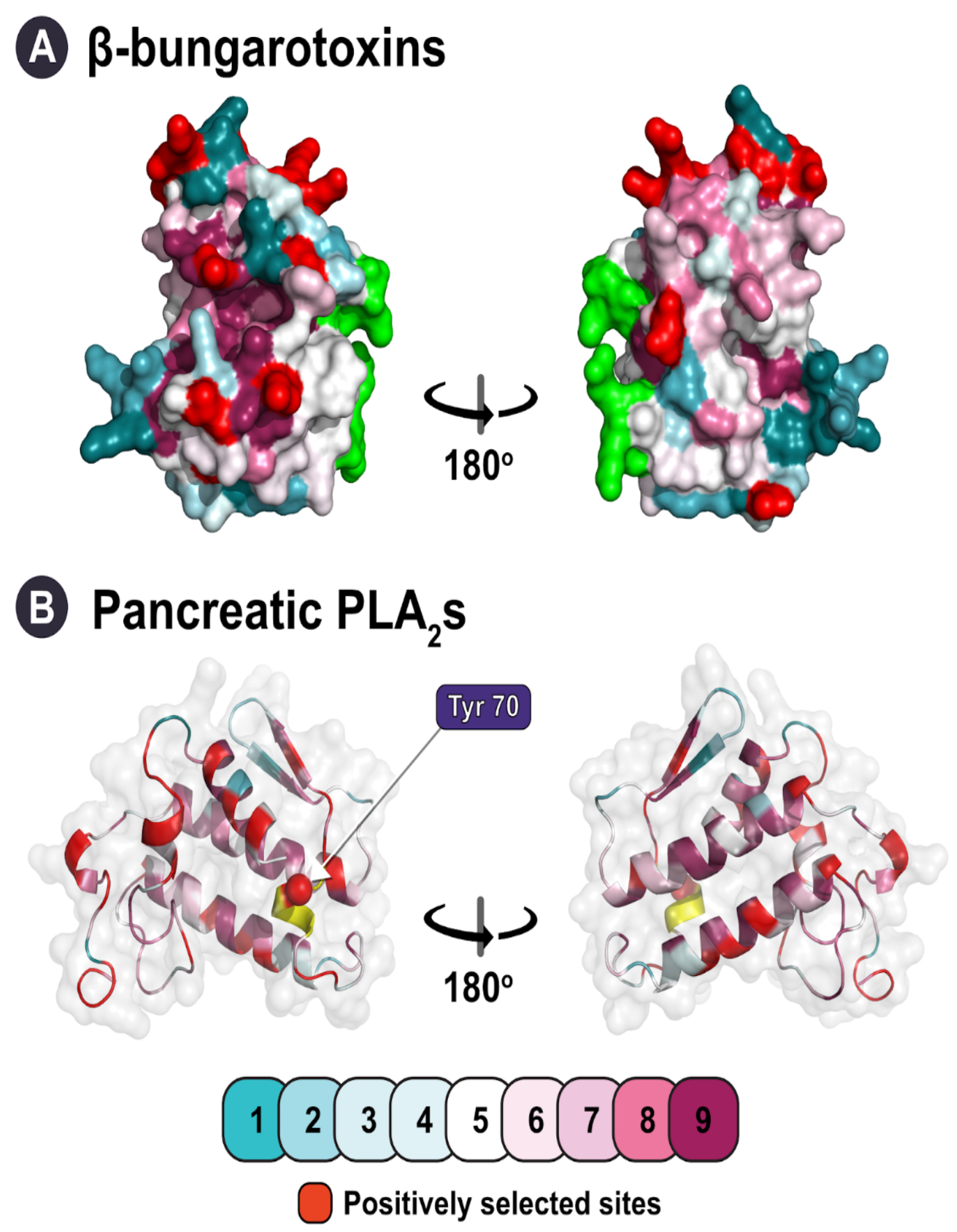
We also identified lineages with signatures of negative selection (ω < 1), which contradicted previous reports of ubiquitous diversifying selection pressures on snake venom PLA2s (Figure 3; Table 2). The evolution of elapid β-bungarotoxins (ω: 0.96; PS: 13) was strongly constrained by purifying selection pressures. Since β-bungarotoxins result from the dimerisation of PLA2 and Kunitz proteins, it is crucial to conserve sites that partake in this process. A similar phenomenon was observed for κ-neurotoxins, 3FTx which form covalently bonded dimeric complexes (and also originate from snakes in the genus Bungarus), in our previous study on the evolution of 3FTx [14]. An accelerated rate of evolution could potentially disrupt structurally important residues and drastically affect these multimeric elapid toxins. Unsurprisingly, amino acid positions that were identified in these toxins as experiencing positive selection were outside of the dimerisation region (Figure 4A). Surprisingly, despite possessing the characteristic loop, which we suspected to be a structural constraint, the pancreatic PLA2s were found to evolve under positive selection and possessed a large number of positively selected sites (ω: 1.37; PS: 33), indicating that such modifications are not deleterious (Figure 3; Table 2). It is interesting to note, however, that only one of the positively selected sites identified in this toxin was found in the pancreatic loop itself (Figure 4B).
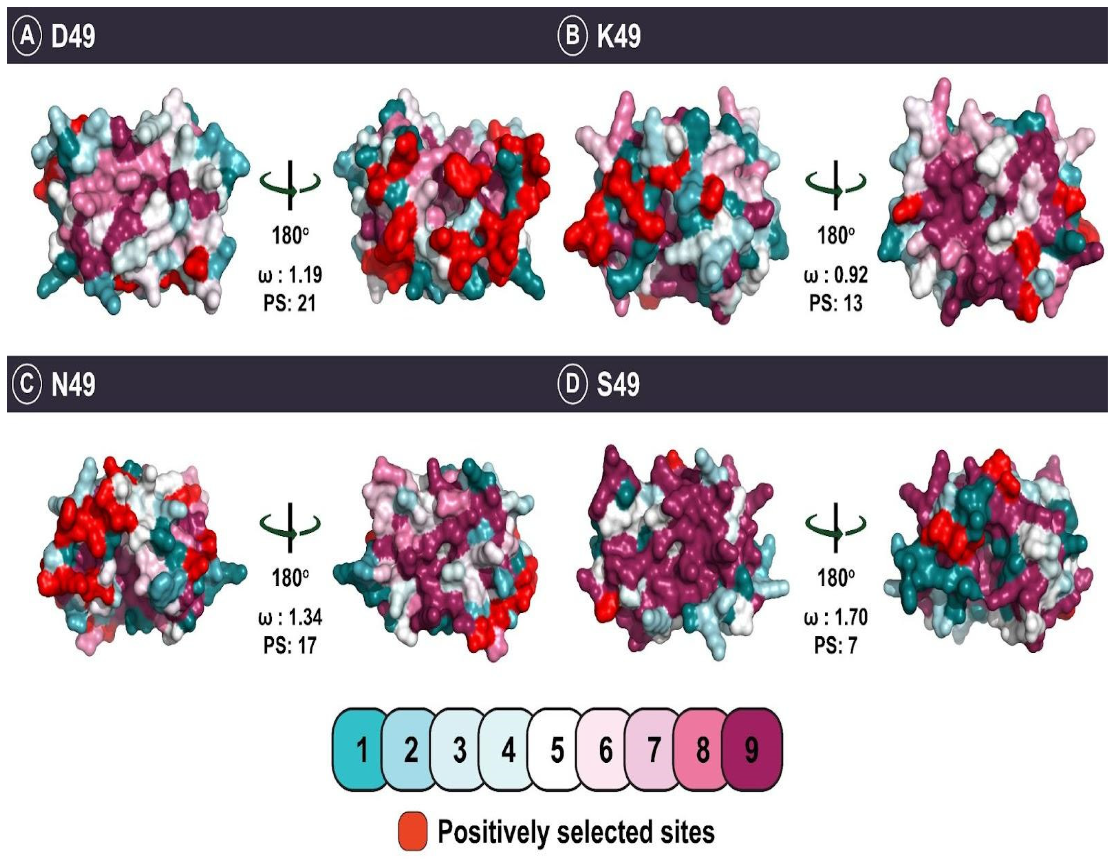
Similar to β-bungarotoxins, the evolution of viperid D49 (ω: 0.83; PS: 21) and K49 (ω: 0.92; PS: 13) PLA2s was also found to be severely constrained by purifying selection (Figure 5). Literature suggests that the D49 viperid PLA2 subtype is a plesiotypic form with haemotoxic activity [52]. When viperid snakes underwent an adaptive radiation [59], their venom PLA2 gene may have also rapidly diversified, resulting in novel toxin forms with apotypic substitutions at the 49th position. Interestingly, these substitutions also correlate with the emergence of atypical functions such as oedematous, neurotoxic, and myotoxic activities [52]. As our understanding of the evolutionary origins, phylogenetic relationships, and ecological relevance of PLA2s with distinct catalytic sites remains limited, future research is required to reconstruct the causal evolutionary pathways leading to the origins of these diverse functions of group II PLA2 venom toxins.
Proteins acquire new functions when they are exposed to novel interaction partners. This can be conceptualised as a change in the protein’s “ecological affordances” [51]. Such changes of a protein’s context within a network of interactions may result from changes in gene expression pattern, inoculation to another organism (as with “exochemical” proteins such as toxins), or changes in functional residues [1]. Rapidly diversifying surface-exposed residues is a primary way in which venom proteins acquire novel functions [14]. In keeping with this general pattern, PLA2s alter their surface chemistry to acquire novel molecular targets and consequently elicit distinct pharmacological effects [18]. The estimation of solvent accessible surface area (SASA) for the amino acid chains of PLA2s from group I and group II sets revealed that a large number of positively selected sites were surface exposed (Table 2), corroborating the results of previous reports [18]. Australian elapid PLA2s contribute to diverse pathologies by acting on a variety of molecular targets. Such a diverse array of activities may have arisen from the evolutionary tinkering of surface chemistry (60 surface exposed sites), while keeping the core intact to maintain structural stability (five buried sites). Sea krait and Micrurus PLA2s too exhibited a similar trend, with a larger number of positively selected sites being surface exposed than buried (exposed: 16 and 15; buried: 3 and 1 sites, respectively). Similarly, all other PLA2 sets were characterised by a great prevalence of positively selected sites amongst solvent accessible regions of the molecule, and very few such sites were buried within the core. Unlike all other PLA2 subgroups, those produced by A. eydouxii were only characterised by a single positively selected solvent accessible site, providing further support to the hypothesis that their accelerated evolution may result from a loss of venom function.
Furthermore, to understand whether the selection pressures driving the evolution of PLA2s are episodic or pervasive in nature, we employed the Mixed Effect Maximum Likelihood Model (MEME) and Fast Unconstrained Bayesian AppRoximation (FUBAR) approaches. In contrast to the site selection analyses in PAML that revealed a strong influence of purifying selection, MEME identified the highest number of episodically diversifying sites for the viperid D49 group (42 sites; Table 2). FUBAR detected 25 amino acid sites under the pervasive influence of positive selection while detecting twice as many sites experiencing the pervasive effects of negative selection. Viperidae snakes are believed to have undergone adaptive radiation nearly 30 million years ago (MYA), which was followed by a marked decrease in the speciation rate [59]. The large number of episodically diversifying sites identified here is perhaps indicative of the changes in group II PLA2s that occurred during the early diversification of these snakes. However, for certain other viperid PLA2s, very few sites were identified that evolved either under episodic or pervasive diversifying selection (K49: 12 and 13; N49: 6 and 17), which could be a consequence of the recent evolution of these forms. MEME failed to identify any such sites for the viperid S49 dataset, while FUBAR identified a single site with pervasive effects of positive selection. The fact that only model 8 of PAML was able to identify this toxin as experiencing diversifying selection could suggest that this toxin form may have begun to diversify only recently.
In congruence with the outcomes of PAML analyses, both MEME and FUBAR identified numerous episodically and pervasively diversifying sites in Australian elapid PLA2 genes (39 and 54 sites, respectively); a lineage of snakes that has recently undergone adaptive radiation. Several diversifying sites (episodic and pervasive) were also detected in Micrurus spp. (33 and 25 sites, respectively). Novel dietary adaptations may have been responsible for the diversifying effects of selection observed in these snakes. β-bungarotoxins and pancreatic PLA2s were also found to have episodic (23 and 21, respectively) and pervasively (14 and 32, respectively) diversifying sites, corroborating the results from PAML analyses, despite the latter analyses indicating that the genes as a whole are constrained by negative selection (see above). Surprisingly, despite the relatively recent shift in habitat for sea kraits, or the dietary specialisation in A. eydouxii, MEME identified a single episodically diversifying site in the former and failed to detect any episodically diversifying sites in the PLA2s of A. eydouxii. As observed in MEME, FUBAR also detected only five pervasively diversifying sites and a single site under the pervasive effects of purifying selection for the sea krait PLA2s. A similar trend was observed in Aipysurus PLA2s, wherein FUBAR failed to identify any sites evolving under pervasive purifying selection and identified a single pervasively diversifying site.

3. Conclusions

The results of the present study are in accordance with the conclusions of a recent investigation of toxin ‘recruitment’ [23] in that our analyses unequivocally demonstrate that there is no ‘one size fits all’ model of toxin evolution, even within a gene family. The impact of structural constraints and the mechanism of action on the evolution of venom toxins has also been documented in 3FTx [14], another major snake venom toxin superfamily, as well as in several other toxins in diverse taxa [47]. On the face of it, this is unsurprising, but it highlights the limitations of abstract models that do not take context—be it ecological, lineage-specific, molecular (in terms of interaction partners), ‘genomic neighbourhood’ (revealed by microsyntenic analyses), etc.—into account [1,47]. Our findings strongly indicate, in contrast to previous claims in the literature, that diversification via positive selection is not the canonical mode of evolution for all PLA2s and that certain lineages in fact evolve under the influence of purifying selection. The unique evolutionary histories of snake lineages, their ecological adaptations, and structural–functional constraints on the snake venom proteins, all contributing to shaping the venom arsenal [47]. By partitioning our datasets in ways that take these contextual factors into account, we have been able to use standard analytical methods to reveal hitherto undocumented complexity in the evolutionary patterns within this widespread, functionally and clinically important, and well-studied toxin family. Our work thus accentuates the importance of investigating venoms through the lens of evolutionary ecology.

4. Material and Methods

4.1. Dataset Assembly and Phylogenetic Reconstructions

Nucleotide datasets for snake venom PLA2 genes were assembled by performing iterative BLAST searches against National Center for Biotechnology Information’s ‘non-redundant’ (NCBI-NR) database: http://www.ncbi.nlm.nih.gov; accessed on 1 November 2019. Acquired sequences were manually curated and aligned using MUSCLE [60]. A Bayesian phylogenetic framework was employed to reconstruct the evolutionary history of snake venom PLA2s. The model for sequence evolution was determined using the ModelFinder program within IQTree [61]. The analysis was performed using MrBayes [62] with GTR + I+G as the preferred model for sequence evolution. Four MonteCarlo chain simulation runs were parallelised across twelve chains to execute the analysis. To terminate the analysis, a convergence diagnostic—standard deviation of split frequency (sdsf) of 0.01 was predefined, and model parameters and trees were sampled every 100th generation. Since the trees never converged for the viperid PLA2s despite multiple runs, the sdsf was reduced to 0.05. Post-simulation, the initial 25% of the sampled trees and their corresponding parameters were discarded as ‘burn-in’. The final topography and node supports (Bayesian posterior probability) were estimated by generating a majority-rule consensus tree using the data retained after burn-in. Subsequently, trees were visualised in Figtree [63].

4.2. Selection Analyses

The nature of selection pressures acting on the snake venom PLA2s was determined using maximum-likelihood based site-selection models implemented in CodeML binaries within the PAML package [64]. The ratio of non-synonymous (nucleotide changes that alter the coded protein) to synonymous (nucleotide changes that do not change the coded protein) substitutions, also known as ‘ω’, was estimated to identify the signatures of selection. The statistical significance of the outcomes was tested by performing a likelihood ratio test (LRT) for the nested models: M7 (null model) and M8 (alternate model). Amino acid sites evolving under the influence of positive selection were identified using the Bayes Empirical Bayes (BEB) method in M8 [65]. The episodic and pervasive effects of natural selection were determined using the Mixed Effect Model of Evolution (MEME) [66], and the Fast Unconstrained Bayesian AppRoximation (FUBAR) [67] packages hosted on the Datamonkey web server [68].

4.3. Structural Analysis

The effects of natural selection on snake venom PLA2s were visualised by generating 3D homology models for various representative sequences using the Phyre2 web server [69]. The evolutionary conservation among various amino acid sites was mapped on the crystal structures using the Consurf web server [70]. PyMOL (Schrodinger, LLC, USA 2010. The PyMOL Molecular Graphics System, Version 2.5.2) was used for the visualisation of homology models. GETAREA web server was used to estimate the solvent accessible surface area (SASA) for the amino acid chains [71]. Additionally, the Adaptive Poisson–Boltzmann Solver (APBS) plug-in was used for determining the electrostatic potential of the solvent accessible area [Connolly surface [72,73]].

Supplementary Materials

The following supporting information can be downloaded at: https://www.mdpi.com/article/10.3390/toxins14060420/s1, Figure S1: Structural and electrostatic features of group I PLA2s (Elapidae); Figure S2: Structural and electrostatic features of group II PLA2s (Viperidae); Figure S3: Bayesian phylogenetic reconstruction of non-pancreatic group I PLA2s (Elapidae); Figure S4: Bayesian phylogenetic reconstruction of pancreatic group I PLA2s (Elapidae); Figure S5: Bayesian phylogenetic reconstruction for Aipysurus group PLA2s; Figure S6: Bayesian phylogenetic reconstruction for β-bungarotoxins; Figure S7: Bayesian phylogenetic reconstruction for Micrurus group PLA2s; Figure S8: Bayesian phylogenetic reconstruction for Naja group PLA2s; Figure S9: Bayesian phylogenetic reconstruction for the sea krait PLA2s; Figure S10: Bayesian phylogenetic reconstruction of group II PLA2s (Viperidae); File S1: Nucleotide dataset for pancreatic group I PLA2s; File S2: Nucleotide dataset for non-pancreatic group I PLA2s; File S3: Nucleotide dataset for group II PLA2s.

Author Contributions

Conceptualization: K.S.; formal analysis: K.S. and V.S.; methodology: K.S. and V.S.; investigation: K.S., V.S. and T.N.W.J.; interpretation: K.S., V.S. and T.N.W.J.; writing—original draft: K.S., T.N.W.J. and V.S.; writing—review and editing: K.S., T.N.W.J. and V.S.; resources: K.S.; data curation: V.S. All authors have read and agreed to the published version of the manuscript.

Funding

K.S. was supported by the Wellcome Trust DBT India Alliance Fellowship (IA/I/19/2/504647). V.S. was supported by the Ministry of Human Resource Development (MHRD) research fellowship. T.N.W.J was supported by a grant from the National Health and medical Council of Australia (13/093/002 AVRU).

Institutional Review Board Statement

Not applicable.

Informed Consent Statement

Not applicable.

Data Availability Statement

All the requisite data are provided as supplemental files.

Conflicts of Interest

The authors have no conflict of interest to declare.

References

  1. Jackson, T.N.; Jouanne, H.; Vidal, N. Snake venom in context: Neglected clades and concepts. Front. Ecol. Evol. 2019, 7, 332. [Google Scholar] [CrossRef] [Green Version]
  2. Fry, B.G.; Roelants, K.; Champagne, D.E.; Scheib, H.; Tyndall, J.D.; King, G.F.; Nevalainen, T.J.; Norman, J.A.; Lewis, R.J.; Norton, R.S. The toxicogenomic multiverse: Convergent recruitment of proteins into animal venoms. Annu. Rev. Genom. Hum. Genet. 2009, 10, 483–511. [Google Scholar] [CrossRef] [PubMed] [Green Version]
  3. Casewell, N.R.; Jackson, T.N.; Laustsen, A.H.; Sunagar, K. Causes and consequences of snake venom variation. Trends Pharmacol. Sci. 2020, 41, 570–581. [Google Scholar] [CrossRef]
  4. Tasoulis, T.; Isbister, G.K. A review and database of snake venom proteomes. Toxins 2017, 9, 290. [Google Scholar] [CrossRef] [Green Version]
  5. Dennis, E.A. 9 Phospholipases. In The Enzymes; Elsevier: Amsterdam, The Netherlands, 1983; Volume 16, pp. 307–353. [Google Scholar]
  6. Dijkstra, B.W.; Kalk, K.H.; Hol, W.G.; Drenth, J. Structure of bovine pancreatic phospholipase A2 at 1.7 Å resolution. J. Mol. Biol. 1981, 147, 97–123. [Google Scholar] [CrossRef]
  7. Fry, M.; Ghosh, S.; East, J.; Franson, R. Role of human sperm phospholipase A2 in fertilization: Effects of a novel inhibitor of phospholipase A2 activity on membrane perturbations and oocyte penetration. Biol. Reprod. 1992, 47, 751–759. [Google Scholar] [CrossRef] [PubMed]
  8. Arita, H.; Hanasaki, K.; Nakano, T.; Oka, S.; Teraoka, H.; Matsumoto, K. Novel proliferative effect of phospholipase A2 in Swiss 3T3 cells via specific binding site. J. Biol. Chem. 1991, 266, 19139–19141. [Google Scholar] [CrossRef]
  9. Murakami, M.; Sato, H.; Taketomi, Y. Updating phospholipase A2 biology. Biomolecules 2020, 10, 1457. [Google Scholar] [CrossRef]
  10. Dennis, E.A.; Rhee, S.G.; Billah, M.M.; Hannun, Y.A. Role of phospholipases in generating lipid second messengers in signal transduction 1. FASEB J. 1991, 5, 2068–2077. [Google Scholar] [CrossRef]
  11. Gutiérrez, J.; Lomonte, B. Venom Phospholipase A2 Enzymes: Structure, Function and Mechanism; Kini, R.M., Ed.; Wiley & Sons: Hoboken, NJ, USA, 1997. [Google Scholar]
  12. Župunski, V.; Kordiš, D. Strong and widespread action of site-specific positive selection in the snake venom Kunitz/BPTI protein family. Sci. Rep. 2016, 6, 37054. [Google Scholar] [CrossRef] [PubMed] [Green Version]
  13. Juárez, P.; Comas, I.; González-Candelas, F.; Calvete, J.J. Evolution of Snake Venom Disintegrins by Positive Darwinian Selection. Mol. Biol. Evol. 2008, 25, 2391–2407. [Google Scholar] [CrossRef] [Green Version]
  14. Sunagar, K.; Jackson, T.N.; Undheim, E.A.; Ali, S.; Antunes, A.; Fry, B.G. Three-fingered RAVERs: Rapid Accumulation of Variations in Exposed Residues of snake venom toxins. Toxins 2013, 5, 2172–2208. [Google Scholar] [CrossRef] [Green Version]
  15. Vonk, F.J.; Casewell, N.R.; Henkel, C.V.; Heimberg, A.M.; Jansen, H.J.; McCleary, R.J.; Kerkkamp, H.M.; Vos, R.A.; Guerreiro, I.; Calvete, J.J. The king cobra genome reveals dynamic gene evolution and adaptation in the snake venom system. Proc. Natl. Acad. Sci. USA 2013, 110, 20651–20656. [Google Scholar] [CrossRef] [Green Version]
  16. Sunagar, K.; Johnson, W.E.; O’Brien, S.J.; Vasconcelos, V.; Antunes, A. Evolution of CRISPs associated with toxicoferan-reptilian venom and mammalian reproduction. Mol. Biol. Evol. 2012, 29, 1807–1822. [Google Scholar] [CrossRef] [PubMed] [Green Version]
  17. Lynch, V.J. Inventing an arsenal: Adaptive evolution and neofunctionalization of snake venom phospholipase A2 genes. BMC Evol. Biol. 2007, 7, 2. [Google Scholar] [CrossRef] [Green Version]
  18. Kini, R.M.; Chan, Y.M. Accelerated evolution and molecular surface of venom phospholipase A2 enzymes. J. Mol. Evol. 1999, 48, 125–132. [Google Scholar] [CrossRef]
  19. Oliveira, A.; Bleicher, L.; Schrago, C.G.; Junior, F.P.S. Conservation analysis and decomposition of residue correlation networks in the phospholipase A2 superfamily (PLA2s): Insights into the structure-function relationships of snake venom toxins. Toxicon 2018, 146, 50–60. [Google Scholar] [CrossRef]
  20. DUFTON, M.J.; HIDER, R.C. Classification of phospholipases A2 according to sequence. Evolutionary and pharmacological implications. Eur. J. Biochem. 1983, 137, 545–551. [Google Scholar] [CrossRef]
  21. Sunagar, K.; Jackson, T.; Reeks, T.; Fry, B. Group I Phospholipase A2 Enzymes; Oxford University Press: New York, NY, USA, 2015. [Google Scholar]
  22. Sunagar, K.; Tsai, I.; Lomonte, B.; Jackson, T.; Fry, B. Group II Phospholipase A2 Enzymes; Oxford University Press: New York, NY, USA, 2015. [Google Scholar]
  23. Jackson, T.N.; Koludarov, I. How the toxin got its toxicity. Front. Pharmacol. 2020, 11, 1893. [Google Scholar] [CrossRef]
  24. Dowell, N.; Giorgianni, M.; Kassner, V.; Selegue, J.; Sanchez, E.; Carroll, S. The deep origin and recent loss of venom toxin genes in rattlesnakes. Curr. Biol. 2016, 26, 2434–2445. [Google Scholar] [CrossRef] [PubMed] [Green Version]
  25. Gibbs, H.L.; Rossiter, W. Rapid evolution by positive selection and gene gain and loss: PLA2 venom genes in closely related Sistrurus rattlesnakes with divergent diets. J. Mol. Evol. 2008, 66, 151–166. [Google Scholar] [CrossRef] [PubMed]
  26. Slowinski, J.B.; Knight, A.; Rooney, A.P. Inferring species trees from gene trees: A phylogenetic analysis of the Elapidae (Serpentes) based on the amino acid sequences of venom proteins. Mol. Phylogenet. Evol. 1997, 8, 349–362. [Google Scholar] [CrossRef] [Green Version]
  27. Kondo, K.; TODA, H.; NARITA, K.; LEE, C.-Y. Amino acid sequence of β2-bungarotoxin from Bungarus multicinctus venom. The amino acid substitutions in the B chains. J. Biochem. 1982, 91, 1519–1530. [Google Scholar] [CrossRef] [PubMed]
  28. Kwong, P.D.; McDonald, N.Q.; Sigler, P.B.; Hendrickson, W.A. Structure of β2-bungarotoxin: Potassium channel binding by Kunitz modules and targeted phospholipase action. Structure 1995, 3, 1109–1119. [Google Scholar] [CrossRef] [Green Version]
  29. Huang, M.Z.; Gopalakrishnakone, P.; Chung, M.C.; Kini, R.M. Complete Amino Acid Sequence of an Acidic, Cardiotoxic Phospholipase A2from the Venom ofOphiophagus hannah (King Cobra): A Novel Cobra Venom Enzyme with “Pancreatic Loop”. Arch. Biochem. Biophys. 1997, 338, 150–156. [Google Scholar] [CrossRef]
  30. Maraganore, J.M.; Heinrikson, R. The lysine-49 phospholipase A2 from the venom of Agkistrodon piscivorus piscivorus. Relation of structure and function to other phospholipases A2. J. Biol. Chem. 1986, 261, 4797–4804. [Google Scholar] [CrossRef]
  31. Van Den Bergh, C.J.; Slotboom, A.J.; Verheij, H.M.; De Haas, G.H. The role of aspartic acid-49 in the active site of phospholipase A2: A site-specific mutagenesis study of porcine pancreatic phospholipase A2 and the rationale of the enzymatic activity of [Iysine49] phospholipase A2 from Agkistrodon piscivorus piscivorus venom. Eur. J. Biochem. 1988, 176, 353–357. [Google Scholar] [PubMed]
  32. Polgár, J.; Magnenat, E.M.; Peitsch, M.C.; Wells, T.N.; Clemetson, K.J. Asp-49 is not an absolute prerequisite for the enzymic activity of low-Mr phospholipases A2: Purification, characterization and computer modelling of an enzymically active Ser-49 phospholipase A2, ecarpholin S, from the venom of Echis carinatus sochureki (saw-scaled viper). Biochem. J. 1996, 319, 961–968. [Google Scholar]
  33. Tsai, I.-H.; Wang, Y.-M.; Chen, Y.-H.; Tsai, T.-S.; Tu, M.-C. Venom phospholipases A2 of bamboo viper (Trimeresurus stejnegeri): Molecular characterization, geographic variations and evidence of multiple ancestries. Biochem. J. 2004, 377, 215–223. [Google Scholar] [CrossRef] [Green Version]
  34. Chijiwa, T.; Tokunaga, E.; Ikeda, R.; Terada, K.; Ogawa, T.; Oda-Ueda, N.; Hattori, S.; Nozaki, M.; Ohno, M. Discovery of novel [Arg49]phospholipase A2 isozymes from Protobothrops elegans venom and regional evolution of Crotalinae snake venom phospholipase A2 isozymes in the southwestern islands of Japan and Taiwan. Toxicon 2006, 48, 672–682. [Google Scholar] [CrossRef]
  35. Gopalakrishnakone, P.; Kochva, E. Venom glands and some associated muscles in sea snakes. J. Morphol. 1990, 205, 85–96. [Google Scholar] [CrossRef] [PubMed]
  36. Li, M.; Fry, B.; Kini, R.M. Eggs-only diet: Its implications for the toxin profile changes and ecology of the marbled sea snake (Aipysurus eydouxii). J. Mol. Evol. 2005, 60, 81–89. [Google Scholar] [CrossRef]
  37. Li, M.; Fry, B.G.; Kini, R.M. Putting the brakes on snake venom evolution: The unique molecular evolutionary patterns of Aipysurus eydouxii (Marbled sea snake) phospholipase A2 toxins. Mol. Biol. Evol. 2005, 22, 934–941. [Google Scholar] [CrossRef] [Green Version]
  38. da Silva Jr, N.J.; Aird, S.D. Prey specificity, comparative lethality and compositional differences of coral snake venoms. Comp. Biochem. Physiol. Part C Toxicol. Pharmacol. 2001, 128, 425–456. [Google Scholar] [CrossRef]
  39. Martins, M.; Oliveira, M.E. Natural history of snakes in forests of the Manaus region, Central Amazonia, Brazil. Herpetol. Nat. Hist. 1998, 6, 78–150. [Google Scholar]
  40. Campbell, J.A.; Lamar, W.W. The venomous reptiles of Latin America. In The Venomous Reptiles of Latin America; Cornell University Press: Ithaca, NY, USA, 1989. [Google Scholar]
  41. Souza, S.M.; Junqueira, A.B.; Jakovac, A.C.C.; Assunção, P.A.; Correia, J.A. Feeding behavior and ophiophagous habits of two poorly known Amazonian coral snakes, Micrurus albicinctus Amaral 1926 and Micrurus paraensis Cunha and Nascimento 1973 (Squamata, Elapidae). Herpetol. Notes 2011, 4, 369–372. [Google Scholar]
  42. Vitt, L.J.; Hulse, A.C. Observations on feeding habits and tail display of the Sonoran coral snake, Micruroides euryxanthus. Herpetologica 1973, 29, 302–304. [Google Scholar]
  43. Marques, O.; Sazima, I. Diet and feeding behavior of the coral snake, Micrurus corallinus, from the Atlantic forest in Brazil. Herpetol. Nat. Hist. 1997, 5, 88–93. [Google Scholar]
  44. Shine, R.; Shetty, S. Moving in two worlds: Aquatic and terrestrial locomotion in sea snakes (Laticauda colubrina, Laticaudidae). J. Evol. Biol. 2001, 14, 338–346. [Google Scholar] [CrossRef]
  45. Greene, H.W. Snakes: The Evolution of Mystery in Nature; University of California Press: Berkeley, CA, USA, 1997. [Google Scholar]
  46. Heatwole, H. Sea Snakes; University of New South Wales Press: Sydney, Australia, 1999. [Google Scholar]
  47. Sunagar, K.; Moran, Y. The rise and fall of an evolutionary innovation: Contrasting strategies of venom evolution in ancient and young animals. PLoS Genet. 2015, 11, e1005596. [Google Scholar] [CrossRef] [Green Version]
  48. Scanlon, J.D.; Lee, M.S. Phylogeny of Australasian venomous snakes (Colubroidea, Elapidae, Hydrophiinae) based on phenotypic and molecular evidence. Zool. Scr. 2004, 33, 335–366. [Google Scholar] [CrossRef]
  49. Wilson, S.K.; Swan, G. A Complete Guide to Reptiles of Australia; New Holland Publishers: Wahroonga, Australia, 2013. [Google Scholar]
  50. Jackson, T.N.; Sunagar, K.; Undheim, E.A.; Koludarov, I.; Chan, A.H.; Sanders, K.; Ali, S.A.; Hendrikx, I.; Dunstan, N.; Fry, B.G. Venom down under: Dynamic evolution of Australian elapid snake toxins. Toxins 2013, 5, 2621–2655. [Google Scholar] [CrossRef] [Green Version]
  51. Jackson, T.N.; Koludarov, I.; Ali, S.A.; Dobson, J.; Zdenek, C.N.; Dashevsky, D.; Op den Brouw, B.; Masci, P.P.; Nouwens, A.; Josh, P. Rapid radiations and the race to redundancy: An investigation of the evolution of Australian elapid snake venoms. Toxins 2016, 8, 309. [Google Scholar] [CrossRef]
  52. Malhotra, A.; Creer, S.; Harris, J.B.; Thorpe, R.S. The importance of being genomic: Non-coding and coding sequences suggest different models of toxin multi-gene family evolution. Toxicon 2015, 107, 344–358. [Google Scholar] [CrossRef]
  53. Koludarov, I.; Jackson, T.N.; Suranse, V.; Pozzi, A.; Sunagar, K.; Mikheyev, A.S. Reconstructing the evolutionary history of a functionally diverse gene family reveals complexity at the genetic origins of novelty. BioRxiv 2020. [Google Scholar] [CrossRef] [Green Version]
  54. Nielsen, R. Molecular signatures of natural selection. Annu. Rev. Genet. 2005, 39, 197–218. [Google Scholar] [CrossRef] [Green Version]
  55. Nei, M.; Gojobori, T. Simple methods for estimating the numbers of synonymous and nonsynonymous nucleotide substitutions. Mol. Biol. Evol. 1986, 3, 418–426. [Google Scholar]
  56. Nielsen, R.; Yang, Z. Estimating the distribution of selection coefficients from phylogenetic data with applications to mitochondrial and viral DNA. Mol. Biol. Evol. 2003, 20, 1231–1239. [Google Scholar] [CrossRef]
  57. Fry, B.; Undheim, E.; Jackson, T. Research methods. In Venomous Reptiles and Their Toxins: Evolution, Pathophysiology and Biodiscovery; Oxford University Press: New York, NY, USA, 2015; pp. 153–214. [Google Scholar]
  58. Lee, M.S.; Sanders, K.L.; King, B.; Palci, A. Diversification rates and phenotypic evolution in venomous snakes (Elapidae). R. Soc. Open Sci. 2016, 3, 150277. [Google Scholar] [CrossRef] [Green Version]
  59. Alencar, L.R.; Quental, T.B.; Grazziotin, F.G.; Alfaro, M.L.; Martins, M.; Venzon, M.; Zaher, H. Diversification in vipers: Phylogenetic relationships, time of divergence and shifts in speciation rates. Mol. Phylogenet. Evol. 2016, 105, 50–62. [Google Scholar] [CrossRef]
  60. Edgar, R.C. MUSCLE: Multiple sequence alignment with high accuracy and high throughput. Nucleic Acids Res. 2004, 32, 1792–1797. [Google Scholar] [CrossRef] [PubMed] [Green Version]
  61. Kalyaanamoorthy, S.; Minh, B.Q.; Wong, T.K.; Von Haeseler, A.; Jermiin, L.S. ModelFinder: Fast model selection for accurate phylogenetic estimates. Nat. Methods 2017, 14, 587–589. [Google Scholar] [CrossRef] [PubMed] [Green Version]
  62. Ronquist, F.; Teslenko, M.; Van Der Mark, P.; Ayres, D.L.; Darling, A.; Höhna, S.; Larget, B.; Liu, L.; Suchard, M.A.; Huelsenbeck, J.P. MrBayes 3.2: Efficient Bayesian phylogenetic inference and model choice across a large model space. Syst. Biol. 2012, 61, 539–542. [Google Scholar] [CrossRef] [Green Version]
  63. Rambaut, A. FigTree v1. 4. Molecular Evolution, Phylogenetics and Epidemiology; University of Edinburgh, Institute of Evolutionary Biology: Edinburgh, UK, 2012. [Google Scholar]
  64. Yang, Z. PAML 4: Phylogenetic analysis by maximum likelihood. Mol. Biol. Evol. 2007, 24, 1586–1591. [Google Scholar] [CrossRef] [Green Version]
  65. Yang, Z.; Wong, W.S.; Nielsen, R. Bayes empirical Bayes inference of amino acid sites under positive selection. Mol. Biol. Evol. 2005, 22, 1107–1118. [Google Scholar] [CrossRef] [PubMed] [Green Version]
  66. Murrell, B.; Wertheim, J.O.; Moola, S.; Weighill, T.; Scheffler, K.; Kosakovsky Pond, S.L. Detecting individual sites subject to episodic diversifying selection. PLoS Genet. 2012, 8, e1002764. [Google Scholar] [CrossRef] [PubMed] [Green Version]
  67. Murrell, B.; Moola, S.; Mabona, A.; Weighill, T.; Sheward, D.; Kosakovsky Pond, S.L.; Scheffler, K. FUBAR: A fast, unconstrained bayesian approximation for inferring selection. Mol. Biol. Evol. 2013, 30, 1196–1205. [Google Scholar] [CrossRef] [Green Version]
  68. Weaver, S.; Shank, S.D.; Spielman, S.J.; Li, M.; Muse, S.V.; Kosakovsky Pond, S.L. Datamonkey 2.0: A modern web application for characterizing selective and other evolutionary processes. Mol. Biol. Evol. 2018, 35, 773–777. [Google Scholar] [CrossRef] [Green Version]
  69. Kelley, L.A.; Mezulis, S.; Yates, C.M.; Wass, M.N.; Sternberg, M.J. The Phyre2 web portal for protein modeling, prediction and analysis. Nat. Protoc. 2015, 10, 845–858. [Google Scholar] [CrossRef] [Green Version]
  70. Ashkenazy, H.; Abadi, S.; Martz, E.; Chay, O.; Mayrose, I.; Pupko, T.; Ben-Tal, N. ConSurf 2016: An improved methodology to estimate and visualize evolutionary conservation in macromolecules. Nucleic Acids Res. 2016, 44, W344–W350. [Google Scholar] [CrossRef] [Green Version]
  71. Fraczkiewicz, R.; Braun, W. Exact and efficient analytical calculation of the accessible surface areas and their gradients for macromolecules. J. Comput. Chem. 1998, 19, 319–333. [Google Scholar] [CrossRef]
  72. Connolly, M.L. Analytical molecular surface calculation. J. Appl. Crystallogr. 1983, 16, 548–558. [Google Scholar] [CrossRef]
  73. Richmond, T.J. Solvent accessible surface area and excluded volume in proteins: Analytical equations for overlapping spheres and implications for the hydrophobic effect. J. Mol. Biol. 1984, 178, 63–89. [Google Scholar] [CrossRef]
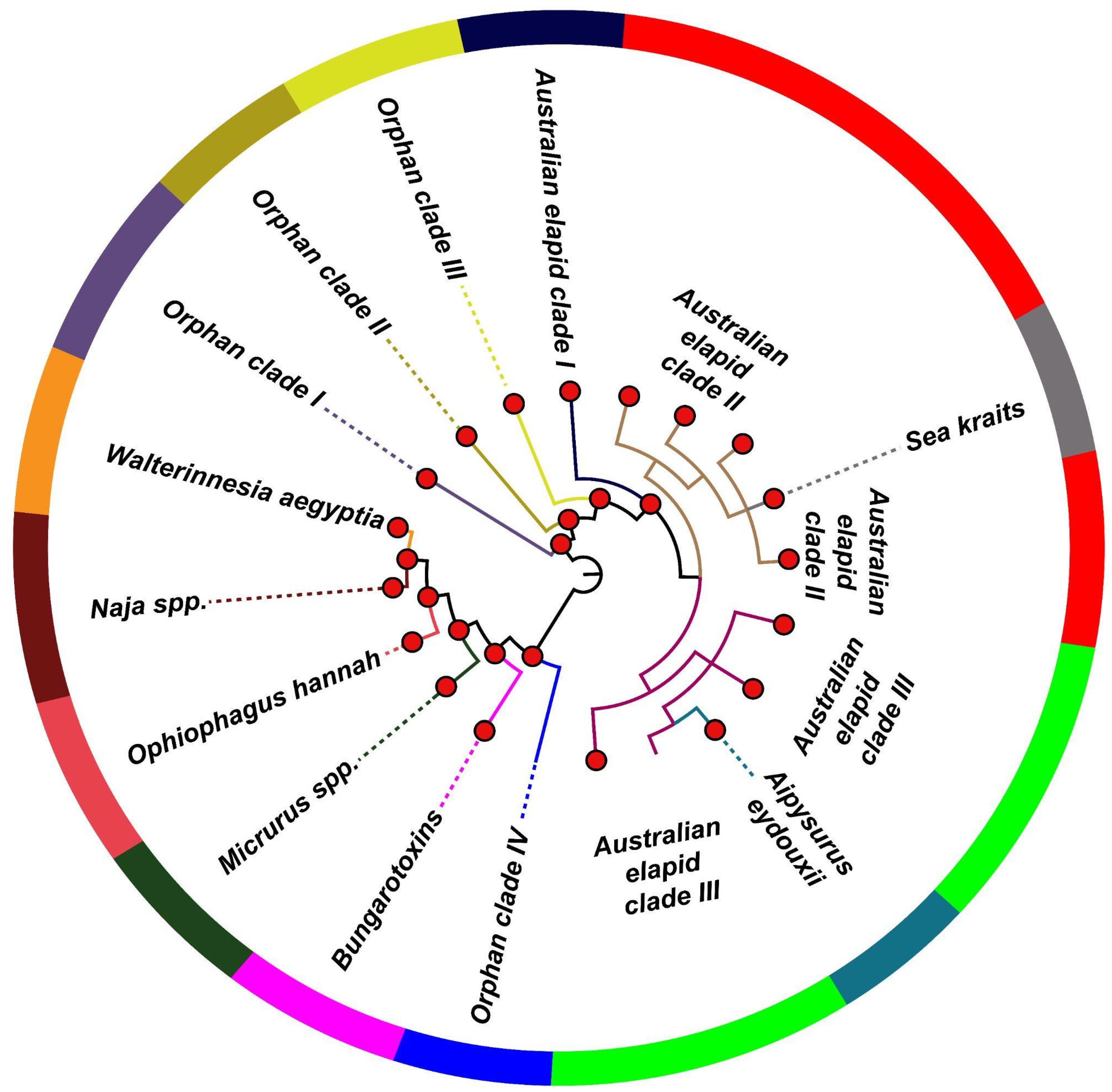
Figure 1. Topology of phylogenetic relationships among group I non-pancreatic PLA2s. A solid red circle represents node support [Bayesian posterior probability (bpp) ≥0.9].
Toxins 14 00420 g001
Figure 2. Topology of phylogenetic relationships among group II PLA2s.
Toxins 14 00420 g002
Figure 3. This figure portrays three-dimensional (3D) homology models depicting the influence of diverse ecologies, structural–functional constraints and evolutionary histories of snake lineages on the evolution of group I PLA2s. The colour-coded scale indicates the strength of evolutionary selection on these PLA2s, shown on a scale of 1 (highly variable) to 9 (highly conserved). Positively selected sites are shown in red.
Toxins 14 00420 g003
Figure 4. This figure depicts the impact of structural constraints on the evolution of group I PLA2s. Here, panel (A) shows a homology model of β-bungarotoxins, highlighting the regions of dimerisation (green shade), while the characteristic loop in pancreatic PLA2s is highlighted in panel (B) (yellow shade). The colour code provided indicates the strength of evolutionary selection acting on PLA2s on a scale of 1 (highly variable) to 9 (highly conserved). Sites identified to be under the significant influence of positive selection are shown in red.
Toxins 14 00420 g004
Figure 5. This figure depicts homology models portraying the evolution of group II PLA2s. The colour-coded scale indicates the strength of evolutionary selection on these PLA2s shown on a scale of 1 (highly variable) to 9 (highly conserved). Positively selected sites are shown in red.
Toxins 14 00420 g005
Table 1. Schema for the classification of snake venom PLA2s.
Feature
Group I (Elapidae)
Naja spp.Phylogenetic clustering
Ophiophagus Hannah *Phylogenetic clustering, ecological constraint
Walterinnesia aegyptia *Phylogenetic clustering, ecological constraint
β-bungarotoxinsPhylogenetic clustering, structural constraint
Australian elapid clades I, II and IIIPhylogenetic clustering, ecological constraint
Sea kraitsPhylogenetic clustering,
ecological constraint
Aipysurus eydouxiiPhylogenetic clustering,
dietary specialisation
Micrurus spp.Phylogenetic clustering,
dietary specialisation
Orphan clades I, II, III and IV *Phylogenetic clustering
PancreaticStructural constraint
Group II (Viperidae)
D49Catalysis
H49 *Catalysis
K49Catalysis
N49Catalysis
R49 *Catalysis
S49Catalysis
T49 *Catalysis
Sets excluded from selection analyses due to limited representation are marked with an asterisk (*).
Table 2. Molecular evolution of snake venom PLA2s.
GroupFUBAR aMEME bPAML cSolvent Accessibility
(Number of Residues)
ExposedBuried
Elapidae
Naja spp.ω > 1 d: 3
ω < 1 e: 4
115120
ω **: 1.19
β-bungarotoxinω > 1 d: 14
ω < 1 e: 11
231390
ω **: 0.96
Australian elapid clade Iω > 1 d: 6
ω < 1 e: 4
118120
ω **: 1.50
Australian elapid clade IIω > 1 d: 24
ω < 1 e: 10
1735272
ω **: 1.71
Australian elapid clade IIIω > 1 d: 24
ω < 1 e: 9
2133213
ω **: 1.64
Sea kraitsω > 1 d: 5
ω < 1 e: 1
126163
ω **: 2.40
A. eydouxiiω > 1 d: 1
ω < 1 e: 0
0211
ω: 2.34
Micrurus spp.ω > 1 d: 25
ω < 1 e: 6
3324151
ω **: 1.34
Pancreatic PLA2sω > 1 d: 32
ω < 1 e: 13
2133211
ω **: 1.37
Viperidae
D49ω > 1 d: 25
ω < 1 e: 50
4221160
ω **: 0.83
K49ω > 1 d: 13
ω < 1 e: 10
121391
ω **: 0.92
N49ω > 1 d: 17
ω < 1 e: 8
617111
ω **: 1.34
S49ω > 1 d: 1
ω < 1 e: 0
0750
ω **: 1.7044
Legend: a: Fast Unconstrained Bayesian AppRoximation to determine pervasive effects of selection; b: Sites experiencing episodic diversifying selection (p ≤ 0.01) based on the Mixed Effects Model Evolution (MEME); c: Positively selected sites detected by the Bayes Empirical Bayes approach implemented in M8; d: Number of sites under pervasive diversifying selection at the posterior probability ≥0.95 (FUBAR); e: Number of sites under pervasive purifying selection at the posterior probability ≥0.95 (FUBAR); ω: mean dN/dS (ω **: p ≤ 0.01).
Publisher’s Note: MDPI stays neutral with regard to jurisdictional claims in published maps and institutional affiliations.