Article

Putative C2H2 Transcription Factor AflZKS3 Regulates Aflatoxin and Pathogenicity in Aspergillus flavus

College of Biological Engineering, Henan University of Technology, Zhengzhou 450001, China
*
Author to whom correspondence should be addressed.
Toxins 2022, 14(12), 883; https://doi.org/10.3390/toxins14120883
Submission received: 12 November 2022 / Revised: 14 December 2022 / Accepted: 15 December 2022 / Published: 17 December 2022
(This article belongs to the Section Mycotoxins)

Abstract

:
Aflatoxin is a carcinogenic secondary metabolite that poses a serious threat to human and animal health. Some C2H2 transcription factors are associated with fungal growth and secondary metabolic regulation. In this study, we characterized the role of AflZKS3, a putative C2H2 transcription factor based on genome annotation, in the growth and aflatoxin biosynthesis of A. flavus and explored its possible mechanisms of action. Surprisingly, the protein was found to be located in the cytoplasm, and gene deletion in A. flavus resulted in defective growth and conidia formation, as well as increased sensitivity to the fluorescent brightener Calcofluor white, Congo red, NaCl, and sorbitol stress. Notably, the biosynthesis of aflatoxin B1 was completely inhibited in the ΔAflZKS3 deletion strain, and its ability to infect peanut and corn seeds was also reduced. RNA sequencing showed that differentially expressed genes in the ΔAflZKS3 strain compared with the control and complementation strains were mainly associated with growth, aflatoxin biosynthesis, and oxidative stress. Thus, AflZKS3 likely contributes to growth, cell development, and aflatoxin synthesis in A. flavus. These findings lay the foundation for a deeper understanding of the roles of C2H2 transcription factors in A. flavus and provide a potential biocontrol target for preventing aflatoxin contamination.
Key Contribution: The C2H2 transcription factor, AflZKS3, affects growth and conidia formation and is involved in aflatoxin biosynthesis in Aspergillus flavus.

1. Introduction

A characteristic of fungi is the ability to produce a wide variety of secondary metabolites, including beneficial compounds such as lovastatin, as well as toxic molecules such as mycotoxins [1]. Aspergillus flavus, a conditional fungal pathogen of important crops in pre- and post-harvest periods, produces carcinogenic aflatoxins (AFs) that cause severe yield reduction and represent a serious threat to animal and human health [2]. A study by the Food and Agriculture Organization proved that about a quarter of the world’s total food production is contaminated by mycotoxins each year, and the main source of pollution is A. flavus and its secondary metabolites [3]. Therefore, exploring the complex mechanism and regulatory network of AF biosynthesis will help to develop effective measures to control the growth of A. flavus and AF contamination, protecting human and animal health and reducing huge economic losses to agricultural production.
The biosynthesis of AFs is regulated by global and pathway-specific transcription factors. Pathway-specific transcription factors, including aflR and aflS within the AF gene cluster, have been studied extensively [4]. AflR is a DNA-binding zinc cluster protein that binds to a palindromic sequence in the promoter region to activate gene expression [5,6]. AflR is necessary for AF synthesis, and the deletion of aflR leads to the downregulation of genes and the complete loss of AF synthesis [7]. AflS regulates genes in the AF synthesis gene cluster by assisting the localization of aflR [8]. Additionally, the biosynthesis of AFs is also regulated by global transcription factors such as zinc finger, bZIP, PHD, homeobox, and APSES transcription factors [9,10,11,12]. Among them, the zinc finger family is the largest and includes the Cys2His2 (C2H2), Cys4 (C4), and Zn(Ⅱ)2C6 subfamilies [13]. Researchers have identified some zinc finger transcription factors with global regulatory functions. The transcription factors nsdC and nsdD, essential for the development of A. nidulans, are also involved in the growth and development of A. flavus, as well as secondary metabolism. AF production is completely lost in nsdC-deleted strains, and aflD, aflM, and aflP genes are not expressed [14]. MtfA encodes a C2H2 zinc finger transcription factor that influences the production of sterigmatocystin, and the overexpression of mtfA can dramatically decrease secondary metabolites such as AFB1 [1]. RsrA, a highly conserved C2H2 transcription factor in A. nidulans, regulates the synthesis of sterigmatocystin, a precursor of AF [15]. These results suggest that C2H2 transcription factors play regulatory roles in mycelia growth development and secondary metabolism. Genome annotation (http://ftfd.snu.ac.kr/index.php?a=view, accessed on 10 October 2022) revealed a putative C2H2 zinc finger transcription factor encoded by AflZKS3 in the genome of A. flavus, which shares 83% homology with the IFM54703_5628 gene in A. lentulus with the property of zinc finger protein with KRAB and SCAN domains 3 (http://FungiDB.org, accessed on 10 October 2022); however, its potential functions in growth and AF biosynthesis remain poorly understood.
In this study, the putative C2H2 zinc finger transcription factor encoded by AflZKS3 in A. flavus was characterized, and its intracellular localization and roles in pathogenicity were investigated. Compared with control and complementation strains, AflZKS3 deletion strains showed a reduced growth rate and conidia number, an inability to produce AF, and increased sensitivity to Calcofluor white (CFW) and NaCl stress. The pathogenicity of the deletion mutant was decreased when infecting peanuts and maize. RNA sequencing (RNA-seq) transcriptomic analysis showed that differentially expressed genes (DEGs) in the AflZKS3 deletion strain were mainly associated with growth, oxidative stress, and the biosynthesis of secondary metabolites, including AF and gliotoxin. Our results reveal the potential regulatory mechanism of AflZKS3 in A. flavus growth, cell development, and AF biosynthesis and provide a potential target for controlling A. flavus and AF contamination.

2. Results

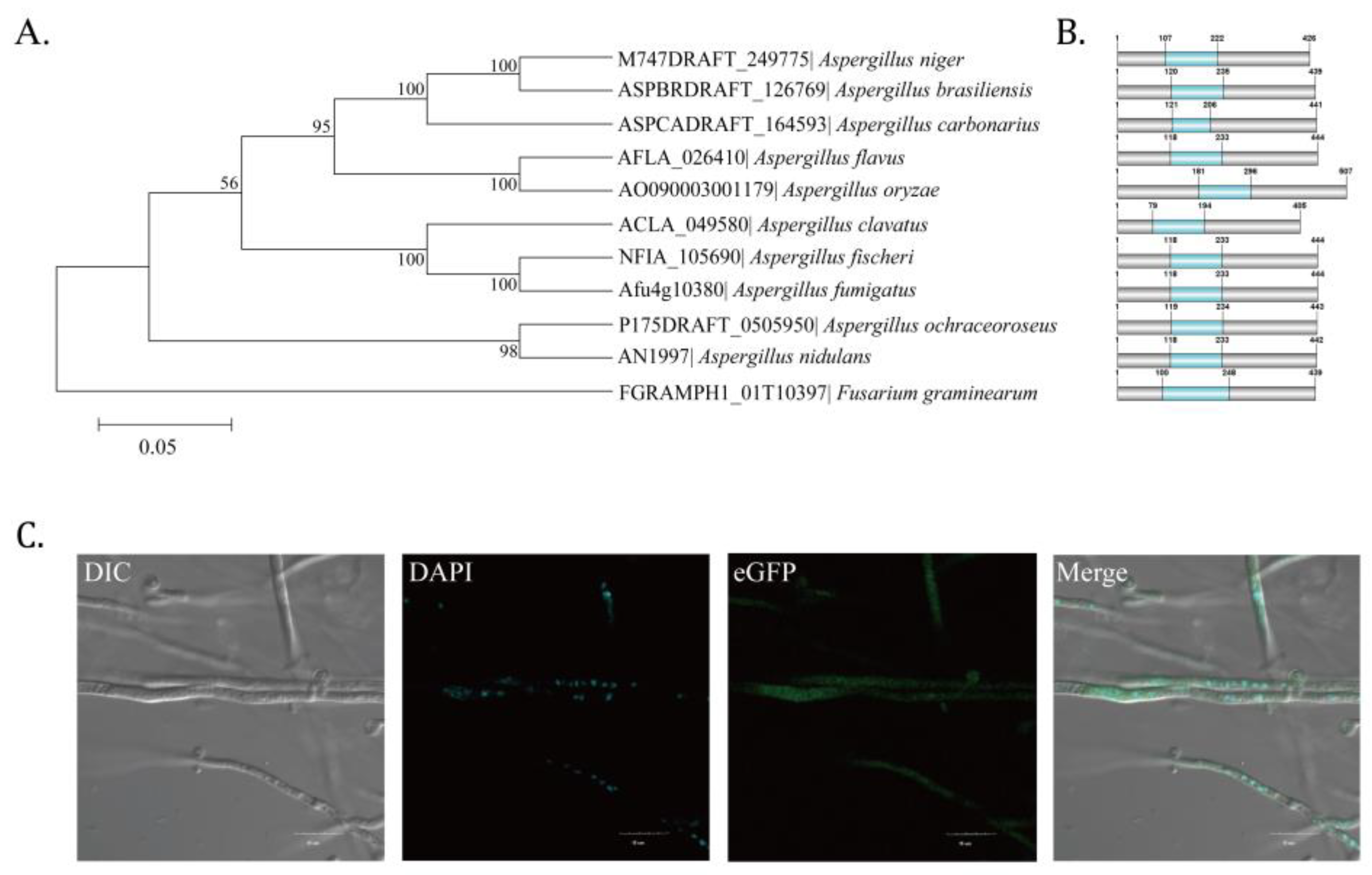
2.1. Identification of Putative C2H2 Zinc Finger Transcription Factor AflZKS3 in A. flavus

Homologous genes of A. flavus AflZKS3 were obtained from NCBI by a BLAST search, and sequences were used to construct a phylogenetic tree using the MEGA 6.0 software, which showed that AflZKS3 was most closely related to AflZKS3 of A. oryzae AO090003001179 (Figure 1A). Protein domain analysis showed that homologs in 10 species harbor C2H2 finger domains (Figure 1B). Unexpectedly, subcellular localization results demonstrated that AflZKS3 was not localized in the nucleus, even though it contains a conserved C2H2 finger domain (Figure 1C).

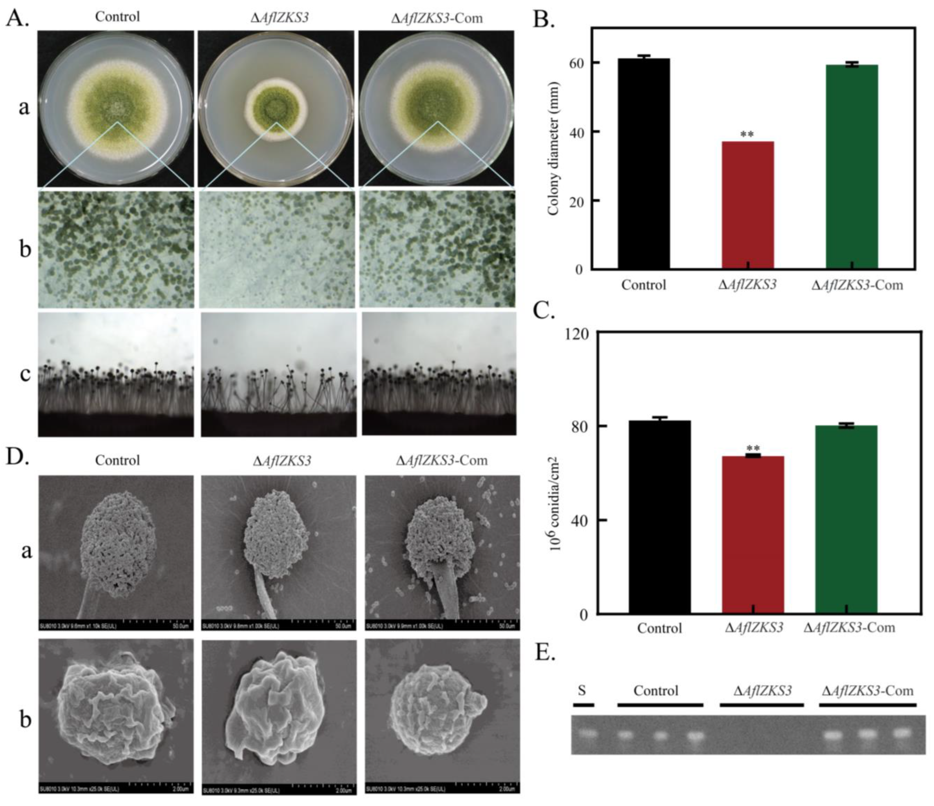
2.2. Deletion of AflZKS3 Affects Growth, Production of Conidia, and AF Biosynthesis

In order to study the roles of AflZKS3 in the pathogenicity of A. flavus, we constructed deletion and complementation strains and verified them using PCR (Figure S1). The role of AflZKS3 in the growth, development, and conidia formation of A. flavus was further studied, and A. flavus control, ΔAflZKS3, and ΔAflZKS3-Com spore suspensions were inoculated and inverted for 5 days at 30 °C. The results demonstrated that compared with the A. flavus control and ΔAflZKS3-Com strains, the mycelia of the ΔAflZKS3 strain were tight, the edges were regular, and the colony diameter was significantly reduced, which indicates that AflZKS3 plays a significant inhibitory role in the growth of A. flavus. Spore analysis indicated that the lack of the AflZKS3 gene reduced the sporogenic ability of A. flavus, consistent with the results observed by stereoscopic microscopy (Figure 2A–C). Additionally, SEM images showed that deletion of the AflZKS3 gene had a minor effect on the morphology of conidia and apical spore heads (Figure 2D). TLC analysis showed that the ΔAflZKS3 strain did not emit fluorescence, indicating that AflZKS3 is essential for AF production in A. flavus (Figure 2E).

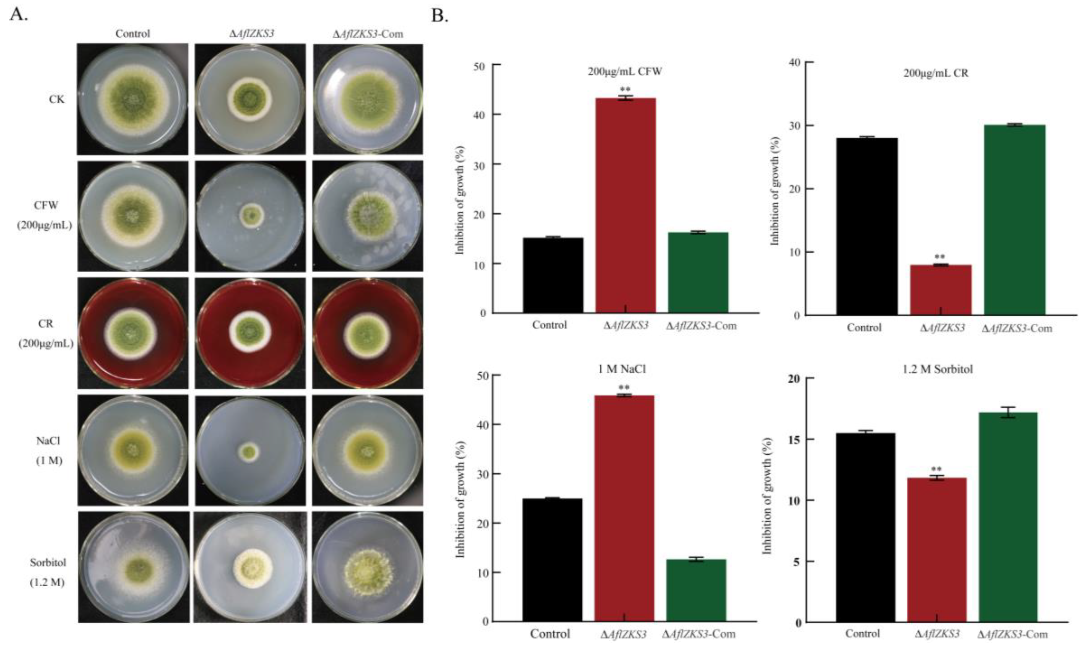
2.3. The ΔAflZKS3 Deletion Mutant Is Highly Sensitive to CFW and NaCl

To investigate the effects of AflZKS3 on the cell wall of A. flavus, strains were cultured in PDA medium supplemented with the cell-wall-stress reagents CFW and CR for 5 days. The results showed that the AflZKS3 deletion strain was sensitive to CFW compared with the control strain and the AflZKS3-Com strain. In addition, similar changes were also observed in the AflZKS3 deletion strain in response to NaCl stress. Specifically, the ΔAflZKS3 strain was much more sensitive to NaCl, and the colony diameter was significantly reduced compared with CR and sorbitol treatment. However, the ∆AflZKS3 mutant showed less sensitivity to CR and sorbitol treatment than the control strain and the AflZKS3-Com strain (Figure 3). These results show that AflZKS3 may have a function in maintaining the cell wall integrity of A. flavus.

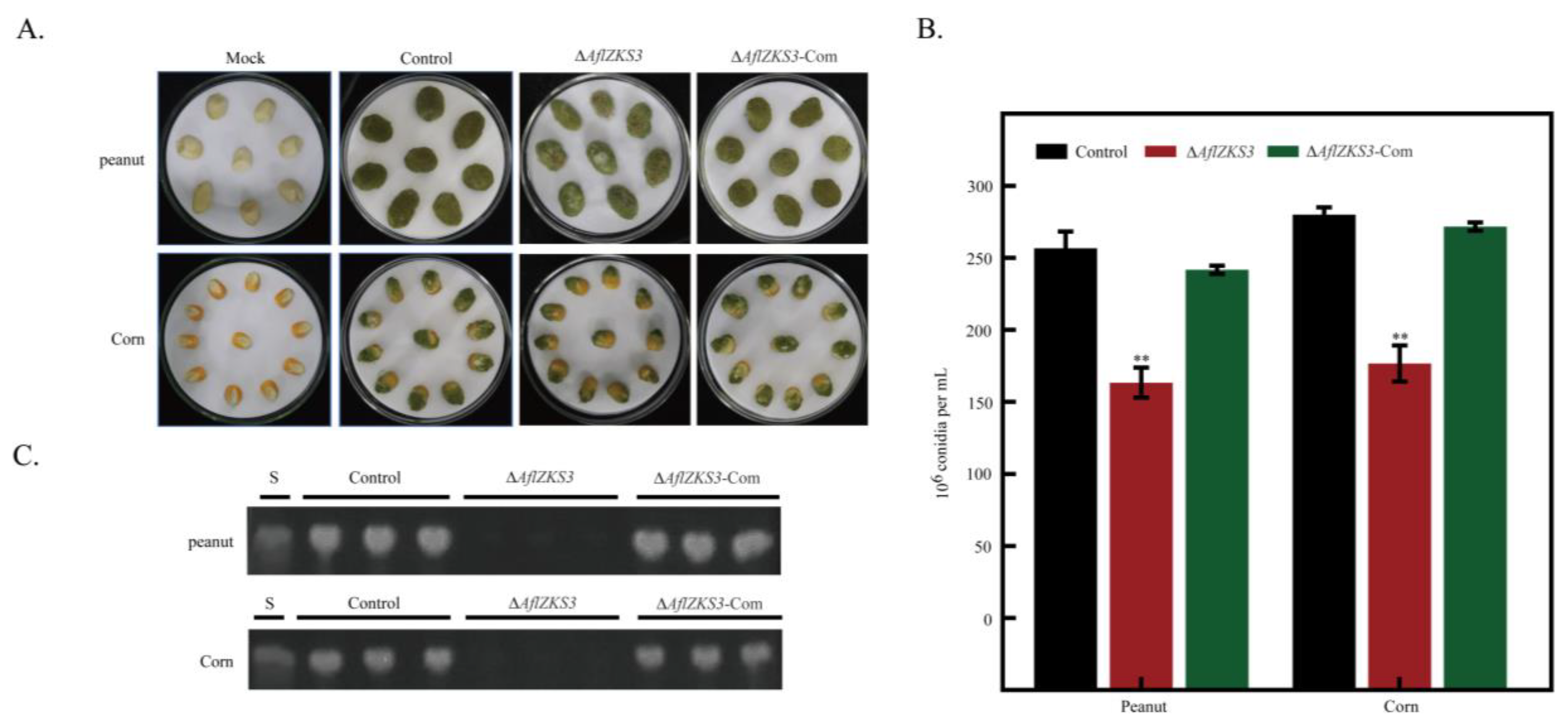
2.4. Effects of AflZKS3 Deletion on the Pathogenicity of A. flavus on Grain Seeds

To study the function of AflZKS3 in the growth and AFB1 biosynthesis of A. flavus in peanut and corn seeds, spore suspensions were inoculated, and AF biosynthesis was characterized. The results showed that the surface of peanuts and corn infected with A. flavus control and ΔAflZKS3-Com strains produced a large number of tight green spores. The surface spores of peanuts and corn infected with the A. flavus ΔAflZKS3 strain were looser, and the yield of conidia decreased by 30.43% and 31.33%, respectively (Figure 4A,B). Additionally, TLC analysis indicated that the deletion of AflZKS3 totally blocked the biosynthesis of AFB1 (Figure 4C). These results suggest that AflZKS3 affected A. flavus pathogenicity by inhibiting A. flavus colonization and AF production.

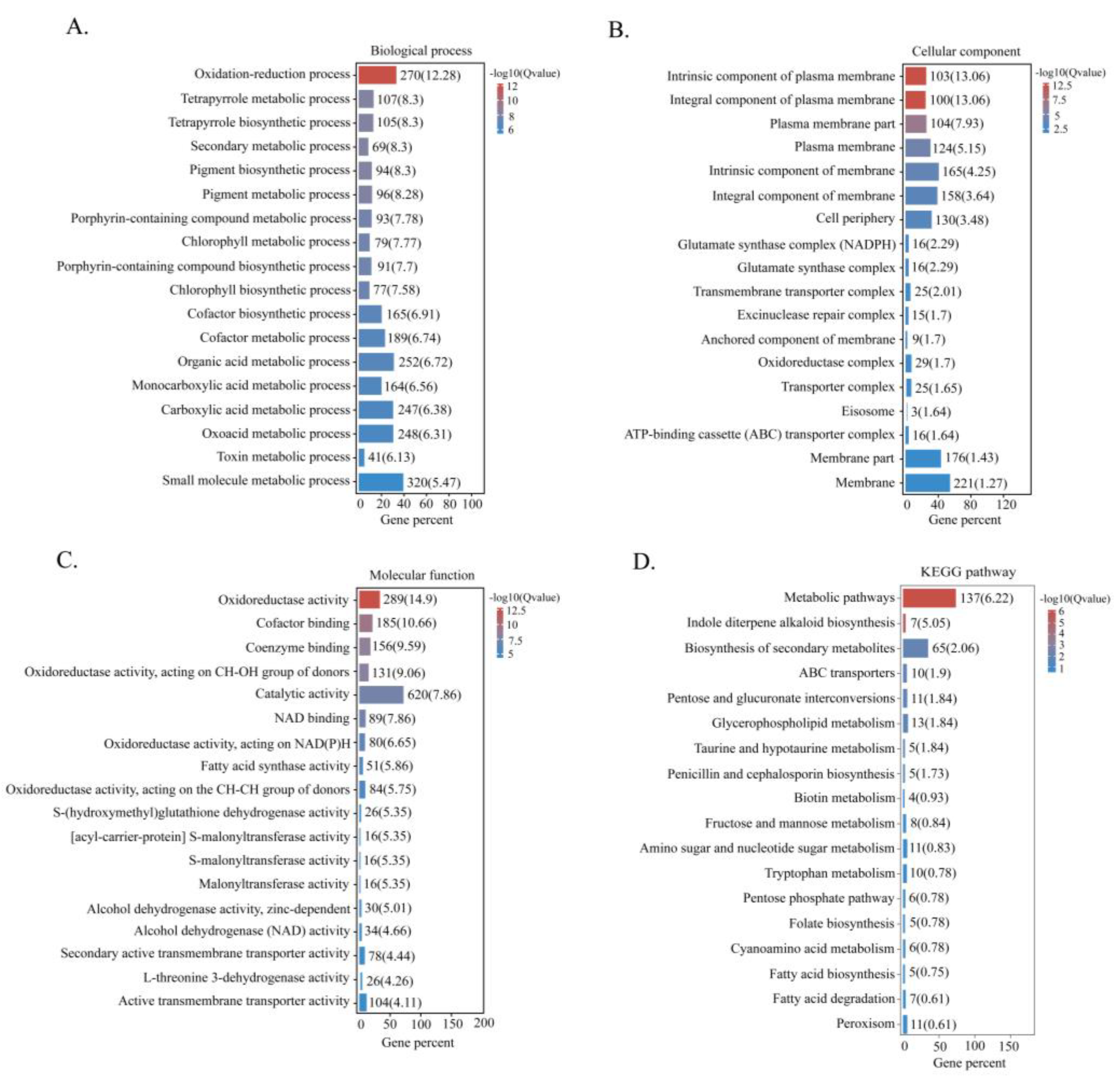
2.5. Transcriptome Analysis

Transcriptome analysis was conducted to investigate the underlying mechanism of AflZKS3 deletion on the growth and AF biosynthesis in A. flavus. The Pearson correlation coefficient was greater than 0.825 between any two replicates, indicating that expression patterns were similar among samples in the groups, and biological replicates were qualified for subsequent analysis (Figure S2A). Expression levels of genes were normalized by FPKM, and DEGs were compared (Figure S2B). The volcano map in Figure S2 shows gene expression fold changes and significance. A total of 1326 significant DEGs were identified, including 476 upregulated genes (35.90%) and 850 downregulated genes (64.10%) (Figure S2D).
GO functional enrichment analysis was performed to further investigate the biological functions of the DEGs. The results revealed that DEGs were mainly associated with oxidation–reduction and metabolic biological processes (Figure 5A). The most enriched cell component categories were the plasma membrane and the cell periphery and membrane (Figure 5B). Molecular functions were mainly related to oxidoreductase activity, catalytic activity, and binding (Figure 5C). Additionally, KEGG pathway enrichment analysis showed that DEGs were mainly linked to metabolic pathways and the biosynthesis of secondary metabolites (Figure 5D).

2.6. Categorisation of DEGs

In order to further unveil the regulatory mechanisms of AflZKS3 in growth and AF biosynthesis, representative DEGs were categorized into four groups: growth, cell wall, secondary metabolism, and oxidative stress (Table 1).
Genes influenced by C2H2 participated in growth and development. The results indicate that the growth-related genes, FLOT1, freB, aspC, and psd2; conidia formation genes, vosA, con-6, cetA, DIT2, AQY1, and betA; and the regulated conidia lipid homeostasis gene, SAY1, were downregulated. Additionally, chiA, agn1, gel2, gel4, glx3, and gpi13, involved in fungal cell wall formation and integrity, were also downregulated.
Alongside defective growth, AF biosynthesis was altered. The results demonstrated that the AFB1 biosynthetic pathway genes, fasA, aflQ, aflB, and aflF, as well as the genes encoding O-methyltransferase (imqG, aclH) and cytochrome P450 (lnaC, BOT4), were downregulated in the AflZKS3 deletion strain. The biosynthesis of other secondary metabolites was also affected; gliotoxin (gliA), polyketide (albA, nscA, and pksCT), and non-ribosomal polypeptide (lnaA) genes were downregulated. Additionally, the deletion of AflZKS3 also downregulated antioxidant-related genes (sodB, cat1, oxr1, and ssuD) and salt stress-related genes (dur3, phoD).

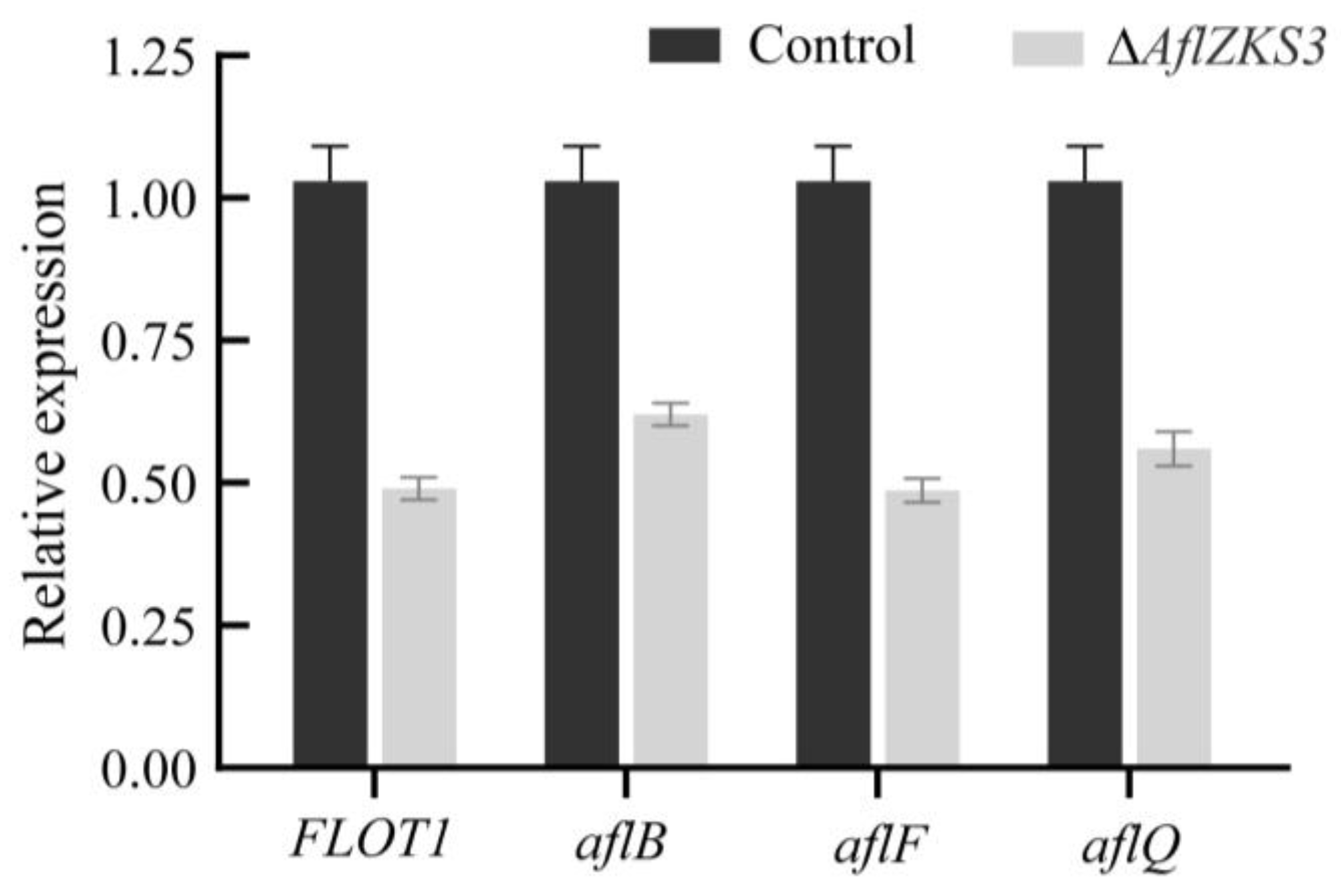
2.7. Validation of RNA-Seq

To further verify the expression levels of the DEGs identified in the transcriptomic analysis, one growth-related gene and three AF synthesis pathway genes were selected, and their expression levels were verified by qRT-PCR. The results showed that the qRT-PCR results were consistent with the transcriptomic results, and the expressions of selected genes were downregulated (Figure 6).

3. Discussion

C2H2 zinc finger transcription factors are known to play vital roles in the development and pathogenicity of microorganisms [16]. In this study, the putative C2H2 zinc finger transcription factor, AflZKS3, annotated in the A. flavus genome, was characterized. The results indicated that this transcription factor is not located in the nucleus and that it plays a major role in the growth and cell development of A. flavus and in AF biosynthesis. Additionally, the potential mechanism was explored by RNA-seq analysis.
AflZKS3 was annotated as a putative C2H2 zinc finger transcription factor in the A. flavus genome. The sequence alignment of homologous Aspergillus, Fusarium, and Saccharomyces proteins revealed that AflZKS3 possesses a conserved C2H2 finger domain, implying similar functions. In S. cerevisiae [17] and A. nidulans [18], C2H2 transcription factors are located in the nucleus, but unexpectedly, our results revealed that AflZKS3 was not located in the nucleus. Previous studies have shown that the localization pattern of the iron deficiency-induced transcription factor, bHLH039, in Arabidopsis varies according to the presence of Fer-like iron deficiency-induced transcription factor (FIT) and that bHLH039 is primarily localized in the cytoplasm when expressed in cells lacking FIT, but localized in the nucleus when FIT is present [19]. These results suggested that the subcellular localization of AflZKS3 might be influenced by other regulatory factors resembling bHLH039 in Arabidopsis. We further determined subcellular localization in the presence of CFW, NaCl, and sorbitol and found that AflZKS3 was not located in the nucleus (Figure S3). However, the specific reasons remain to be further explored.
C2H2 transcription factors have been shown to play crucial roles in plant and fungal growth and development [20]. Previous studies have reported that the membrane microdomain-associated protein Flotillin 1 (Flot1) is involved in plant growth and development in A. thaliana [21]. In A. fumigatus, the freB gene encoding iron reductase mediates iron metabolism, and the disruption of freB reduces the fungal growth rate, iron reductase activity, and tolerance to oxidative stress [22]. Septins are a conserved GTPase family that play vital roles in growth, meristem, and cell wall integrity. In A. fumigatus, the loss of aspC led to septation, cell wall stress, and meristem defects [23]. Phosphatidylserine decarboxylases (PSDs) are responsible for catalyzing the production of phosphatidylethanolamine, an important phospholipid in homeostasis, growth, and the development of fungi. In A. nidulans, the loss of psdB resulted in severe growth defects, impaired conidia development, and abnormal conidia structure [24]. The present study found that growth-related genes such as FLOT1, freB, aspC, and psd2 were downregulated after the deletion of AflZKS3, indicating that iron metabolism, GTPases, and phospholipid homeostasis might be regulated by AflZKS3, and thereby affect mycelia growth. Additionally, con-6, a conidia-related gene, is relatively conserved in filamentous fungi and preferentially expressed during conidia development [25]. The vosA gene encodes a key regulator of Aspergillus spores and is essential for the morphological development and metabolic integrity of conidia. Previous studies found that a vosA mutant strain displayed defective growth on media supplemented with Congo red, sodium chloride, and sorbitol [26]. Herein, we found that genes associated with spore development in the strain ΔAflZKS3, such as con-6, vosA, cetA, betA, AQY1, spore wall-specific gene DIT2 [27], and conidia lipid homeostasis-related gene SAY1 [28] were all downregulated. Furthermore, previous studies indicated that CFW is specifically bound to chitin, while CR is bound to β-1, 3-glucan, thus obstructing the normal assembly of the cell wall, resulting in cell wall stress, and inhibiting the growth of the cell [29]. Our results demonstrated that the AflZKS3 deletion strain showed different sensitivity to CFW and CR compared with A. flavus control and the AflZKS3-Com strains, which might be attributed to their different mechanisms of action and the cell wall defects caused by AflZKS3 deletion. Additionally, the AflZKS3 deletion strain was much more sensitive to NaCl than sorbitol compared with the control and AflZKS3-Com strains. The possible reason might be that NaCl belongs to the category of ionic and cell penetrating agent, which can induce ionic stress and produce specific ionic toxicity, while sorbitol belongs to non-ionic osmotic stress agent. Previous research also demonstrated that the induced effect of NaCl is more profound than that of sorbitol in Japonica rice [30,31].
The cell wall of fungi is a complex structure composed mainly of chitin and glucan and plays a vital role in morphogenesis and protection from various environmental stresses [32]. ChiA is a class III chitinase involved in spore germination and mycelial growth [33]. Agn1, which encodes 1, 3-α-glucanase, is involved in cell division [34]. Gel2 and glx3 are associated with cell wall integrity [35,36]. These results indicate that AflZKS3 might affect fungal morphogenesis, defense responses, and cell division by downregulating cell wall-related genes chiA, agn1, gel2, gel4, glx3, and gpi13. We found that the AFLA_02641 deletion mutant displayed increased sensitivity to CFW, similar to the glx3 deletion strain in Candida albicans [36].
A. flavus growth has been reported to be closely related to AF biosynthesis [37]. Impaired growth and conidia development are often accompanied by secondary metabolism disruption. The biosynthesis of AFs is a complex enzymatic process involving 21 enzymes encoded by a gene cluster ~70 kb in size [38]. Studies have shown that the biosynthesis of fatty acids is involved in the initial stage of biosynthesis, fatty acid synthase is involved in the formation of polyketide initiation units of AFs, and high fatty acid synthase activity can promote AFB1 production [39]. Furthermore, fas-1, which encodes fatty acid synthase, is required for the biosynthesis of norsolorinic acid and AFs [40]. AflQ encodes an oxidoreductase involved in the formation of the AFB1 precursor hydroxyl-methylsterigmatocystin, and it plays a role in the latter stages of the biosynthetic pathway [41]. There is a strong linear relationship between aflQ expression and the AF-producing capacity of A. flavus and A. parasiticus [42]. In this study, the deletion of AflZKS3 downregulated the AF biosynthesis-related genes, fasA, aflQ, aflB, and aflF. O-methyltransferase, another key enzyme in AFB1 synthesis, catalyzes the transformation of sterigmatocystin to O-methylsterigmatocystin and dihydrosterigmatocystin to dihydro-O-methylsterigmatocystin [38]. Cytochrome P450 enzymes are involved in the formation of sterigmatocystin, a late intermediate in the AFB1 synthesis pathway [43]. We found that genes associated with O-methyltransferase (imqG, aclH) and cytochrome P450 (lnaC, BOT4) were downregulated in AflZKS3 mutants. These results indicate that AFB1 production can be inhibited by AflZKS3 through the regulation of multiple genes involved in AF biosynthesis.
In addition to AF biosynthesis, genes involved in other secondary metabolic pathways were also affected. Gliotoxin is synthesized by a biosynthetic gene cluster of 12 genes in A. fumigatus [44]. GliA is involved in gliotoxin biosynthesis and has important functions in gliotoxin export and fungal self-protection. It was found that the disruption of gliA greatly reduced the production of gliotoxin [45]. We found that the gliA gene related to gliotoxin biosynthesis was downregulated. Additionally, polyketide synthases and non-ribosomal peptide synthetases are large multimodular enzymes that participate in the biosynthesis of polyketides and peptide secondary metabolites [46]. Among them, polyketides are the most abundant fungal secondary metabolites, and they are synthesized by a type I diketone synthase [43]. Previous studies have revealed that the deletion of the pksCT gene in Monascus decreased citrinin production capacity by >98% [47]. In this study, genes associated with polyketide synthase and non-ribosomal peptide synthase (albA, nscA, pksCT, and lnaA) were also downregulated. These results suggest that AflZKS3 might play a global regulatory role in mycotoxin export and the self-protection of fungi.
Oxidative stress in filamentous fungi is often associated with secondary metabolism, and it is also one of the prerequisites for AF production. Studies have found that low concentrations of reactive oxygen species (ROS) can stimulate the synthesis of secondary metabolites; conversely, high concentrations of ROS are toxic to cells, even causing cell death, and they are detrimental to the biosynthesis of secondary metabolites [48]. A variety of antioxidant enzymes produced by cells, such as superoxide dismutase, peroxidase, and catalase, remove excess ROS to protect cells from oxidative stress [49]. Previous studies have indicated that oxr1 encodes an antioxidant regulator that protects against intracellular H2O2-induced oxidative stress [50]. SsuD encodes an alkane sulfonate monooxygenase that also protects cells from oxidative stress [51]. In our current study, transcriptome data showed that the deletion of AflZKS3 downregulated antioxidant-related genes sodB, cat1, oxr1, and ssuD, and salt stress-related genes dur3 and phoD [52,53], which might be responsible for the observed changes in AFB1 and other secondary metabolites.
In conclusion, we investigated the putative C2H2 zinc finger transcription factor AflZKS3 in A. flavus, and the results indicated that deletion of AflZKS3 inhibited cell growth, conidia formation, and AFB1 biosynthesis ability. RNA-seq was used to further investigate its underlying regulatory mechanism, and the analysis of DEGs indicated that growth-related genes (FLOT1, psd2, vosA, con-6, and gel2), secondary metabolism-related genes (aflB, aflF, aflQ, and pksCT), and oxygen stress-related genes (sodB, cat1, and oxr1) were downregulated. Therefore, the putative C2H2 zinc finger transcription factor AflZKS3 regulates growth, cell development, and oxidative stress-related genes, and affects the secondary metabolism in A. flavus. These results further our understanding of the functions of C2H2 zinc finger transcription factors in fungal pathogenicity regulation and provide a potential target for developing novel control strategies in A. flavus.

4. Materials and Methods

4.1. Strains, Media, and Culture Conditions

A. flavus strain CA14 (kusA, pyrG+) served as the control strain, and AflZKS3 deletion (ΔAflZKS3), AflZKS3 complementation (ΔAflZKS3-Com), and AflZKS3-eGFP strains were explored in this study. Potato dextrose agar (PDA) was used to evaluate growth rate, conidia number, and AFB1 yield, with a final concentration of 10 mM uridine added if necessary. CFW, NaCl, and sorbitol were added to PDA medium to assess sensitivity to stress. A. flavus transformation was carried out according to previous methods [54]. All experiments were independently repeated three times.

4.2. Sequence Homology Analysis

The sequence of AflZKS3 was searched against the NCBI database, and BLAST comparison was performed to obtain homologous sequences. Relationships were analyzed using MEGA 6.0. software (Mega Limited, Auckland, New Zealand). Protein-related information was downloaded to explore AflZKS3 domains, and a protein domain comparison map was generated using DOG 2.0 software (University of Science & Technology of China, Anhui, China).

4.3. Construction of Deletion, Complementation, and Localization Strains

Deletion, complementation, and localization strains were constructed using the primers listed in Table S1. For the deletion strain, the pyrG gene was used to replace the AflZKS3 target gene in the A. flavus genome. Primers AflZKS3-del-1 and AflZKS3-del-2 were used to amplify the 1424 bp upstream flanking region of the target gene, and primers AflZKS3-del-3 and AflZKS3-del-4 were used to amplify the 1423 bp downstream flanking region. PyrG screening marker genes were amplified from plasmid ANIp7 with primers pyrG-F and pyrG-R. The flanking regions, pyrG screening marker gene, and downstream homologous arm were ligated according to a previous study [54], and products were purified and transferred into the A. flavus CA14 (kusA, pyrG) strain. Transformants were verified using primers AflZKS3-iden-1 and AflZKS3-iden-2.
For the construction of the complementation strain, the native promoter, coding sequence, and terminator were amplified using primers AflZKS3-com-1 and AflZKS3-com-2, and ligated to the pPTRI plasmid after double digestion with HindIII and SmaI. After ligation, an ampicillin antibiotic was used to identify successfully constructed recombinant plasmids, and these were transferred to ΔAflZKS3 protoplasts according to a previous study [55]. Pyrithiamin-resistant transformants were selected, and PCR was used for verification.
For construction of the AflZKS3-eGFP localization strain, linker, enhanced green fluorescent protein (eGFP), and TglaA (primers AflZKS3-eGFP-3, AflZKS3-eGFP-4) were connected to the AFLA_026410 gene in sequence according to a previous study [54], and fused PCR products were purified and linked to the pPTRI plasmid after HindIII and SmaI double digestion. The successfully ligated plasmid was transferred into ΔAflZKS3 protoplasts, and transformants were selected for PCR verification.

4.4. Localization Analysis of AflZKS3 in A. flavus

To assess the subcellular localization of AflZKS3, mycelium was grown for 12 h, collected, stained with 4′,6-diamidino-2-phenylindole (DAPI) according to a previous method [56], and analyzed using an Olympus FV1000 laser confocal microscope (Olympus, Beijing, China). DAPI and eGFP-labeled cells were sequentially imaged by dual-channel imaging.

4.5. Morphological and Physiological Analysis

In order to study the morphological effects of AflZKS3 on A. flavus, 2 μL of spore suspension (106 spores/mL) was inoculated on the surface of PDA medium, and after 5 days of incubation at 30 °C, colony size was observed, colony diameter was measured, the conidia number was calculated, conidia head morphology was assessed by stereoscopic microscopy, and the amount of toxin synthesis evaluated by thin layer chromatography (TLC). The number of conidia was used to calculate the conidia number of the whole plate, and then the area was obtained based on the colony diameter, and finally, the conidia number per cm2 was obtained. PDA medium was supplemented with 200 μg/mL CFW, 200 μg/mL Congo red (CR), 1 M NaCl, and 1.2 M sorbitol for stress testing. Additionally, scanning electron microscopy (SEM) was used to photograph the spore and conidia microstructure of the A. flavus control, ΔAflZKS3 deletion, and ΔAflZKS3-Com strains, as previously described [57].

4.6. Extraction and Detection of AFs

A. flavus control, ΔAflZKS3 deletion, and ΔAflZKS3-Com strains were inoculated in the middle of PDA medium and cultured in the dark for 5 days at 30 °C. Solid samples were collected, and AF was extracted from the culture using chloroform and separated via TLC. The developing solvent was chloroform: acetone (85:15). When the developing solvent migrated to 2/3 of the silica gel plate, the plate was removed to dry, and the fluorescence intensity of AFB1 was observed at a UV wavelength of 365 nm.

4.7. Evaluation of the Effect of AflZKS3 Deletion on the Growth of A. flavus Infecting Peanut and Corn

A. flavus was propagated on peanut and corn. Seeds were treated according to previous methods [54]. A. flavus control, ΔAflZKS3 deletion, and ΔAflZKS3-com spore suspensions were inoculated with 106 spores/ml and incubated in the dark for 5 days at 30 °C. A known amount of sterile water was added to the Petri dishes every day. Spore fluid was collected, and the number of conidia was counted by a hemocytometer according to a previous method [54]. Then, 20 mL of chloroform was added to detect the AFs as described above.

4.8. Transcriptome Analysis

Total RNA was extracted using a TRIzol kit (Thermo Fisher, Shanghai, China) according to the manufacturer’s instructions. RNA quality and RNA integrity were assessed, cDNA libraries were constructed, and RNA-seq analysis was performed by Guangzhou Gene Denovo Biotechnology (Guangzhou, China). Three biological replicates were set up for the RNA-seq analysis, and the accuracy and reliability of sample selection were analyzed with the Pearson correlation coefficient. DESeq was then used to analyze differences in FPKM value, and genes were considered differentially expressed when |log2 (fold change)| > 1 and p-value < 0.05 criteria were met. Gene Ontology (GO) terms and Kyoto Encyclopedia of Genes and Genomes (KEGG) pathways of DEGs were analyzed using GO and KEGG databases (http://www.genome.jp/kegg/, accessed on 12 October 2022).

4.9. Quantitative Real-Time PCR (qRT-PCR) Verification

To detect the expression of AF biosynthesis-related genes, spore suspensions (106) were spread on a PDA plate and cultured at 30°C for 3 days. Total RNA was extracted using the above methods, and cDNA was synthesized by reverse transcription using the PrimeScrip™ RT reagent kit (Takara, Japan). qRT-PCR was performed using the Step One system (Applied Biosystems, Waltham, MA, USA), in which the β-actin housekeeping gene was used as the internal reference for normalization. Relative gene expression was calculated by 2−ΔΔCt. The qRT-PCR primers are listed in Table S1.

4.10. Data Analysis

Analysis of variance and least significant difference (LSD) tests were used for statistical analysis to determine the significance of differences between means, and p < 0.05 was considered statistically significant.

Supplementary Materials

The following supporting information can be downloaded at: https://www.mdpi.com/article/10.3390/toxins14120883/s1, Figure S1: Construction and verification of ΔAflZKS3, ΔAflZKS3-Com, and AflZKS3-eGFP strains; Figure S2: Quality control of samples and overview of DEGs; Figure S3: Location of AflZKS3-eGFP in A. flavus in the presence of CFW, NaCl, and sorbitol; Table S1: Primers used in this study.

Author Contributions

Methodology, L.L.; validation, H.Y.; data curation, S.W.; writing—original draft preparation, L.L.; writing—review and editing, S.Z., L.C. and Y.L.; project administration, Y.H. All authors have read and agreed to the published version of the manuscript.

Funding

This work was sponsored by grants from the Natural Science Foundation of China (31972176), Natural Science Foundation of Henan Province (222300420037), Cultivation Programme for Young Backbone Teachers in Henan University of Technology (21420114), the Innovative Funds Plan of Henan University of Technology (2020ZKCJ01), the earmarked fund for CARS-13.

Institutional Review Board Statement

Not applicable.

Informed Consent Statement

Not applicable.

Data Availability Statement

The raw transcriptome read data are available in the SRA database under accession number SUB12290388.

Conflicts of Interest

The authors declare no conflict of interest.

References

  1. Zhuang, Z.; Lohmar, J.M.; Satterlee, T.; Cary, J.W.; Calvo, A.M. The master transcription factor mtfA governs aflatoxin production, morphological development and pathogenicity in the fungus Aspergillus flavus. Toxins 2016, 8, 29. [Google Scholar] [CrossRef] [PubMed] [Green Version]
  2. Omara, T.; Nassazi, W.; Omute, T.; Awath, A.; Laker, F.; Kalukusu, R.; Musau, B.; Nakabuye, B.V.; Kagoya, S.; Otim, G.; et al. Aflatoxins in uganda: An encyclopedic review of the etiology, epidemiology, detection, quantification, exposure assessment, reduction, and control. Int. J. Microbiol. 2020, 2020, 4723612. [Google Scholar] [CrossRef] [Green Version]
  3. Moretti, A.; Logrieco, A.F.; Susca, A. Mycotoxins: An underhand food problem. Methods Mol. Biol. 2017, 1542, 3–12. [Google Scholar] [CrossRef] [PubMed]
  4. Brakhage, A.A. Regulation of fungal secondary metabolism. Nat. Rev. Microbiol. 2013, 11, 21–32. [Google Scholar] [CrossRef] [PubMed]
  5. Ehrlich, K.C.; Montalbano, B.G.; Cary, J.W. Binding of the C6-zinc cluster protein, AFLR, to the promoters of aflatoxin pathway biosynthesis genes in Aspergillus parasiticus. Gene 1999, 230, 249–257. [Google Scholar] [CrossRef] [PubMed]
  6. Wang, P.; Xu, J.; Chang, P.K.; Liu, Z.; Kong, Q. New insights of transcriptional regulator AflR in Aspergillus flavus physiology. Microbiol. Spectr. 2022, 10, e0079121. [Google Scholar] [CrossRef]
  7. Price, M.S.; Yu, J.; Nierman, W.C.; Kim, H.S.; Pritchard, B.; Jacobus, C.A.; Bhatnagar, D.; Cleveland, T.E.; Payne, G.A. The aflatoxin pathway regulator AflR induces gene transcription inside and outside of the aflatoxin biosynthetic cluster. Fems Microbiol. Lett. 2006, 255, 275–279. [Google Scholar] [CrossRef] [Green Version]
  8. Ehrlich, K.C.; Mack, B.M.; Wei, Q.; Li, P.; Roze, L.V.; Dazzo, F.; Cary, J.W.; Bhatnagar, D.; Linz, J.E. Association with AflR in endosomes reveals new functions for AflJ in aflatoxin biosynthesis. Toxins 2012, 4, 1582–1600. [Google Scholar] [CrossRef]
  9. Chen, J.F.; Tan, J.J.; Wang, J.Y.; Mao, A.J.; Xu, X.P.; Zhang, Y.; Zheng, X.L.; Liu, Y.; Jin, D.; Li, X.B.; et al. The zinc finger transcription factor BbCmr1 regulates conidium maturation in Beauveria bassiana. Microbiol. Spectr. 2022, 10, e0206621. [Google Scholar] [CrossRef]
  10. Jun, S.C.; Choi, Y.H.; Lee, M.W.; Yu, J.H.; Shin, K.S. The putative APSES transcription factor RgdA governs growth, development, toxigenesis, and virulence in Aspergillus fumigatus. Msphere 2020, 5, e00998-20. [Google Scholar] [CrossRef]
  11. Guan, X.; Zhao, Y.; Liu, X.; Shang, B.; Xing, F.; Zhou, L.; Wang, Y.; Zhang, C.; Bhatnagar, D.; Liu, Y. The bZIP transcription factor Afap1 mediates the oxidative stress response and aflatoxin biosynthesis in Aspergillus flavus. Rev. Argent. Microbio. 2019, 51, 292–301. [Google Scholar] [CrossRef]
  12. Hu, Y.; Yang, G.; Zhang, D.; Liu, Y.; Li, Y.; Lin, G.; Guo, Z.; Wang, S.; Zhuang, Z. The PHD transcription factor Rum1 regulates morphogenesis and aflatoxin biosynthesis in Aspergillus flavus. Toxins 2018, 10, 301. [Google Scholar] [CrossRef] [PubMed] [Green Version]
  13. Shelest, E. Transcription factors in fungi. FEMS Microbiol. Lett. 2008, 286, 145–151. [Google Scholar] [CrossRef] [PubMed] [Green Version]
  14. Cary, J.W.; Harris-Coward, P.Y.; Ehrlich, K.C.; Mack, B.M.; Kale, S.P.; Larey, C.; Calvo, A.M. NsdC and NsdD affect Aspergillus flavus morphogenesis and aflatoxin production. Eukaryot. Cell 2012, 11, 1104–1111. [Google Scholar] [CrossRef] [PubMed] [Green Version]
  15. Bok, J.W.; Wiemann, P.; Garvey, G.S.; Lim, F.Y.; Haas, B.; Wortman, J.; Keller, N.P. Illumina identification of RsrA, a conserved C2H2 transcription factor coordinating the NapA mediated oxidative stress signaling pathway in Aspergillus. BMC Genom. 2014, 15, 1011. [Google Scholar] [CrossRef] [Green Version]
  16. Wang, P.; Li, B.; Pan, Y.T.; Zhang, Y.Z.; Li, D.W.; Huang, L. Calcineurin-responsive transcription factor CgCrzA is required for cell wall integrity and infection-related morphogenesis in Colletotrichum gloeosporioides. Plant Pathol. J. 2020, 36, 385–397. [Google Scholar] [CrossRef]
  17. Görner, W.; Durchschlag, E.; Martinez-Pastor, M.T.; Estruch, F.; Ammerer, G.; Hamilton, B.; Ruis, H.; Schüller, C. Nuclear localization of the C2H2 zinc finger protein Msn2p is regulated by stress and protein kinase A activity. Genes Dev. 1998, 12, 586–597. [Google Scholar] [CrossRef] [Green Version]
  18. Kwon, N.J.; Garzia, A.; Espeso, E.A.; Ugalde, U.; Yu, J.H. FlbC is a putative nuclear C2H2 transcription factor regulating development in Aspergillus nidulans. Mol. Microbiol. 2010, 77, 1203–1219. [Google Scholar] [CrossRef]
  19. Trofimov, K.; Ivanov, R.; Eutebach, M.; Acaroglu, B.; Mohr, I.; Bauer, P.; Brumbarova, T. Mobility and localization of the iron deficiency-induced transcription factor bHLH039 change in the presence of FIT. Plant Direct. 2019, 3, e00190. [Google Scholar] [CrossRef] [Green Version]
  20. Xiong, D.; Wang, Y.; Deng, C.; Hu, R.; Tian, C. Phylogenic analysis revealed an expanded C2H2-homeobox subfamily and expression profiles of C2H2 zinc finger gene family in Verticillium dahliae. Gene 2015, 562, 169–179. [Google Scholar] [CrossRef]
  21. Cao, Y.; He, Q.; Qi, Z.; Zhang, Y.; Lu, L.; Xue, J.; Li, J.; Li, R. Dynamics and endocytosis of Flot1 in Arabidopsis require CPI1 function. Int. J. Mol. Sci. 2020, 21, 1552. [Google Scholar] [CrossRef] [PubMed] [Green Version]
  22. Rehman, L.; Su, X.; Li, X.; Qi, X.; Guo, H.; Cheng, H. FreB is involved in the ferric metabolism and multiple pathogenicity-related traits of Verticillium dahliae. Curr. Genet. 2018, 64, 645–659. [Google Scholar] [CrossRef] [PubMed]
  23. Vargas-Muñiz, J.M.; Renshaw, H.; Richards, A.D.; Lamoth, F.; Soderblom, E.J.; Moseley, M.A.; Juvvadi, P.R.; Steinbach, W.J. The Aspergillus fumigatus septins play pleiotropic roles in septation, conidiation, and cell wall stress, but are dispensable for virulence. Fungal Genet. Biol. 2015, 81, 41–51. [Google Scholar] [CrossRef] [PubMed] [Green Version]
  24. Takagi, K.; Kikkawa, A.; Iwama, R.; Fukuda, R.; Horiuchi, H. Type II phosphatidylserine decarboxylase is crucial for the growth and morphogenesis of the filamentous fungus Aspergillus nidulans. J. Biosci. Bioeng. 2021, 131, 139–146. [Google Scholar] [CrossRef] [PubMed]
  25. Ma, L.; Li, X.; Ma, X.; Yu, Q.; Yu, X.; Liu, Y.; Nie, C.; Zhang, Y.; Xing, F. The regulatory mechanism of water activities on aflatoxins biosynthesis and conidia development, and transcription Factor AtfB is involved in this regulation. Toxins 2021, 13, 431. [Google Scholar] [CrossRef]
  26. Zhang, G.; Zheng, Y.; Ma, Y.; Yang, L.; Xie, M.; Zhou, D.; Niu, X.; Zhang, K.Q.; Yang, J. The velvet proteins VosA and VelB play different roles in conidiation, trap formation, and pathogenicity in the nematode-trapping fungus Arthrobotrys oligospora. Front. Microbiol. 2019, 10, 1917. [Google Scholar] [CrossRef] [Green Version]
  27. Friesen, H.; Hepworth, S.R.; Segall, J. An Ssn6-Tup1-dependent negative regulatory element controls sporulation-specific expression of DIT1 and DIT2 in Saccharomyces cerevisiae. Mol. Cell. Biol. 1997, 17, 123–134. [Google Scholar] [CrossRef] [Green Version]
  28. Peng, Y.J.; Zhang, H.; Feng, M.G.; Ying, S.H. Sterylacetyl hydrolase 1 (BbSay1) links lipid homeostasis to conidiogenesis and virulence in the entomopathogenic fungus Beauveria bassiana. J. Fungi 2022, 8, 292. [Google Scholar] [CrossRef]
  29. Ram, A.F.; Klis, F.M. Identification of fungal cell wall mutants using susceptibility assays based on Calcofluor white and Congo red. Nat. Protoc. 2006, 1, 2253–2256. [Google Scholar] [CrossRef]
  30. Kiełkowska, A. Allium cepa root meristem cells under osmotic (sorbitol) and salt (NaCl) stress in vitro. Acta Bot. Croat. 2017, 76, 146–153. [Google Scholar] [CrossRef]
  31. Wankhade, S.D.; Bahaji, A.; Mateu-Andrés, I.; Cornejo, M.D. Phenotypic indicators of NaCl tolerance levels in rice seedlings: Variations in development and leaf anatomy. Acta Physiol. Plant. 2010, 32, 1161–1169. [Google Scholar] [CrossRef] [Green Version]
  32. Yamazaki, H.; Tanaka, A.; Kaneko, J.; Ohta, A.; Horiuchi, H. Aspergillus nidulans ChiA is a glycosylphosphatidylinositol (GPI)-anchored chitinase specifically localized at polarized growth sites. Fungal Genet. Biol. 2008, 45, 963–972. [Google Scholar] [CrossRef] [PubMed]
  33. Takaya, N.; Yamazaki, D.; Horiuchi, H.; Ohta, A.; Takagi, M. Cloning and characterization of a chitinase-encoding gene (chiA) from Aspergillus nidulans, disruption of which decreases germination frequency and hyphal growth. Biosci. Biotechno. Biochem. 1998, 62, 60–65. [Google Scholar] [CrossRef] [PubMed] [Green Version]
  34. Villalobos-Duno, H.; San-Blas, G.; Paulinkevicius, M.; Sánchez-Martín, Y.; Nino-Vega, G. Biochemical characterization of Paracoccidioides brasiliensis α-1,3-glucanase Agn1p, and its functionality by heterologous expression in Schizosaccharomyces pombe. PLoS ONE 2013, 8, e66853. [Google Scholar] [CrossRef] [PubMed] [Green Version]
  35. Zhao, W.; Lü, Y.; Ouyang, H.; Zhou, H.; Yan, J.; Du, T.; Jin, C. N-Glycosylation of Gel1 or Gel2 is vital for cell wall β-glucan synthesis in Aspergillus fumigatus. Glycobiology 2013, 23, 955–968. [Google Scholar] [CrossRef] [PubMed] [Green Version]
  36. Cabello, L.; Gómez-Herreros, E.; Fernández-Pereira, J.; Maicas, S.; Martínez-Esparza, M.C.; de Groot, P.; Valentín, E. Deletion of GLX3 in Candida albicans affects temperature tolerance, biofilm formation and virulence. Fems Yeast Res. 2019, 19, 124. [Google Scholar] [CrossRef] [Green Version]
  37. Bayram, O.; Braus, G.H. Coordination of secondary metabolism and development in fungi: The velvet family of regulatory proteins. Fems Yeast Res. 2012, 36, 1–24. [Google Scholar] [CrossRef] [Green Version]
  38. Yu, J.; Chang, P.K.; Ehrlich, K.C.; Cary, J.W.; Bhatnagar, D.; Cleveland, T.E.; Payne, G.A.; Linz, J.E.; Woloshuk, C.P.; Bennett, J.W. Clustered pathway genes in aflatoxin biosynthesis. Appl. Environ. Microb. 2004, 70, 1253–1262. [Google Scholar] [CrossRef] [Green Version]
  39. Reding, C.L.; Harrison, M.A. Possible relationship of succinate dehydrogenase and fatty acid synthetase activities to Aspergillus parasiticus (NRRL 5139) growth and aflatoxin production. Mycopathologia 1994, 127, 175–181. [Google Scholar] [CrossRef]
  40. Payne, G.A.; Brown, M.P. Genetics and physiology of aflatoxin biosynthesis. Annu. Rev. Phytopathol. 1998, 36, 329–362. [Google Scholar] [CrossRef]
  41. Ren, X.; Branà, M.T.; Haidukowski, M.; Gallo, A.; Zhang, Q.; Logrieco, A.F.; Li, P.; Zhao, S.; Altomare, C. Potential of Trichoderma spp. for biocontrol of aflatoxin-producing Aspergillus flavus. Toxins 1998, 14, 86. [Google Scholar] [CrossRef] [PubMed]
  42. Jamali, M.; Karimipour, M.; Shams-Ghahfarokhi, M.; Amani, A.; Razzaghi-Abyaneh, M. Expression of aflatoxin genes aflO (omtB) and aflQ (ordA) differentiates levels of aflatoxin production by Aspergillus flavus strains from soils of pistachio orchards. Res. Microbiol. 2013, 164, 293–299. [Google Scholar] [CrossRef] [PubMed]
  43. Keller, N.P.; Turner, G.; Bennett, J.W. Fungal secondary metabolism-from biochemistry to genomics. Nat. Rev. Microbiol. 2005, 3, 937–947. [Google Scholar] [CrossRef] [PubMed]
  44. Kwon-Chung, K.J.; Sugui, J.A. What do we know about the role of gliotoxin in the pathobiology of Aspergillus fumigatus? Med. Mycol. 2009, 47 (Suppl. S1), 97–103. [Google Scholar] [CrossRef] [Green Version]
  45. Wang, D.N.; Toyotome, T.; Muraosa, Y.; Watanabe, A.; Wuren, T.; Bunsupa, S.; Aoyagi, K.; Yamazaki, M.; Takino, M.; Kamei, K. GliA in Aspergillus fumigatus is required for its tolerance to gliotoxin and affects the amount of extracellular and intracellular gliotoxin. Med. Mycol. 2014, 52, 506–518. [Google Scholar] [CrossRef] [Green Version]
  46. Gallo, A.; Ferrara, M.; Perrone, G. Phylogenetic study of polyketide synthases and nonribosomal peptide synthetases involved in the biosynthesis of mycotoxins. Toxins 2013, 5, 717–742. [Google Scholar] [CrossRef] [Green Version]
  47. Huang, Z.; Su, B.; Xu, Y.; Li, L.; Li, Y. Determination of two potential toxicity metabolites derived from the disruption of the pksCT gene in Monascus aurantiacus Li As3.4384. J. Sci. Food Agr. 2017, 97, 4190–4197. [Google Scholar] [CrossRef]
  48. Zhu, Z.; Yang, M.; Bai, Y.; Ge, F.; Wang, S. Antioxidant-related catalase CTA1 regulates development, aflatoxin biosynthesis, and virulence in pathogenic fungus Aspergillus flavus. Environ. Microbiol. 2020, 22, 2792–2810. [Google Scholar] [CrossRef]
  49. Domènech, A.; Ayté, J.; Antunes, F.; Hidalgo, E. Using in vivo oxidation status of one- and two-component redox relays to determine H2O2 levels linked to signaling and toxicity. BMC Biol. 2018, 16, 61. [Google Scholar] [CrossRef]
  50. Su, L.D.; Zhang, Q.L.; Lu, Z. Oxidation resistance 1 (OXR1) participates in silkworm defense against bacterial infection through the JNK pathway. Insect Sci. 2017, 24, 17–26. [Google Scholar] [CrossRef]
  51. Park, C.; Shin, B.; Park, W. Protective role of bacterial alkanesulfonate monooxygenase under oxidative stress. Appl. Environ. Microb. 2020, 86, e00692-20. [Google Scholar] [CrossRef]
  52. Dos Santos, T.B.; Baba, V.Y.; Vieira, L.; Pereira, L.; Domingues, D.S. The urea transporter DUR3 is differentially regulated by abiotic and biotic stresses in coffee plants. Physiol. Mol. Biol. Plants 2021, 27, 203–212. [Google Scholar] [CrossRef] [PubMed]
  53. Kageyama, H.; Tripathi, K.; Rai, A.K.; Cha-Um, S.; Waditee-Sirisattha, R.; Takabe, T. An alkaline phosphatase/phosphodiesterase, PhoD, induced by salt stress and secreted out of the cells of Aphanothece halophytica, a halotolerant cyanobacterium. Appl. Environ. Microb. 2011, 77, 5178–5183. [Google Scholar] [CrossRef] [PubMed] [Green Version]
  54. Lv, Y.; Yang, H.; Wang, J.; Wei, S.; Zhai, H.; Zhang, S.; Hu, Y. Afper1 contributes to cell development and aflatoxin biosynthesis in Aspergillus flavus. Int. J. Food Microbiol. 2022, 377, 109828. [Google Scholar] [CrossRef] [PubMed]
  55. Zhi, Q.Q.; He, L.; Li, J.Y.; Li, J.; Wang, Z.L.; He, G.Y.; He, Z.M. The kinetochore protein spc105, a novel interaction partner of LaeA, regulates development and secondary metabolism in Aspergillus flavus. Front. Microbiol. 2019, 10, 1881. [Google Scholar] [CrossRef] [Green Version]
  56. Mengjuan, Z.; Guanglan, L.; Xiaohua, P.; Weitao, S.; Can, T.; Xuan, C.; Yanling, Y.; Zhenhong, Z. The PHD transcription factor Cti6 is involved in the fungal colonization and aflatoxin B1 biological synthesis of Aspergillus flavus. IMA Fungus 2021, 12, 12. [Google Scholar] [CrossRef] [PubMed]
  57. Zhang, W.; Lv, Y.; Lv, A.; Wei, S.; Zhang, S.; Li, C.; Hu, Y. Sub3 inhibits Aspergillus flavus growth by disrupting mitochondrial energy metabolism, and has potential biocontrol during peanut storage. J. Sci. Food Agric. 2021, 101, 486–496. [Google Scholar] [CrossRef]
Figure 1. Bioinformatics analyses and subcellular localization of AflZKS3. (A) Construction of phylogenetic trees of AflZKS3. (B) Functional domain of AflZKS3. The blue area represents the C2H2 finger domain. (C) Localization of AflZKS3-eGFP in A. flavus.
Toxins 14 00883 g001
Figure 2. AFLA 026410 affects growth, as well as the conidial and AF biosynthesis of A. flavus. (A) The colonies: (a) stereoscopic microscope, (b) conidiophores, and (c) analysis of the A. flavus control, ΔAflZKS3, and ΔAflZKS3-Com strains. (B) Colony diameter. (C) Conidial production. (D) SEM analysis of (a) conidia and (b) conidial heads. (E) TLC analysis of AFB1 production. ** represents p < 0.001.
Toxins 14 00883 g002
Figure 3. Comparison of multiple stress sensitivity of A. flavus control, ΔAflZKS3, and ΔAflZKS3-Com strains. (A) The growth of A. flavus strains on PDA media supplemented with CK, CFW, CR, NaCl, and sorbitol, respectively. (B) Colony diameter. ** represents p < 0.001.
Toxins 14 00883 g003
Figure 4. Effect of AflZKS3 deletion on the ability of A. flavus to infect peanut and corn seeds. (A) Colonization of A. flavus control, ΔAflZKS3, and ΔAflZKS3-Com strains on peanut and corn seeds. (B) Conidia number. (C) TLC analysis of AFB1 production. ** represents p < 0.001.
Toxins 14 00883 g004
Figure 5. Functional enrichment analysis of DEGs. (A) Biological process. (B) Cellular component. (C) Molecular function. (D) KEGG pathway.
Toxins 14 00883 g005
Figure 6. qRT-PCR analysis to validate four genes with RNA-seq data (growth-related gene FLOT1, and AF biosynthesis-related genes aflB, aflF, and aflQ).
Toxins 14 00883 g006
Table 1. Representative DEG classification in ΔAflZKS3 strain vs. A. flavus control.
Gene CategoryLog2(fc)NameDescription
Growth
AFLA_046830−2.37FLOT1flotillin domain protein
AFLA_089670−1.38freBferric reductase transmembrane component 4 precursor
AFLA_061400−1.25aspCaminotransferase
AFLA_014230−1.49psd2phosphatidylserine decarboxylase
AFLA_074470−2.25vosAnuclear division Rft1 protein
AFLA_044800−1.70con-6conidiation protein Con-6
AFLA_085140−1.18cetAextracellular thaumatin domain protein
AFLA_058960−1.03DIT2hypothetical protein AFLA_058960
AFLA_041620−6.80AQY1aquaporin
AFLA_016100−5.30betAglucose-methanol-choline (gmc) oxidoreductase
AFLA_122440−1.40SAY1lipase/thioesterase family protein
Cell wall
AFLA_006590−1.19chiAclass III chitinase ChiA1
AFLA_077910−2.41agn1alpha-1,3-glucanase
AFLA_108860−1.03gel21,3-beta-glucanosyltransferase Gel2
AFLA_0649201.96gel41,3-beta-glucanosyltransferase gel4 precursor
AFLA_124160−1.39glx3intracellular protease/amidase
AFLA_018750−1.09gpi13phosphatidylinositol glycan
Secondary metabolism
AFLA_038640−1.12fasAfatty acid synthase alpha subunit
AFLA_139370−1.31aflBaflB/fas-1/fatty acid synthase beta subunit
AFLA_093600−2.77aflFoxidoreductase
AFLA_002920−1.94aflQflavonoid 3-hydroxylase
AFLA_064290−4.26imqGO-methyltransferase
AFLA_059990−3.93aclHO-methyltransferase
AFLA_101720−2.06lnaCcytochrome P450
AFLA_097510−8.69BOT4cytochrome P450 monooxygenase
AFLA_118990−2.08gliAefflux pump antibiotic resistance protein
AFLA_006170−1.43albApolyketide synthetase PksP
AFLA_060010−4.70nscAPKS-like enzyme
AFLA_127090−1.43pksCTpolyketide synthase
AFLA_101700−2.34lnaANRPS-like enzyme
Oxidative stress
AFLA_033420−2.26sodBMn superoxide dismutase MnSOD
AFLA_034380−2.25cat1catalase
AFLA_124620−9.14oxr1disulfide oxidoreductase
AFLA_117020−3.24ssuDalkanesulfonate monooxygenase
AFLA_089810−2.34dur3sodium/solute symporter
AFLA_075170−2.26phoDalkaline phosphatase
Publisher’s Note: MDPI stays neutral with regard to jurisdictional claims in published maps and institutional affiliations.

