Review

Homeostasis in the Gut Microbiota in Chronic Kidney Disease

1
Institute of Molecular Cardiovascular Research, Medical Faculty, RWTH Aachen University, 52062 Aachen, Germany
2
Institute of Home Economics, Delhi University, Delhi 110021, India
3
Experimental Vascular Pathology, Cardiovascular Research Institute Maastricht (CARIM), University of Maastricht, 6211 Maastricht, The Netherlands
*
Author to whom correspondence should be addressed.
Toxins 2022, 14(10), 648; https://doi.org/10.3390/toxins14100648
Submission received: 16 August 2022 / Revised: 10 September 2022 / Accepted: 15 September 2022 / Published: 20 September 2022
(This article belongs to the Special Issue Kidney Disease-Gut Dysbiosis: What Is the Role of Uremic Toxins?)

Abstract

:
The gut microbiota consists of trillions of microorganisms, fulfilling important roles in metabolism, nutritional intake, physiology and maturation of the immune system, but also aiding and abetting the progression of chronic kidney disease (CKD). The human gut microbiome consists of bacterial species from five major bacterial phyla, namely Firmicutes, Bacteroidetes, Actinobacteria, Proteobacteria, and Verrucomicrobia. Alterations in the members of these phyla alter the total gut microbiota, with a decline in the number of symbiotic flora and an increase in the pathogenic bacteria, causing or aggravating CKD. In addition, CKD-associated alteration of this intestinal microbiome results in metabolic changes and the accumulation of amines, indoles and phenols, among other uremic metabolites, which have a feedforward adverse effect on CKD patients, inhibiting renal functions and increasing comorbidities such as atherosclerosis and cardiovascular diseases (CVD). A classification of uremic toxins according to the degree of known toxicity based on the experimental evidence of their toxicity (number of systems affected) and overall experimental and clinical evidence was selected to identify the representative uremic toxins from small water-soluble compounds, protein-bound compounds and middle molecules and their relation to the gut microbiota was summarized. Gut-derived uremic metabolites accumulating in CKD patients further exhibit cell-damaging properties, damage the intestinal epithelial cell wall, increase gut permeability and lead to the translocation of bacteria and endotoxins from the gut into the circulatory system. Elevated levels of endotoxins lead to endotoxemia and inflammation, further accelerating CKD progression. In recent years, the role of the gut microbiome in CKD pathophysiology has emerged as an important aspect of corrective treatment; however, the mechanisms by which the gut microbiota contributes to CKD progression are still not completely understood. Therefore, this review summarizes the current state of research regarding CKD and the gut microbiota, alterations in the microbiome, uremic toxin production, and gut epithelial barrier degradation.
Key Contribution: This review summarizes the current state of research in regard to chronic kidney disease and the intestinal microflora, the alterations of the gut microbiome, production of uremic toxins and degradation of the intestinal epithelial barrier.

1. Introduction

1.1. Mutualism between the Gut Microbiota in Healthy Individuals

The gut microbiome has recently emerged as a significant contributing factor to the overall health of an individual, contributing to both the prevention and progression of various diseases such as chronic kidney disease (CKD) and its associated complications including cardiovascular diseases (CVD) [1,2].
The human gut microbiome consists of 10–100 trillion microorganisms with more than 400 bacterial species from five major bacterial phyla, namely Firmicutes, Bacteroidetes, Actinobacteria, Proteobacteria, and Verrucomicrobia [3,4,5]. Firmicutes (Ruminococcus, Clostridium, Lactobacillus, Eubacterium, Faecalibacterium, and Roseburia) and Bacteroidetes (Bacteroides, Prevotella, and Xylanibacter) are the most predominant in healthy individuals [6].
The Firmicutes include Gram-positive bacteria with a low guanine/cytosine (G/C) content in their DNA. Most of the species have a rod-shaped morphology (straight or slightly curved) and cell walls containing muramic acid. Many members of this phylum break down complex carbohydrates in the gut that cannot be digested by endogenous enzymes [7]. Lactobacillus, a probiotic bacterium, found in fermented dairy products, belongs to this phylum and leads to short-chain fatty acid (SCFA) production, such as acetate, lactate, and antimicrobial molecules that prevent the colonization of the gut by pathogens [8]. Besides Lactobacillus, other mutualistic bacteria of the Firmicutes phylum, such as Faecalibacterium, Eubacterium, Roseburia, and Anaerostipes ferment carbohydrates to produce butyrate which acts as an energy source for the host and has anti-carcinogenic, and anti-inflammatory effects [7].
Bacteroidetes are anaerobic Gram-negative bacteria that colonize the entire gastrointestinal tract. One of the most prevalent genus of this phylum, Bacteroides species are anaerobic, bile-resistant, non-spore-forming, Gram-negative rods [9]. They ferment complex carbohydrates and produce volatile fatty acids which are a source of energy for the host [9]. Another significant bacterial genus in the gut belonging to the phylum Bacteroidetes is, Prevotella. Prevalence of Prevotella is higher in individuals consuming a plant-based diet as compared to those consuming largely animal diets [10]. This high-fiber diet acts as a prebiotic and stimulates the growth of mutualistic bacteria. The gut microbiomes of Americans and Europeans consuming a Western diet tend to be dominated by Bacteroides and Clostridiales, while rural populations with a high-fiber, low-protein diet tend to be dominated by Prevotella [11]. However, studies have also shown that Prevotella colonization results in metabolic changes in the microbiota, leading to reduced IL 18 production and consequent increase in susceptibility to mucosal inflammation, and potential systemic autoimmunity [12].
The ratio of Firmicutes/Bacteroidetes (F/B) plays an important role in maintaining intestinal homeostasis [13] and a deviation of F/B ratio is regarded as dysbiosis which leads to pathological conditions [14]. While some members of both Firmicutes and Bacteroidetes are probiotics, an overall increase in Firmicutes coincides with obesity and an increase in Bacteroidetes coincides with inflammatory bowel disease [14]. The F/B ratio is of importance as some members of one phyla help maintain normal numbers of potential pathogenic bacteria from the other phyla [14]. While the ideal F/B ratio is 1, it is known to increase with age in both males and females [15]. The F/B ratio is highly significant in CKD patients and related complications such as hypertension and obesity [16,17,18]. There is a decrease in microbial diversity, in addition to an increase in F/B ratio under CKD and hypertensive conditions [16,18]. In addition to the increase in F/B ratio, the abundance of acetate- and butyrate-producing bacteria was reduced [16]. Under CKD conditions, a high-fiber diet increased microbial biodiversity and the abundance of Bacteroidetes leading to a lower F/B ratio. This was accompanied by lower concentrations of indoxyl sulfate and p-cresol sulfate. This leads to improved kidney function highlighting the importance of a balanced F/B ratio in maintaining kidney health [18]. High-fiber diet intake leads to enrichment of SCFA-producing bacteria, balancing the F/B ratio by increasing the Bacteroidetes [19].
Actinobacteria are a phylum of mostly Gram-positive bacteria which comprise a small percentage of the gut microbiota but are essential for gut homeostasis. An Actinobacteria, Bifidobacteria is widely used as a probiotic, validating the importance of these phyla in the maintenance of gut homeostasis [20].
Proteobacteria constitute a small percentage of the gut microbiota in healthy individuals and are characteristically facultative anaerobes. Proteobacteria, including common bacteria such as Escherichia coli and Salmonella, are increased in dysbiosis conditions. An increase in Proteobacteria often coincide with a compromised gut microbiota and inflammation [21].
Verrucomicrobia is in comparison a small phylum of Gram-negative bacteria with the mucus-degrading bacterium Akkermansia muciniphila as the only member of this phylum identified in the human gut microbiota [22]. An increase in Akkermansia muciniphila is associated with a healthy gut due to its ability to improve gut barrier function and has anti-inflammatory properties [22]. The potential members and the functions of the five major bacterial phyla of the human gut are summarized in Table 1.
Under non-pathogenic conditions these microorganisms have a symbiotic relationship with their host, contributing to metabolism, nutrition and physiology, leading to various health benefits; a symbiotic gut microbiota prevents pathogenic bacterial colonization in the host by competing for nutrients, triggering and enhancing host immune responses and even by directly killing the pathogenic microbes by mechanisms such as phagocytosis [3]. The gut microbiota produces vitamin B12, riboflavin, biotin, nicotinic acid, thiamine and vitamin K among other metabolites essential for survival. The SCFAs produced by gut bacteria are a source of energy for the host, improving glucose homeostasis, maintaining gut integrity and function and are anti-inflammatory and anti-carcinogenic. The gut–colonizing bacteria also benefit the host by stimulating both humoral and cell-mediated immunity [23]. The gut microbiota furthermore modulates brain development, behavior and neural health [24]. This interaction is facilitated through the enteric nervous system and by defining the host immune cell’s function [24]. Alterations in the gut microbiota alter the hormonal secretions from intestinal epithelial cells which signal the enteric nervous system contributing to complications such as eating disorders, gastroparesis and irritable bowel syndrome [24].
The gut microbiota modifies the fermentation of substrates such as dietary fibers, production of SCFAs and gases (CO2, CH4, and H2) [25], and activation of intestinal gluconeogenesis in the gut [26]. SCFAs prevent obesity via ’G protein-coupled receptor’ GPR41 receptor by promoting the expression and activity of anorectic hormones such as glucagon-like peptide-1 and peptide YY, which are produced by colonic L-cells [27]. These hormones lead to perceived satiety and reduce food intake by acting on the hypothalamus [28]. SCFAs such as butyrate, propionate and acetate interact with metabolite-sensing ‘G protein-coupled receptors’ GPR41, GPR43 and GPR109A present in the gut epithelium and on immune cells contributing to host gut homeostasis [29] and maintain the intestinal epithelial barrier integrity [30].

1.2. Gut Microbiota Dysbiosis in Chronic Kidney Disease

Indigenous microbiota can change non-pathological microenvironments within the organism to disease-prone conditions by changing the balance between symbiotic microbes by pathogenic changes in the abundance of gut microflora belonging to the 5 major phyla [1]. Disease conditions such as a low glomerular filtration rate in CKD, in turn, lead to alterations of gut microflora by means of metabolite accumulation, consequently altering the intestinal epithelial barrier to mediate and promote a bias in gut microflora in an exponential feedforward loop [31]. Gut microbiota alterations in CKD conditions promote an increase in intestinal permeability facilitating an increment of endotoxins such as lipopolysaccharides (LPS) in blood [1], disrupting the blood homeostasis leading to atherosclerosis and an increase in mortality of CKD patients [32].
The fermentation of non-digestible substrates has certain health benefits, e.g., energy provision and contributing to glucose homeostasis [1]. However, protein substrates lead to the release of amines, indoles and phenols, the accumulation of which has adverse effects on CKD patients [1]. An increase in uremic metabolites due to gut-microflora leads to an increase in inflammation, oxidative stress and deimmunization [1,26,32]. In CKD patients, activation of the NF-ĸB pathway due to elevated levels of microbial metabolites such as p-cresol, trimethylamine and indole propionic acid leads to systemic inflammation [33,34] which translates to a high risk of atherosclerosis. The accumulation of uremic metabolites leads to reactive oxygen species (ROS) formation in the kidney epithelial cells by inflammasome-mediated IL-1β production [35].
Gut microbiota-mediated fermentation of substrates such as dietary phosphatidylcholine and carnitine leads to the production of trimethylamine in the gut [26]. Indole propionic acid, which is correlated with dietary fibers intake, is also produced by the gut microbiota [26]. While oxidized trimethylamine (trimethylamine N-oxide (TMAO)) disturbs the blood homeostasis in CKD patients by causing platelet hyperactivity and lipid disorders [32] by promoting metabolic bacteremia and endotoxemia and reducing the expression of angiopoietin-like protein 4 which inhibits lipoprotein lipase activity and stimulates white adipose tissue lipolysis [30], indole propionic acid is associated with a reduced risk of type 2 diabetes demonstrating the importance of the gut microbiota in determining the risk of chronic disorders [36].
It is essential to know the homeostasis of the intestinal microbiota in CKD. Therefore, this review summarizes the current state of research regarding CKD and the gut microbiota, alterations in the microbiome, uremic toxin production, and gut epithelial barrier degradation. A literature search was done for the gut microbiota in healthy individuals and alterations in the gut microbiota in CKD to form the basis of this review. Additionally, a list of gut-derived uremic toxins which have the highest global toxicity score in terms of several biological systems affected and overall experimental and clinical evidence was found in a recent study by Vanholder et al. [37]. A second literature search was carried out for each of the uremic metabolites from this list in terms of the microbes which influence these metabolites and their role in CKD and the impact on gut-derived uremic metabolites on gut health in terms of deimmunization and damage to the intestinal epithelial barrier.

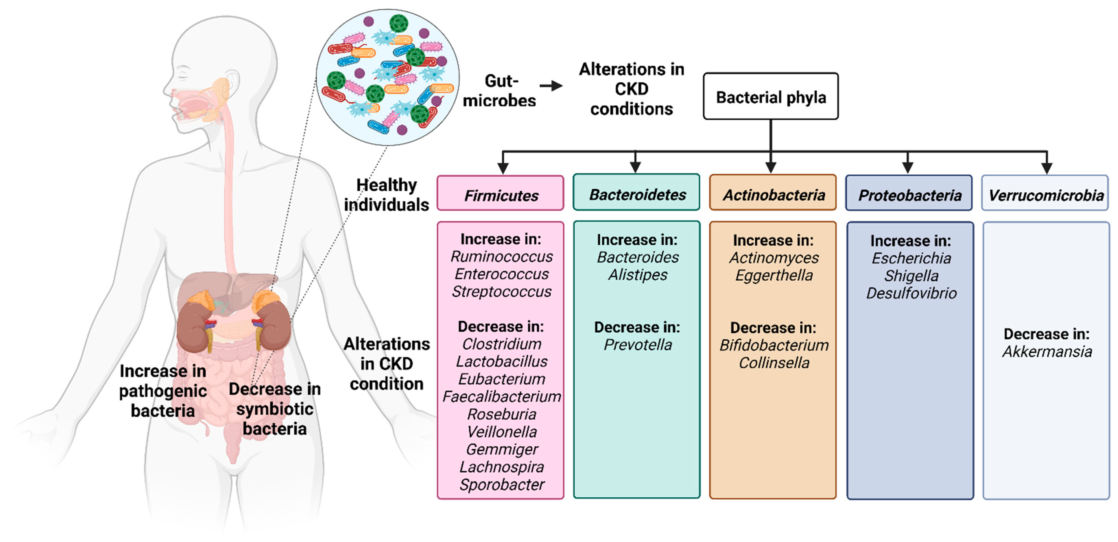
2. Alterations in the Intestinal Microflora in Chronic Kidney Disease

Dysbiosis of intestinal flora in patients with CKD is associated with a decline in the number of symbiotic flora and an increase in certain indigenous microbes leading to pathogenic conditions, which may cause or aggravate CKD due to disorders of the metabolic, endocrine or immune system [38]. This alters the composition, diversity and richness of the microflora: phyla Proteobacteria, Actinobacteria and Firmicutes show overgrowth in the duodenum and jejunum [38].
The gut microbe Akkermansia from the phyla Verrucomicrobia plays a pivotal role in increasing gut-barrier function and thickness of the mucus [4] and helps in the detoxification of hydrogen sulfide [39]. It supports the growth of bacteria-producing SCFAs such as butyrate, by providing them with carbon, nitrogen and energy produced as a result of mucus degradation [40]. Studies on fecal microbial communities in CKD patients have revealed that the abundance of probiotic bacteria Akkermansia was decreased as compared to healthy controls [40].
The gut bacteria from phyla Proteobacteria are responsible for causing an inflammatory response, alteration of gut mucosal permeability and increasing the cell ratio of intestinal T helper 17 cells to T regulatory cells and promoting the LPS translocation. The gut microbiota of CKD patients showed an increase in members of the phylum Bacteroidetes and Proteobacteria and a decrease in Lactobacillus, which belongs to the phyla Firmicutes, compared to healthy individuals [41]. Lower abundance of Lactobacillus is associated with the development of hypertension and linked to adverse outcomes in patients with CKD [42].
Patients with CKD show enhanced numbers of phylum Proteobacteria, genus Escherichia, Shigella, Desulfovibrio, and phylum Firmicutes, genus Streptococcus, while a lower abundance of the phylum Firmicutes, genus Roseburia, Faecalibacterium, and Prevotella [43]. A less prominent bacteria, Pyramidobacter from the phylum Synergistetes, is also reduced [43]. Lower numbers of these bacteria lead to less production of butyrate a compound known to protect kidneys [44]. Butyrate inhibits the histone deacetylases, thereby reducing fibrosis and attenuating acute kidney injury-mediated damage [45]. It furthermore exhibits anti-inflammatory properties as an agonist for ´G protein-coupled receptors´, which is involved in inflammation regulation [22].
A comparison between the CKD group and the healthy controls showed that Ruminococcus and Roseburia from the phyla Firmicutes have the best diagnostic performance within the thirty-one species identified with a differentiated prevalence [46]. Ruminococcus promotes inflammatory bowel syndrome, promoting CKD-associated complications by producing inflammatory polysaccharides such as glucorhamnan which induce inflammatory cytokine tumor necrosis factor alpha secretion by dendritic cells [47]. Roseburia is reduced in inflammatory and metabolic diseases and produces butyrate, which has anti-inflammatory and immunoregulatory functions [48] leading to damaged local gastrointestinal tract function and thereby aggravating inflammation in CKD patients [49].
Members of the phylum Verrucomicrobia were reduced and Actinobacteria were increased in CKD than in the healthy control groups [4]. Minor differences were observed in the numbers of several other bacterial genera in CKD patients, including Parasutterella from phyla Proteobacteria, Paraprevotella from the phyla Bacteroidetes, Clostridium IV from the phyla Firmicutes, and Alloprevotella from the phyla Bacteroidetes [39,50].
In CKD patients, Bifidobacterium from the phyla Actinobacteria was depleted and supplementing Bifidobacterium in CKD specific diets reduced serum creatinine, urea nitrogen, and p-cresyl sulfate, demonstrating its role in reducing the accumulation of these uremic toxins [51]. Patients with advanced stages of CKD had increased numbers of Eggerthella lenta from the phyla Actinobacteria, Fusobacterium nucleatum from the phyla Fusobacteriota, and Alistipes shahii from the phyla Bacteroidetes [42].
CKD patients are predisposed to immune-mediated inflammatory disease [39,52]. An increase in Actinomyces and Eggerthella from the phyla Actinobacteria, Clostridium III, Faecalicoccus, and Streptococcus from the phyla Firmicutes coincides with inflammatory diseases, while Gemmiger, Lachnospira, and Sporobacter from the phyla Firmicutes are decimated [48]. In addition, in CKD the bacterial composition changes towards favouring proteolytic bacteria, which can produce protease enzyme and a reduction in the number of saccharolytic bacteria, which can break down sugars [53]. The major changes in gut microbial compositions and their pathophysiological functions under chronic kidney disease conditions are summarized in Table 2.
Diabetic kidney disease (DKD) is another complication often accompanying CKD. The gut microbiota modifies the endocrine functions of the gut and vice versa. Additionally, hyperglycemia moderates the alterations of the gut microbiota in DKD patients. The phyla Proteobacteria, Verrucomicrobia, and Fusobacteria are relatively more abundant in patients with DKD. Dysbiosis in both DKD and CKD patients results in similar downstream processes of uremic toxin accumulation-mediated progression of renal impairment [54]. Akkermansia muciniphila supplementation has various health benefits including aiding weight loss [55].
A systemic analysis of twenty-five studies shows that Escherichia Shigella and Desulfovibrio from the phylum Proteobacteria and Streptococcus from the phylum Firmicutes show an increased abundance in CKD patients. In addition, Roseburia, Faecalibacterium and Prevotella_9 from the phyla Firmicutes, Pyramidobacter from the phyla Synergistota, and Prevotellaceae_UCG-001 from the phyla Bacteroidales have a reduced abundance under CKD conditions [43].
Furthermore, antibiotics are administered to patients with CKD which alter the intestinal microflora [1]. The major CKD-induced changes in the gut microbiota are summarized in Figure 1.

3. Effects of Alterations in the Intestinal Flora in Chronic Kidney Disease

3.1. Production of Gut-Derived Metabolites

Alteration of intestinal microflora in CKD is a major source of alteration in the uremic metabolite profile [56,57,58]. Known uremic metabolites have been classified into three categories as proposed by the ´European Uremic Toxin Work Group´ (EUTox) based on their solubility and molecular weight: (a) small water-soluble molecules which are typically <500 Da, (b) protein-bound uremic toxins (PBUTs) which are typically more than 500 Da and (c) middle molecules which are ≥500 Da in size.
The two most investigated metabolites in CKD are p-cresyl sulfate and indoxyl sulfate. As the renal function declines, the concentration of these gut-derived uremic metabolites increases [56,57]. P-cresol/p-cresyl sulfate is produced by intestinal anaerobic bacteria due to the metabolism of phenylalanine and tyrosine [59]. Tryptophan is metabolized to indole by intestinal bacteria, which is then metabolized by the liver to indoxyl sulfate [60]. The concentrations of these two metabolites in the serum increase considerably (10-fold for p-cresyl sulfate and 50-fold for indoxyl sulfate) among patients with CKD [59]. This has severe negative impact on the health of the individual causing renal tubular cell damage, coagulation disturbances and endothelial dysfunction [61] and these two metabolites play a vital role in CKD progression [62]. Further, phenol and phenylacetic acid are produced by intestinal bacteria by degradation of tyrosine and phenylalanine, respectively [63]. Levels of polyamines such as spermine, and spermidine induce the maturation of small intestine mucosa and are cytotoxic in high quantities and are also elevated in CKD patients [63,64,65].
An increase in proteolytic bacteria and a reduction in saccharolytic bacteria lead to enhanced production of ammonia and uremic metabolites such as phenols and indoles, and a decrease in SCFAs [53]. This can be observed in CKD patients, where SCFAs concentration is decreased whereas p-cresyl sulfate and TMAO are increased [43].
Quaternary amines, such as choline/phosphatidylcholine, betaine, or l-carnitine, are metabolized by intestinal bacteria to produce TMAO [62]. In experimental animal models for CKD (C57BL6J mice) [66], a diet rich in choline or TMAO produces progressive tubulointerstitial fibrosis and renal dysfunction [67].

3.2. The Accumulation of Gut-Derived Metabolites

Upon the production of the gut-derived uremic metabolites, a reduction in the filtration capacity of the kidneys increases the accumulation of these uremic, gut-derived metabolites in the serum, leading to toxicity, significantly increasing accelerated CKD progression and its accompanying co-morbidities such as atherosclerosis, obesity, and cardiovascular diseases [37]. The characteristic representatives of gut-derived metabolites are listed in Figure 2.

3.2.1. Small Water-Soluble Molecules

Currently there are more than 30 known uremic metabolites which are classified as small water-soluble molecules. Characteristic representatives of this group include asymmetric dimethylarginine (ADMA), TMAO and uric acid which have the highest global toxicity score in terms of several biological systems affected and overall experimental and clinical evidence [37].
Asymmetric dimethylarginine: ADMA, an analogue of L-arginine, is produced by gut microbes and metabolized by dimethylarginine dimethylaminohydrolases-like enzymes in Streptomyces coelicolor, Mycobacterium tuberculosis from the phylum Actinobacteria and Pseudomonas aeruginosa from the phylum Proteobacteria among others [68]. ADMA downregulates nitric oxide (NO) production [69]: a vasoactive compound, which is essential for the maintenance of endothelial homeostasis. Low levels of NO are associated with impaired endothelial function impacting the lining of all major organs including the gut. Elevated ADMA in CKD patients leads to endothelial dysfunction and consequently cardiovascular events and is therefore identified as an independent risk factor for progression of atherosclerosis, cardiovascular death, and all-cause mortality [70].
Trimethylamine-N-oxide: TMAO is formed in the liver by oxidation of the gut microbe metabolite trimethylamine (TMA), which is produced by the fermentation of dietary nutrients such as choline, betaine or L-carnitine [71,72]. Even though the mechanism of TMAO production and its link to the gut microbiota is well established, the exact gut microbe species responsible are not known [73]; however, Clostridium XIVa strains, Eubacterium sp. strain AB3007 from the phylum Firmicutes and Gammaproteobacteria from the phylum Proteobacteria are likely candidates [74]. TMAO hinders mitochondrial function and energy metabolism by reducing the pyruvate and fatty acid oxidation and activates the release of IL-1β and IL-18 in endothelial cells, thereby promoting endothelial dysfunction which could in turn degrade the intestinal epithelial barrier [75]. Elevated TMAO production in CKD patients increases the risk of mortality by increasing endothelial dysfunction and cardiovascular events [72,75].
Urea: Urea is produced in the liver via the urea cycle, a catabolite of purine metabolism [76] from dietary amino acids and was traditionally considered to be biologically inert. Urea is then excreted to the gastrointestinal tract [77], where it is utilized by E. coli from the phylum Proteobacteria [78]. Escherichia-Shigella from the phylum Proteobacteria and Bacteroides from the phylum Bacteroidetes are opportunistic pathogens which increase in the gut with the increase in urea [79].
Urea has recently re-emerged as a highly relevant gut-derived toxin which triggers molecular changes leading to insulin resistance [80,81]. An increased concentration of urea leads to endothelial dysfunction by promoting free radical production, inhibiting glyceraldehyde-3-phosphate dehydrogenase (GAPDH) and upregulating protein kinase C isoform activity, which negatively impacts the gut endothelial lining. Endothelial pro-inflammatory pathways are initiated by urea through an increase in hexosamine pathway activity and inactivating an anti-atherosclerotic enzyme PGI2 synthase [82].

3.2.2. Gut-Derived, Protein-Bound Uremic Toxins (PBUTs)

There are 25 known gut-derived PBUTs [83]. PBUTs are accumulated in CKD patients despite dialysis and hemofiltration due to their association with proteins which hinders their clearance from the plasma [84]. Characteristic representatives of this group include advanced glycation end products (AGEs), p-cresyl sulfate, indoxyl sulfate, indole acetic acid, kynurenines and phenylacetic acid which have the highest global toxicity score in terms of several biological systems affected and overall experimental and clinical evidence [37].
Advanced glycation end products: AGEs are characterized by non-enzymatically modified amino groups of proteins or lipids by monosaccharides. AGEs such as fructoselysine and N-ε-carboxymethyl lysine can be utilized by the gut microbes E. coli from the phylum Proteobacteria, Intestinimonas spp from the phylum Firmicutes and Cloacibacillus from the phylum Synergistota and potentially Oscillibacter spp from the phylum Firmicutes, respectively [85]. However, it is not known if these microbes are lower in CKD patients as compared to controls. AGEs cause sarcopenia and frailty in CKD patients [86]. The production and circulation of AGEs to tubular cells lead to high expression of transforming growth factor-beta (TGFβ), plasminogen activator inhibitor-1, tissue transglutaminase, and MCP1 contributing to tubulointerstitial fibrosis [87,88].
p-Cresyl sulfate: p-Cresyl sulfate is formed by the metabolism of aromatic amino acids by the gut microbiota. Bacteroidaceae, Clostridiaceae, Enterobacteriaceae, Ruminococcaceae, Veillonellaceae, Lactobacillaceae, Staphylococcaceae, Lachnospiraceae from the phylum Proteobacteria, Bifidobacteriaceae from the phylum Actinobacteria, Enterococcaceae, Eubacteriaceae from the phylum Firmicutes, Porphyromonadaceae from the phylum Bacteroidetes and Fusobacteriaceae from the phylum Fusobacteriota are the potential gut microbiota families which play a role in the production of p-cresyl sulfate [89].
p-Cresyl sulfate is less removed from circulation by dialysis as it is protein-bound, thereby significantly accumulating in serum of CKD patients [89]. In addition to causing cardiovascular complications p-cresyl sulfate is known to cause tubular cell damage, tubular epithelial–mesenchymal transition, tubulointerstitial inflammation and fibrosis, or whole-kidney damage by stimulating the renin–angiotensin–aldosterone system, TGFβ, intercellular adhesion molecule-1, ROS, and DNA methyltransferase 1 among other pathways [61].
Indoxyl sulfate: Indoxyl sulfate results from the metabolism of aromatic amino acids by numerous tryptophanase-producing bacteria in the gut, including E. coli from the phylum Proteobacteria [90], and Lactobacillus spp. from the phylum Firmicutes [91]. Indoxyl sulfate is not cleared from circulation by dialysis as more than 90% of this small molecule is bound to plasma proteins [92]. Indoxyl sulfate hinders glomerular filtration by promoting mesangial cell cytotoxicity by stimulating ROS production [93]. In addition, indoxyl sulfate is known to mediate renal fibrosis via the organic anion transporters/NADPH oxidase/ROS pathway through the mechanistic target of rapamycin complex 1 (mTORC1). mTORC1 mediates epithelial–mesenchymal transition of tubular epithelial cells (HK-2 cells), differentiation of fibroblasts into myofibroblasts (NRK-49F cells), and inflammatory response of macrophages [94].
Indole acetic acid: Indole acetic acid is a metabolite of aromatic amino acids metabolism [95], which is produced by numerous bacteria including E.coli from the phylum Proteobacteria, and is metabolized to Skatole by Clostridium from the phylum Firmicutes, Bacteroides from the phylum Bacteroidetes and to indole-3-aldehyde by Lactobacillus acidophilus, Lactobacillus murinus, Lactobacillus reuteri from the phylum Firmicutes, respectively [96,97,98]. In CKD patients, indole acetic acid positively correlates with inflammation and oxidative stress markers [99]. In addition to increasing ROS production, it activates the aryl hydrocarbon receptor (AhR)/p38MAPK/NF-κB pathway that induces proinflammatory enzymes [99].
Kynurenines: Kynurenine is a metabolite of tryptophan metabolism released via the kynurenine pathway. In silico analysis predicts the phylum Actinobacteria, Firmicutes and Proteobacteria, and genus Bacteroides from the phylum Bacteroidetes, Fusobacteria from the phylum Fusobacteriota as kynurenine-producing gut microbes [100].
Kynurenine promotes CKD pathophysiology through the inflammation-induced activity of indoleamine 2,3-dioxygenase [71] by regulating the AhR [101]. Proinflammatory factors such as interferon-gamma (IFN-γ) and TNF-α induce and transcriptionally enhance indoleamine dioxygenase [71] expression [102]. The accumulation of the kynurenine pathway metabolites leads to a depletion of tryptophan, which activates T regulatory cells inducing apoptosis of T cells inhibiting their proliferation and acting as a counter-reaction to inflammation [103].
Phenylacetic acid: Phenylacetic acid is formed from the microbial fermentation of aromatic amino acids, in addition to dietary intake [104]. Bacteroides thetaiotaomicron, Bacteroides eggerthii, Bacteroides ovatus, Bacteroides fragilis, Parabacteroides distasonis from the phylum Bacteroidetes and Eubacterium hallii and Clostridium bartlettii from the phylum Firmicutes found among the gut microbiota lead to significant phenylacetic acid production by fermentation [104].
Phenylacetic acid inhibits inducible nitric oxide synthase (iNOS) gene expression, reducing nitric oxide production, promoting cell proliferation and cytokine-induced endothelial expression of adhesion molecules and proinflammatory cytokines [105]. In addition, iNOS inhibition by phenylacetic acid reduces macrophage-killing aggravating immunodeficiency in CKD patients [106].

3.2.3. Middle Molecules

There are 58 known middle molecules [107]. Characteristic representatives of this group include β₂-microglobulin, ghrelin and parathyroid hormone which have the highest global toxicity score in terms of a number of biological systems affected and overall experimental and clinical evidence [37]. However, ghrelin and the impact of microbiota on this uremic metabolite have not been studied in the pathogenesis of CKD conditions until now. A majority of the middle molecules are endogenously generated, as native proteins or in response to other gut-derived uremic metabolites [37]. These can be removed by dialysis but upon the use of high-flux membranes which can lead to conflicting results due to the loss of other unspecific metabolites [37].
Β₂-microglobulin: β₂-microglobulin is an essential endogenous protein produced by all nucleated cells. Animal studies show an increase in Prevotella spp. And Bacteroides vulgatus from the phylum Bacteroidetes and a decrease in Rikenellaceae also from the phylum Bacteroidetes, in transgenic animals expressing high β₂-microglobulin [108]. The 9 kDa from the β₂-microglobulin-derived metabolite, named shed β₂-microglobulin has antibacterial activity against Staphylococcus aureus from the phylum Firmicutes, leading to the production of ‘Staphylococcus aureus-shed β₂-microglobulin’ clumps which promote the migration of THP-1 monocytes [109]. Cell migration is associated with higher inflammation under CKD conditions [110]. Β₂-microglobulin causes dialysis-related amyloidosis in end-stage CKD, contributing to bone and joint deterioration in these patients [111]. The accumulation of β₂-microglobulin in the musculoskeletal system leads to amyloid fibrils formation instead of globular, roughly natively folded protein leading to loss of function in the musculoskeletal system [112].
Parathyroid hormone: Parathyroid hormone is a polypeptide that is synthesized and cleaved by the parathyroid gland into its active form which regulates calcium metabolism under normal physiological conditions [113]. Parathyroid hormone-dependent mineral metabolism requires butyrate production by intestinal microbiota [114]; however, the exact microbes involved and the mechanism by which this process is hindered in CKD patients have not been established so far [115]. In CKD patients, parathyroid hormone promotes serum fibroblast growth factor 23 (FGF23) and reduces vitamin D production and activity, resulting in CKD-mineral bone disorder [116]. The representative uremic toxins and the potential microbes which impact their concentrations in human gut are summarized in Table 3.

3.3. Deimmunization: Inflammation and Immunosuppression

Even though the human body contains numerous microbes they are kept outside the body by various inherent barriers such as the mucosal and epithelial layers. The overgrowth of pathogenic bacteria leads to increased secretion of endotoxins and other bacterial products such as lipopolysaccharides, peptidoglycans, outer membrane proteins and bacterial DNA, into the blood through the gut lining. This leads to alteration of intestinal permeability, the release of pro-inflammatory molecules and activation of the immune system associated with intestinal mucosa [38]. Along with an increase in pathogenic bacteria, there is enhanced production of inflammatory factors such as interleukin (IL)-6, IFN-γ and TNF-α, which would promote inflammation along the gut barrier [117]. This leads to chronic systemic inflammation, thereby elucidating the role of gut bacterial dysbiosis, leading to an increase in uremic toxins in circulation, promoting CKD progression [10]. Persistent systemic inflammation in CKD result from LPS-induced monocyte/macrophage activation and systemic inflammation by sepsis from Gram-negative bacterial species [52].
Contact with different members of the gut microbiota induces different types of immune responses in the host gut. Experimentally induced uremia in complementarity to bacterial DNA treatment leads to an increase in intestinal permeability, plasma hs-CRP, pentraxin-3, proinflammatory cytokines and IL-6 which are known biomarkers of inflammation [118,119]. While anaerobic Clostridium spp. from the phylum Firmicutes, usually found in the human gut, induces FoxP3+ regulatory T cells, an uncultivated segmented filamentous bacteria of the Clostridia family promotes T helper type 17 cell differentiation [120]. Lower levels of Akkermansia from the phylum Verrucomicrobiota in the gut of CKD patients coincide with higher levels of IL-10 and lower levels of Lactobacillus from the phylum Firmicutes are accompanied by higher levels of IL-4 and IL-10, demonstrating the impact of these gut microbes in modulating immune response in CKD patients [40]. The microbes involved in inflammation and immunosuppression and the possible mechanism are summarized in Table 4.
Peptidoglycan, a component of the cell wall of most Gram-negative and Gram-positive bacteria, translocates from the intestinal microflora triggering the nucleotide-binding oligomerization domain-containing protein 1 (NOD1) receptor which leads to activation of bone marrow neutrophils causing an immune response [121]. NOD1 is a soluble cytosolic receptor expressed in macrophages, vascular endothelial and smooth muscle cells. As pattern-recognition receptors, they recognize bacterial components such as peptidoglycan and lead to activation of pro-inflammatory components such as NF-ĸB [122]. Bacterial fermentation products such as SCFAs trigger G-protein-coupled receptors 41 and 43 on intestinal epithelial cells causing an enhanced production of cytokines and chemokines from these cells [123] and reducing the production of LPS-induced production of TNF-α, IL-1β and IL-6 [124].
Therefore, gut microbes and their products induce inflammation and mediate immunosuppression by interacting with various receptors on both endothelial cells and macrophages, which has been summarized in Figure 3.

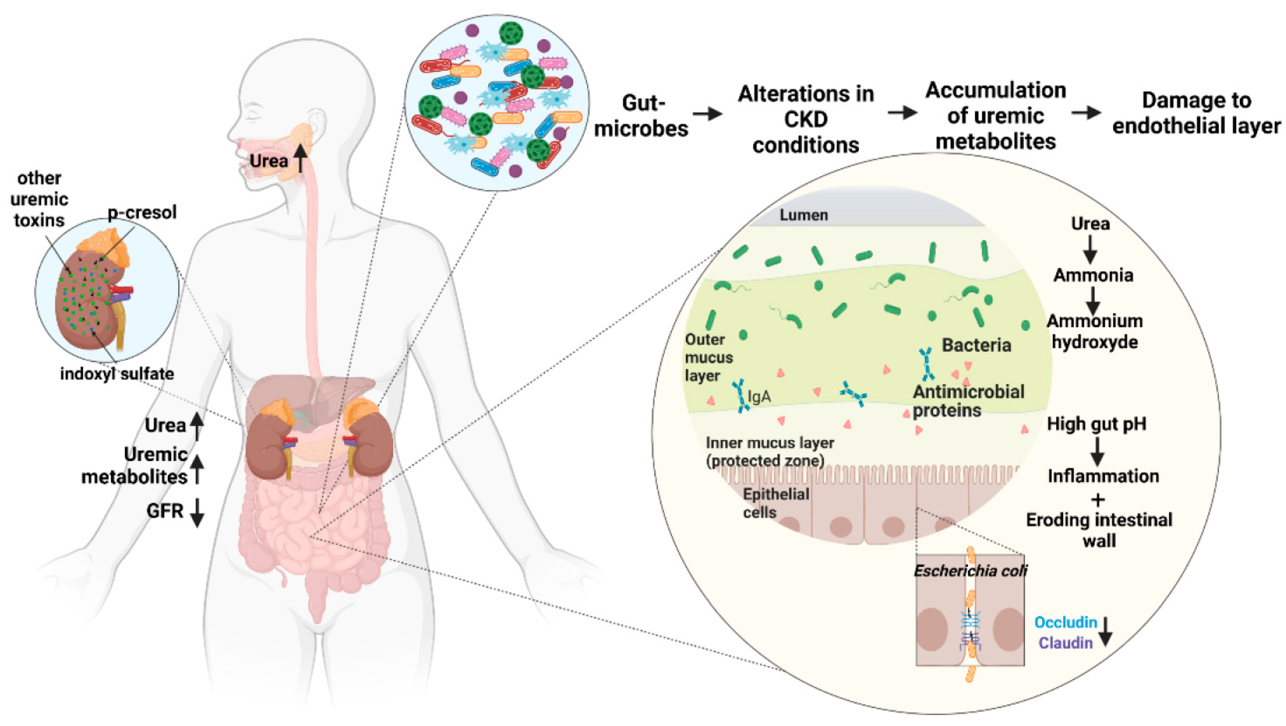
3.4. Damage to Intestinal Epithelial Barrier

Impaired kidney function and reduced glomerular filtration rate in CKD patients lead to an increase in waste products of the kidney. Increased urea concentrations can be found in glandular secretions, e.g., saliva or gastric juice [1,125,126]. This leads to an influx of urea in the gastrointestinal tract, increasing hydrolyzation of urea to ammonia [CO(NH2)2 + H2O→CO2 + 2 NH3] and further conversion to ammonium hydroxide [NH3 +H2O→NH4OH]. Urea is hydrolyzed both spontaneously and by the microbial urease enzyme, resulting in an increase in gut pH, which promotes inflammation and eroding of the intestinal wall [125,126]. The urea-rich gut milieu further favors the growth of urease-containing microbial families. Twelve such families were identified in greater abundance in patients with end-stage renal disease. Alteromonadaceae, Cellulomonadaceae, Clostridiaceae, Dermabacteraceae, Enterobacteriaceae, Halomonadaceae, Methylococcaceae, Micrococcaceae, Moraxellaceae, Polyangiaceae, Pseudomonadaceae and Xanthomonadaceae [127]. Most bacterial urease occurs as inactive holoenzyme, consisting of a triple trimer with three active centers, each in one of the α-subunits. Nickel-ions act as a cofactor and are transported into the cells and integrated into the active site via UreD, UreE, UreF, UreG and UreH proteins [128].
Ammonia dampens the acid-dependent reinforcement of the epithelial barrier in gastric epithelial (he20) cells [129,130]. Ammonium hydroxide interacts with tight junction proteins facing the lumen and leads to the depletion of tight junction proteins occludin, claudin-1, and ZO-1 [131], lowering transepithelial electrical resistance, and increasing permeability of the intestinal wall. This metabolite further promotes the influx of pro-inflammatory leukocytes, promoting endocytosis of transcellular tight junction proteins [125,132]. The lowered concentration of butyrate-producing gut bacteria and consequently lowered levels of butyrate in CKD patients further destabilize the intestinal wall. Butyrate promotes the production of mucin and tight junction proteins [133,134,135] and possesses anti-inflammatory and antioxidant attributes; its absence thus promotes inflammation [134]. Macrophages in the intestine contribute to inflammation and bacterial translocation across the intestinal wall: LPS activate macrophages via TLR4. Activated macrophages produce the pro-inflammatory cytokines IL-6 and TNF-α, promoting microinflammation and contributing to the weakening of the integrity of the intestinal wall. One key function of macrophages is the phagocytosis of, for example, cellular debris or pathogens [136].
Phagocytosis is the process through which a cell engulfs an extracellular particle with its cell membrane and digests it. In conditions of disrupted intestinal barrier, macrophages pass through the barrier into the circulatory system, where they release waste products and possible endotoxins of phagocytized bacteria through exocytosis [137]. This would ultimately release endotoxins into the circulatory system, promoting endotoxemia and systemic inflammation [137]. The degradation of the epithelial tight junction complex weakens the transcellular connection, makes the epithelial cells more liable to mechanical stress, and has detrimental effects on cell polarity as well as the passive transcellular transport of ions and molecules, leading to an increase in luminal nutrients and water, promoting pathogen growth and dispersal [130,138].
Tight junction proteins from the apical junctional regions are dislocated by pathogens, resulting in their downward movement along the lateral membrane to intracellular locations. Alterations in tight junction fencing can lead to the flipping of basolateral proteins to apical sites, making new receptors available for microbes or toxins [130,138]. For example, Escherichia coli lines up along the junctions of epithelial cells and may use junctional proteins as receptors. The resulting alterations in pathogen-mediated signaling leads to actin perijunctional ring contraction, leading to enhanced paracellular permeability. Enhanced paracellular permeability provides an opportunity for the translocation of molecules by diffusion. In a parallel mechanism, cytoskeletal changes by microbes such as myosin light-chain phosphorylation trigger actomyosin contraction, also altering paracellular permeability [130,138].
In addition, fluid retention is a symptom of CKD which leads to adverse renal outcomes [139], with hemodialysis induced circulatory stress leading to increased endotoxin translocation from the gut [138]. The increased permeability of the intestinal wall results in the translocation of endotoxins and bacterial components such as DNA, LPS, and metabolites into the local and systemic tissue. This promotes activation of the pro-inflammatory cytokines IL-1 and IL-6 as well as expression of soluble TNF receptors which promote endothelial dysfunction, disturbing the endothelial barrier. The continuous activation of the immune system through the influx of bacterial components leads to an overactivation of the immune system and acquired immunodeficiency, as commonly seen in sepsis [140]. Products of bacterial catabolism in the circulatory system may also produce various uremic metabolites. The bacterial metabolites indole, trimethylamine, and p-cresyl glucoronidate are further metabolized in the liver to indoxyl sulfate, trimethylamine N-oxide, and p-cresyl glucuronide, respectively, contributing to kidney-related diseases [132].
In addition, CKD patients undergoing extracorporeal treatment show increased levels of circulating endotoxins compared to CKD patients in a similar stage of renal failure but without extracorporeal treatment. Endotoxemia and chronic inflammation through intestinal barrier dysfunction are promoted by hypotension, which is a concomitant of hemodialysis [141] and promote bowel edema and ischemia [142]. Extracorporeal treatment-induced ischemia and hypoxia are additional risk factors since the intestinal wall is sensitive to low oxygen milieu due to epithelial injuries resulting from hypoxic villus tips [143,144]. Hemodialysis furthermore leads to a rise in core temperature, which could disturb the intestinal barrier. Diuretics and anticoagulants used in hemodialysis are potential risk factors, promoting hypotension and micro-bleeding, respectively [125,138,144]. The mechanisms by which the gut microbiota damages the intestinal epithelial barrier have been summarized in Figure 4.
These results emphasize the influence of CKD on the intestinal barrier and how dysfunction of the latter can in turn worsen the effect of CKD, creating a feedforward loop.

4. Discussion

The human gut microbiome consists of bacterial species from five major bacterial phyla, Firmicutes, Bacteroidetes, Actinobacteria, Proteobacteria and Verrucomicrobia. The ratio of Firmicutes/Bacteroidetes, which are the most abundant phyla in the human gut, plays an important role in maintaining intestinal homeostasis. Some species of Actinobacteria have been used as probiotics. An increase in Verrucomicrobia coincides with a healthy gut, while an increase in Proteobacteria coincides with dysbiosis and pathogenic conditions.
In CKD patients among members of the phylum Firmicutes, there is an increase in Ruminococcus, Enterococcus and Streptococcus and a decrease in Clostridium, Lactobacillus, Eubacterium, Faecalibacterium, Roseburia, Veillonella, Gemmiger, Lachnospira and Sporobacter. Among members of the phylum Bacteroidetes, there is an increase in Bacteroides and Alistipes and a decrease in Prevotella. Among members of the phylum Actinobacteria there is an increase in Actinomyces and Eggerthella, along with a decrease in Bifidobacterium and Collinsella. There is an increase in the members of the phylum Proteobacteria, especially genus Escherichia, Shigella and Desulfovibrio. From the phylum Verrucomicrobia the genus Akkermansia is decreased in CKD patients.
In addition, CKD-associated alteration of this intestinal microbiome results in metabolic changes and the accumulation of uremic metabolites, which have a feedforward adverse effect on CKD patients, inhibiting renal functions and increasing comorbidities such as atherosclerosis and CVD. Characteristic representatives of small water-soluble uremic toxins include ADMA, TMAO and urea which are most influenced by members of the phyla Firmicutes, Actinobacteria and Bacteriodetes. Characteristic representatives of protein-bound uremic toxins include AGEs, p-cresyl sulfate, indoxyl sulfate, indole acetic acid, the kynurenines and phenylacetic acid which are most influenced by members of the phyla Firmicutes, Proteobacteria and Bacteriodetes. Characteristic representatives of middle molecules include β₂-microglobulin, ghrelin and parathyroid hormone which are most influenced by members of the phyla Firmicutes and Bacteriodetes. Elevated levels of endotoxins in the gut lead to endotoxemia and inflammation, further accelerating CKD progression. Gut microbes and their metabolites induce inflammation and mediate immunosuppression by interacting with various receptors leading to macrophage activation, dysregulation of chemokines and cytokines and promoting a change in membrane permeability. Gut microbiota-derived metabolites damage the intestinal epithelial barrier by an influx of urea in the gastrointestinal tract, increasing hydrolyzation of urea to ammonia and ammonium hydroxide, leading to depletion of tight junction proteins occludin and claudin-1, lowering transepithelial electrical resistance, and increasing permeability of the intestinal wall.
Even though the gut microbiota and their importance in the progression of CKD have gained traction in recent years, there is still a knowledge gap that hinders widespread therapeutic application. Most clinical studies focus on the links between CKD, uremic metabolites and the microbes dysregulated without focusing on the mechanisms by which these microbes regulate CKD progression. Moreover, the studies linking microbes to uremic metabolite concentrations are limited by lack of standardized techniques for quantifying the large array of diverse uremic metabolites. Comprehensive systematic studies are required to completely understand the role of the gut microbiota in CKD progression, followed by gut microbiota-corrective studies to restore healthy gut conditions. In addition, pathogen-induced deimmunization and intestinal barrier disruption present novel opportunities for mitigating CKD progression.

5. Conclusions

Alterations in the members of these phyla alter the total gut microbiota, with a decline in the number of symbiotic flora and an increase in the pathogenic bacteria, causing or aggravating CKD. Numerous studies focus on the alterations of the gut microbiota in CKD and the alteration of metabolic profile of CKD patients. However, there is a lack of studies connecting these two modifications in high-throughput studies. Gut-derived uremic metabolites exhibit cell-damaging properties, damage the intestinal epithelial cell wall, increase gut permeability and lead to the translocation of bacteria and endotoxins from the gut into the circulatory system. To limit CKD progression, it is imperative to address the changes in the gut microbiome; therefore, it is necessary to identify and characterize the role of different microbes in the progression of CKD.

Author Contributions

The author S.B. conceived and planned the review. S.B., E.M. and A.V. wrote the manuscript. H.N. and J.J. corrected the manuscript and provided guidance. All authors discussed the findings and contributed to the final manuscript. All authors have read and agreed to the published version of the manuscript.

Funding

The authors are supported by grants from the German Research Foundation (DFG) (SFB/TRR 219 project-ID: 322900939 and SFB 1382 Project-ID 403224013) as well as by the European Union’s Horizon 2020 research and innovation programme under the Marie Skłodowska-Curie grant agreement No 764474 (CaReSyAn) and No 860329 (Strategy-CKD).

Institutional Review Board Statement

Not applicable.

Informed Consent Statement

Not applicable.

Data Availability Statement

Not applicable.

Acknowledgments

All figures were created using the program Biorender.

Conflicts of Interest

The authors have no conflict of interest.

References

  1. Guldris, S.C.; Parra, E.G.; Amenos, A.C. Gut microbiota in chronic kidney disease. Nefrologia 2017, 37, 9–19. [Google Scholar] [CrossRef]
  2. Diez, J.; Ortiz, A. The need for a cardionephrology subspecialty. Clin. Kidney J. 2021, 14, 1491–1494. [Google Scholar] [CrossRef] [PubMed]
  3. Pickard, J.M.; Zeng, M.Y.; Caruso, R.; Nunez, G. Gut microbiota: Role in pathogen colonization, immune responses, and inflammatory disease. Immunol. Rev. 2017, 279, 70–89. [Google Scholar] [CrossRef] [PubMed]
  4. Ottman, N.; Geerlings, S.Y.; Aalvink, S.; de Vos, W.M.; Belzer, C. Action and function of Akkermansia muciniphila in microbiome ecology, health and disease. Best Pract. Res. Clin. Gastroenterol. 2017, 31, 637–642. [Google Scholar] [CrossRef]
  5. Backhed, F.; Ley, R.E.; Sonnenburg, J.L.; Peterson, D.A.; Gordon, J.I. Host-bacterial mutualism in the human intestine. Science 2005, 307, 1915–1920. [Google Scholar] [CrossRef]
  6. Mahmoodpoor, F.; Rahbar Saadat, Y.; Barzegari, A.; Ardalan, M.; Zununi Vahed, S. The impact of gut microbiota on kidney function and pathogenesis. Biomed Pharm. 2017, 93, 412–419. [Google Scholar] [CrossRef]
  7. Jenson, I. BACILLUS | Introduction. In Encyclopedia of Food Microbiology, 2nd ed.; Academic Press: Cambridge, MA, USA, 2014. [Google Scholar] [CrossRef]
  8. Castro-Gonzalez, J.M.; Castro, P.; Sandoval, H.; Castro-Sandoval, D. Probiotic lactobacilli precautions. Front. Microbiol. 2019, 10, 375. [Google Scholar] [CrossRef]
  9. Wexler, H.M. Bacteroides: The good, the bad, and the nitty-gritty. Clin. Microbiol. Rev. 2007, 20, 593–621. [Google Scholar] [CrossRef]
  10. Clemente, J.C.; Pehrsson, E.C.; Blaser, M.J.; Sandhu, K.; Gao, Z.; Wang, B.; Magris, M.; Hidalgo, G.; Contreras, M.; Noya-Alarcon, O.; et al. The microbiome of uncontacted Amerindians. Sci. Adv. 2015, 1, e1500183. [Google Scholar] [CrossRef] [Green Version]
  11. Gorvitovskaia, A.; Holmes, S.P.; Huse, S.M. Interpreting Prevotella and Bacteroides as biomarkers of diet and lifestyle. Microbiome 2016, 4, 15. [Google Scholar] [CrossRef]
  12. Iljazovic, A.; Roy, U.; Galvez, E.J.C.; Lesker, T.R.; Zhao, B.; Gronow, A.; Amend, L.; Will, S.E.; Hofmann, J.D.; Pils, M.C.; et al. Perturbation of the gut microbiome by Prevotella spp. enhances host susceptibility to mucosal inflammation. Mucosal Immunol. 2021, 14, 113–124. [Google Scholar] [CrossRef] [PubMed]
  13. Li, W.; Ma, Z.S. FBA ecological guild: Trio of firmicutes-bacteroidetes alliance against actinobacteria in human oral microbiome. Sci. Rep. 2020, 10, 287. [Google Scholar] [CrossRef]
  14. Stojanov, S.; Berlec, A.; Strukelj, B. The influence of probiotics on the firmicutes/bacteroidetes ratio in the treatment of obesity and inflammatory bowel disease. Microorganisms 2020, 8, 1715. [Google Scholar] [CrossRef] [PubMed]
  15. Vaiserman, A.; Romanenko, M.; Piven, L.; Moseiko, V.; Lushchak, O.; Kryzhanovska, N.; Guryanov, V.; Koliada, A. Differences in the gut Firmicutes to Bacteroidetes ratio across age groups in healthy Ukrainian population. BMC Microbiol. 2020, 20, 221. [Google Scholar] [CrossRef] [PubMed]
  16. Yang, T.; Santisteban, M.M.; Rodriguez, V.; Li, E.; Ahmari, N.; Carvajal, J.M.; Zadeh, M.; Gong, M.; Qi, Y.; Zubcevic, J.; et al. Gut dysbiosis is linked to hypertension. Hypertension 2015, 65, 1331–1340. [Google Scholar] [CrossRef] [PubMed]
  17. Ley, R.E.; Turnbaugh, P.J.; Klein, S.; Gordon, J.I. Microbial ecology: Human gut microbes associated with obesity. Nature 2006, 444, 1022–1023. [Google Scholar] [CrossRef]
  18. Kieffer, D.A.; Piccolo, B.D.; Vaziri, N.D.; Liu, S.; Lau, W.L.; Khazaeli, M.; Nazertehrani, S.; Moore, M.E.; Marco, M.L.; Martin, R.J.; et al. Resistant starch alters gut microbiome and metabolomic profiles concurrent with amelioration of chronic kidney disease in rats. Am. J. Physiol. Renal. Physiol. 2016, 310, F857–F871. [Google Scholar] [CrossRef]
  19. Marques, F.Z.; Nelson, E.; Chu, P.Y.; Horlock, D.; Fiedler, A.; Ziemann, M.; Tan, J.K.; Kuruppu, S.; Rajapakse, N.W.; El-Osta, A.; et al. High-fiber diet and acetate supplementation change the gut microbiota and prevent the development of hypertension and heart failure in hypertensive mice. Circulation 2017, 135, 964–977. [Google Scholar] [CrossRef]
  20. Binda, C.; Lopetuso, L.R.; Rizzatti, G.; Gibiino, G.; Cennamo, V.; Gasbarrini, A. Actinobacteria: A relevant minority for the maintenance of gut homeostasis. Dig. Liver Dis. 2018, 50, 421–428. [Google Scholar] [CrossRef]
  21. Shin, N.R.; Whon, T.W.; Bae, J.W. Proteobacteria: Microbial signature of dysbiosis in gut microbiota. Trends Biotechnol. 2015, 33, 496–503. [Google Scholar] [CrossRef]
  22. Fujio-Vejar, S.; Vasquez, Y.; Morales, P.; Magne, F.; Vera-Wolf, P.; Ugalde, J.A.; Navarrete, P.; Gotteland, M. The gut microbiota of healthy chilean subjects reveals a high abundance of the phylum verrucomicrobia. Front. Microbiol. 2017, 8, 1221. [Google Scholar] [CrossRef] [PubMed]
  23. Gomaa, E.Z. Human gut microbiota/microbiome in health and diseases: A review. Antonie Van Leeuwenhoek 2020, 113, 2019–2040. [Google Scholar] [CrossRef] [PubMed]
  24. Chen, X.; D’Souza, R.; Hong, S.T. The role of gut microbiota in the gut-brain axis: Current challenges and perspectives. Protein Cell 2013, 4, 403–414. [Google Scholar] [CrossRef] [PubMed]
  25. Topping, D.L.; Clifton, P.M. Short-chain fatty acids and human colonic function: Roles of resistant starch and nonstarch polysaccharides. Physiol. Rev. 2001, 81, 1031–1064. [Google Scholar] [CrossRef]
  26. Valdes, A.M.; Walter, J.; Segal, E.; Spector, T.D. Role of the gut microbiota in nutrition and health. BMJ 2018, 361, k2179. [Google Scholar] [CrossRef]
  27. Tolhurst, G.; Heffron, H.; Lam, Y.S.; Parker, H.E.; Habib, A.M.; Diakogiannaki, E.; Cameron, J.; Grosse, J.; Reimann, F.; Gribble, F.M. Short-chain fatty acids stimulate glucagon-like peptide-1 secretion via the G-protein-coupled receptor FFAR2. Diabetes 2012, 61, 364–371. [Google Scholar] [CrossRef]
  28. Carvalho, B.M.; Saad, M.J. Influence of gut microbiota on subclinical inflammation and insulin resistance. Mediat. Inflamm. 2013, 2013, 986734. [Google Scholar] [CrossRef]
  29. Sivaprakasam, S.; Prasad, P.D.; Singh, N. Benefits of short-chain fatty acids and their receptors in inflammation and carcinogenesis. Pharmacol. Ther. 2016, 164, 144–151. [Google Scholar] [CrossRef]
  30. Amabebe, E.; Robert, F.O.; Agbalalah, T.; Orubu, E.S.F. Microbial dysbiosis-induced obesity: Role of gut microbiota in homoeostasis of energy metabolism. Br. J. Nutr. 2020, 123, 1127–1137. [Google Scholar] [CrossRef]
  31. Hobby, G.P.; Karaduta, O.; Dusio, G.F.; Singh, M.; Zybailov, B.L.; Arthur, J.M. Chronic kidney disease and the gut microbiome. Am. J. Physiol. Renal. Physiol. 2019, 316, F1211–F1217. [Google Scholar] [CrossRef]
  32. Duttaroy, A.K. Role of gut microbiota and their metabolites on atherosclerosis, hypertension and human blood platelet function: A review. Nutrients 2021, 13, 144. [Google Scholar] [CrossRef] [PubMed]
  33. Anders, H.J.; Andersen, K.; Stecher, B. The intestinal microbiota, a leaky gut, and abnormal immunity in kidney disease. Kidney Int. 2013, 83, 1010–1016. [Google Scholar] [CrossRef]
  34. Glorieux, G.; Gryp, T.; Perna, A. Gut-derived metabolites and their role in immune dysfunction in chronic kidney disease. Toxins 2020, 12, 245. [Google Scholar] [CrossRef]
  35. Mihajlovic, M.; Krebber, M.M.; Yang, Y.; Ahmed, S.; Lozovanu, V.; Andreeva, D.; Verhaar, M.C.; Masereeuw, R. Protein-bound uremic toxins induce reactive oxygen species-dependent and inflammasome-mediated il-1beta production in kidney proximal tubule cells. Biomedicines 2021, 9, 1326. [Google Scholar] [CrossRef] [PubMed]
  36. de Mello, V.D.; Paananen, J.; Lindstrom, J.; Lankinen, M.A.; Shi, L.; Kuusisto, J.; Pihlajamaki, J.; Auriola, S.; Lehtonen, M.; Rolandsson, O.; et al. Indolepropionic acid and novel lipid metabolites are associated with a lower risk of type 2 diabetes in the Finnish Diabetes Prevention Study. Sci. Rep. 2017, 7, 46337. [Google Scholar] [CrossRef] [PubMed]
  37. Vanholder, R.; Pletinck, A.; Schepers, E.; Glorieux, G. Biochemical and clinical impact of organic uremic retention solutes: A comprehensive update. Toxins 2018, 10, 33. [Google Scholar] [CrossRef]
  38. Sabatino, A.; Regolisti, G.; Brusasco, I.; Cabassi, A.; Morabito, S.; Fiaccadori, E. Alterations of intestinal barrier and microbiota in chronic kidney disease. Nephrol. Dial. Transplant. 2015, 30, 924–933. [Google Scholar] [CrossRef]
  39. Li, F.; Wang, M.; Wang, J.; Li, R.; Zhang, Y. Alterations to the gut microbiota and their correlation with inflammatory factors in chronic kidney disease. Front. Cell Infect. Microbiol. 2019, 9, 206. [Google Scholar] [CrossRef]
  40. Hanninen, A.; Toivonen, R.; Poysti, S.; Belzer, C.; Plovier, H.; Ouwerkerk, J.P.; Emani, R.; Cani, P.D.; De Vos, W.M. Akkermansia muciniphila induces gut microbiota remodelling and controls islet autoimmunity in NOD mice. Gut 2018, 67, 1445–1453. [Google Scholar] [CrossRef] [Green Version]
  41. Kanbay, M.; Onal, E.M.; Afsar, B.; Dagel, T.; Yerlikaya, A.; Covic, A.; Vaziri, N.D. The crosstalk of gut microbiota and chronic kidney disease: Role of inflammation, proteinuria, hypertension, and diabetes mellitus. Int. Urol. Nephrol. 2018, 50, 1453–1466. [Google Scholar] [CrossRef]
  42. Wehedy, E.; Shatat, I.F.; Al Khodor, S. The human microbiome in chronic kidney disease: A double-edged sword. Front. Med. 2021, 8, 790783. [Google Scholar] [CrossRef] [PubMed]
  43. Zhao, J.; Ning, X.; Liu, B.; Dong, R.; Bai, M.; Sun, S. Specific alterations in gut microbiota in patients with chronic kidney disease: An updated systematic review. Ren. Fail. 2021, 43, 102–112. [Google Scholar] [CrossRef] [PubMed]
  44. Felizardo, R.J.F.; de Almeida, D.C.; Pereira, R.L.; Watanabe, I.K.M.; Doimo, N.T.S.; Ribeiro, W.R.; Cenedeze, M.A.; Hiyane, M.I.; Amano, M.T.; Braga, T.T.; et al. Gut microbial metabolite butyrate protects against proteinuric kidney disease through epigenetic- and GPR109a-mediated mechanisms. FASEB J. 2019, 33, 11894–11908. [Google Scholar] [CrossRef]
  45. Brilli, L.L.; Swanhart, L.M.; de Caestecker, M.P.; Hukriede, N.A. HDAC inhibitors in kidney development and disease. Pediatr. Nephrol. 2013, 28, 1909–1921. [Google Scholar] [CrossRef]
  46. Hu, Q.; Wu, K.; Pan, W.; Zeng, Y.; Hu, K.; Chen, D.; Huang, X.; Zhang, Q. Intestinal flora alterations in patients with early chronic kidney disease: A case-control study among the Han population in southwestern China. J. Int. Med. Res. 2020, 48, 300060520926033. [Google Scholar] [CrossRef]
  47. Henke, M.T.; Kenny, D.J.; Cassilly, C.D.; Vlamakis, H.; Xavier, R.J.; Clardy, J. Ruminococcus gnavus, a member of the human gut microbiome associated with Crohn’s disease, produces an inflammatory polysaccharide. Proc. Natl. Acad. Sci. USA 2019, 116, 12672–12677. [Google Scholar] [CrossRef] [PubMed]
  48. Forbes, J.D.; Chen, C.Y.; Knox, N.C.; Marrie, R.A.; El-Gabalawy, H.; de Kievit, T.; Alfa, M.; Bernstein, C.N.; Van Domselaar, G. A comparative study of the gut microbiota in immune-mediated inflammatory diseases-does a common dysbiosis exist? Microbiome 2018, 6, 221. [Google Scholar] [CrossRef]
  49. Chung, S.; Barnes, J.L.; Astroth, K.S. Gastrointestinal microbiota in patients with chronic kidney disease: A systematic review. Adv. Nutr. 2019, 10, 888–901. [Google Scholar] [CrossRef]
  50. Chen, Y.J.; Wu, H.; Wu, S.D.; Lu, N.; Wang, Y.T.; Liu, H.N.; Dong, L.; Liu, T.T.; Shen, X.Z. Parasutterella, in association with irritable bowel syndrome and intestinal chronic inflammation. J. Gastroenterol. Hepatol. 2018, 33, 1844–1852. [Google Scholar] [CrossRef]
  51. Iwashita, Y.; Ohya, M.; Yashiro, M.; Sonou, T.; Kawakami, K.; Nakashima, Y.; Yano, T.; Iwashita, Y.; Mima, T.; Negi, S.; et al. Dietary changes involving bifidobacterium longum and other nutrients delays chronic kidney disease progression. Am. J. Nephrol. 2018, 47, 325–332. [Google Scholar] [CrossRef]
  52. Stearns-Kurosawa, D.J.; Osuchowski, M.F.; Valentine, C.; Kurosawa, S.; Remick, D.G. The pathogenesis of sepsis. Annu. Rev. Pathol. 2011, 6, 19–48. [Google Scholar] [CrossRef] [PubMed]
  53. Noce, A.; Marchetti, M.; Marrone, G.; Di Renzo, L.; Di Lauro, M.; Di Daniele, F.; Albanese, M.; Di Daniele, N.; De Lorenzo, A. Link between gut microbiota dysbiosis and chronic kidney disease. Eur. Rev. Med. Pharmacol. Sci. 2022, 26, 2057–2074. [Google Scholar] [CrossRef] [PubMed]
  54. Lin, J.R.; Wang, Z.T.; Sun, J.J.; Yang, Y.Y.; Li, X.X.; Wang, X.R.; Shi, Y.; Zhu, Y.Y.; Wang, R.T.; Wang, M.N.; et al. Gut microbiota and diabetic kidney diseases: Pathogenesis and therapeutic perspectives. World J. Diabetes 2022, 13, 308–318. [Google Scholar] [CrossRef]
  55. Corb Aron, R.A.; Abid, A.; Vesa, C.M.; Nechifor, A.C.; Behl, T.; Ghitea, T.C.; Munteanu, M.A.; Fratila, O.; Andronie-Cioara, F.L.; Toma, M.M.; et al. Recognizing the benefits of pre-/probiotics in metabolic syndrome and type 2 diabetes mellitus considering the influence of Akkermansia muciniphila as a key gut bacterium. Microorganisms 2021, 9, 618. [Google Scholar] [CrossRef] [PubMed]
  56. Aronov, P.A.; Luo, F.J.; Plummer, N.S.; Quan, Z.; Holmes, S.; Hostetter, T.H.; Meyer, T.W. Colonic contribution to uremic solutes. J. Am. Soc. Nephrol. 2011, 22, 1769–1776. [Google Scholar] [CrossRef]
  57. Ramezani, A.; Raj, D.S. The gut microbiome, kidney disease, and targeted interventions. J. Am. Soc. Nephrol. 2014, 25, 657–670. [Google Scholar] [CrossRef]
  58. Rysz, J.; Franczyk, B.; Lawinski, J.; Olszewski, R.; Cialkowska-Rysz, A.; Gluba-Brzozka, A. The impact of CKD on uremic toxins and gut microbiota. Toxins 2021, 13, 252. [Google Scholar] [CrossRef]
  59. Dou, L.; Bertrand, E.; Cerini, C.; Faure, V.; Sampol, J.; Vanholder, R.; Berland, Y.; Brunet, P. The uremic solutes p-cresol and indoxyl sulfate inhibit endothelial proliferation and wound repair. Kidney Int. 2004, 65, 442–451. [Google Scholar] [CrossRef]
  60. Cao, X.S.; Chen, J.; Zou, J.Z.; Zhong, Y.H.; Teng, J.; Ji, J.; Chen, Z.W.; Liu, Z.H.; Shen, B.; Nie, Y.X.; et al. Association of indoxyl sulfate with heart failure among patients on hemodialysis. Clin. J. Am. Soc. Nephrol. 2015, 10, 111–119. [Google Scholar] [CrossRef] [Green Version]
  61. Vanholder, R.; Schepers, E.; Pletinck, A.; Nagler, E.V.; Glorieux, G. The uremic toxicity of indoxyl sulfate and p-cresyl sulfate: A systematic review. J. Am. Soc. Nephrol. 2014, 25, 1897–1907. [Google Scholar] [CrossRef]
  62. Wu, I.W.; Hsu, K.H.; Lee, C.C.; Sun, C.Y.; Hsu, H.J.; Tsai, C.J.; Tzen, C.Y.; Wang, Y.C.; Lin, C.Y.; Wu, M.S. p-Cresyl sulphate and indoxyl sulphate predict progression of chronic kidney disease. Nephrol. Dial. Transplant. 2011, 26, 938–947. [Google Scholar] [CrossRef] [PubMed]
  63. Lutz, W. A uremic peptide containing polyamine: Formation and possible role in uremic hypertriglyceridemia. Physiol. Chem. Phys. 1980, 12, 451–456. [Google Scholar] [PubMed]
  64. Farhadian, S.; Shareghi, B.; Saboury, A.A.; Babaheydari, A.K.; Raisi, F.; Heidari, E. Molecular aspects of the interaction of spermidine and alpha-chymotrypsin. Int. J. Biol. Macromol. 2016, 92, 523–532. [Google Scholar] [CrossRef]
  65. Dufour, C.; Dandrifosse, G.; Forget, P.; Vermesse, F.; Romain, N.; Lepoint, P. Spermine and spermidine induce intestinal maturation in the rat. Gastroenterology 1988, 95, 112–116. [Google Scholar] [CrossRef]
  66. Tang, W.H.; Wang, Z.; Kennedy, D.J.; Wu, Y.; Buffa, J.A.; Agatisa-Boyle, B.; Li, X.S.; Levison, B.S.; Hazen, S.L. Gut microbiota-dependent trimethylamine N-oxide (TMAO) pathway contributes to both development of renal insufficiency and mortality risk in chronic kidney disease. Circ. Res. 2015, 116, 448–455. [Google Scholar] [CrossRef] [PubMed]
  67. Stubbs, J.R.; House, J.A.; Ocque, A.J.; Zhang, S.; Johnson, C.; Kimber, C.; Schmidt, K.; Gupta, A.; Wetmore, J.B.; Nolin, T.D.; et al. Serum trimethylamine-N-oxide is elevated in CKD and correlates with coronary atherosclerosis burden. J. Am. Soc. Nephrol. 2016, 27, 305–313. [Google Scholar] [CrossRef]
  68. Santa Maria, J.; Vallance, P.; Charles, I.G.; Leiper, J.M. Identification of microbial dimethylarginine dimethylaminohydrolase enzymes. Mol. Microbiol. 1999, 33, 1278–1279. [Google Scholar] [CrossRef]
  69. Fiedler, L. The DDAH/ADMA pathway is a critical regulator of NO signalling in vascular homeostasis. Cell Adhes. Migr. 2008, 2, 149–150. [Google Scholar] [CrossRef]
  70. Meinitzer, A.; Seelhorst, U.; Wellnitz, B.; Halwachs-Baumann, G.; Boehm, B.O.; Winkelmann, B.R.; Marz, W. Asymmetrical dimethylarginine independently predicts total and cardiovascular mortality in individuals with angiographic coronary artery disease (the Ludwigshafen Risk and Cardiovascular Health study). Clin. Chem. 2007, 53, 273–283. [Google Scholar] [CrossRef] [Green Version]
  71. Wang, Z.; Klipfell, E.; Bennett, B.J.; Koeth, R.; Levison, B.S.; Dugar, B.; Feldstein, A.E.; Britt, E.B.; Fu, X.; Chung, Y.M.; et al. Gut flora metabolism of phosphatidylcholine promotes cardiovascular disease. Nature 2011, 472, 57–63. [Google Scholar] [CrossRef]
  72. He, S.; Jiang, H.; Zhuo, C.; Jiang, W. Trimethylamine/trimethylamine-N-oxide as a key between diet and cardiovascular diseases. Cardiovasc. Toxicol. 2021, 21, 593–604. [Google Scholar] [CrossRef]
  73. Rath, S.; Rud, T.; Pieper, D.H.; Vital, M. Potential TMA-producing bacteria are ubiquitously found in mammalia. Front. Microbiol. 2019, 10, 2966. [Google Scholar] [CrossRef]
  74. Rath, S.; Heidrich, B.; Pieper, D.H.; Vital, M. Uncovering the trimethylamine-producing bacteria of the human gut microbiota. Microbiome 2017, 5, 54. [Google Scholar] [CrossRef] [PubMed]
  75. Zhang, Y.; Wang, Y.; Ke, B.; Du, J. TMAO: How gut microbiota contributes to heart failure. Transl. Res. 2021, 228, 109–125. [Google Scholar] [CrossRef] [PubMed]
  76. Barr, W.G. Uric acid. In Clinical Methods: The History, Physical, and Laboratory Examinations, 3rd ed.; Walker, H.K., Hall, W.D., Hurst, J.W., Eds.; Butterworths: Boston, MA, USA, 1990. [Google Scholar]
  77. Ramezani, A.; Massy, Z.A.; Meijers, B.; Evenepoel, P.; Vanholder, R.; Raj, D.S. Role of the gut microbiome in uremia: A potential therapeutic target. Am. J. Kidney Dis. 2016, 67, 483–498. [Google Scholar] [CrossRef] [PubMed]
  78. Liu, H.; Zhuang, J.; Tang, P.; Li, J.; Xiong, X.; Deng, H. The role of the gut microbiota in coronary heart disease. Curr. Atheroscler. Rep. 2020, 22, 77. [Google Scholar] [CrossRef]
  79. Xu, X.; Wang, H.; Guo, D.; Man, X.; Liu, J.; Li, J.; Luo, C.; Zhang, M.; Zhen, L.; Liu, X. Curcumin modulates gut microbiota and improves renal function in rats with uric acid nephropathy. Ren. Fail. 2021, 43, 1063–1075. [Google Scholar] [CrossRef]
  80. D’Apolito, M.; Du, X.; Zong, H.; Catucci, A.; Maiuri, L.; Trivisano, T.; Pettoello-Mantovani, M.; Campanozzi, A.; Raia, V.; Pessin, J.E.; et al. Urea-induced ROS generation causes insulin resistance in mice with chronic renal failure. J. Clin. Investig. 2010, 120, 203–213. [Google Scholar] [CrossRef]
  81. Koppe, L.; Nyam, E.; Vivot, K.; Manning Fox, J.E.; Dai, X.Q.; Nguyen, B.N.; Trudel, D.; Attane, C.; Moulle, V.S.; MacDonald, P.E.; et al. Urea impairs beta cell glycolysis and insulin secretion in chronic kidney disease. J. Clin. Investig. 2016, 126, 3598–3612. [Google Scholar] [CrossRef] [Green Version]
  82. D’Apolito, M.; Du, X.; Pisanelli, D.; Pettoello-Mantovani, M.; Campanozzi, A.; Giacco, F.; Maffione, A.B.; Colia, A.L.; Brownlee, M.; Giardino, I. Urea-induced ROS cause endothelial dysfunction in chronic renal failure. Atherosclerosis 2015, 239, 393–400. [Google Scholar] [CrossRef]
  83. Graboski, A.L.; Redinbo, M.R. Gut-derived protein-bound uremic toxins. Toxins 2020, 12, 590. [Google Scholar] [CrossRef] [PubMed]
  84. Rocchetti, M.T.; Cosola, C.; Ranieri, E.; Gesualdo, L. Protein-bound uremic toxins and immunity. Methods Mol. Biol. 2021, 2325, 215–227. [Google Scholar] [CrossRef] [PubMed]
  85. de Vos, W.M.; Tilg, H.; Van Hul, M.; Cani, P.D. Gut microbiome and health: Mechanistic insights. Gut 2022, 71, 1020–1032. [Google Scholar] [CrossRef]
  86. Yabuuchi, J.; Ueda, S.; Yamagishi, S.I.; Nohara, N.; Nagasawa, H.; Wakabayashi, K.; Matsui, T.; Yuichiro, H.; Kadoguchi, T.; Otsuka, T.; et al. Association of advanced glycation end products with sarcopenia and frailty in chronic kidney disease. Sci. Rep. 2020, 10, 17647. [Google Scholar] [CrossRef]
  87. Mallipattu, S.K.; Uribarri, J. Advanced glycation end product accumulation: A new enemy to target in chronic kidney disease? Curr. Opin. Nephrol. Hypertens. 2014, 23, 547–554. [Google Scholar] [CrossRef]
  88. Sasai, Y.; Iwakawa, K.; Yanagida, K.; Shen, Y.; Hosono, T.; Ariga, T.; Seki, T. Advanced glycation endproducts stimulate renal epithelial cells to release chemokines that recruit macrophages, leading to renal fibrosis. Biosci. Biotechnol. Biochem. 2012, 76, 1741–1745. [Google Scholar] [CrossRef]
  89. Gryp, T.; Vanholder, R.; Vaneechoutte, M.; Glorieux, G. p-Cresyl Sulfate. Toxins 2017, 9, 52. [Google Scholar] [CrossRef] [PubMed]
  90. Qayed, M.; Michonneau, D.; Socie, G.; Waller, E.K. Indole derivatives, microbiome and graft versus host disease. Curr. Opin. Immunol. 2021, 70, 40–47. [Google Scholar] [CrossRef]
  91. Roager, H.M.; Licht, T.R. Microbial tryptophan catabolites in health and disease. Nat. Commun. 2018, 9, 3294. [Google Scholar] [CrossRef] [Green Version]
  92. Leong, S.C.; Sirich, T.L. Indoxyl sulfate—Review of toxicity and therapeutic strategies. Toxins 2016, 8, 358. [Google Scholar] [CrossRef]
  93. Lu, C.L.; Zheng, C.M.; Lu, K.C.; Liao, M.T.; Wu, K.L.; Ma, M.C. Indoxyl-sulfate-induced redox imbalance in chronic kidney disease. Antioxidants 2021, 10, 936. [Google Scholar] [CrossRef] [PubMed]
  94. Nakano, T.; Watanabe, H.; Imafuku, T.; Tokumaru, K.; Fujita, I.; Arimura, N.; Maeda, H.; Tanaka, M.; Matsushita, K.; Fukagawa, M.; et al. Indoxyl sulfate contributes to mTORC1-induced renal fibrosis via the OAT/NADPH Oxidase/ROS pathway. Toxins 2021, 13, 909. [Google Scholar] [CrossRef]
  95. Gryp, T.; Huys, G.R.B.; Joossens, M.; Van Biesen, W.; Glorieux, G.; Vaneechoutte, M. Isolation and quantification of uremic toxin precursor-generating gut bacteria in chronic kidney disease patients. Int. J. Mol. Sci. 2020, 21, 1986. [Google Scholar] [CrossRef]
  96. Zelante, T.; Puccetti, M.; Giovagnoli, S.; Romani, L. Regulation of host physiology and immunity by microbial indole-3-aldehyde. Curr. Opin. Immunol. 2021, 70, 27–32. [Google Scholar] [CrossRef] [PubMed]
  97. Zelante, T.; Iannitti, R.G.; Cunha, C.; De Luca, A.; Giovannini, G.; Pieraccini, G.; Zecchi, R.; D’Angelo, C.; Massi-Benedetti, C.; Fallarino, F.; et al. Tryptophan catabolites from microbiota engage aryl hydrocarbon receptor and balance mucosal reactivity via interleukin-22. Immunity 2013, 39, 372–385. [Google Scholar] [CrossRef] [PubMed]
  98. Wesoly, R.; Weiler, U. Nutritional influences on skatole formation and skatole metabolism in the pig. Animals 2012, 2, 221–242. [Google Scholar] [CrossRef]
  99. Dou, L.; Sallee, M.; Cerini, C.; Poitevin, S.; Gondouin, B.; Jourde-Chiche, N.; Fallague, K.; Brunet, P.; Calaf, R.; Dussol, B.; et al. The cardiovascular effect of the uremic solute indole-3 acetic acid. J. Am. Soc. Nephrol. 2015, 26, 876–887. [Google Scholar] [CrossRef]
  100. Kaur, H.; Bose, C.; Mande, S.S. Tryptophan metabolism by gut microbiome and gut-brain-axis: An in silico analysis. Front. Neurosci. 2019, 13, 1365. [Google Scholar] [CrossRef]
  101. Cernaro, V.; Loddo, S.; Macaione, V.; Ferlazzo, V.T.; Cigala, R.M.; Crea, F.; De Stefano, C.; Genovese, A.R.R.; Gembillo, G.; Bolignano, D.; et al. RAS inhibition modulates kynurenine levels in a CKD population with and without type 2 diabetes mellitus. Int. Urol. Nephrol. 2020, 52, 1125–1133. [Google Scholar] [CrossRef]
  102. Robinson, C.M.; Hale, P.T.; Carlin, J.M. The role of IFN-gamma and TNF-alpha-responsive regulatory elements in the synergistic induction of indoleamine dioxygenase. J. Interferon Cytokine Res. 2005, 25, 20–30. [Google Scholar] [CrossRef]
  103. Mezrich, J.D.; Fechner, J.H.; Zhang, X.; Johnson, B.P.; Burlingham, W.J.; Bradfield, C.A. An interaction between kynurenine and the aryl hydrocarbon receptor can generate regulatory T cells. J. Immunol. 2010, 185, 3190–3198. [Google Scholar] [CrossRef] [PubMed]
  104. Russell, W.R.; Duncan, S.H.; Scobbie, L.; Duncan, G.; Cantlay, L.; Calder, A.G.; Anderson, S.E.; Flint, H.J. Major phenylpropanoid-derived metabolites in the human gut can arise from microbial fermentation of protein. Mol. Nutr. Food Res. 2013, 57, 523–535. [Google Scholar] [CrossRef]
  105. Jankowski, J.; van der Giet, M.; Jankowski, V.; Schmidt, S.; Hemeier, M.; Mahn, B.; Giebing, G.; Tolle, M.; Luftmann, H.; Schluter, H.; et al. Increased plasma phenylacetic acid in patients with end-stage renal failure inhibits iNOS expression. J. Clin. Investig. 2003, 112, 256–264. [Google Scholar] [CrossRef] [PubMed]
  106. Schmidt, S.; Westhoff, T.H.; Krauser, P.; Ignatius, R.; Jankowski, J.; Jankowski, V.; Zidek, W.; van der Giet, M. The uraemic toxin phenylacetic acid impairs macrophage function. Nephrol. Dial. Transplant. 2008, 23, 3485–3493. [Google Scholar] [CrossRef] [PubMed]
  107. Wolley, M.; Jardine, M.; Hutchison, C.A. Exploring the clinical relevance of providing increased removal of large middle molecules. Clin. J. Am. Soc. Nephrol. 2018, 13, 805–814. [Google Scholar] [CrossRef] [PubMed]
  108. Lin, P.; Bach, M.; Asquith, M.; Lee, A.Y.; Akileswaran, L.; Stauffer, P.; Davin, S.; Pan, Y.; Cambronne, E.D.; Dorris, M.; et al. HLA-B27 and human beta2-microglobulin affect the gut microbiota of transgenic rats. PLoS ONE 2014, 9, e105684. [Google Scholar] [CrossRef]
  109. Chiou, S.J.; Wang, C.C.; Tseng, Y.S.; Lee, Y.J.; Chen, S.C.; Chou, C.H.; Chuang, L.Y.; Hong, Y.R.; Lu, C.Y.; Chiu, C.C.; et al. A novel role for beta2-microglobulin: A precursor of antibacterial chemokine in respiratory epithelial cells. Sci. Rep. 2016, 6, 31035. [Google Scholar] [CrossRef]
  110. Musial, K.; Zwolinska, D. New markers of cell migration and inflammation in children with chronic kidney disease. Biomarkers 2019, 24, 295–302. [Google Scholar] [CrossRef]
  111. Winchester, J.F.; Salsberg, J.A.; Levin, N.W. Beta-2 microglobulin in ESRD: An in-depth review. Adv. Ren. Replace. Ther. 2003, 10, 279–309. [Google Scholar] [CrossRef]
  112. Corlin, D.B.; Heegaard, N.H. Beta(2)-microglobulin amyloidosis. Subcell. Biochem. 2012, 65, 517–540. [Google Scholar] [CrossRef]
  113. Goltzman, D. Physiology of parathyroid hormone. Endocrinol. Metab. Clin. N. Am. 2018, 47, 743–758. [Google Scholar] [CrossRef] [PubMed]
  114. Li, J.Y.; Yu, M.; Pal, S.; Tyagi, A.M.; Dar, H.; Adams, J.; Weitzmann, M.N.; Jones, R.M.; Pacifici, R. Parathyroid hormone-dependent bone formation requires butyrate production by intestinal microbiota. J. Clin. Investig. 2020, 130, 1767–1781. [Google Scholar] [CrossRef]
  115. Kermgard, E.; Chawla, N.K.; Wesseling-Perry, K. Gut microbiome, parathyroid hormone, and bone. Curr. Opin. Nephrol. Hypertens. 2021, 30, 418–423. [Google Scholar] [CrossRef] [PubMed]
  116. Hruska, K.A.; Sugatani, T.; Agapova, O.; Fang, Y. The chronic kidney disease—Mineral bone disorder (CKD-MBD): Advances in pathophysiology. Bone 2017, 100, 80–86. [Google Scholar] [CrossRef] [PubMed]
  117. Rossi, M.; Campbell, K.L.; Johnson, D.W.; Stanton, T.; Vesey, D.A.; Coombes, J.S.; Weston, K.S.; Hawley, C.M.; McWhinney, B.C.; Ungerer, J.P.; et al. Protein-bound uremic toxins, inflammation and oxidative stress: A cross-sectional study in stage 3–4 chronic kidney disease. Arch. Med. Res. 2014, 45, 309–317. [Google Scholar] [CrossRef]
  118. Wang, F.; Zhang, P.; Jiang, H.H.; Cheng, S. Gut bacterial translocation contributes to microinflammation in experimental uremia. Dig. Dis. Sci. 2012, 57, 2856–2862. [Google Scholar] [CrossRef]
  119. Stenvinkel, P. Inflammation in end-stage renal disease—A fire that burns within. Contrib. Nephrol. 2005, 149, 185–199. [Google Scholar] [CrossRef]
  120. Ivanov, I.I.; Atarashi, K.; Manel, N.; Brodie, E.L.; Shima, T.; Karaoz, U.; Wei, D.; Goldfarb, K.C.; Santee, C.A.; Lynch, S.V.; et al. Induction of intestinal Th17 cells by segmented filamentous bacteria. Cell 2009, 139, 485–498. [Google Scholar] [CrossRef]
  121. Clarke, T.B.; Davis, K.M.; Lysenko, E.S.; Zhou, A.Y.; Yu, Y.; Weiser, J.N. Recognition of peptidoglycan from the microbiota by Nod1 enhances systemic innate immunity. Nat. Med. 2010, 16, 228–231. [Google Scholar] [CrossRef] [Green Version]
  122. Vlacil, A.K.; Schuett, J.; Ruppert, V.; Soufi, M.; Oberoi, R.; Shahin, K.; Wachter, C.; Tschernig, T.; Lei, Y.; Liu, F.; et al. Deficiency of Nucleotide-binding oligomerization domain-containing proteins (NOD) 1 and 2 reduces atherosclerosis. Basic Res. Cardiol. 2020, 115, 47. [Google Scholar] [CrossRef]
  123. Kim, M.H.; Kang, S.G.; Park, J.H.; Yanagisawa, M.; Kim, C.H. Short-chain fatty acids activate GPR41 and GPR43 on intestinal epithelial cells to promote inflammatory responses in mice. Gastroenterology 2013, 145, 396–406.e10. [Google Scholar] [CrossRef] [PubMed]
  124. Vinolo, M.A.; Rodrigues, H.G.; Festuccia, W.T.; Crisma, A.R.; Alves, V.S.; Martins, A.R.; Amaral, C.L.; Fiamoncini, J.; Hirabara, S.M.; Sato, F.T.; et al. Tributyrin attenuates obesity-associated inflammation and insulin resistance in high-fat-fed mice. Am. J. Physiol. Endocrinol. Metab. 2012, 303, E272–E282. [Google Scholar] [CrossRef] [PubMed]
  125. Vaziri, N.D.; Zhao, Y.Y.; Pahl, M.V. Altered intestinal microbial flora and impaired epithelial barrier structure and function in CKD: The nature, mechanisms, consequences and potential treatment. Nephrol. Dial. Transplant. 2016, 31, 737–746. [Google Scholar] [CrossRef] [PubMed]
  126. Vaziri, N.D. CKD impairs barrier function and alters microbial flora of the intestine: A major link to inflammation and uremic toxicity. Curr. Opin. Nephrol. Hypertens. 2012, 21, 587–592. [Google Scholar] [CrossRef] [PubMed]
  127. Wong, J.; Piceno, Y.M.; DeSantis, T.Z.; Pahl, M.; Andersen, G.L.; Vaziri, N.D. Expansion of urease- and uricase-containing, indole- and p-cresol-forming and contraction of short-chain fatty acid-producing intestinal microbiota in ESRD. Am. J. Nephrol. 2014, 39, 230–237. [Google Scholar] [CrossRef]
  128. Konieczna, I.; Zarnowiec, P.; Kwinkowski, M.; Kolesinska, B.; Fraczyk, J.; Kaminski, Z.; Kaca, W. Bacterial urease and its role in long-lasting human diseases. Curr. Protein Pept. Sci. 2012, 13, 789–806. [Google Scholar] [CrossRef]
  129. Marcus, E.A.; Vagin, O.; Tokhtaeva, E.; Sachs, G.; Scott, D.R. Helicobacter pylori impedes acid-induced tightening of gastric epithelial junctions. Am. J. Physiol.-Gastrointest. Liver Physiol. 2013, 305, G731–G739. [Google Scholar] [CrossRef]
  130. Roxas, J.L.; Viswanathan, V.K. Modulation of intestinal paracellular transport by bacterial pathogens. Compr. Physiol. 2018, 8, 823–842. [Google Scholar] [CrossRef]
  131. Vaziri, N.D.; Yuan, J.; Norris, K. Role of urea in intestinal barrier dysfunction and disruption of epithelial tight junction in chronic kidney disease. Am. J. Nephrol 2013, 37, 1–6. [Google Scholar] [CrossRef] [Green Version]
  132. Lau, W.L.; Chang, Y.; Vaziri, N.D. The consequences of altered microbiota in immune-related chronic kidney disease. Nephrol. Dial. Transplant. 2021, 36, 1791–1798. [Google Scholar] [CrossRef]
  133. Wang, H.B.; Wang, P.Y.; Wang, X.; Wan, Y.L.; Liu, Y.C. Butyrate enhances intestinal epithelial barrier function via up-regulation of tight junction protein Claudin-1 transcription. Dig. Dis. Sci. 2012, 57, 3126–3135. [Google Scholar] [CrossRef] [PubMed]
  134. Gonzalez, A.; Krieg, R.; Massey, H.D.; Carl, D.; Ghosh, S.; Gehr, T.W.B.; Ghosh, S.S. Sodium butyrate ameliorates insulin resistance and renal failure in CKD rats by modulating intestinal permeability and mucin expression. Nephrol. Dial. Transplant. 2019, 34, 783–794. [Google Scholar] [CrossRef] [PubMed]
  135. Ikee, R.; Sasaki, N.; Yasuda, T.; Fukazawa, S. Chronic kidney disease, gut dysbiosis, and constipation: A burdensome triplet. Microorganisms 2020, 8, 1862. [Google Scholar] [CrossRef]
  136. O’Callaghan, A.A.; Dempsey, E.; Iyer, N.; Stiegeler, S.; Mercurio, K.; Corr, S.C. Intestinal metabolites influence macrophage phagocytosis and clearance of bacterial infection. Front. Cell Infect. Microbiol. 2021, 11, 622491. [Google Scholar] [CrossRef] [PubMed]
  137. Sun, L.; Liu, H.; Jiang, H.; Wei, M.; Liang, S.; Wang, M.; Shi, K.; He, Q. Macrophages are involved in gut bacterial translocation and reversed by lactobacillus in experimental uremia. Dig. Dis. Sci. 2016, 61, 1534–1544. [Google Scholar] [CrossRef] [PubMed]
  138. McIntyre, C.W.; Harrison, L.E.; Eldehni, M.T.; Jefferies, H.J.; Szeto, C.C.; John, S.G.; Sigrist, M.K.; Burton, J.O.; Hothi, D.; Korsheed, S.; et al. Circulating endotoxemia: A novel factor in systemic inflammation and cardiovascular disease in chronic kidney disease. Clin. J. Am. Soc. Nephrol. 2011, 6, 133–141. [Google Scholar] [CrossRef]
  139. Hung, S.C.; Kuo, K.L.; Peng, C.H.; Wu, C.H.; Wang, Y.C.; Tarng, D.C. Association of fluid retention with anemia and clinical outcomes among patients with chronic kidney disease. J. Am. Heart Assoc. 2015, 4, e001480. [Google Scholar] [CrossRef]
  140. Meijers, B.; Evenepoel, P.; Anders, H.J. Intestinal microbiome and fitness in kidney disease. Nat. Rev. Nephrol. 2019, 15, 531–545. [Google Scholar] [CrossRef]
  141. Caplin, B.; Kumar, S.; Davenport, A. Patients’ perspective of haemodialysis-associated symptoms. Nephrol. Dial. Transplant. 2011, 26, 2656–2663. [Google Scholar] [CrossRef]
  142. Rossi, U.G.; Petrocelli, F.; Seitun, S.; Ferro, C. Nonocclusive mesenteric ischemia in a dialysis patient with extensive vascular calcification. Am. J. Kidney Dis. 2012, 60, 843–846. [Google Scholar] [CrossRef]
  143. Blikslager, A.T.; Moeser, A.J.; Gookin, J.L.; Jones, S.L.; Odle, J. Restoration of barrier function in injured intestinal mucosa. Physiol. Rev. 2007, 87, 545–564. [Google Scholar] [CrossRef] [PubMed]
  144. March, D.S.; Graham-Brown, M.P.; Stover, C.M.; Bishop, N.C.; Burton, J.O. Intestinal Barrier Disturbances in Haemodialysis Patients: Mechanisms, Consequences, and Therapeutic Options. Biomed. Res. Int. 2017, 2017, 5765417. [Google Scholar] [CrossRef] [PubMed] [Green Version]
Figure 1. The human gut microbiome consists of five major phyla: Firmicutes, Bacteroidetes, Actinobacteria, Proteobacteria, and Verrucomicrobia within which there are alterations under CKD conditions. (Figure based on [3,4,39,40,41,42,43,44,46,48,53]).
Toxins 14 00648 g001
Figure 2. Known uremic metabolites have been classified into three categories based on their solubility and molecular weight: small water-soluble molecules, protein-bound uremic toxins (PBUTs) and middle molecules. Each category has characteristic representatives which have the highest global toxicity score in terms of the number of biological systems affected and overall experimental and clinical evidence [37].
Toxins 14 00648 g002
Figure 3. Gut microbes and their metabolites induce inflammation and mediate immunosuppression by interacting with various receptors leading to macrophage activation, dysregulation of chemokines and cytokines and promoting a change in membrane permeability. (Figure based on [1,38,40,52,117,123,124]).
Toxins 14 00648 g003
Figure 4. Gut microbiota-derived metabolites damage the intestinal epithelial barrier by an influx of urea in the gastrointestinal tract, increasing hydrolyzation of urea to ammonia and ammonium hydroxide, leading to depletion of tight junction proteins occludin and claudin-1, lowering transepithelial electrical resistance, and increasing permeability of the intestinal wall. (Figure based on [1,125,126,130,132,135]).
Toxins 14 00648 g004
Table 1. Functions of gut microbes from the five most abundant phyla in the human gut.
PhylaProminent MembersFunctions
FirmicutesRuminococcus, Clostridium, Lactobacillus, Anaerostipes, Eubacterium, Faecalibacterium, and Roseburia- Break down complex carbohydrates in the gut that cannot be digested by endogenous enzymes [7]
- SCFA production [8]
- Production of antimicrobial, anti-carcinogenic and anti-inflammatory molecules and peptides [7]
- Increase in firmicutes coincides with obesity [14]
BacteroidetesBacteroides, Prevotella, Clostridiales and Xylanibacter- Ferment complex carbohydrates and produce volatile fatty acids which are a source of energy for the host [9]
- Promote the growth of mutualistic bacteria upon high-fiber consumption [11]
- Metabolic changes in the microbiota, leading to reduced IL 18 production, mucosal inflammation, and potential systemic autoimmunity [12]
- An increase in Bacteroidetes coincides with inflammatory bowel disease [14]
ActinobacteriaBifidobacteria- Essential for gut homeostasis
- Probiotic [20]
ProteobacteriaEscherichia coli and Salmonella- A dysbiotic increase leads to a compromised gut microbiota and inflammation [21]
VerrucomicrobiaAkkermansia muciniphila- Improves gut barrier function and has anti-inflammatory properties [22]
Table 2. Major changes in gut microbial compositions and their pathophysiological functions under chronic kidney disease conditions.
PhylaChanges in MicrobesChanges in Functions
FirmicutesLower Lactobacillus, Roseburia, Faecalibacterium, Prevotella, Gemmiger, Lachnospira, and Sporobacter
Increase in Streptococcus, Clostridium III, Faecalicoccus
- Lower Lactobacillus is associated with hypertension and linked to adverse outcomes in patients with CKD [42].
- Less production of butyrate a compound known to protect kidneys [44].
- Ruminococcus promotes inflammatory bowel syndrome, produces inflammatory polysaccharides such as glucorhamnan [47].
- Higher protease production, lower saccharolysis [53].
- Leading to damaged local gastrointestinal tract function and aggravating inflammation [49].
BacteroidetesMinor differences in Paraprevotella Alloprevotella - Lower levels of Bacteroidetes are associated with lower SCFA production [16].
ActinobacteriaDecrease in Bifidobacteria
Increase in Eggerthella lenta and Actinomyces
- Supplementing Bifidobacterium reduced serum creatinine, urea nitrogen, and p-cresyl sulfate [51].
ProteobacteriaIncrease in Escherichia, Shigella, Desulfovibrio- Inflammatory response, alteration of gut mucosal permeability and increasing the cell ratio of intestinal T helper 17 cells to T regulatory cells and promoting the LPS translocation [41].
VerrucomicrobiaDecrease in Akkermansia muciniphila- Proportionate reduction in functions such as gut-barrier function and thickness of the mucus [4] and the detoxification of hydrogen sulfide [39], the growth of bacteria-producing SCFAs and energy produced as a result of mucus degradation [40].
Table 3. Characteristic representatives, of the three uremic toxin classes, based on their global toxicity score in terms of the number of biological systems affected and overall experimental and clinical evidence [37] and the microbes which influence their abundance in the human gut.
Uremic Metabolite ClassRepresentative MoleculesPotential Microbes Involved
Small water-soluble moleculesAsymmetric dimethylarginineStreptomyces coelicolor, Mycobacterium tuberculosis from the phylum Actinobacteria and Pseudomonas aeruginosa from the phylum Proteobacteria among others [65]
Trimethylamine-N-oxideClostridium XIVa strains, Eubacterium sp. strain AB3007 from the phylum Firmicutes and Gammaproteobacteria from the phylum Proteobacteria are likely candidates [67]
UreaUtilized by E. coli from the phylum Proteobacteria [71]. Escherichia-Shigella from the phylum Proteobacteria and Bacteroides from the phylum Bacteroidetes increase with the increase in urea [72].
Protein-bound uremic toxinsAdvanced glycation end productsUtilized by the gut microbes E. coli from the phylum Proteobacteria, Intestinimonas spp from the phylum Firmicutes and Cloacibacillus from the phylum Synergistota and potentially Oscillibacter spp from the phylum Firmicutes [78]
P-cresyl sulfateBacteroidaceae, Clostridiaceae, Enterobacteriaceae, Ruminococcaceae, Veillonellaceae, Lactobacillaceae, Staphylococcaceae, Lachnospiraceae from the phylum Proteobacteria, Bifidobacteriaceae from the phylum Actinobacteria, Enterococcaceae, Eubacteriaceae from the phylum Firmicutes, Porphyromonadaceae from the phylum Bacteroidetes and Fusobacteriaceae from the phylum Fusobacteriota play a role in the production of p-cresyl sulfate [82]
Indoxyl sulfateProduced by E. coli from the phylum Proteobacteria [87], and Lactobacillus spp. from the phylum Firmicutes [88]
indole acetic acidProduced by numerous bacteria including E.coli from the phylum Proteobacteria, and is metabolized to Skatole by Clostridium from the phylum Firmicutes, Bacteroides from the phylum Bacteroidetes and to indole-3-aldehyde by Lactobacillus acidophilus, Lactobacillus murinus, Lactobacillus reuteri from the phylum Firmicutes, respectively [89,90,91]
KynureninesIn silico analysis predicts the phylum Actinobacteria, Firmicutes and Proteobacteria, and genus Bacteroides from the phylum Bacteroidetes, Fusobacteria from the phylum Fusobacteriota as kynurenine-producing gut microbes [97]
Phenylacetic acidBacteroides thetaiotaomicron, Bacteroides eggerthii, Bacteroides ovatus, Bacteroides fragilis, Parabacteroides distasonis from the phylum Bacteroidetes and Eubacterium hallii, Clostridium bartlettii from the phylum Firmicutes lead to higher phenylacetic acid production [101]
Middle moleculesβ₂-microglobulinAn increase in Prevotella spp. And Bacteroides vulgatus from the phylum Bacteroidetes and a decrease in Rikenellaceae also from the phylum Bacteroidetes [101]
Parathyroid hormoneThe exact microbes involved have not been established so far [112]
Table 4. The microbes involved in inflammation and immunosuppression.
MicrobesRole in Inflammation and Immunosuppression
Clostridium spp. from the phylum FirmicutesInduces FoxP3+ regulatory T cells [120]
Uncultivated segmented filamentous bacteria of the Clostridia familyPromotes T helper type 17 cell differentiation [120]
Akkermansia from the phylum VerrucomicrobiotaHigher levels of IL-10 [40]
Lower levels of Lactobacillus from the phylum FirmicutesHigher levels of IL-4 and IL-10 [40]
Publisher’s Note: MDPI stays neutral with regard to jurisdictional claims in published maps and institutional affiliations.

