Intravenous Vipera berus Venom-Specific Fab Fragments and Intramuscular Vipera ammodytes Venom-Specific F(ab’)2 Fragments in Vipera ammodytes-Envenomed Patients

 , , ,
, , ,  , and
, and
Abstract
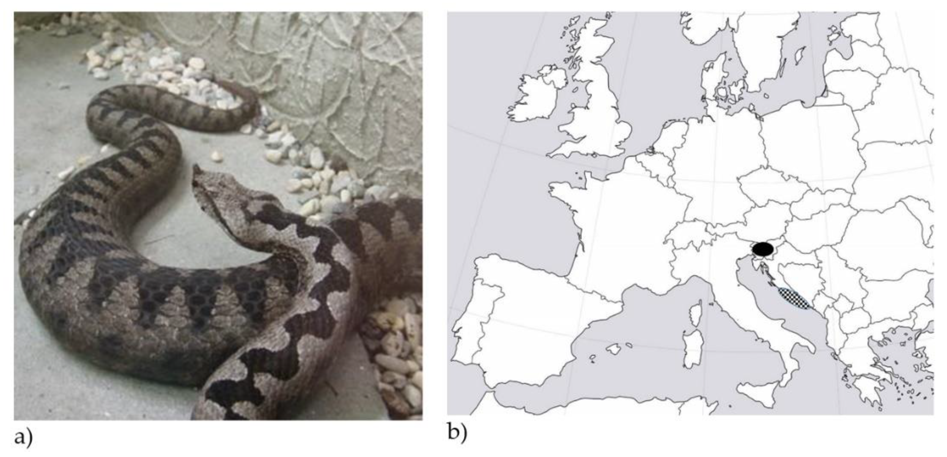
1. Introduction
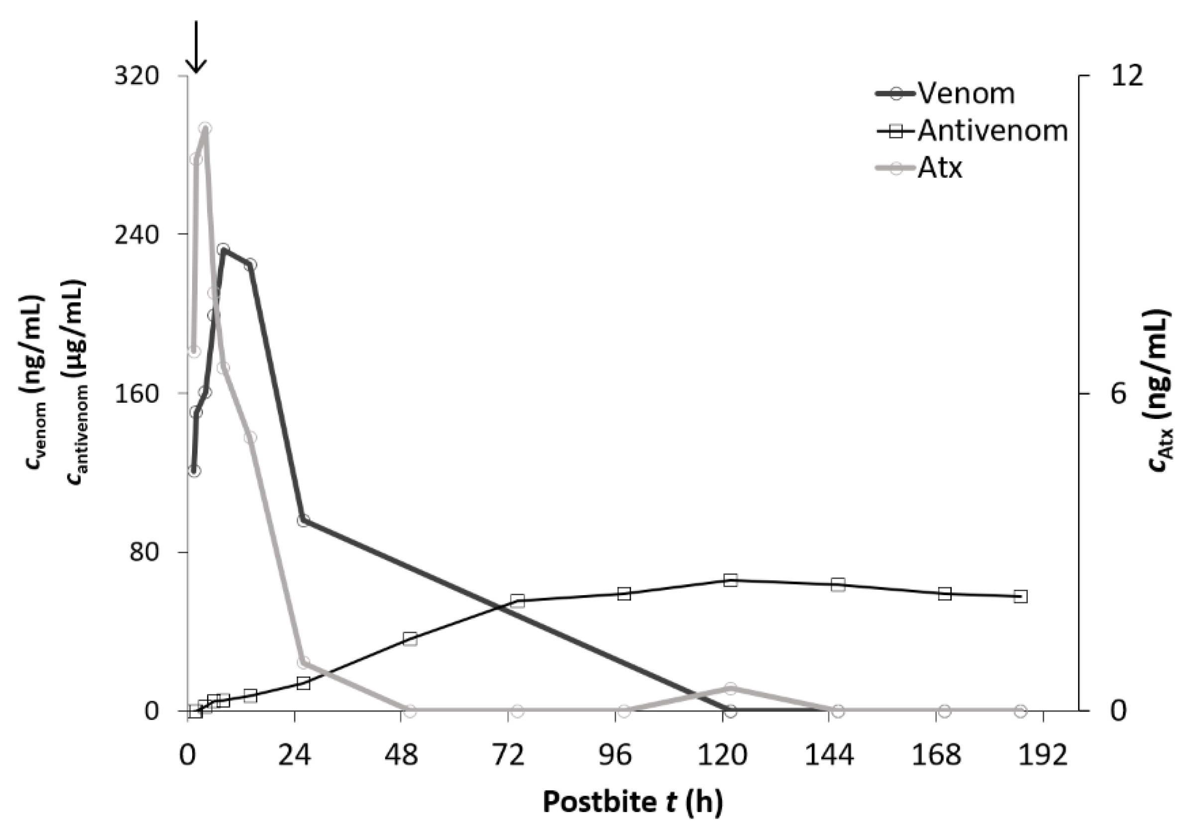
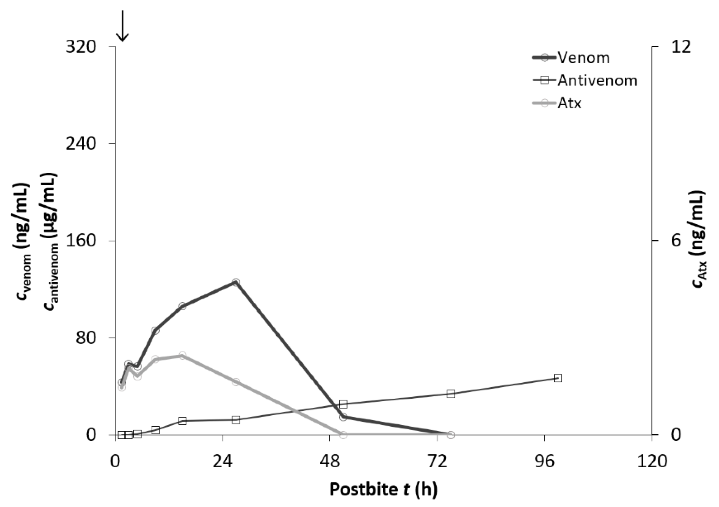
2. Results
3. Discussion
3.1. Fab and F(ab’)2 Pharmacokinetics Analysis
3.2. The Clinical Picture and Outcome of the Envenomation and Antivenom Therapy
4. Conclusions
5. Materials and Methods
5.1. Patients
5.2. Antivenom Therapy
5.3. Blood Samples
5.4. Reagents and Chemicals
5.5. Quantification of V. ammodytes Venom in Sera Samples
5.6. Quantification of Atxs in Sera Samples
5.7. Quantification of Antivenom in Sera Samples
5.8. Pharmacokinetic Analysis
5.9. Statistical Analysis
Author Contributions
Funding
Institutional Review Board Statement
Informed Consent Statement
Data Availability Statement
Acknowledgments
Conflicts of Interest
References
- Lamb, T.; de Haro, L.; Lonati, D.; Brvar, M.; Eddleston, M. Antivenom for European Vipera species envenoming. Clin. Toxicol. 2017, 55, 557–568. [Google Scholar] [CrossRef] [PubMed]
- Chippaux, J.P. Epidemiology of snakebites in Europe: A systematic review of the literature. Toxicon 2012, 59, 86–99. [Google Scholar] [CrossRef] [PubMed]
- Mallow, D.; Ludwig, D.; Nilson, G. True Vipers: Natural History and Toxinology of Old-World Vipers; Krieger Publishing Company: Malabar, FL, USA, 2003; p. 359. [Google Scholar]
- Latinović, Z.; Leonardi, A.; Šribar, J.; Sajevic, T.; Žužek, M.C.; Frangež, R.; Halassy, B.; Trampuš-Bakija, A.; Pungerčar, J.; Križaj, I. Venomics of Vipera berus berus to explain differences in pathology elicited by Vipera ammodytes ammodytes envenomation: Therapeutic implications. J. Proteom. 2016, 146, 34–47. [Google Scholar] [CrossRef] [PubMed]
- Kurtović, T.; Brvar, M.; Grenc, D.; Lang-Balija, M.; Križaj, I.; Halassy, B. A single dose of Viperfav™ may be inadequate for Vipera ammodytes snake bite: A case report and pharmacokinetic evaluation. Toxins 2016, 8, 244. [Google Scholar] [CrossRef] [PubMed]
- Luksić, B.; Bradarić, N.; Prgomet, S. Venomous snakebites in Southern Croatia. Coll. Antropol. 2006, 30, 191–197. [Google Scholar] [PubMed]
- Karabuva, S.; Vrkić, I.; Brizić, I.; Ivić, I.; Lukšić, B. Venomous snakebites in children in Southern Croatia. Toxicon 2016, 112, 8–15. [Google Scholar] [CrossRef] [PubMed]
- Casewell, N.R.; Al-Abdulla, I.; Smith, D.; Coxon, R.; Landon, J. Immunological crossreactivity and neutralisation of European viper venoms with the monospecific Vipera berus antivenom ViperaTAb. Toxins 2014, 6, 2471–2482. [Google Scholar] [CrossRef] [PubMed]
- Boels, D.; Hamel, J.F.; Bretaudeau-Deguigne, M.; Harry, P. European viper envenomings: Assessment of Viperfav and other symptomatic treatments. Clin. Toxicol. 2012, 50, 189–196. [Google Scholar] [CrossRef] [PubMed]
- Audebert, F.; Sorkine, M.; Robbe-Vincent, A.; Bon, C. Viper bites in France: Clinical and biological evaluation; kinetics of envenomations. Hum. Exp. Toxicol. 1994, 13, 683–688. [Google Scholar] [CrossRef] [PubMed]
- Rivière, G.; Choumet, V.; Saliou, B.; Debray, M.; Bon, C. Absorption and elimination of viper venom after antivenom administration. J. Pharmacol. Exp. Ther. 1998, 285, 490–495. [Google Scholar] [PubMed]
- Audebert, F.; Urtizberea, M.; Sabouraud, A.; Scherrmann, J.M.; Bon, C. Pharmacokinetics of Vipera aspis venom after experimental envenomation in rabbits. J. Pharmacol. Exp. Ther. 1994, 268, 1512–1517. [Google Scholar] [PubMed]
- Paniagua, D.; Vergara, I.; Román, R.; Romero, C.; Benard-Valle, M.; Calderón, A.; Jiménez, L.; Bernas, M.J.; Witte, M.H.; Boyer, L.V.; et al. Antivenom effect on lymphatic absorption and pharmacokinetics of coral snake venom using a large animal model. Clin. Toxicol. 2019, 57, 727–734. [Google Scholar] [CrossRef] [PubMed]
- Paniagua, G.; Jiménez, L.; Romero, C.; Vergara, I.; Calderón, A.; Benard, M.; Bernas, M.J.; Rilo, H.; de Roodt, A.; D’Suze, G.; et al. Lymphatic route of transport and pharmacokinetics of Micrurus fulvius (coral snake) venom in sheep. Lymphology 2012, 45, 144–153. [Google Scholar] [PubMed]
- Paniagua, D.; Vergara, I.; Boyer, L.; Alagón, A. Role of Lymphatic System on Snake Venom Absorption. In Snake Venoms Toxinology; Gopalakrishnakone, P., Inagaki, H., Vogel, C.W., Mukherjee, A., Rahmy, T., Eds.; Springer: Dordrecht, The Netherlands, 2017. [Google Scholar] [CrossRef]
- Seifert, S.A.; Boyer, L.V. Recurrence phenomena after immunoglobulin therapy for snake envenomations: Part 1. Pharmacokinetics and pharmacodynamics of immunoglobulin antivenoms and related antibodies. Ann. Emerg. Med. 2001, 37, 189–195. [Google Scholar] [CrossRef] [PubMed]
- Pepin, S.; Lutsch, C.; Grandgeorge, M.; Scherrmann, J.M. Snake F(ab’)2 antivenom from hyperimmunized horse: Pharmacokinetics following intravenous and intramuscular administrations in rabbits. Pharm. Res. 1995, 12, 1470–1473. [Google Scholar] [CrossRef] [PubMed]
- Laustsen, A.H.; María-Gutiérrez, J.; Knudsen, C.; Johansen, K.H.; Bermúdez-Méndez, E.; Cerni, F.A.; Jürgensen, J.A.; Ledsgaard, L.; Martos-Esteban, A.; Øhlenschlæger, M.; et al. Pros and cons of different therapeutic antibody formats for recombinant antivenom development. Toxicon 2018, 146, 151–175. [Google Scholar] [CrossRef] [PubMed]
- Boels, D.; Hamel, J.F.; le Roux, G.; Labadie, M.; Paret, N.; Delcourt, N.; Langrand, J.; Puskarczyk, E.; Nisse, P.; Sinno-Tellier, S.; et al. Snake bites by European vipers in Mainland France in 2017–2018: Comparison of two antivenoms Viperfav® and Viperatab®. Clin. Toxicol. 2020, 58, 1050–1057. [Google Scholar] [CrossRef] [PubMed]
- Lian, N.S.; Pungerčar, J.; Križaj, I.; Štrukelj, B.; Gubenšek, F. Expression of fully active ammodytoxin A, a potent presynaptically neuro-toxic phospholipase A2, in Escherichia coli. FEBS Lett. 1993, 334, 55–59. [Google Scholar] [CrossRef]
- Zhang, Y.; Huo, M.; Zhou, J.; Xie, S. PKSolver: An add-in program for pharmacokinetic and pharmacodynamic data analysis in Microsoft Excel. Comput. Methods Programs Biomed. 2010, 99, 306–314. [Google Scholar] [CrossRef] [PubMed]


| General Characteristics of the V. ammodytes Envenomed Patients | ViperaTAb (n = 9 Patients) | “Zagreb” Antivenom (n = 9 Patients) | p |
|---|---|---|---|
| Age (median, IQR) (year) | 40 (35–60) | 58 (36–67) | 0.44 |
| Gender (male) | 8/9 | 7/9 | 1.00 |
| Weight (median, IQR) (kg) | 72 (70–75) | 93 (81–98) | 0.11 |
| Comorbidities | 4/9 | 4/9 | 0.34 |
| Distance from the scene to the ED (median, IQR) (km) | 69 (61–98) | 44 (40–50) | 0.09 |
| Time from bite to admission at the ED (median, IQR) (h) | 4 (3–4) | 1.5 (1.5–1.75) | 0.01 |
| Symptoms and Laboratory Results | ViperaTAb (n = 9 Patients) | “Zagreb” Antivenom (n = 9 Patients) | OR (95% CI) | p |
|---|---|---|---|---|
| Bite location | ||||
| Arm | 7/9 | 7/9 | 1.0 (0.11–9.23) | 1.00 |
| Leg | 2/9 | 2/9 | 1.0 (0.11–9.23) | 1.00 |
| Venom concentration (median, IQR) (ng/mL) | 52.5 (27.3–106.3) | 47.3 (37.8–90.3) | NA | 0.65 |
| Atx concentration (median, IQR) (ng/mL) | 2.3 (1.6–5.9) | 3.2 (1.8–5.1) | NA | 0.85 |
| Local pain | 9/9 | 9/9 | 1.0 (0.02–55.80) | 1.00 |
| Local oedema | 9/9 | 9/9 | 1.0 (0.02–55.80) | 1.00 |
| Ecchymosis | 4/9 | 9/9 | 23.22 (1.04–517.96) | 0.13 |
| Nausea | 5/9 | 2/9 | 0.23 (0.03–1.77) | 0.13 |
| Vomiting | 3/9 | 1/9 | 0.25 (0.02–3.04) | 0.13 |
| Dizziness | 4/9 | 0/9 | 0.06 (0.01–1.44) | 0.01 |
| Syncope | 1/9 | 0/9 | 0.29 (0.01–8.34) | 0.13 |
| Cranial nerve palsies | 1/9 | 2/9 | 0.30 (0.01–8.35) | 1.00 |
| Tachycardia | 5/9 | 2/9 | 0.23 (0.03–1.77) | 0.57 |
| Hypotension | 3/9 | 0/9 | 0.10 (0.01–2.23) | 0.02 |
| Shock | 1/9 | 0/9 | 0.30 (0.01–8.35) | 0.13 |
| Acute respiratory failure | 1/9 | 0/9 | 0.30 (0.01–8.35) | 0.13 |
| Rhabdomyolysis | 5/9 | 1/9 | 0.10 (0.01–1.17) | 0.13 |
| Acute renal failure | 0/9 | 0/9 | 1.00 (0.02–55.80) | 1.00 |
| Acute myocardial injury | 1/9 | 0/9 | 0.30 (0.01–8.35) | 0.13 |
| Disseminated intravascular coagulation | 0/9 | 0/9 | 1.00 (0.02–55.80) | 1.00 |
| D-dimer (>500 µg/L) | 7/9 | 4/9 | 0.23 (0.03–1.77) | 0.06 |
| International normalized ratio (INR) (>1.3) | 2/9 | 0/9 | 0.16 (0.01–3.81) | 0.13 |
| Activated partial thromboplastin time (>36 s) | 2/9 | 0/9 | 0.16 (0.01–3.81) | 0.13 |
| Fibrinogen (<1.8 g/L) | 0/9 | 0/9 | 1.00 (0.02–55.80) | 1.00 |
| Thrombocytopenia (<150 × 109) | 6/9 | 3/9 | 0.25 (0.04–1.77) | 0.57 |
| Procalcitonin (>0.24 µg/L) | 2/9 | 0/9 | 0.16 (0.01–3.81) | 0.02 |
| Leucocytosis (>11 × 109/L) | 7/9 | 4/9 | 0.23 (0.03–1.77) | 0.13 |
| Therapy | ViperaTAb (n = 9 Patients) | “Zagreb” Antivenom (n = 9 Patients) | OR (95% CI) | p |
|---|---|---|---|---|
| Antivenom Therapy | ||||
| Time from bite to first dose (median, IQR) (h) | 6 (4.5–10) | 2.5 (2–3) | NA | 0.03 |
| Time from admission at the ED to first dose (median, IQR) (h) | 2 (1.5–6.5) | 1 (0.5–1.5) | NA | 0.13 |
| Multiple doses | 5/9 | 0/9 | 0.04 (0.01–0.96) | 0.02 |
| Number of doses (median, IQR) | 2 (1–2) | 1 (1–1) | NA | 0.03 |
| Anaphylactic reaction | 0/9 | 0/9 | 1.00 (0.02–55.80) | 1.00 |
| Serum sickness | 0/9 | 0/9 | 1.00 (0.02–55.80) | 1.00 |
| Other Therapy | ||||
| Corticosteroids | 1/9 | 9/9 | 107.66 (3.85–3013.31) | 0.01 |
| Antihistamines | 2/9 | 9/9 | 57.00 (2.36–1375.85) | 0.02 |
| Analgesics | 7/9 | 3/9 | 0.14 (0.02–1.16) | 0.02 |
| Antiemetics | 8/9 | 1/9 | 0.02 (0.01–0.30) | 0.01 |
| Antibiotics | 1/9 | 4/9 | 6.40 (0.55–78.89) | 1.00 |
| Low-molecular-weight heparin | 1/9 | 0/9 | 0.30 (0.01–8.35) | 0.13 |
| Platelet transfusion | 1/9 | 0/9 | 0.30 (0.01–8.35) | 0.13 |
| Red blood cell transfusion | 1/9 | 0/9 | 0.30 (0.01–8.35) | 0.13 |
| Oxygen | 2/9 | 0/9 | 0.16 (0.01–3.81) | 0.13 |
| Mechanical ventilation | 1/9 | 0/9 | 0.30 (0.01–8.35) | 0.13 |
| Noradrenaline infusion | 1/9 | 0/9 | 0.30 (0.01–8.35) | 0.13 |
| Symptoms and Laboratory Results | ViperaTAb (n = 9 Patients) | “Zagreb” Antivenom (n = 9 Patients) | OR (95% CI) | p |
|---|---|---|---|---|
| Maximum venom concentration (median, IQR) (ng/mL) | 52.5 (29.7–106.3) | 84.0 (47.3–125.7) | NA | 0.36 |
| Maximum Atx concentration (median, IQR) (ng/mL) | 2.4 (1.6–6.0) | 3.8 (2.7–8.3) | NA | 0.65 |
| Time from bite to maximum venom concentration (median, IQR) (h) | 5.5 (4–7) | 6.5 (1.5–8) | NA | 0.22 |
| Local pain | 9/9 | 9/9 | 1.00 (0.02–55.80) | 1.00 |
| Local oedema | 9/9 | 9/9 | 1.00 (0.02–55.80) | 1.00 |
| Local lymphadenitis | 8/9 | 4/9 | 0.10 (0.01–1.17) | 0.13 |
| Oedema spread to trunk | 4/9 | 4/9 | 1.00 (0.16–6.42) | 0.34 |
| Ecchymosis | 5/9 | 9/9 | 15.5 (0.70–346.74) | 0.13 |
| Nausea | 7/9 | 2/9 | 0.08 (0.01–0.75) | 0.02 |
| Vomiting | 3/9 | 1/9 | 0.25 (0.02–3.04) | 0.13 |
| Hypotension | 4/9 | 0/9 | 0.06 (0.01–1.43) | 0.01 |
| Shock | 1/9 | 0/9 | 0.30 (0.01–8.35) | 0.13 |
| Tachycardia | 5/9 | 1/9 | 0.10 (0.01–1.17) | 0.13 |
| Dizziness | 4/9 | 0/9 | 0.06 (0.01–1.43) | 0.01 |
| Syncope | 1/9 | 0/9 | 0.30 (0.01–8.35) | 0.13 |
| Cranial nerve palsies | 1/9 | 2/9 | 2.29 (0.17–30.96) | 1.00 |
| Acute respiratory failure | 2/9 | 0/9 | 0.16 (0.01–3.81) | 0.13 |
| Acute renal failure | 0/9 | 0/9 | 1.00 (0.02–55.80) | 1.00 |
| Rhabdomyolysis | 7/9 | 1/9 | 0.04 (0.01–0.48) | 0.02 |
| Acute myocardial injury | 1/9 | 0/9 | 0.30 (0.01–8.35) | 0.13 |
| Disseminated intravascular coagulation | 1/9 | 0/9 | 0.30 (0.01–8.35) | 0.13 |
| Thrombocytopenia (<150 × 109) | 6/9 | 3/9 | 0.25 (0.04–1.77) | 0.57 |
| D-dimer (>500 µg/L) | 9/9 | 5/9 | 0.06 (0.01–1.44) | 0.13 |
| International normalized ratio (INR) (>1.3) | 4/9 | 0/9 | 0.06 (0.01–1.43) | 0.02 |
| Activated partial thromboplastin time (>36 s) | 3/9 | 0/9 | 0.10 (0.01–2.23) | 0.13 |
| Fibrinogen (<1.8 g/L) | 1/9 | 0/9 | 0.30 (0.01–8.35) | 0.13 |
| Procalcitonin (>0.24 µg/L) | 6/9 | 0/9 | 0.03 (0.01–0.65) | 0.01 |
| Leucocytosis (>11 × 109/L) | 8/9 | 6/9 | 0.25 (0.02–3.04) | 0.26 |
| Length of hospital stay (median, IQR, days) | 4 (2–9) | 7 (4–7) | NA | 0.14 |
| Death | 0/9 | 0/9 | 1.00 (0.02–55.80) | 1.00 |
| Pharmacokinetic Values | Intravenous ViperaTAb (n = 11 Doses) |
|---|---|
| tmax (h) | 2 (2–2.3) |
| cmax (μg/mL) | 25.0 (23.3–33.8) |
| t1/2 (h) | 24.5 (9.0–50.0) |
| AUC∞ (μg·h/mL) | 250.3 (218.9–326.0 |
| Vss (mL/kg) | 317.5 (182.3–415.1) |
| Vz (mL/kg) | 473.1 (230.0–651.5) |
| MRT (h) | 22.1 (11.4–49.9) |
| CL (mL/h·kg) | 9.8 (8.9–13.3) |
| Pharmacokinetic Values | Intramuscular “Zagreb” Antivenom (n = 1 Dose) |
|---|---|
| tmax (h) | 120.0 |
| cmax (μg/mL) | 66.1 |
| t1/2 (h) | 317.2 |
| AUC∞ (μg·h/mL) | 3.5 × 10⁴ |
| Vz (mL/kg) | 190.9 |
| MRT (h) | 510.2 |
| CL (mL/h·kg) | 0.4 |
Publisher’s Note: MDPI stays neutral with regard to jurisdictional claims in published maps and institutional affiliations. |
© 2021 by the authors. Licensee MDPI, Basel, Switzerland. This article is an open access article distributed under the terms and conditions of the Creative Commons Attribution (CC BY) license (https://creativecommons.org/licenses/by/4.0/).
Share and Cite
Kurtović, T.; Karabuva, S.; Grenc, D.; Dobaja Borak, M.; Križaj, I.; Lukšić, B.; Halassy, B.; Brvar, M. Intravenous Vipera berus Venom-Specific Fab Fragments and Intramuscular Vipera ammodytes Venom-Specific F(ab’)2 Fragments in Vipera ammodytes-Envenomed Patients. Toxins 2021, 13, 279. https://doi.org/10.3390/toxins13040279
Kurtović T, Karabuva S, Grenc D, Dobaja Borak M, Križaj I, Lukšić B, Halassy B, Brvar M. Intravenous Vipera berus Venom-Specific Fab Fragments and Intramuscular Vipera ammodytes Venom-Specific F(ab’)2 Fragments in Vipera ammodytes-Envenomed Patients. Toxins. 2021; 13(4):279. https://doi.org/10.3390/toxins13040279
Chicago/Turabian StyleKurtović, Tihana, Svjetlana Karabuva, Damjan Grenc, Mojca Dobaja Borak, Igor Križaj, Boris Lukšić, Beata Halassy, and Miran Brvar. 2021. "Intravenous Vipera berus Venom-Specific Fab Fragments and Intramuscular Vipera ammodytes Venom-Specific F(ab’)2 Fragments in Vipera ammodytes-Envenomed Patients" Toxins 13, no. 4: 279. https://doi.org/10.3390/toxins13040279
APA StyleKurtović, T., Karabuva, S., Grenc, D., Dobaja Borak, M., Križaj, I., Lukšić, B., Halassy, B., & Brvar, M. (2021). Intravenous Vipera berus Venom-Specific Fab Fragments and Intramuscular Vipera ammodytes Venom-Specific F(ab’)2 Fragments in Vipera ammodytes-Envenomed Patients. Toxins, 13(4), 279. https://doi.org/10.3390/toxins13040279