Review

Undercover Agents of Infection: The Stealth Strategies of T4SS-Equipped Bacterial Pathogens

Institut de Recherche en Infectiologie de Montpellier (IRIM), Université de Montpellier, CNRS, CEDEX 5, 34293 Montpellier, France
*
Authors to whom correspondence should be addressed.
Toxins 2021, 13(10), 713; https://doi.org/10.3390/toxins13100713
Submission received: 29 July 2021 / Revised: 4 October 2021 / Accepted: 6 October 2021 / Published: 9 October 2021

Abstract

:
Intracellular bacterial pathogens establish their replicative niches within membrane-encompassed compartments, called vacuoles. A subset of these bacteria uses a nanochannel called the type 4 secretion system (T4SS) to inject effector proteins that subvert the host cell machinery and drive the biogenesis of these compartments. These bacteria have also developed sophisticated ways of altering the innate immune sensing and response of their host cells, which allow them to cause long-lasting infections and chronic diseases. This review covers the mechanisms employed by intravacuolar pathogens to escape innate immune sensing and how Type 4-secreted bacterial effectors manipulate host cell mechanisms to allow the persistence of bacteria.
Key Contribution: Bacterial pathogens have developed elaborated means to escape the immune response of their host and establish their replicative niche. Some bacteria use a type 4 secretion system to translocate effector proteins in the host cell cytoplasm. These effectors can then modulate and attenuate different pathways, such as inflammation, apoptosis or autophagy, to favor bacterial persistence.

1. Introduction

Gram-negative bacteria use complex nanomachines, called secretion systems, to translocate effector proteins, molecules and DNA in order to modulate the microbial response to the environment. Nine secretion systems have been identified to date: five span the inner and outer bacterial membrane, while the remaining four are embedded in the outer membrane [1]. Initially described in the context of bacterial conjugation and horizontal gene transfer, type 4 secretion systems (T4SS) are large protein complexes that span through the inner and outer membranes of several Gram-negative and Gram-positive bacteria [2]. These highly versatile nanomachines are characterised by an internal channel that mediates the translocation of proteins, toxins and genetic material. Cytoplasmic ATPases typically energise translocation. Depending on their function, T4SS can be divided into three functional groups [3]. T4SSs belonging to the first group mediate the conjugative transfer of DNA and transposon among bacteria, or from bacteria to plants, fungi and mammalian cells. Notably, DNA exchange has been associated with genome plasticity, the acquisition of eukaryotic domains and the acquisition of antibiotic resistance traits [3]. T4SSs belonging to the second group mediate the release and uptake of DNA directly from the extracellular milieu. Finally, the third group of T4SS mediates the translocation of proteins. These are primarily found in pathogenic bacteria and are essential virulence factors that coordinate host/pathogen interactions. Interestingly, T4SSs are not exclusive, and some bacteria, including Helicobacter pylori, encode both a DNA exchange system and a protein delivery system [4]. Genes encoding T4SSs are usually arranged in operons or clusters of operons [2]. The type 4a secretion system (T4SSa, also known as VirB/D4 secretion system) was initially discovered in the plant pathogen Agrobacterium tumefaciens and is usually composed of 12 genes [5]. Bacterial pathogens, including Orientia tsutsugamushi, Brucella spp., Bartonella spp., Anaplasma spp. and Ehrlichia spp., possess a T4SSa [6]. Bartonella species, except B. bacilliformis, can also express additional T4SSs, such as the Vbh/TraG and Trw secretion systems, that play a role in bacterial conjugation and erythrocyte invasion, respectively [7]. The T4SSb (also known as the Dot/Icm secretion system) is composed of 23 to 25 genes, and it is only found in Legionella and Coxiella species [8].
Notably, many bacterial pathogens equipped with a type 4 secretion system (T4SS) have evolved to colonise host cells from the safe haven of replicative vacuoles. These membrane-bound compartments derive from the initial internalisation vacuole that hosts the pathogen and are de facto intracellular compartments of eukaryotic composition, which only exist in the context of infection. Life in a membrane-enclosed compartment presents several advantages, including access to nutrients and escape from cytosolic immune surveillance. On the other hand, without bacterial intervention, internalisation vacuoles fuse with lysosomes, which in most cases is fatal for the pathogen itself. Thus, these pathogens rely on their T4SS to translocate effector proteins through the membrane of the replicative vacuole, directly into the host cytoplasm, where they subvert membrane trafficking to deviate their replicative niche off the tracks of the endocytic pathway [9]. Each replicative niche is remarkably specific to its pathogen and is characterised by a unique protein and lipid signature and morphology. Furthermore, the “fate” of these compartments may differ depending on the pathogen’s lifestyle: some remain intact and host non-replicative bacteria for an extended time, whereas others rupture to release bacteria and facilitate the infection of bystander cells. Importantly, infected cells are not entirely blind to these pathogens, which are readily recognised by cell surface and endosomal Toll-like receptors (TLRs) that transduce signals leading to the activation of the antimicrobial response [10]. Later, during the intracellular cycle, cytosolic receptors sense pathogen-derived ligands, which may include the effector proteins themselves [11]. Furthermore, infected cells can destabilise the membrane of the bacterial replicative niche, thereby exposing microbes to immune surveillance [12]. Finally, innate immunity can also be alerted by recognition of the hybrid membranes of replicative vacuoles as foreign bodies [13]. Thus, several T4SS pathogens have evolved stealth strategies to remain undetected within infected cells and avoid triggering inflammation.
This review illustrates the diverse strategies developed by stealth pathogens equipped with a T4SS to elude innate immune recognition and/or silence the multiple signalling pathways coordinating the inflammatory response.

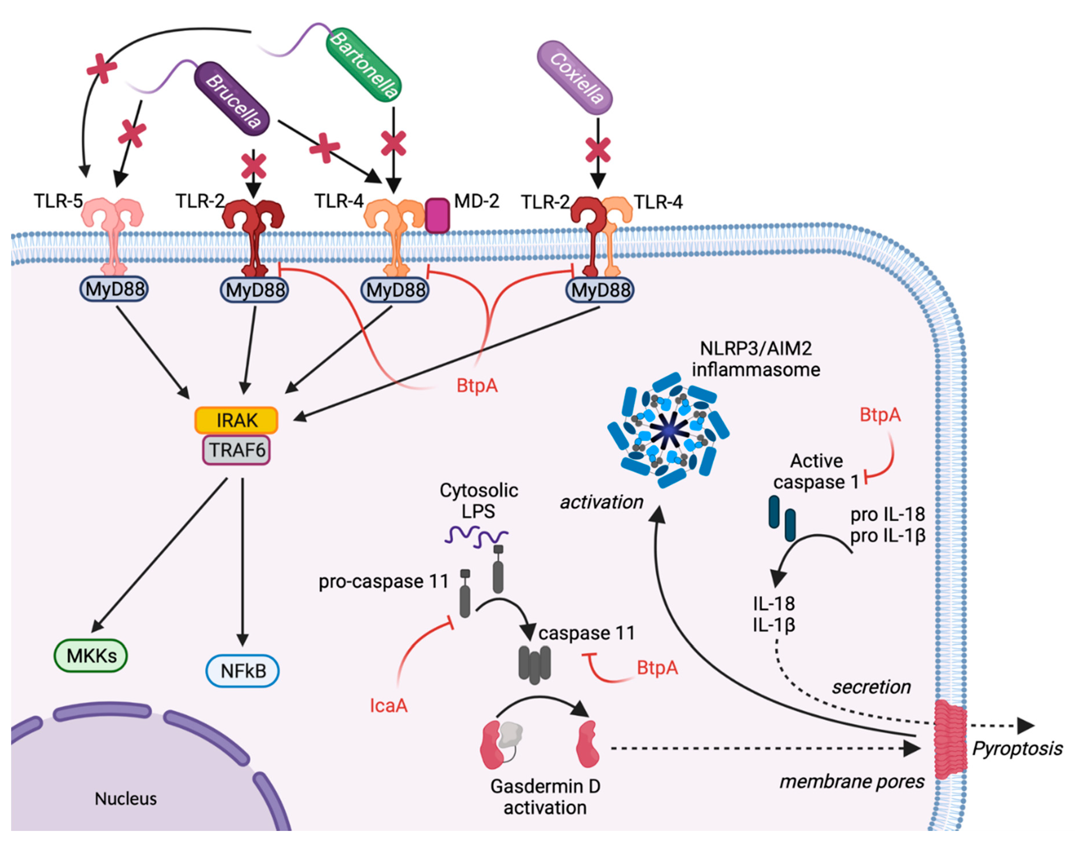
2. Escape from Host Sensing

In immune cells, the surveillance and detection of invading bacterial pathogens are ensured by pattern-recognition receptors (PRR), such as surface and endosomal TLRs and cytosolic Nucleotide oligomerization domain (NOD)-like receptors (NLRs) [14]. The NLR family of proteins comprises 22 members, who are critical in triggering and modulating the inflammatory response of immune cells. TLRs are transmembrane proteins that localise either at the cell surface or at the membranes of intracellular compartments, such as the endoplasmic reticulum (ER), endosomes, lysosomes, or endolysosomes. The human genome encodes 10 proteins of the TLR family. They recognise distinct or overlapping pathogens associated molecular patterns (PAMPs), such as lipids, lipoproteins, proteins, and nucleic acid [15]. Each TLR comprises an ectodomain with leucine-rich repeats (LRRs) that mediate PAMPs recognition, a transmembrane domain, and a cytoplasmic Toll/Interleukin-1 (IL-1) receptor (TIR) domain that initiates downstream signalling. Some stealth bacteria adapted their microbe-associated molecular patterns (MAMPs) to escape recognition by cell sensors. For example, the Bartonella lipid A is poorly recognised by TLR4 as its structure is characterised by a short glycosyl backbone (di-aminoglucose), a pentaacylation, and a very long fatty acid side chain [16]. This lipid A impedes a broad inflammatory response [17]. Furthermore, B. quintana Lipopolysaccharide (LPS) is a potent antagonist of TLR4 signalling (Figure 1) [18,19].
Brucella LPS exhibits low toxicity, and its atypical structure was postulated to delay the host immune response, favouring the establishment of chronic disease [20]. Brucella Lipid A is a 2,3-diaminoglucose disaccharide substituted with C16, C18, C28 and other very long acyl chains [21]. This peculiar structure is a poor agonist of TLR4/myeloid differentiation-2 (MD-2), and a paradigm has emerged proposing Brucella LPS as a crucial virulence factor that hampers recognition by PRRs and plays essential roles during infection (Figure 1) [16]. One study showed that Brucella LPS does not induce inflammatory responses in macrophages and dendritic cells (DCs), two of the most important immune system sentinels. This was attributed to its poor recognition by TLR4/MD-2, which is widely considered the major receptor complex for LPS binding and signalling [22].
As previously mentioned, the activation of TLR4 and TLR2 is dependent on strong affinity and direct interaction with bacterial LPS. In macrophages challenged with avirulent Coxiella burnetii LPS, TLR-4 and TLR-2 co-immunoprecipitate, indicating that the avirulent strain can be recognised by these receptors. However, this association was absent in cells challenged by the LPS of virulent C. burnetii (Figure 1). The disruption makes TLRs unable to signal during the recognition of the LPS of pathogenic C. burnetii [23]. The re-organization of the macrophage cytoskeleton induced the disruption of TLR-2 and TLR-4 by C. burnetii LPS. Interestingly, blocking the actin cytoskeleton re-organization relieved the disruption of the association TLR-2/TLR-4 by pathogenic C. burnetii and rescued the p38α-mitogen-activated protein kinase (MAPK) activation by C. burnetii [23].
Bacterial flagellin is another major MAMP that can be sensed by TLR5 and NLR family CARD domain containing 4 (NLRC4), leading to a robust pro-inflammatory response. While flagellin from Brucella melitensis is weakly detected by TLR5 and might play an essential role in immune escape [24], Bartonella bacilliformis synthesises flagellin molecules that escape TLR5 recognition entirely (Figure 1). This is due to the presence of aspartic acid and serine amino acids in the 89–96 region of N-terminal D1 domain of flagellin, which is usually recognised by TLR5. These changes in amino acids prevent TLR sensing while preserving bacterial motility [25].
Besides eluding TLR recognition, Brucella uses additional evasion strategies by suppressing innate immune signalling. The B. abortus Btp1 effector protein and its B. melitensis homolog TcpB (now both referred to as BtpA) encode a TIR domain that competes with myeloid differentiation response gene 88 (MyD88) for binding with TIR domain-containing adaptor protein (TIRAP, Figure 1), which ultimately facilitates the ubiquitination and degradation of MyD88 adaptor-like (Mal) and inhibits both TLR4 and TLR2 signalling [26,27]. TcpB seems to be important in the bacterial immune escape, as the B. melitensis tcpB mutant displays a markedly retarded systemic spread in a mouse model of infection. The weakened phenotype of tcpB mutants for immune-competent mice suggests that Brucella can evade innate immunity by using multiple strategies [28]. Finally, the Brucella effector BtpB inhibits TLR signalling and contributes to the control of dendritic cell activation [29]. Recently, BtpA and BtpB have been shown to modulate energy metabolism through NAD+ hydrolysis, which could in part explain the down-modulation of innate immune signalling in Brucella-infected cells [30].

3. Inhibition of Inflammasome

Upon detection of PAMPs, several NLRs (such as NLR family Pyrin Domain containing 1 (NLRP1), NLR family Pyrin Domain containing 3 (NRLP3) or Pyrin) oligomerise with the apoptosis-associated speck-like protein containing a CARD (ASC) to activate inflammatory Caspase-1 [11]. The major Caspase-1 substrates are Gasdermin-D (GSDMD), pro-IL-1β, and pro-IL-18. Cleaved GSDMD targets and permeabilizes the plasma membrane, thus triggering ion imbalance and necrotic cell death, which is termed “pyroptosis” [31]. During this process, cleaved IL-1β and IL-18 are also secreted and stimulate inflammatory responses in neighbouring cells. Cytosolic LPS can be sensed directly by Caspase-11 (in mouse cells) and Caspase-4 and -5 (in human cells), which leads to GSDMD cleavage and pyroptosis [32]. These caspases belong to the non-canonical inflammasome pathway as they cannot directly process the inflammatory cytokines IL-1β and IL-18 [33].
Brucella effector BtpA interacts with Caspase-4 and induces the ubiquitination of inflammatory caspases-1, 4, and 11, leading to their degradation [34]. BtpA expression leads to a reduction of IL-1β secretion, induced by the non-canonical inflammasome in Brucella-infected macrophages (Figure 1). BtpA also attenuates pyroptosis and proinflammatory cytokine secretion in macrophages infected with Salmonella enterica [34].
C. burnetii does not stimulate inflammasome activation in primary mouse macrophages, as Caspase-1 is not activated during infection [35]. The secretion of the effector protein IcaA inhibits the non-canonical caspase-11-mediated activation of the NLRP3 inflammasome (Figure 1). However, the molecular targets of this effector protein remain unknown [35]. Other C. burnetii mechanisms may be involved in the inhibition of inflammation as several Coxiella transposon mutants displayed a cytotoxic phenotype during infection [36].

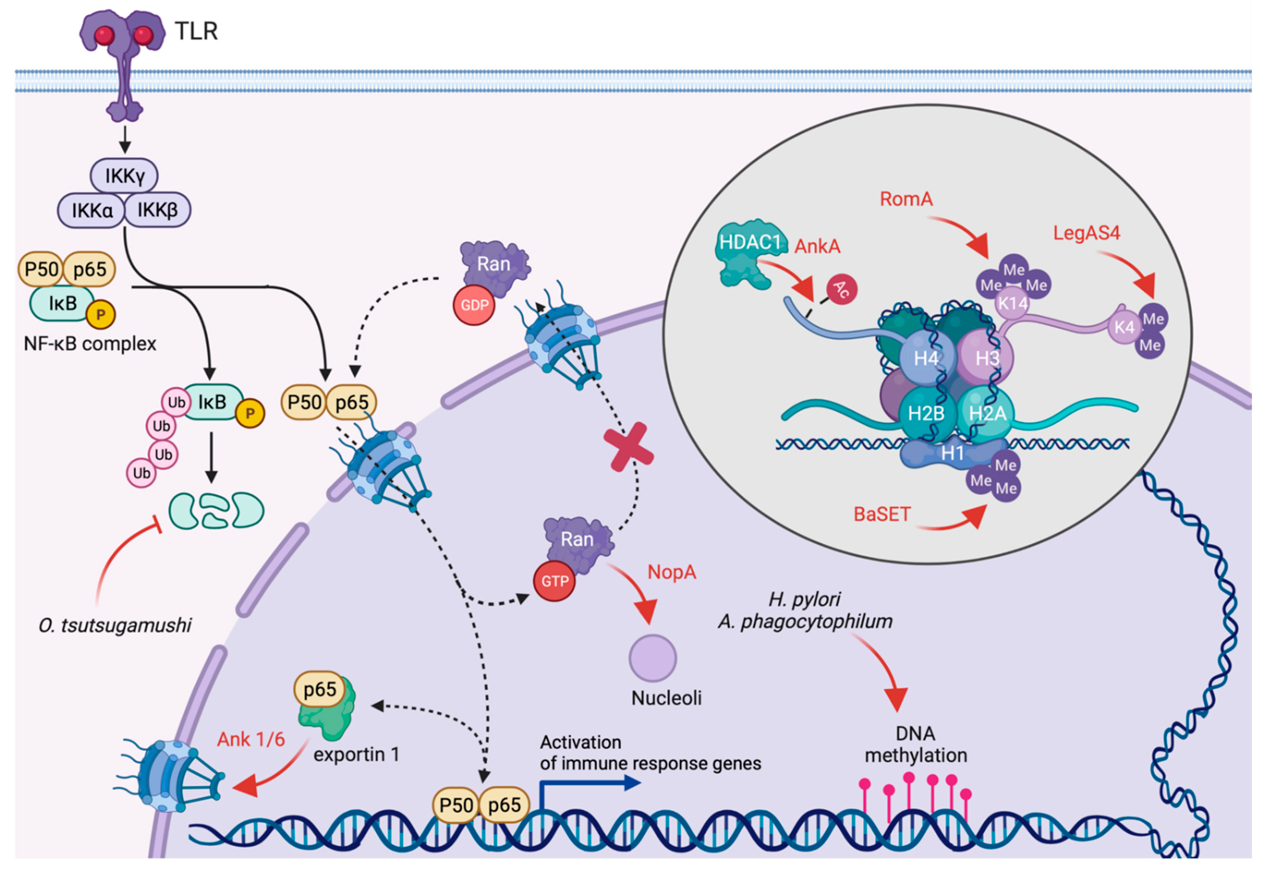
4. Inhibition of Transcription and Translation

The recognition of danger-associated molecular patterns (DAMPs) by infected cells triggers an important transcriptional reprogramming, which is essential to mount a specific proinflammatory response. The three primary inflammation signal-transducing pathways are the signal transducers and activators of transcription (STATs), the interferon regulatory factors (IRFs), and the nuclear factor κB (NFκB) [37]. To counter this, stealth pathogens effectively subvert the host transcriptional response, either by epigenetic silencing or by perturbing the nuclear accumulation of the main transcriptional regulators.
Controlling DNA methylation and histones after -translational modifications are efficient strategies to control the transcriptional landscape of infected cells. Thus, over the past decade, epigenetic silencing of host defences has emerged as a common strategy of several bacterial pathogens. DNA methylation interferes with the recruitment of transcription factors and leads to transcriptional repression [38]. Global changes in the methylation state of intergenic DNA regions have been described in cells infected by Helicobacter pylori [39] or Anaplasma phagocytophilum [40] (Figure 2). In the latter case, this is required for optimal intracellular replication. However, whether DNA methylation is actively triggered by bacterial effectors or reflects the cellular response to infection remains to be defined.
Histone modifications include acetylation, methylation and phosphorylation. Collectively, they regulate the accessibility of DNA to transcription factors, thus regulating gene expression [41]. Enzymatically active bacterial effectors can modulate epigenetic histone marks directly. Examples include an emerging class of bacterial effectors encoding a eukaryotic-like suppression of variegation, enhancer of zeste and trithorax (SET) domain with methyltransferase activity. Legionella pneumophila translocates the effector protein RomA/LegAS4 [42,43], which localises to the nucleus of cells infected with the Paris strain, an endemic Legionella strain predominant in France [44], but displays a marked nucleolar localisation when cells are infected with L. pneumophila Lp02, a virulent thymine auxotroph derived from the serogroup 1 Philadelphia-1 strain [45]. Interestingly, despite their high homology, RomA catalyses the tri-methylation of histone H3 at lysine 14 (Figure 2), which results in the repression of host innate immune genes, such as Tumor Necrosis Factor (TNF), IL-6, TLR5 and NLRP3 [42]. On the contrary, LegAS4 catalyses a di-methylation of histone H3 at lysine 4, leading to increased transcription of rDNA genes [43] (Figure 2). Similar to RomA, the Bacillus anthracis protein BaSET encodes a SET domain and localises to the nucleus of infected cells and specifically tri-methylates histone H1 [46] (Figure 2). The ectopic expression of BaSET represses the activity of NF-kB response elements, including the pro-inflammatory cytokines IL-6 and IL-8 [46]. Furthermore, BaSET is important for virulence as assessed using a murine bacteremia model [46]. However, whether BaSET is a T4SS substrate remains to be defined. Of note, SET domain proteins have been identified in other bacterial pathogens, including Chlamydia spp. [47] and Burkholderia thailandensis [43], which encode type 3 and type 6 secretion systems, respectively.
Other bacterial effector proteins indirectly manipulate histone marks. Among T4SS-equipped pathogens, the best example is the A. phagocytophilum effector protein AnkA, which translocates into the nucleus of infected cells and directly interacts with the histone deacetylase HDAC1 [48] (Figure 2). The removal of acetyl groups from histones mediated by HDACs favours a closed conformation of chromatin, thereby terminating transcription. Importantly, AnkA also binds host DNA at ATC-rich intergenic regions upstream of genes involved in the antimicrobial immune response [49], targeting HDAC1 activity towards the specific silencing of host defence genes [50]. Interestingly, A. phagocytophilum infections also increase the expression of HDAC through an unknown mechanism [50].
The mammalian NF-κB complex consists of five proteins that function by forming homo- or heterodimers. Among these, the transcription factor p65 is retained in the cytosol by the interaction with members of the NF-κB inhibitor (IκB) family, which hides its nuclear localisation signal (NLS). Exposure to PAMPs or TNFα activates a signalling cascade that leads to the phosphorylation and proteasomal degradation of IκB proteins. This exposes the NLS on p65, which translocates into the nucleus and coordinates the expression of antimicrobial and pro-inflammatory genes, such as Interleukin-1α (IL-1α), Interleukin-1β (IL-1β), Interleukin-6 (IL-6), etc (Figure 2). Several bacterial pathogens interfere directly with this signalling cascade at different levels, thereby activating or inhibiting the NF-κB pathway [51].
Emerging evidence indicates that T4SS-equipped microbes translocate effector proteins that manipulate the NF-κB pathway indirectly, by subverting the host nucleocytoplasmic transport. The Orientia tsutsugamushi ankyrin repeat-containing effectors Ank1 and Ank6 are transported to the nucleus of infected cells in an importin β1-dependent manner [52]. There, they interact with and rely on the function of exportin 1 to promote p65 nuclear export [52] (Figure 2). The cytoplasmic accumulation of p65 should be sufficient to reduce NF-κB-dependent transcriptional activation. Interestingly, however, preventing nuclear export in O. tsutsugamushi-infected cells does not restore NF-κB-dependent transcription [52], suggesting the existence of redundant strategies to dampen the innate immune response to Orientia infections. Indeed, it has recently been reported that O. tsutsugamushi also stabilises the intracellular levels of the IκB family member p105, thereby fostering p65 cytoplasmic retention (Figure 2). While the molecular mechanisms regulating this phenomenon remain to be defined, p105 stabilisation depends on the bacterial load and protein synthesis, suggesting the implication of effector proteins [53]. Similar to Orientia, C. burnetii also perturbs nucleocytoplasmic traffic to dampen the NF-κB-dependent transcriptional response to infection. The effector protein NopA (for nucleolar protein A) localises at the nucleoli of infected cells, interacts with the eukaryotic small GTPase Ran, and sequesters it within the nucleus [54] (Figure 2). As nucleocytoplasmic transport depends on a protein concentration gradient of Ran between the cytoplasm and the nucleus [55], NopA activity effectively perturbs the nuclear import of proteins, including the transcription factors p65 and interferon regulatory transcription factor 3 (IRF3) (Figure 2). Consequently, cytokines expression is strongly attenuated in cells infected by wild-type Coxiella, whereas this effect is completely lost in cells infected either with nopA or T4SS-defective dotA mutants [54]. Notably, despite their subcellular localisation, neither Ank1, Ank6, nor NopA encode canonical nuclear localisation signals (NLS), suggesting that bacterial effector proteins have evolved alternative strategies to target subcellular compartments.
Parallel to the NF-κB pathway, and besides its role in cell development and differentiation, the Janus Kinase (JAK)-Signal Transducer and Activator of Transcription (STAT) signalling pathway is central to the host cell’s response to viral and bacterial infections. Signal transducer and activator of transcription 3 (STAT3) is a key regulator of the switch between pro- and anti-inflammatory signalling in cells [56]. In its inactive form, STAT3 is found as a monomer in the cytoplasm. Cellular stressors, including cytokines, activate surface receptors and transduce the signal to cytoplasmic Janus kinases (JAKs), which phosphorylate STAT3, triggering its dimerisation and nuclear translocation. Notably, STAT3 can also be phosphorylated by non-receptor tyrosine kinases, including MAP kinases, Src and c-Abl. The Bartonella henselae effector protein BepD possesses several EPIYA-related motifs that can be phosphorylated by host Src-family tyrosine kinases. Importantly, this leads to the recruitment of STAT3 and facilitates its activation by c-Abl-dependent phosphorylation, thereby bypassing the canonical JAK pathway. BepD-mediated activation of the STAT3 pathway leads to impairment of pro-inflammatory TNFα secretion and stimulation of the anti-inflammatory cytokine Interleukine-10 (IL-10) [57].

5. Modulation of the Unfolded Protein Response

The unfolded protein response (UPR) also plays a relevant role in the interplay between pathogens and inflammation. UPR is triggered by stress conditions at the level of the ER in response to the accumulation of unfolded or misfolded proteins in the ER lumen due to an increase of protein secretion or a direct disruption of ER protein folding [58]. UPR includes multiple signal transduction pathways that regulate gene transcription, protein modifications, and mRNA translation to restore cellular homeostasis. Rapid modifications of the transcriptional profile of cells exposed to pathogens can trigger UPR. Three main pathways have been identified and depend on three ER transmembrane protein sensors: inositol-requiring enzyme-1α (IRE1α), protein kinase RNA-like endoplasmic reticulum kinase (PERK), and activating transcription factor 6α (ATF6α) [59]. The failure of the UPR to restore cellular homeostasis results in apoptosis. Notably, some intravacuolar pathogens, including Brucella spp. and L. pneumophila, escape the endocytic maturation pathway by favouring interactions between the membranes of their replicative niches with those of the ER, which can be sensed as an ER stress. UPR induction may be triggered explicitly by effector proteins and may be required for intracellular replication, as shown for the Brucella effector proteins VceC and TcpB [60,61]. During L. pneumophila infections, the IRE1α pathway is activated by TLRs upon extracellular pathogen sensing. However, L. pneumophila silences the downstream UPR pathways by two different mechanisms involving the effector proteins Lgt1 and Lgt2, which inhibit the splicing of XBP1u to XBP1s, thereby blocking the IRE1 pathway and unidentified effector proteins that inhibit the translation of ATF6-upregulated genes, leading to an inhibition of the cell response [62,63]. Interestingly, infections by C. burnetii also activate the UPR, as indicated by an increase in the intracellular levels of CCAAT/enhancer binding proteins (C/EBPs) homologous protein (CHOP). As for Brucella infections, UPR activation is required for optimal Coxiella-containing vacuole (CCV) expansion. However, unidentified C. burnetii effectors actively prevent CHOP nuclear translocation and downstream apoptosis [64]. Furthermore, the intracellular localisation of the C. burnetii effector CaeB has been recently re-assessed, and it has been shown that the effector localises at the ER upon ectopic expression. CaeB specifically modulates IRE1 signalling in cells exposed to tunicamycin, which ultimately leads to the inhibition of apoptosis [65]. This is in line with previous observations on the anti-apoptotic properties of CaeB in cells exposed to staurosporine, which is mediated by an inhibition of mitochondrial outer membrane permeabilisation (MOMP) [66]. Finally, another aspect of the UPR pathway is the stress response leading to the ER-associated degradation (ERAD) of misfolded proteins. Ank4, an effector protein of O. tsutsugamushi, has been shown to interact with HLA-B-associated transcript 3 (Bat3) to impair ERAD mechanism during the early stages of infection. Bat3 is an essential protein of ERAD that acts as a chaperone to mediate the translocation of misfolded proteins to the cytosol, where they can be ubiquitinated prior to degradation by the proteasome [67]. O. tsutsugamushi temporarily blocks ERAD until ERAD-derived amino acids are needed to support its growth [68].

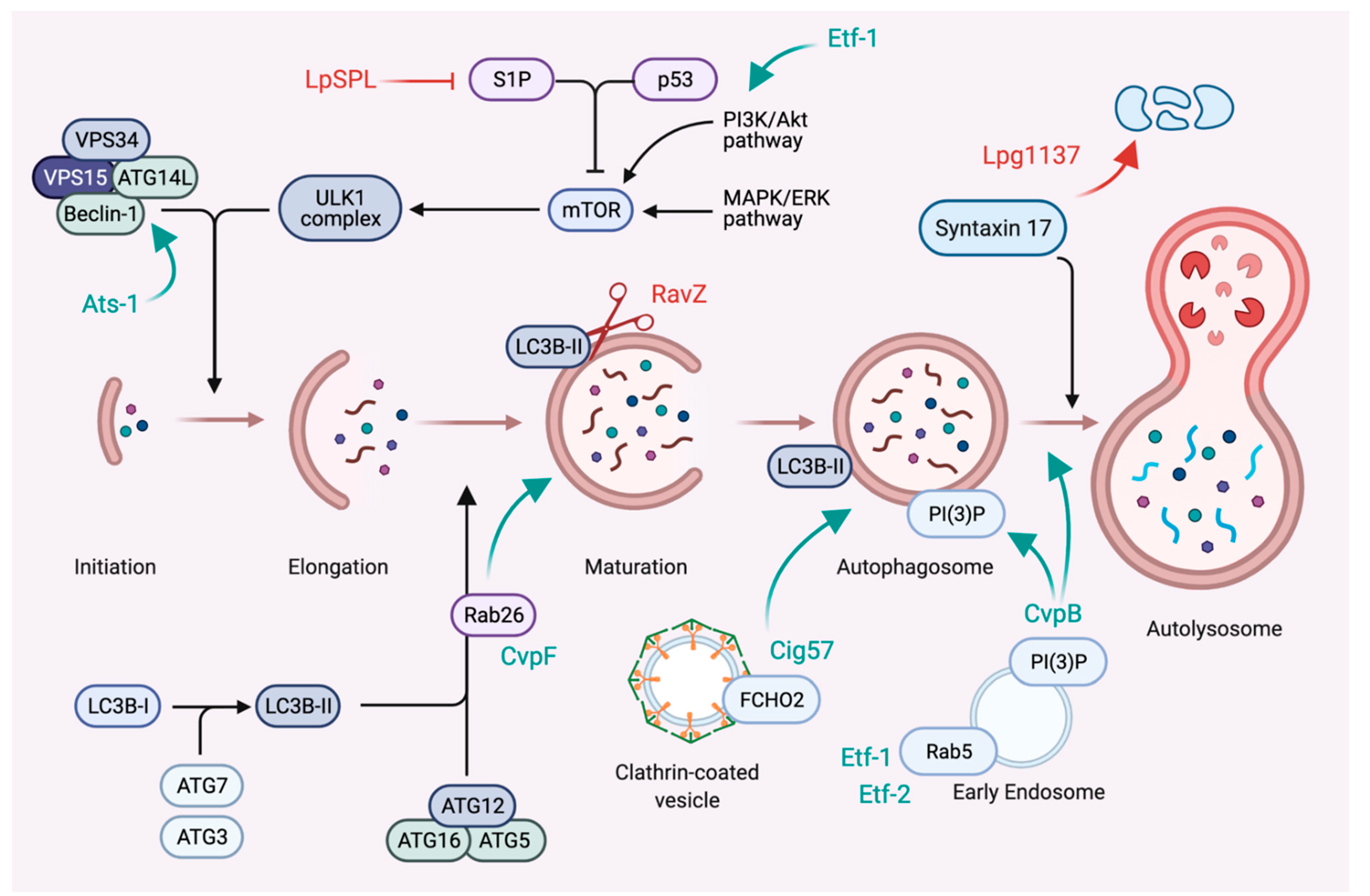
6. Subversion of Autophagy

Macroautophagy (hereafter referred to as autophagy) is a eukaryotic process for the engulfment and degradation of cytosolic material, including misfolded proteins, damaged organelles and macromolecules [69]. While basal autophagy is crucial to maintain cellular homeostasis by providing energy substrates to the cell, it plays an important role in the innate immune response against intracellular pathogens. Intravacuolar pathogens developed strategies to either avoid degradation by the autophagic machinery or divert autophagy elements to generate their replicative niche. L. pneumophila uses effector proteins to inhibit autophagy and avoid destruction in lytic autolysosomes. The bacterium secretes the cysteine protease RavZ that specifically and irreversibly cleaves the autophagy adapter protein LC3B from autophagosomes, allowing the Legionella-containing vacuoles (LCVs) to escape degradation by the autophagy machinery (Figure 3) [70]. Interestingly, LCVs generated by ΔravZ mutants still lack the autophagy marker LC3B, strongly suggesting that the bacterium secretes additional effectors capable of diverting the autophagy machinery away from the Legionella replicative niche. L. pneumophila also secretes Lpg1137, a serine protease that triggers the degradation of Syntaxin 17 (Stx17), a SNARE involved in vesicle trafficking and autophagy [71]. Additionally, some Legionella strains exert additional control over the autophagy machinery via host sphingosine-1-phosphate lipid levels (Figure 3). This lipid mediates the balance between sphingolipid-induced autophagy and cell death. L. pneumophila secretes the effector LpSPL (Sphingosine-1 phosphate lyase) to downregulate host sphingolipid levels, which causes a delay in the autophagic response to L. pneumophila infections [72]. A. phagocytophilum and Ehrlichia chaffeensis hijack the autophagosome initiation machinery to develop inside vacuoles with autophagosomal properties. A. phagocytophilum secretes the effector protein Ats-1, which interacts with Beclin-1 and ATG14L to induce autophagosome formation [73]. E. chaffeensis secrete Etf-1, which stimulates autophagy by activating class III phosphatidylinositol 3-kinase and Rab5 [74], and Etf-2, which binds to active Rab5 and delays endosomal maturation (Figure 3) [75]. For these two intracellular bacteria, the diversion of autophagy seems devoted to the acquisition of nutrients and membranes, as their respective vacuoles do not fuse with lysosomes and avoid being acidified. C. burnetii appears unique in the group of intravacuolar pathogens as the biogenesis of its vacuole relies on the autophagy machinery and fusion with lysosomes [76,77]. C. burnetii secretes several effector proteins to manipulate autophagy and promote CCV biogenesis and expansion. Coxiella vacuolar protein B (CvpB) localises to CCVs, where it stabilises the host lipid phosphatidylinositol 3-phosphate (PI(3)P) to stimulate autophagy and the homotypic fusion of CCVs [78,79,80]. Recently, effector protein CvpF was shown to stimulate Rab26-dependent autophagy to expand Coxiella-containing vacuoles [81]. Finally, the effector protein Cig57 interacts with the clathrin accessory protein FCHO2 [82] and stimulates the clathrin-dependent recruitment of autophagy markers to CCVs (Figure 3) [83]. In the case of Brucella, the autophagy initiation machinery intervenes at the end of the bacterial cell cycle to facilitate egress of the bacterium [84]. Interestingly, the autophagy adapter LC3B is upregulated during Brucella infections of human peripheral blood mononuclear cells (PBMCs). Brucella-induced autophagy leads to a defect in monocyte polarisation, as these cells fail to differentiate into M1 (pro-inflammatory) or M2 (anti-inflammatory) macrophages [85], suggesting that autophagy upregulation could favour the bacterial escape from a controlled immune response.

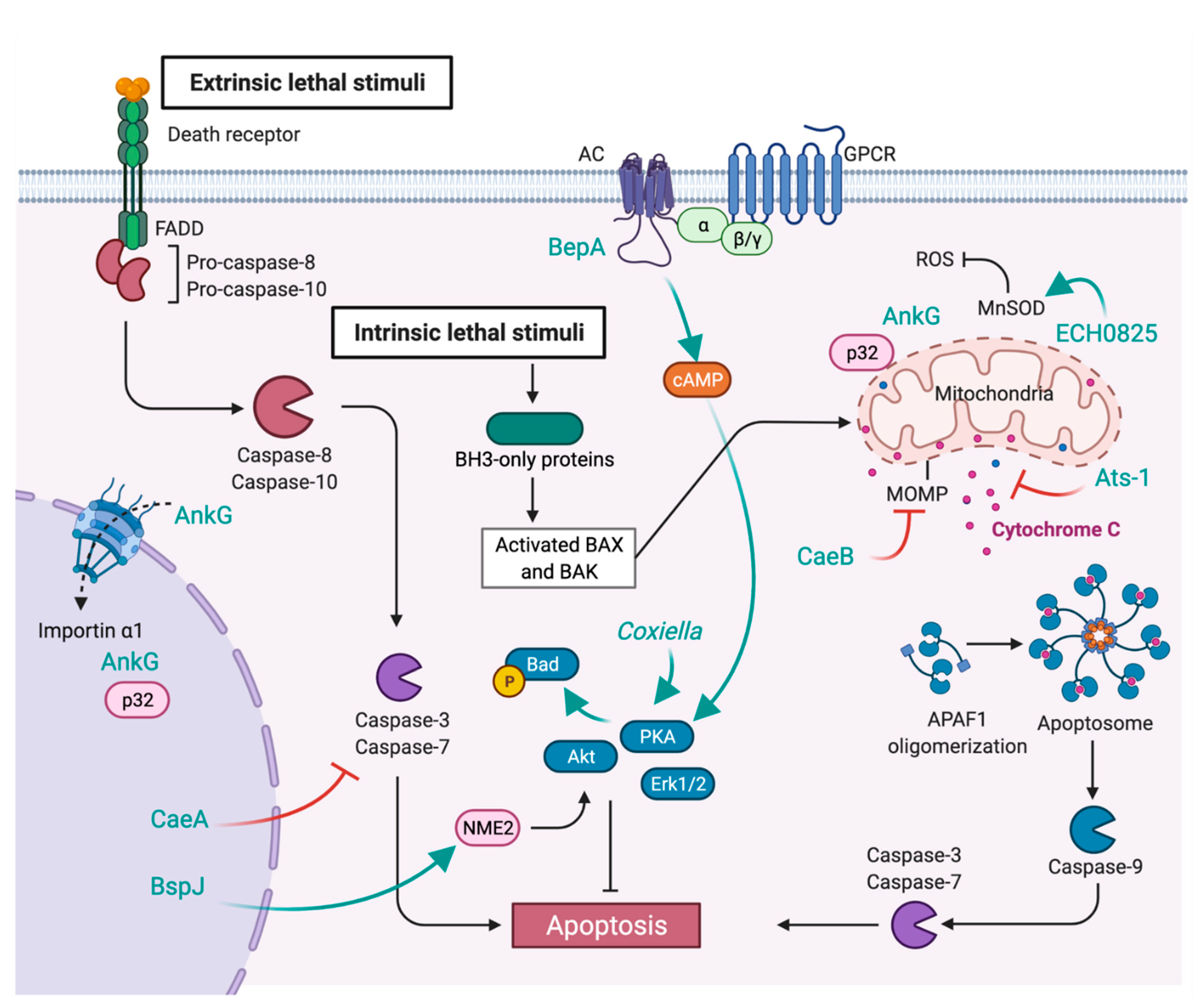
7. Inhibition of Apoptosis

Programmed cell death is common to all multicellular organisms. It is essential to dispose of unwanted cells, either during development or disease. Apoptosis can be activated by the extrinsic pathway upon sensing environmental stress by cell surface-exposed death receptors or by the intrinsic pathway, which detects intracellular stress (DNA damage, starvation, hypoxia, etc.) (Figure 4) [86]. Both pathways lead to the activation of caspases to coordinate intracellular proteolytic events. The intrinsic pathway also promotes mitochondrial outer membrane permeabilisation (MOMP), followed by cytochrome C release, which initiates the apoptosome assembly. Several intravacuolar pathogens promote the lysis of infected cells to propagate infections; however, host cell death by apoptosis is important to control intracellular bacteria replication and survival. Thus, stealth pathogens translocate effector proteins to prevent untimely cell death and preserve their replicative niche during chronic infections. The inhibition of apoptosis by C. burnetii is particularly well detailed and stands as a remarkable example of how important host/pathogen interaction hubs can be multilayered and regulated by redundant mechanisms. Indeed, C. burnetii infections trigger an anti-apoptotic transcriptional program [87] and the activation of pro-survival kinases, including Akt, Erk1/2, and cAMP-dependent protein kinase (PKA) [88,89]. Activated PKA phosphorylates the pro-apoptotic protein Bad, which is sequestered at CCVs during infections. While this process relies on a functional T4SS, the effector protein/s involved remain unidentified [88]. C. burnetii possesses at least 3 effector proteins capable of inhibiting apoptosis. AnkG inhibits apoptosis by interacting with the proapoptotic mammalian protein p32 (Figure 4) [90]. Ectopically expressed AnkG associates with mitochondria but traffics to the nucleus after staurosporine-induced apoptosis. AnkG lacks typical NLS; however, binding to p32 and importin-α1 is required for nuclear translocation, suggesting that the effector protein might piggyback on host proteins [91,92]. How AnkG prevents apoptosis remains to be defined. Besides AnkG, Coxiella anti-apoptotic effectors A and B (CaeA and CaeB, respectively) block both intrinsic and extrinsic apoptosis (Figure 4). As already discussed above, CaeB localises at the ER, targets the ER stress sensors IRE1 [65] and prevents MOMP [66]. Ectopically expressed CaeA localises at the nucleus and prevents the activation of executioner caspase 7 [93]. Although the molecular mechanisms regulating CaeA function remain to be described, its antiapoptotic effect depends on the number of EK repetitions embedded in its protein sequence, which is variable among Coxiella isolates [93]. While the secretion of this protein still needs to be confirmed, the Brucella abortus protein BspJ also localises at nuclei upon ectopic expression and is important for the intracellular survival of the bacterium. Preliminary experiments indicate that BspJ could interact with NME/NM23 nucleoside diphosphate kinase 2 (NME2) and creatine kinase B (CKB) to inhibit macrophage apoptosis [94]. Ehrlichia and Anaplasma target mitochondria to prevent apoptosis during infections. The Ehrlichia type IV effector ECH0825 is highly upregulated during exponential growth in human monocytes and localises to the mitochondria upon T4SS-mediated translocation (Figure 4). ECH0825 upregulates mitochondrial manganese superoxide dismutase (MnSOD) to decrease reactive oxygen species (ROS) and dampen ROS-mediated cellular damages and apoptosis [95]. The Anaplasma effector protein Ats-1 has a dual role in apoptosis and autophagy. The protein translocates to the mitochondria, where it is cleaved by host proteases and inhibits cytochrome c release and poly ADP-ribose polymerase (PARP) cleavage [96]. Finally, the Bartonella BepA effector binds the C2 catalytic domain of host adenylyl cyclase (AC) and stabilises its interaction with G protein-coupled receptor (GPCR) subunit Gαs. The latter stimulates the activity of AC, leading to an increase in cellular cAMP levels, which protect endothelial cells from apoptosis [97,98].

8. Concluding Remarks

Infections by pathogens are typically characterised by two possible outcomes: pathogens are either cleared by an efficient innate immune response, pre-existing immunity and, of course, medical treatment; or pathogens overcome host defences, eventually killing their hosts. Either way, infections typically manifest with detectable symptoms that allow a rapid response. However, a number of pathogens, including viruses, fungi and bacteria, have adapted to their hosts in such a way that they evade innate immune recognition and/or suppress the downstream signalling pathways that lead to the mounting of an antimicrobial response. These microorganisms are thus called stealth pathogens and can be the cause of long-lasting chronic infections that often remain undiagnosed. Notably, many stealth bacteria are equipped with a T4SS and replicate within membrane-bound compartments whose biogenesis relies on the subversion of host cell membrane trafficking by bacterial effector proteins. Interestingly, however, despite their shared lifestyle and secretion system, stealth bacteria target an extremely diverse variety of host pathways involved in mounting an inflammatory response. In some cases, multiple effector proteins target the same pathway, suggesting the existence of fail-safe mechanisms. Understanding how stealth pathogens manipulate the host immune response is of utmost importance to counter latent chronic infections, both by developing specialised diagnostic tools to improve detection, and by specifically boosting adaptive immunity to clear these pathogens. On the other hand, understanding the mode of action of stealth pathogen effector proteins may provide us with innovative tools to modulate the immune system in the context of non-infectious chronic diseases.

Author Contributions

Conceptualization, E.M. and M.B.; writing—original draft preparation, A.B., E.M. and M.B.; writing—review and editing, A.B., E.M. and M.B. All authors have read and agreed to the published version of the manuscript.

Funding

This research was funded by Agence Nationale de la Recherche (ANR; ANR-17-CE15-0021, project QPiD; ANR-21-CE15-0015, project ST-Health).

Institutional Review Board Statement

Not applicable.

Informed Consent Statement

Not applicable.

Data Availability Statement

Not applicable.

Acknowledgments

Figures were created with BioRender.com. We apologize to all colleagues whose work was not discussed in this review.

Conflicts of Interest

The authors declare no conflict of interest.

References

  1. Green, E.R.; Mecsas, J. Bacterial Secretion Systems: An Overview. Microbiol. Spectr. 2016, 4. [Google Scholar] [CrossRef] [PubMed] [Green Version]
  2. Wallden, K.; Rivera-Calzada, A.; Waksman, G. Microreview: Type IV Secretion Systems: Versatility and Diversity in Function. Cell. Microbiol. 2010, 12, 1203–1212. [Google Scholar] [CrossRef] [Green Version]
  3. Costa, T.R.D.; Felisberto-Rodrigues, C.; Meir, A.; Prevost, M.S.; Redzej, A.; Trokter, M.; Waksman, G. Secretion Systems in Gram-Negative Bacteria: Structural and Mechanistic Insights. Nat. Rev. Microbiol. 2015, 13, 343–359. [Google Scholar] [CrossRef] [PubMed]
  4. Fischer, W.; Tegtmeyer, N.; Stingl, K.; Backert, S. Four Chromosomal Type IV Secretion Systems in Helicobacter pylori: Composition, Structure and Function. Front. Microbiol. 2020, 11, 1592. [Google Scholar] [CrossRef]
  5. Li, Y.G.; Christie, P.J. Agrobacterium Biology, from Basic Science to Biotechnology. Curr. Top Microbiol. 2018, 418, 233–260. [Google Scholar] [CrossRef]
  6. Gonzalez-Rivera, C.; Bhatty, M.; Christie, P.J. Mechanism and Function of Type IV Secretion during Infection of the Human Host. Microbiol. Spectr. 2016, 4. [Google Scholar] [CrossRef] [PubMed] [Green Version]
  7. Wagner, A.; Dehio, C. Role of Distinct Type-IV-secretion Systems and Secreted Effector Sets in Host Adaptation by Pathogenic Bartonella Species. Cell. Microbiol. 2019, 21, e13004. [Google Scholar] [CrossRef] [Green Version]
  8. Kubori, T.; Nagai, H. The Type IVB Secretion System: An Enigmatic Chimera. Curr. Opin. Microbiol. 2016, 29, 22–29. [Google Scholar] [CrossRef] [PubMed]
  9. Martinez, E.; Siadous, F.A.; Bonazzi, M. Tiny Architects: Biogenesis of Intracellular Replicative Niches by Bacterial Pathogens. FEMS Microbiol. Rev. 2018, 42, 425–447. [Google Scholar] [CrossRef]
  10. Fitzgerald, K.A.; Kagan, J.C. Toll-like Receptors and the Control of Immunity. Cell 2020, 180, 1044–1066. [Google Scholar] [CrossRef]
  11. Broz, P.; Dixit, V.M. Inflammasomes: Mechanism of Assembly, Regulation and Signalling. Nat. Rev. Immunol. 2016, 16, 407–420. [Google Scholar] [CrossRef] [PubMed]
  12. Santos, J.C.; Broz, P. Sensing of Invading Pathogens by GBPs: At the Crossroads between Cell-autonomous and Innate Immunity. J. Leukoc. Biol. 2018, 104, 729–735. [Google Scholar] [CrossRef] [PubMed]
  13. Coers, J. Self and Non-Self Discrimination of Intracellular Membranes by the Innate Immune System. PLoS Pathog. 2013, 9, e1003538. [Google Scholar] [CrossRef] [PubMed] [Green Version]
  14. Kumar, S.; Ingle, H.; Prasad, D.V.R.; Kumar, H. Recognition of Bacterial Infection by Innate Immune Sensors. Crit. Rev. Microbiol. 2012, 39, 229–246. [Google Scholar] [CrossRef]
  15. Amarante-Mendes, G.P.; Adjemian, S.; Branco, L.M.; Zanetti, L.C.; Weinlich, R.; Bortoluci, K.R. Pattern Recognition Receptors and the Host Cell Death Molecular Machinery. Front. Immunol. 2018, 9, 2379. [Google Scholar] [CrossRef] [Green Version]
  16. Ben-Tekaya, H.; Gorvel, J.-P.; Dehio, C. Bartonella and Brucella—Weapons and Strategies for Stealth Attack. Csh. Perspect. Med. 2013, 3, a010231. [Google Scholar] [CrossRef] [PubMed] [Green Version]
  17. Miller, S.I.; Ernst, R.K.; Bader, M.W. LPS, TLR4 and Infectious Disease Diversity. Nat. Rev. Microbiol. 2005, 3, 36–46. [Google Scholar] [CrossRef]
  18. Popa, C.; Abdollahi-Roodsaz, S.; Joosten, L.A.B.; Takahashi, N.; Sprong, T.; Matera, G.; Liberto, M.C.; Foca, A.; van Deuren, M.; Kullberg, B.J.; et al. Bartonella quintana Lipopolysaccharide Is a Natural Antagonist of Toll-Like Receptor 4. Infect. Immun. 2007, 75, 4831–4837. [Google Scholar] [CrossRef] [Green Version]
  19. Malgorzata-Miller, G.; Heinbockel, L.; Brandenburg, K.; van der Meer, J.W.M.; Netea, M.G.; Joosten, L.A.B. Bartonella quintana Lipopolysaccharide (LPS): Structure and Characteristics of a Potent TLR4 Antagonist for in-Vitro and in-Vivo Applications. Sci. Rep. 2016, 6, 34221. [Google Scholar] [CrossRef] [Green Version]
  20. Smith, J.A. Brucella Lipopolysaccharide and Pathogenicity: The Core of the Matter. Virulence 2018, 9, 379–382. [Google Scholar] [CrossRef] [Green Version]
  21. Conde-Álvarez, R.; Arce-Gorvel, V.; Iriarte, M.; Manček-Keber, M.; Barquero-Calvo, E.; Palacios-Chaves, L.; Chacón-Díaz, C.; Chaves-Olarte, E.; Martirosyan, A.; von Bargen, K.; et al. The Lipopolysaccharide Core of Brucella abortus Acts as a Shield Against Innate Immunity Recognition. PLoS Pathog. 2012, 8, e1002675. [Google Scholar] [CrossRef] [Green Version]
  22. Park, B.S.; Song, D.H.; Kim, H.M.; Choi, B.-S.; Lee, H.; Lee, J.-O. The Structural Basis of Lipopolysaccharide Recognition by the TLR4–MD-2 Complex. Nature 2009, 458, 1191–1195. [Google Scholar] [CrossRef] [PubMed]
  23. Conti, F.; Boucherit, N.; Baldassarre, V.; Trouplin, V.; Toman, R.; Mottola, G.; Mege, J.-L.; Ghigo, E. Coxiella burnetii Lipopolysaccharide Blocks P38α-MAPK Activation through the Disruption of TLR-2 and TLR-4 Association. Front. Cell. Infect. Microbiol. 2015, 4, 182. [Google Scholar] [CrossRef] [PubMed] [Green Version]
  24. Terwagne, M.; Ferooz, J.; Rolán, H.G.; Sun, Y.; Atluri, V.; Xavier, M.N.; Franchi, L.; Núñez, G.; Legrand, T.; Flavell, R.A.; et al. Innate Immune Recognition of Flagellin Limits Systemic Persistence of Brucella. Cell. Microbiol. 2013, 15, 942–960. [Google Scholar] [CrossRef] [Green Version]
  25. Andersen-Nissen, E.; Smith, K.D.; Strobe, K.L.; Barrett, S.L.R.; Cookson, B.T.; Logan, S.M.; Aderem, A. Evasion of Toll-like Receptor 5 by Flagellated Bacteria. Proc. Natl. Acad. Sci. USA 2005, 102, 9247–9252. [Google Scholar] [CrossRef] [Green Version]
  26. Snyder, G.A.; Deredge, D.; Waldhuber, A.; Fresquez, T.; Wilkins, D.Z.; Smith, P.T.; Durr, S.; Cirl, C.; Jiang, J.; Jennings, W.; et al. Crystal Structures of the Toll/Interleukin-1 Receptor (TIR) Domains from the Brucella Protein TcpB and Host Adaptor TIRAP Reveal Mechanisms of Molecular Mimicry. J. Biol. Chem. 2014, 289, 669–679. [Google Scholar] [CrossRef] [Green Version]
  27. Sengupta, D.; Koblansky, A.; Gaines, J.; Brown, T.; West, A.P.; Zhang, D.; Nishikawa, T.; Park, S.-G.; Roop, R.M.; Ghosh, S. Subversion of Innate Immune Responses by Brucella through the Targeted Degradation of the TLR Signaling Adapter, MAL. J. Immunol. 2010, 184, 956–964. [Google Scholar] [CrossRef] [Green Version]
  28. Radhakrishnan, G.K.; Yu, Q.; Harms, J.S.; Splitter, G.A. Brucella TIR Domain-Containing Protein Mimics Properties of the Toll-like Receptor Adaptor Protein TIRAP. J. Biol. Chem. 2009, 284, 9892–9898. [Google Scholar] [CrossRef] [PubMed] [Green Version]
  29. Salcedo, S.P.; Marchesini, M.I.; Degos, C.; Terwagne, M.; Bargen, K.V.; Lepidi, H.; Herrmann, C.K.; Lacerda, T.L.S.; Imbert, P.R.C.; Pierre, P.; et al. BtpB, a Novel Brucella TIR-Containing Effector Protein with Immune Modulatory Functions. Front. Cell. Infect. Microbiol. 2013, 3, 28. [Google Scholar] [CrossRef] [Green Version]
  30. Coronas-Serna, J.M.; Louche, A.; Rodríguez-Escudero, M.; Roussin, M.; Imbert, P.R.C.; Rodríguez-Escudero, I.; Terradot, L.; Molina, M.; Gorvel, J.-P.; Cid, V.J.; et al. The TIR-Domain Containing Effectors BtpA and BtpB from Brucella abortus Impact NAD Metabolism. PLoS Pathog. 2020, 16, e1007979. [Google Scholar] [CrossRef] [PubMed] [Green Version]
  31. Krakauer, T. Inflammasomes, Autophagy, and Cell Death: The Trinity of Innate Host Defense against Intracellular Bacteria. Mediat. Inflamm. 2019, 2019, 2471215. [Google Scholar] [CrossRef] [PubMed] [Green Version]
  32. Ding, J.; Shao, F. SnapShot: The Noncanonical Inflammasome. Cell 2017, 168, 544. [Google Scholar] [CrossRef]
  33. Broz, P. Recognition of Intracellular Bacteria by Inflammasomes. Microbiol. Spectr. 2019, 7. [Google Scholar] [CrossRef]
  34. Jakka, P.; Namani, S.; Murugan, S.; Rai, N.; Radhakrishnan, G. The Brucella Effector Protein TcpB Induces Degradation of Inflammatory Caspases and Thereby Subverts Non-Canonical Inflammasome Activation in Macrophages. J. Biol. Chem. 2017, 292, 20613–20627. [Google Scholar] [CrossRef] [Green Version]
  35. Cunha, L.D.; Ribeiro, J.M.; Fernandes, T.D.; Massis, L.M.; Khoo, C.A.; Moffatt, J.H.; Newton, H.J.; Roy, C.R.; Zamboni, D.S. Inhibition of Inflammasome Activation by Coxiella burnetii Type IV Secretion System Effector IcaA. Nat. Commun. 2015, 6, 10205. [Google Scholar] [CrossRef]
  36. Martinez, E.; Cantet, F.; Fava, L.; Norville, I.; Bonazzi, M. Identification of OmpA, a Coxiella burnetii Protein Involved in Host Cell Invasion, by Multi-Phenotypic High-Content Screening. PLoS Pathog. 2014, 10, e1004013. [Google Scholar] [CrossRef] [PubMed] [Green Version]
  37. Platanitis, E.; Decker, T. Regulatory Networks Involving STATs, IRFs, and NFκB in Inflammation. Front. Immunol. 2018, 9, 2542. [Google Scholar] [CrossRef] [PubMed] [Green Version]
  38. Zhu, H.; Wang, G.; Qian, J. Transcription Factors as Readers and Effectors of DNA Methylation. Nat. Rev. Genet. 2016, 17, 551–565. [Google Scholar] [CrossRef] [PubMed]
  39. Maekita, T.; Nakazawa, K.; Mihara, M.; Nakajima, T.; Yanaoka, K.; Iguchi, M.; Arii, K.; Kaneda, A.; Tsukamoto, T.; Tatematsu, M.; et al. High Levels of Aberrant DNA Methylation in Helicobacter pylori–Infected Gastric Mucosae and Its Possible Association with Gastric Cancer Risk. Clin. Cancer Res. 2006, 12, 989–995. [Google Scholar] [CrossRef] [Green Version]
  40. Sinclair, S.H.G.; Yegnasubramanian, S.; Dumler, J.S. Global DNA Methylation Changes and Differential Gene Expression in Anaplasma phagocytophilum-Infected Human Neutrophils. Clin. Epigenet. 2015, 7, 77. [Google Scholar] [CrossRef] [PubMed] [Green Version]
  41. Grabiec, A.M.; Potempa, J. Epigenetic Regulation in Bacterial Infections: Targeting Histone Deacetylases. Crit. Rev. Microbiol. 2017, 44, 336–350. [Google Scholar] [CrossRef] [Green Version]
  42. Rolando, M.; Sanulli, S.; Rusniok, C.; Gomez-Valero, L.; Bertholet, C.; Sahr, T.; Margueron, R.; Buchrieser, C. Legionella pneumophila Effector RomA Uniquely Modifies Host Chromatin to Repress Gene Expression and Promote Intracellular Bacterial Replication. Cell Host Microbe 2013, 13, 395–405. [Google Scholar] [CrossRef] [Green Version]
  43. Li, T.; Lu, Q.; Wang, G.; Xu, H.; Huang, H.; Cai, T.; Kan, B.; Ge, J.; Shao, F. SET-domain Bacterial Effectors Target Heterochromatin Protein 1 to Activate Host RDNA Transcription. EMBO Rep. 2013, 14, 733–740. [Google Scholar] [CrossRef] [Green Version]
  44. Cazalet, C.; Rusniok, C.; Brüggemann, H.; Zidane, N.; Magnier, A.; Ma, L.; Tichit, M.; Jarraud, S.; Bouchier, C.; Vandenesch, F.; et al. Evidence in the Legionella pneumophila Genome for Exploitation of Host Cell Functions and High Genome Plasticity. Nat. Genet. 2004, 36, 1165–1173. [Google Scholar] [CrossRef] [Green Version]
  45. Joshi, A.D.; Swanson, M.S. Comparative Analysis of Legionella pneumophila and Legionella micdadei Virulence Traits. Infect. Immun. 1999, 67, 4134–4142. [Google Scholar] [CrossRef] [Green Version]
  46. Mujtaba, S.; Winer, B.Y.; Jaganathan, A.; Patel, J.; Sgobba, M.; Schuch, R.; Gupta, Y.K.; Haider, S.; Wang, R.; Fischetti, V.A. Anthrax SET Protein a potential virulence determinant that epigenetically represses NF-kB activation in infected macrophages. J. Biol. Chem. 2013, 288, 23458–23472. [Google Scholar] [CrossRef] [PubMed] [Green Version]
  47. Pennini, M.E.; Perrinet, S.; Dautry-Varsat, A.; Subtil, A. Histone Methylation by NUE, a Novel Nuclear Effector of the Intracellular Pathogen Chlamydia trachomatis. PLoS Pathog. 2010, 6, e1000995. [Google Scholar] [CrossRef]
  48. Rennoll-Bankert, K.E.; Garcia-Garcia, J.C.; Sinclair, S.H.; Dumler, J.S. Chromatin-bound Bacterial Effector Ankyrin A Recruits Histone Deacetylase 1 and Modifies Host Gene Expression. Cell. Microbiol. 2015, 17, 1640–1652. [Google Scholar] [CrossRef] [PubMed] [Green Version]
  49. Garcia-Garcia, J.C.; Rennoll-Bankert, K.E.; Pelly, S.; Milstone, A.M.; Dumler, J.S. Silencing of Host Cell CYBB Gene Expression by the Nuclear Effector AnkA of the Intracellular Pathogen Anaplasma phagocytophilum. Infect. Immun. 2009, 77, 2385–2391. [Google Scholar] [CrossRef] [Green Version]
  50. Garcia-Garcia, J.C.; Barat, N.C.; Trembley, S.J.; Dumler, J.S. Epigenetic Silencing of Host Cell Defense Genes Enhances Intracellular Survival of the Rickettsial Pathogen Anaplasma phagocytophilum. PLoS Pathog. 2009, 5, e1000488. [Google Scholar] [CrossRef] [PubMed]
  51. Dorrington, M.G.; Fraser, I.D.C. NF-ΚB Signaling in Macrophages: Dynamics, Crosstalk, and Signal Integration. Front. Immunol. 2019, 10, 705. [Google Scholar] [CrossRef]
  52. Evans, S.M.; Rodino, K.G.; Adcox, H.E.; Carlyon, J.A. Orientia tsutsugamushi Uses Two Ank Effectors to Modulate NF-ΚB P65 Nuclear Transport and Inhibit NF-ΚB Transcriptional Activation. PLoS Pathog. 2018, 14, e1007023. [Google Scholar] [CrossRef] [PubMed] [Green Version]
  53. Wangsanut, T.; Brann, K.R.; Adcox, H.E.; Carlyon, J.A. Orientia tsutsugamushi Modulates Cellular Levels of NF-kB Inhibitor P105. PLoS Neglect. Trop. Dis. 2021, 15, e0009339. [Google Scholar] [CrossRef]
  54. Burette, M.; Allombert, J.; Lambou, K.; Maarifi, G.; Nisole, S.; Case, E.D.R.; Blanchet, F.P.; Hassen-Khodja, C.; Cabantous, S.; Samuel, J.; et al. Modulation of Innate Immune Signaling by a Coxiella burnetii Eukaryotic-like Effector Protein. Proc. Natl. Acad. Sci. USA 2020, 117, 13708–13718. [Google Scholar] [CrossRef] [PubMed]
  55. Stewart, M. Molecular Mechanism of the Nuclear Protein Import Cycle. Nat. Rev. Mol. Cell Biol. 2007, 8, 195–208. [Google Scholar] [CrossRef]
  56. Hillmer, E.J.; Zhang, H.; Li, H.S.; Watowich, S.S. STAT3 Signaling in Immunity. Cytokine Growth Factor Rev. 2016, 31, 1–15. [Google Scholar] [CrossRef] [PubMed] [Green Version]
  57. Sorg, I.; Schmutz, C.; Lu, Y.-Y.; Fromm, K.; Siewert, L.K.; Bögli, A.; Strack, K.; Harms, A.; Dehio, C. A Bartonella Effector Acts as Signaling Hub for Intrinsic STAT3 Activation to Trigger Anti-Inflammatory Responses. Cell Host Microbe 2020, 27, 476–485.e7. [Google Scholar] [CrossRef]
  58. Hetz, C. The Unfolded Protein Response: Controlling Cell Fate Decisions under ER Stress and Beyond. Nat. Rev. Mol. Cell Biol. 2012, 13, 89–102. [Google Scholar] [CrossRef]
  59. Hetz, C.; Zhang, K.; Kaufman, R.J. Mechanisms, Regulation and Functions of the Unfolded Protein Response. Nat. Rev. Mol. Cell Biol. 2020, 21, 421–438. [Google Scholar] [CrossRef]
  60. De Jong, M.F.; Starr, T.; Winter, M.G.; den Hartigh, A.B.; Child, R.; Knodler, L.A.; van Dijl, J.M.; Celli, J.; Tsolis, R.M. Sensing of Bacterial Type IV Secretion via the Unfolded Protein Response. Mbio 2013, 4, e00418-12. [Google Scholar] [CrossRef] [Green Version]
  61. Smith, J.A.; Khan, M.; Magnani, D.D.; Harms, J.S.; Durward, M.; Radhakrishnan, G.K.; Liu, Y.-P.; Splitter, G.A. Brucella Induces an Unfolded Protein Response via TcpB That Supports Intracellular Replication in Macrophages. PLoS Pathog. 2013, 9, e1003785. [Google Scholar] [CrossRef] [Green Version]
  62. Hempstead, A.D.; Isberg, R.R. Inhibition of Host Cell Translation Elongation by Legionella pneumophila Blocks the Host Cell Unfolded Protein Response. Proc. Natl. Acad. Sci. USA 2015, 112, E6790–E6797. [Google Scholar] [CrossRef] [PubMed] [Green Version]
  63. Treacy-Abarca, S.; Mukherjee, S. Legionella Suppresses the Host Unfolded Protein Response via Multiple Mechanisms. Nat. Commun. 2015, 6, 7887. [Google Scholar] [CrossRef] [Green Version]
  64. Brann, K.R.; Fullerton, M.S.; Voth, D.E. Coxiella burnetii Requires Host Eukaryotic Initiation Factor 2α Activity for Efficient Intracellular Replication. Infect. Immun. 2020, 88, e00096-20. [Google Scholar] [CrossRef] [PubMed]
  65. Friedrich, A.; Beare, P.A.; Schulze-Luehrmann, J.; Cordsmeier, A.; Pazen, T.; Sonnewald, S.; Lührmann, A. The Coxiella burnetii Effector Protein CaeB Modulates Endoplasmatic Reticulum (ER) Stress Signalling and Is Required for Efficient Replication in Galleria mellonella. Cell. Microbiol. 2021, 23, e13305. [Google Scholar] [CrossRef] [PubMed]
  66. Klingenbeck, L.; Eckart, R.A.; Berens, C.; Lührmann, A. The Coxiella burnetii Type IV Secretion System Substrate CaeB Inhibits Intrinsic Apoptosis at the Mitochondrial Level. Cell. Microbiol. 2013, 15, 675–687. [Google Scholar] [CrossRef]
  67. Lee, J.; Ye, Y. Bag6/Bat3/Scythe: A Novel Chaperone Activity with Diverse Regulatory Functions in Protein Biogenesis and Degradation. Bioessays 2013, 35, 377–385. [Google Scholar] [CrossRef]
  68. Rodino, K.G.; VieBrock, L.; Evans, S.M.; Ge, H.; Richards, A.L.; Carlyon, J.A. Orientia tsutsugamushi Modulates Endoplasmic Reticulum-Associated Degradation to Benefit Its Growth. Infect. Immun. 2018, 86, e00596-17. [Google Scholar] [CrossRef] [Green Version]
  69. Yin, Z.; Pascual, C.; Klionsky, D.J. Autophagy: Machinery and Regulation. Microb. Cell 2016, 3, 588. [Google Scholar] [CrossRef]
  70. Choy, A.; Dancourt, J.; Mugo, B.; O’Connor, T.J.; Isberg, R.R.; Melia, T.J.; Roy, C.R. The Legionella Effector RavZ Inhibits Host Autophagy Through Irreversible Atg8 Deconjugation. Science 2012, 338, 1072–1076. [Google Scholar] [CrossRef] [PubMed] [Green Version]
  71. Arasaki, K.; Mikami, Y.; Shames, S.R.; Inoue, H.; Wakana, Y.; Tagaya, M. Legionella Effector Lpg1137 Shuts down ER-Mitochondria Communication through Cleavage of Syntaxin 17. Nat. Commun. 2017, 8, 15406. [Google Scholar] [CrossRef] [PubMed]
  72. Rolando, M.; Escoll, P.; Nora, T.; Botti, J.; Boitez, V.; Bedia, C.; Daniels, C.; Abraham, G.; Stogios, P.J.; Skarina, T.; et al. Legionella pneumophila S1P-Lyase Targets Host Sphingolipid Metabolism and Restrains Autophagy. Proc. Natl. Acad. Sci. USA 2016, 113, 1901–1906. [Google Scholar] [CrossRef] [PubMed] [Green Version]
  73. Niu, H.; Xiong, Q.; Yamamoto, A.; Hayashi-Nishino, M.; Rikihisa, Y. Autophagosomes Induced by a Bacterial Beclin 1 Binding Protein Facilitate Obligatory Intracellular Infection. Proc. Natl. Acad. Sci. USA 2012, 109, 20800–20807. [Google Scholar] [CrossRef] [Green Version]
  74. Lin, M.; Liu, H.; Xiong, Q.; Niu, H.; Cheng, Z.; Yamamoto, A.; Rikihisa, Y. Ehrlichia Secretes Etf-1 to Induce Autophagy and Capture Nutrients for Its Growth through RAB5 and Class III Phosphatidylinositol 3-Kinase. Autophagy 2016, 12, 2145–2166. [Google Scholar] [CrossRef] [Green Version]
  75. Yan, Q.; Lin, M.; Huang, W.; Teymournejad, O.; Johnson, J.M.; Hays, F.A.; Liang, Z.; Li, G.; Rikihisa, Y. Ehrlichia Type IV Secretion System Effector Etf-2 Binds to Active RAB5 and Delays Endosome Maturation. Proc. Natl. Acad. Sci. USA 2018, 115, 201806904. [Google Scholar] [CrossRef] [PubMed] [Green Version]
  76. Gutierrez, M.G.; Vázquez, C.L.; Munafó, D.B.; Zoppino, F.C.M.; Berón, W.; Rabinovitch, M.; Colombo, M.I. Autophagy Induction Favours the Generation and Maturation of the Coxiella-replicative Vacuoles. Cell. Microbiol. 2005, 7, 981–993. [Google Scholar] [CrossRef]
  77. Newton, P.; Thomas, D.R.; Reed, S.C.O.; Lau, N.; Xu, B.; Ong, S.Y.; Pasricha, S.; Madhamshettiwar, P.B.; Edgington-Mitchell, L.E.; Simpson, K.J.; et al. Lysosomal Degradation Products Induce Coxiella burnetii Virulence. Proc. Natl. Acad. Sci. USA 2020, 117, 6801–6810. [Google Scholar] [CrossRef] [Green Version]
  78. Newton, H.J.; Kohler, L.J.; McDonough, J.A.; Temoche-Diaz, M.; Crabill, E.; Hartland, E.L.; Roy, C.R. A Screen of Coxiella burnetii Mutants Reveals Important Roles for Dot/Icm Effectors and Host Autophagy in Vacuole Biogenesis. PLoS Pathog. 2014, 10, e1004286. [Google Scholar] [CrossRef] [Green Version]
  79. Kohler, L.J.; Reed, S.R.; Sarraf, S.A.; Arteaga, D.D.; Newton, H.J.; Roy, C.R. Effector Protein Cig2 Decreases Host Tolerance of Infection by Directing Constitutive Fusion of Autophagosomes with the Coxiella-Containing Vacuole. Mbio 2016, 7, e01127-16. [Google Scholar] [CrossRef] [Green Version]
  80. Martinez, E.; Allombert, J.; Cantet, F.; Lakhani, A.; Yandrapalli, N.; Neyret, A.; Norville, I.H.; Favard, C.; Muriaux, D.; Bonazzi, M. Coxiella burnetii Effector CvpB Modulates Phosphoinositide Metabolism for Optimal Vacuole Development. Proc. Natl. Acad. Sci. USA 2016, 113, E3260–E3269. [Google Scholar] [CrossRef] [PubMed] [Green Version]
  81. Siadous, F.A.; Cantet, F.; Schaik, E.V.; Burette, M.; Allombert, J.; Lakhani, A.; Bonaventure, B.; Goujon, C.; Samuel, J.; Bonazzi, M.; et al. Coxiella Effector Protein CvpF Subverts RAB26-Dependent Autophagy to Promote Vacuole Biogenesis and Virulence. Autophagy 2020, 17, 706–722. [Google Scholar] [CrossRef] [Green Version]
  82. Latomanski, E.A.; Newton, P.; Khoo, C.A.; Newton, H.J. The Effector Cig57 Hijacks FCHO-Mediated Vesicular Trafficking to Facilitate Intracellular Replication of Coxiella burnetii. PLoS Pathog. 2016, 12, e1006101. [Google Scholar] [CrossRef]
  83. Latomanski, E.A.; Newton, H.J. Interaction between Autophagic Vesicles and the Coxiella-Containing Vacuole Requires CLTC (Clathrin Heavy Chain). Autophagy 2018, 14, 1710–1725. [Google Scholar] [CrossRef] [PubMed] [Green Version]
  84. Starr, T.; Child, R.; Wehrly, T.D.; Hansen, B.; Hwang, S.; López-Otin, C.; Virgin, H.W.; Celli, J. Selective Subversion of Autophagy Complexes Facilitates Completion of the Brucella Intracellular Cycle. Cell Host Microbe 2012, 11, 33–45. [Google Scholar] [CrossRef] [PubMed] [Green Version]
  85. Wang, Y.; Li, Y.; Li, H.; Song, H.; Zhai, N.; Lou, L.; Wang, F.; Zhang, K.; Bao, W.; Jin, X.; et al. Brucella Dysregulates Monocytes and Inhibits Macrophage Polarization through LC3-Dependent Autophagy. Front. Immunol. 2017, 8, 691. [Google Scholar] [CrossRef] [PubMed]
  86. Bertheloot, D.; Latz, E.; Franklin, B.S. Necroptosis, Pyroptosis and Apoptosis: An Intricate Game of Cell Death. Cell. Mol. Immunol. 2021, 18, 1106–1121. [Google Scholar] [CrossRef] [PubMed]
  87. Voth, D.E.; Howe, D.; Heinzen, R.A. Coxiella burnetii Inhibits Apoptosis in Human THP-1 Cells and Monkey Primary Alveolar Macrophages. Infect. Immun. 2007, 75, 4263–4271. [Google Scholar] [CrossRef] [Green Version]
  88. MacDonald, L.J.; Graham, J.G.; Kurten, R.C.; Voth, D.E. Coxiella burnetii Exploits Host CAMP-dependent Protein Kinase Signalling to Promote Macrophage Survival. Cell. Microbiol. 2014, 16, 146–159. [Google Scholar] [CrossRef] [Green Version]
  89. Voth, D.E.; Heinzen, R.A. Sustained Activation of Akt and Erk1/2 Is Required for Coxiella burnetii Antiapoptotic Activity. Infect. Immun. 2009, 77, 205–213. [Google Scholar] [CrossRef] [Green Version]
  90. Lührmann, A.; Nogueira, C.V.; Carey, K.L.; Roy, C.R. Inhibition of Pathogen-Induced Apoptosis by a Coxiella burnetii Type IV Effector Protein. Proc. Natl. Acad. Sci. USA 2010, 107, 18997–19001. [Google Scholar] [CrossRef] [Green Version]
  91. Eckart, R.A.; Bisle, S.; Schulze-Luehrmann, J.; Wittmann, I.; Jantsch, J.; Schmid, B.; Berens, C.; Lührmann, A. Antiapoptotic Activity of Coxiella burnetii Effector Protein AnkG Is Controlled by P32-Dependent Trafficking. Infect. Immun. 2014, 82, 2763–2771. [Google Scholar] [CrossRef] [Green Version]
  92. Schäfer, W.; Eckart, R.A.; Schmid, B.; Cagköylü, H.; Hof, K.; Muller, Y.A.; Amin, B.; Lührmann, A. Nuclear Trafficking of the Anti-apoptotic Coxiella burnetii Effector Protein AnkG Requires Binding to P32 and Importin-α1. Cell. Microbiol. 2016, 19, e12634. [Google Scholar] [CrossRef] [PubMed]
  93. Bisle, S.; Klingenbeck, L.; Borges, V.; Sobotta, K.; Schulze-Luehrmann, J.; Menge, C.; Heydel, C.; Gomes, J.P.; Lührmann, A. The Inhibition of the Apoptosis Pathway by the Coxiella burnetii Effector Protein CaeA Requires the EK Repetition Motif, but Is Independent of Survivin. Virulence 2016, 7, 400–412. [Google Scholar] [CrossRef] [Green Version]
  94. Ma, Z.; Li, R.; Hu, R.; Deng, X.; Xu, Y.; Zheng, W.; Yi, J.; Wang, Y.; Chen, C. Brucella abortus BspJ Is a Nucleomodulin That Inhibits Macrophage Apoptosis and Promotes Intracellular Survival of Brucella. Front. Microbiol. 2020, 11, 599205. [Google Scholar] [CrossRef]
  95. Liu, H.; Bao, W.; Lin, M.; Niu, H.; Rikihisa, Y. Ehrlichia Type IV Secretion Effector ECH0825 Is Translocated to Mitochondria and Curbs ROS and Apoptosis by Upregulating Host MnSOD. Cell. Microbiol. 2012, 14, 1037–1050. [Google Scholar] [CrossRef] [Green Version]
  96. Niu, H.; Kozjak-Pavlovic, V.; Rudel, T.; Rikihisa, Y. Ats-1 Is Imported into Host Cell Mitochondria and Interferes with Apoptosis Induction. PLoS Pathog. 2010, 6, e1000774. [Google Scholar] [CrossRef]
  97. Schmid, M.C.; Scheidegger, F.; Dehio, M.; Balmelle-Devaux, N.; Schulein, R.; Guye, P.; Chennakesava, C.S.; Biedermann, B.; Dehio, C. A Translocated Bacterial Protein Protects Vascular Endothelial Cells from Apoptosis. PLoS Pathog. 2006, 2, e115. [Google Scholar] [CrossRef] [PubMed] [Green Version]
  98. Pulliainen, A.T.; Pieles, K.; Brand, C.S.; Hauert, B.; Böhm, A.; Quebatte, M.; Wepf, A.; Gstaiger, M.; Aebersold, R.; Dessauer, C.W.; et al. Bacterial Effector Binds Host Cell Adenylyl Cyclase to Potentiate Gαs-Dependent CAMP Production. Proc. Natl. Acad. Sci. USA 2012, 109, 9581–9586. [Google Scholar] [CrossRef] [PubMed] [Green Version]
Figure 1. Pathogen evasion of host cell sensing. Pathogen recognition is essential to initiate an inflammatory response. Due to their specific LPS structure, Brucella spp., Bartonella spp. and C. burnetii are poorly recognised by TLRs. Furthermore, Brucella melitensis and Bartonella bacilliformis flagellin are weak activators of TLR5. By competing with the interaction of TLRs with MyD88, the Brucella effector protein BtpA is the only bacterial protein currently known to directly inhibit the TLR signalling pathway. BtpA also induces caspases 1, 4 and 11 degradation, impairing interleukin secretion. The C. burnetii effector protein IcaA actively modulates the inflammatory response by inhibiting the activation of caspase 11.
Toxins 13 00713 g001
Figure 2. Modulation of the host transcriptional landscape by bacterial pathogens. T4SS-equipped bacterial pathogens manipulate the host transcriptional response to infection by subverting the NF-kB signalling pathway or remodelling the host epigenome. Unidentified O. tsutsugamushi effector proteins stabilise IkB, thereby preventing NF-kB translocation to the nucleus. Furthermore, this microbe also translocates the ankyrin repeat effector proteins Ank1 and Ank6, promoting the nuclear export of p65. On the contrary, the C. burnetii effector protein NopA sequesters Ran at nucleoli, thereby perturbing nucleocytoplasmic traffic preventing the nuclear import of p65 and IRF3 transcription factors. Infections by H. pylori and A. phagocytophilum trigger an increase in DNA methylation, silencing transcription, whereas other bacteria specifically target histones after translational modifications. The L. pneumophila effector RomA/LegAS4 targets histone H3 methylation. RomA promotes trimethylation at lysine 14, whereas LegAS4 promotes the dimethylation of lysine 4. The B. anthracis effector BaSET promotes trimethylation of histone H1, whereas the A. phagocytophilum effector AnkA subverts HDAC1 function, promoting histone de-acetylation.
Toxins 13 00713 g002
Figure 3. Subversion of autophagy by stealth bacterial pathogens. Stealth pathogens equipped with a T4SS translocate effector proteins to either activate or inhibit autophagy and establish infections. L. pneumophila inhibits autophagy, thus allowing LCVs to escape the degradative pathway. LpSPL downregulates host sphingolipid levels to perturb autophagy initiation; RavZ irreversibly cleaves the autophagy adapter protein LC3B from autophagosomes to inhibit autophagosomes maturation, and Lpg1137 degrades Syntaxin 17 to inhibit the fusion of autophagosomes with lysosomes. C. burnetii promotes autophagy, probably to reroute membranes and nutrients to CCVs. The effector protein CvpF enhances the Rab26-mediated recruitment of LC3B to autophagosomes; Cig57 stimulates the clathrin-dependent recruitment of autophagy markers to CCVs and CvpB manipulates PI(3)P metabolism to enhance autophagosomes fusion with CCVs, thereby favouring their homotypic fusion. E. chaffeensis also stimulates autophagy by secreting the effectors Etf-1 and Etf-2, which activate class III phosphatidylinositol 3-kinase and Rab5. Finally, the A. phagocytophilum effector protein Ats-1 interacts with Beclin-1 and ATG14L to stimulate autophagosome formation.
Toxins 13 00713 g003
Figure 4. Subversion of apoptosis by stealth bacterial pathogens. Stealth pathogens inhibit apoptosis of infected cells to establish persistent infections. Unidentified C. burnetii effectors stimulate the activity of the anti-apoptotic factors PKA, Akt and Erk1/2. The effector protein AnkG localises at mitochondria but can translocate to the nucleus where it interacts with the proapoptotic factor p32. The effector proteins CaeA and B inhibit extrinsic and intrinsic apoptosis by preventing the activation of caspase 7 and by inhibiting mitochondrial outer membrane permeabilisation, respectively. The Brucella effector BspJ could interact with NME2 and CKB (not shown) to inhibit macrophage apoptosis. The Ehrlichia effector ECH0825 localises to the mitochondria, where it upregulates MnSOD, thereby decreasing ROS-mediated apoptosis. The Anaplasma effector protein Ats-1 translocates to mitochondria, where it inhibits cytochrome c release. Finally, the Bartonella BepA effector binds the adenylyl cyclase (AC), thereby stabilising its interaction with the G-protein-coupled receptor (GPCR) subunit, Gαs. This leads to an increase in cellular cAMP levels, protecting endothelial cells from apoptosis.
Toxins 13 00713 g004
Publisher’s Note: MDPI stays neutral with regard to jurisdictional claims in published maps and institutional affiliations.

