Article

Ciguatera-Causing Dinoflagellate Gambierdiscus spp. (Dinophyceae) in a Subtropical Region of North Atlantic Ocean (Canary Islands): Morphological Characterization and Biogeography

Centro Oceanográfico de Vigo, Instituto Español de Oceanografía (IEO), Subida a Radio Faro 50, 36390 Vigo, Spain
*
Author to whom correspondence should be addressed.
Current address: Praza Mestra Manuela 1, 36340 Nigrán, Spain.
Toxins 2019, 11(7), 423; https://doi.org/10.3390/toxins11070423
Submission received: 24 June 2019 / Revised: 16 July 2019 / Accepted: 17 July 2019 / Published: 19 July 2019

Abstract

:
Dinoflagellates belonging to the genus Gambierdiscus produce ciguatoxins (CTXs), which are metabolized in fish to more toxic forms and subsequently cause ciguatera fish poisoning (CFP) in humans. Five species of Gambierdiscus have been described from the Canary Islands, where CTXs in fish have been reported since 2004. Here we present new data on the distribution of Gambierdiscus species in the Canary archipelago and specifically from two islands, La Palma and La Gomera, where the genus had not been previously reported. Gambierdiscus spp. concentrations were low, with maxima of 88 and 29 cells·g−1 wet weight in samples from La Gomera and La Palma, respectively. Molecular analysis (LSUrRNA gene sequences) revealed differences in the species distribution between the two islands: only G. excentricus was detected at La Palma whereas four species, G. australes, G. caribaeus, G. carolinianus, and G. excentricus, were identified from La Gomera. Morphometric analyses of cultured cells of the five Canary Islands species and of field specimens from La Gomera included cell size and a characterization of three thecal arrangement traits: (1) the shape of the 2′ plate, (2) the position of Po in the anterior suture of the 2′ plate, and (3) the length–width relationship of the 2″″ plate. Despite the wide morphological variability within the culture and field samples, the use of two or more variables allowed the discrimination of two species in the La Gomera samples: G. cf. excentricus and G. cf. silvae. A comparison of the molecular data with the morphologically based classification demonstrated important coincidences, such as the dominance of G. excentricus, but also differences in the species composition of Gambierdiscus, as G. caribaeus was detected in the study area only by using molecular methods.
Key Contribution: Molecular analysis (LSUrRNA gene sequences) revealed differences in the Gambierdiscus species composition between two islands, La Palma and La Gomera, in the Canary archipelago. Only G. excentricus was detected at La Palma, whereas four species, G. australes, G. caribaeus, G. carolinianus, and G. excentricus, were identified from La Gomera. The parameters described herein, based on the size and shape of the second apical (2′) and second antapical (2″″) plates, were very useful for discriminating among field specimens of a Gambierdiscus population in which G. excentricus and G. silvae prevailed.

1. Introduction

Gambierdiscus is a genus of benthic dinoflagellates that produces ciguatoxins (CTXs) and maitotoxins (MTXs). The transfer of CTXs up the food chain results in their metabolism and accumulation in the tissues of fish, including edible species, thus potentially causing ciguatera fish poisoning (CFP) in humans. However, the dynamics of the trophic transfer of CTXs remains unclear. Biotransformations that occur in fish have been reported to play a role in the accumulation and retention of toxins identified in G. polynesiensis from the Pacific Ocean [1,2]. However, with the exception of this species, there is no information on the profile of CTXs in Gambierdiscus. Investigations of the occurrence of CFP requires accurate knowledge of the species present in areas where the disease appears. Thus, determinations of the biogeography of this genus must be complemented by a detailed morphological comparison between Gambierdiscus species.
The presence of CTX-producing dinoflagellates in tropical and subtropical regions, such as the Caribbean Sea, Hawaiian Islands, Southeast Asia, French Polynesia, Indian Ocean, and tropical and subtropical Australia, is well documented [3,4,5]. Over the last decade, however, the incidence of CFP in some of those areas has been increasing, as reported for the Pacific Islands [6]. Moreover, new observations of Gambierdiscus in subtropical-temperate regions such as the Canary Islands, where CFP has been reported since 2004, is an emerging problem [7,8]. In addition, Gambierdiscus species have recently been detected in areas characterized by a temperate climate, including the Mediterranean Sea, Gulf of Mexico, temperate areas of Japan, Brazil, and the coast of North Carolina [9,10,11,12,13]. The reasons for this apparent expansion are still under discussion. Although it no doubt in part reflects a more geographically intense sampling during the last few years. Climate change has been implicated as well [14], and an association between CFP incidence and a warm sea surface temperature has been proposed [15]. Consequently, global warming can be expected to cause important changes in the diversity, abundance and distribution of Gambierdiscus species during this century.
Among the 16 species of Gambierdiscus described so far, five have been detected in the Canary Islands: G. australes, G. caribaeus, G. carolinianus, G. excentricus, and G. silvae [12]; the two last species were originally described from those islands [16,17]. Although the first recorded case of ciguatera in the region was relatively recent, in 2004 [8,18], and Gambierdiscus spp. (subsequently identified as G. excentricus by [17]) was also detected for the first time in Tenerife in 2004 [9], both the diversity and the concentrations of Gambierdiscus spp. (average of up to 2200 cells·g−1 in one location) within the archipelago resulted to be quite high, as noted by [12]. The increase in CFP outbreaks, positive CTXs detection in fish, and the presence of a high diversity and concentration of Gambierdiscus have generated considerable concern regarding the regional persistence and spread of CFP [8,12]. Although more specific studies are needed, Caribbean CTX (C-CTX1) has been reported to be mainly responsible for CFP contamination in fish samples from the East Atlantic coast related to ciguatera [7,19,20,21].
Progress in studies of the ecology and biogeography of Gambierdiscus has been hindered by the difficulty in morphologically differentiating among species, especially using traditional light or electron microscopy methods [22,23]. Rather, the unequivocal identification of Gambierdiscus cells requires genetic sequencing techniques, most of which are currently based on the ribosomal DNA sequences of cultured cells. Semi-quantitative molecular techniques have been used for five species of Gambierdiscus [24], but quantitative determinations of specimens in field samples remains challenging. However, because Gambierdiscus species differ in their toxicity [25,26], an assessment of the overall risk of CFP rests on the ability to differentiate among them. In fact, G. excentricus, first described in Canary Islands in 2004 and the most abundant species reported in the region so far, has been recently shown as one of the most toxic species known to date, both for CTXs and MTXs [25,26,27]. Thus, an objective of the present study was to provide a morphological comparison of the species that have been detected in the Canary Islands and therefore evaluate the extent to which a morphologically based taxonomy can be used in the identification of Gambierdiscus species in the waters of the region.
The morphological identification of thecate dinoflagellates is commonly based on cell size and shape together with the architecture of the thecal plates. However, the morphological differences among Gambierdiscus species are often very subtle [23,28]. A detailed morphological comparison of nine species was provided by [23]. Three of those species were included in the present study, G. australes, G. caribaeus, and G. carolinianus, together with two others also identified in the Canary Islands. Both cultures and field specimens were used in our analysis. In addition, for a more complete characterization of the biogeography of Gambierdiscus in the Canary archipelago, the diversity and abundance of this genus in La Gomera and La Palma were determined. These are the only two Canary Islands where this genus has not yet been reported—with the exception of G. excentricus in La Palma, detected during opportunistic sampling (Fraga, unpublished observations).

2. Results

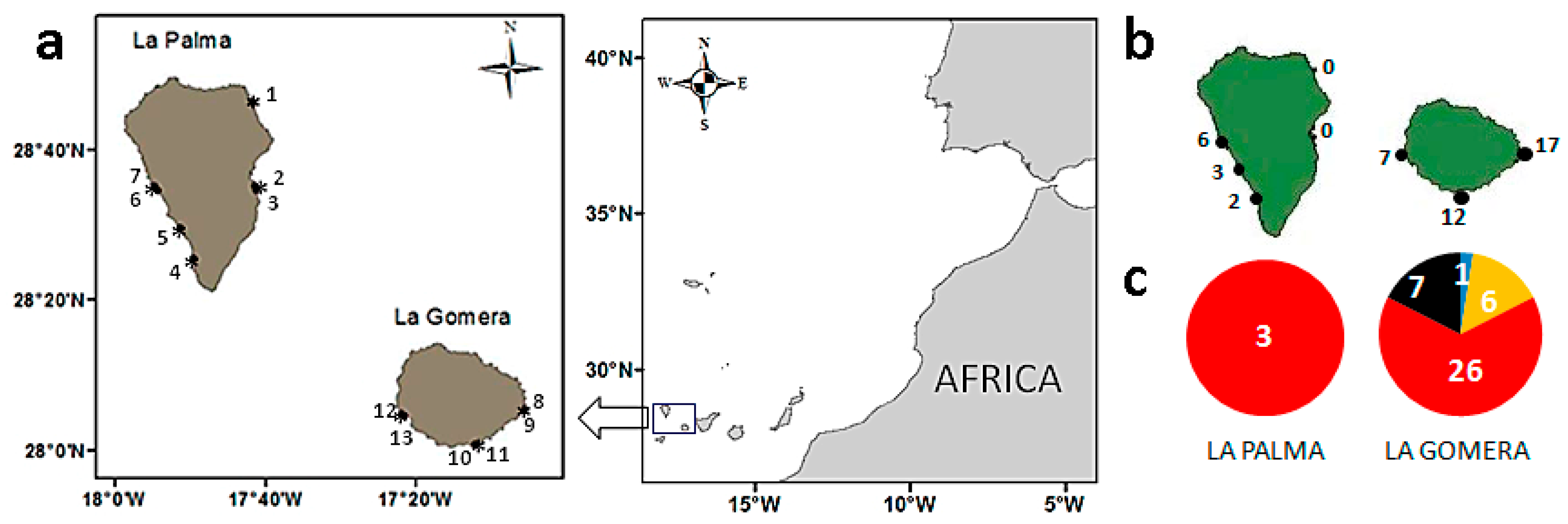
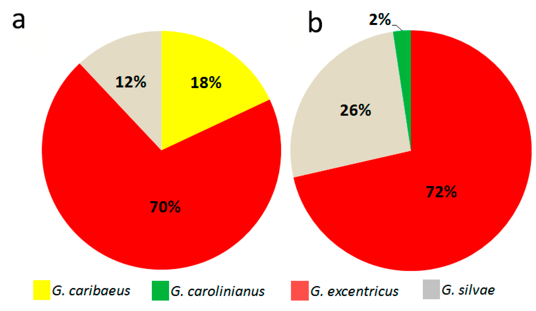
2.1. Diversity and Abundance of Gambierdiscus from La Palma and La Gomera

LSUrRNA gene sequencing of 61 isolates, 3 from La Gomera and 58 from La Palma, showed that G. excentricus was the only species identified from La Palma, whereas four species, G. australes, G. caribaeus, G. carolinianus, and G. excentricus, were identified from La Gomera, albeit in very different proportions (Figure 1). Thus, of the 40 Gambierdiscus strains isolated from macrophytes from this last island, 65% were G. excentricus, followed by G. silvae (18%), G. caribaeus (15%), and G. australes (2%) (Figure 1c). In addition, G. caribaeus (18 isolates) was detected in a net sample collected in the harbor of Playa Santiago, in La Gomera. No Gambierdiscus cells were detected in any of the samples taken from other harbors (Table 1).
The average concentration of Gambierdiscus spp. in macrophytes was 2 cells·g−1 in samples from La Palma (SD = 5, n = 25) and 12 cells·g−1 in those from La Gomera (SD = 21, n = 28). The maximum concentrations were also observed in the latter with 88 cells·g−1 in La Cueva beach (La Gomera) by contrast with 29 cells·g−1 in Tazacorte beach (La Palma) (Table 1, Figure 1). The average abundance at each station is shown in Figure 1b. Gambierdiscus cells appeared in 17 of the 28 La Gomera samples but in only 3 of the 25 La Palma samples. A comparison of macrophyte weights measured as the wet weight vs. the dry-blot weight indicated a wet-weight loss of 62% (SD = 14, n = 53), which translated into an increase in the concentrations when expressed as the blot-dried weight. Accordingly, the maximum Gambierdiscus concentration of 88 cells·g−1 wet weight corresponds to 130–155 cells·g−1 blot-dried weight of macrophytes.

2.2. Epiphytic Dinoflagellate Composition

The concentrations of dinoflagellates of other genera besides Gambierdiscus were also quantified in the epiphytic samples from La Gomera and La Palma and included Prorocentrum, Coolia, Sinophysis, Ostreopsis, Vulcanodinium, Heterocapsa, and Scrippsiella (Table S1). The most abundant dinoflagellates were those of the Ostreopsis genus, which reached concentrations of 2.8 × 10³ and 1.6 × 10³ cells·g−1 in samples from Tazacorte beach (La Palma) and Charco Condesa (La Gomera), respectively. While the concentrations of Gambierdiscus were low, the cells were detected in a higher number of samples than either Heterocapsa or Scripsiella (Table S1). The principal component analysis (PCA) showed significant differences between islands with respect to the composition and abundances of the studied dinoflagellates (p < 0.001), with the two principal components explaining 46% of the variance. The rotated component matrix is shown in Table 2 (absolute values <0.5 were excluded). In the first component, the highest correlation was between Gambierdiscus and Sinophysis, and secondly between Prorocentrum, Coolia, and Vulcanodinium. Based on the correlation coefficients, the first component was significantly more important in La Gomera than in La Palma. The second component assembled the remaining genera, Ostreopsis, Heterocapsa, and Scrippsiella (Table 2), but without a clear correlation with any of the islands.

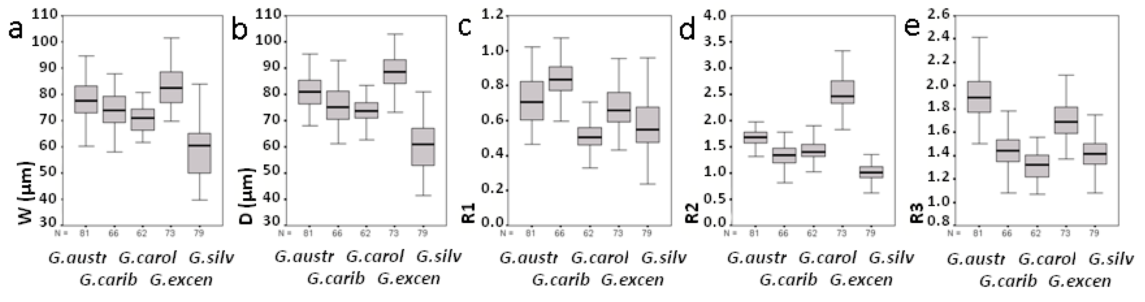
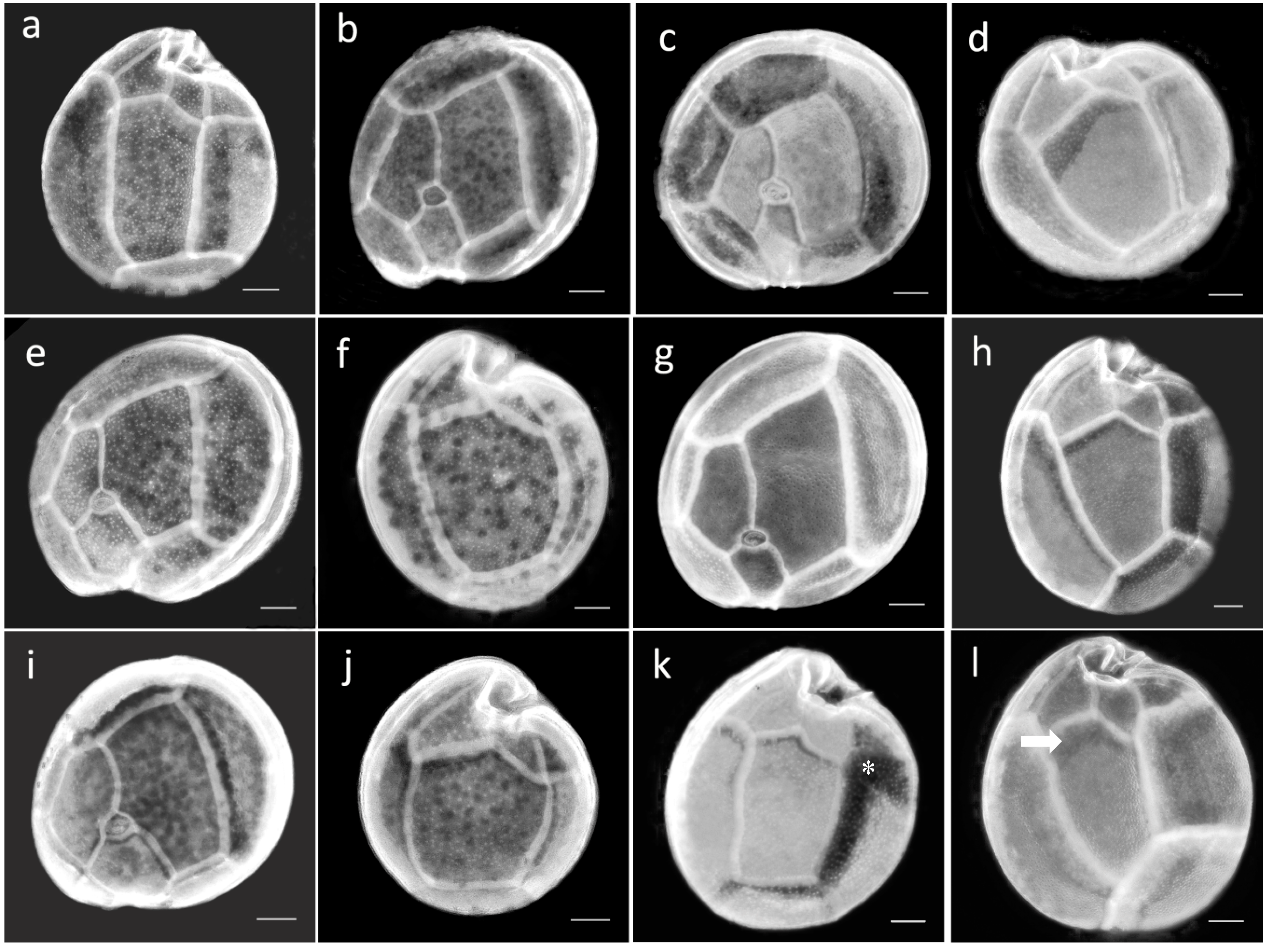
2.3. Morphological Study of Cultured Gambierdiscus Cells

Cells of cultures of the five Gambierdiscus species from the Canary Islands were anteroposteriorly compressed, and their thecal plate formula was typical of the genus: Po, 4′, 0a, 6″, 6c, 5‴, 0p, 2″″. The cell sizes and thecal plate measurements of the species were compared. The very small cells of G. silvae allowed this species to be differentiated based on a definition of >50% of the cells being ≤60 µm in diameter (Table 3, Figure 2a,b). The cell size distributions of the other species also differed significantly (p < 0.01); the exceptions were G. caribaeus and G. carolinianus, as indicated by their very similar mean ranks (Table S2). Observations of the shape of the 2′ and 2″″ plates based on R1, R2, and R3 (see Materials and Methods) also revealed significant differences among species (p < 0.01). Only the comparison of the R3 distribution according to the mean rank showed no significant differences between G. caribaeus and G. silvae (p > 0.01) (Table S2). The values of the three parameters are box-plotted in Figure 2c–e.
G. australes cells had a mean (± SD) depth of 81 ± 6.3 µm and a mean width of 78 ± 7.5 µm. Among the studied species, the 2″″ plate of G. australes was the most elongated (Figure 2e and Figure 3a), resulting in the highest R3 value (R3 > 1.5, mean ± SD = 1.91 ± 0.20) (Table 3). For the 2′ plate of G. australes, the R1 value was close to 1 (mean ± SD = 0.72 ± 0.13, Table 3) and second highest after the R1 of G. caribaeus (mean ± SD = 0.83 ± 0.11, Table 3), which indicated the more rectangular shape of this plate in these two species (Figure 3b,c) than in the others, in which the smaller R1 values reflected a more hatchet-shaped 2′ plate. The R2 values of G. australes (mean ± SD = 1.67 ± 0.16) differed significantly from those of G. excentricus, with only a 5% overlap (Table 3 and Figure 2d).
G. caribaeus cells had a mean (± SD) depth and width of 76 ± 7.2 µm and 74 ± 6.7 µm, respectively. Other distinctive characteristics of this species, in addition to its more rectangular 2′ plate, as described above, was its R3 value (mean ± SD = 1.44 ± 0.14) indicating less elongation of the 2″″ plate compared to that of G. australes (Figure 3a,d). Lower R3 values distinguished not only G. caribaeus but also G. carolinianus and G. silvae from G. australes and to a lesser extent from G. excentricus (Table 3 and Figure 3a,d,f,h,j). Furthermore, the R2 values of G. caribaeus (mean ± SD = 1.34 ± 0.22, Table 3) significantly differed from those of G. excentricus, G. australes, and G. silvae but were very similar to the R2 values of G. carolinianus, as it is indicated by the similar mean ranks (Table S2).
G. carolinianus cells had a mean (± SD) depth of 73 ± 4.6 µm and a mean width of 71 ± 4.9 µm. This species along with G. silvae had the smallest R1 mean values (mean ± SD = 0.50 ± 0.08) (Table 3 and Figure 2c), indicating a hatchet-shaped 2′ plate (Figure 3e,i) but different from the more rectangular of G. australes (Figure 3b) and G. caribaeus (Figure 3c). However, as stated above, the R2 values of G. carolinianus (mean ± SD = 1.44 ± 0.19) differed from those of G. silvae and G. excentricus, the species with the least and most eccentric Po, respectively (Table 3). The R3 values of G. carolinianus (mean ± SD = 1.31 ± 0.12) indicated less elongation of the 2″″ plate (Figure 3f), a characteristic shared, as noted above, with G. caribaeus and G. silvae (Table 3 and Figure 3d,j) but different from G. australes and G. excentricus (Figure 3a,h).
G. excentricus had the largest cells among the studied species (88 ± 6.8 µm depth and 83 ± 7.9 µm width, mean ± SD). A distinguishing feature of this species was its highly eccentric Po, evidenced by R2 ≥ 2.1 (mean ± SD = 2.55 ± 0.31, Table 3 and Figure 2d and Figure 3g). The sole species with an overlapping R2 was G. australes, but the overlap was only by 5%, as mentioned previously. A comparison of these two species showed that the hatchet-shaped 2′ plate frequently seen in G. excentricus (Figure 3g) (mean ± SD of R1 = 0.67 ± 0.11) contrasted with the mostly rectangular 2′ plate of G. australes (Figure 3b). In addition, the 2″″ plate was significantly more elongated in G. australes (Figure 3a) than in G. excentricus (mean ± SD of R3 = 1.71 ± 0.15, Table 3 and Figure 3h) though with a degree of overlapping values as shown in Figure 2e.
G. silvae was characterized by the smallest cells (60 ± 9.9 µm depth and 59 ± 9.7 µm width, mean ± SD) and an R2 value close to 1 (mean ± SD = 1.02 ± 0.16), indicating a nearly central position of Po in the anterior suture of the plate and representing a less eccentric Po (Table 3 and Figure 3i). This feature clearly distinguished G. silvae from G excentricus and G. australes (Figure 2d). The R3 values of G. silvae (mean ± SD = 1.41 ± 0.14, Table 3) were consistent with a less elongated 2″″ plate (Figure 3j), a feature shared with G. caribaeus and G. carolinianus but not with G. australes and G. excentricus, as has already been mentioned previously.

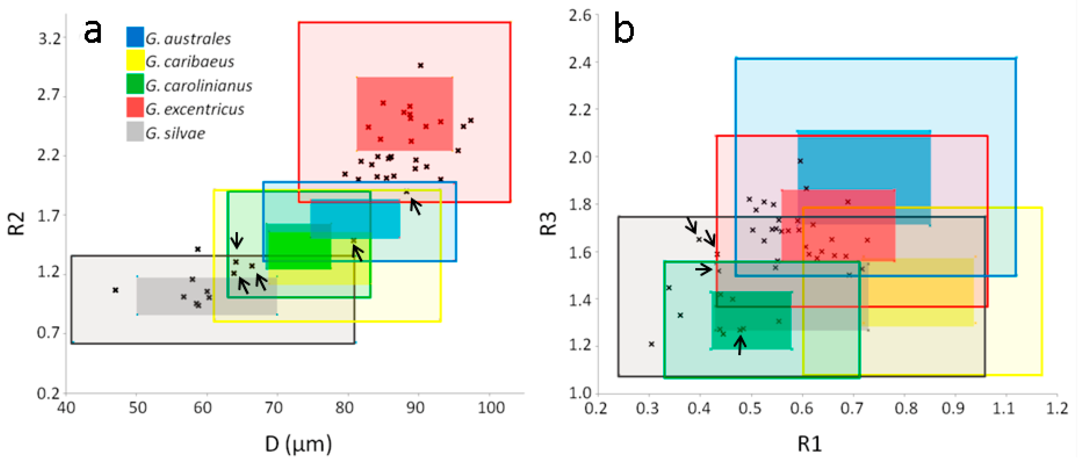
2.4. Morphological Study of Field Cells of Gambierdiscus

Gambierdiscus specimens from field samples showed the same general morphology as the cultured cells (i.e., anteroposterior compression) and the same thecal plate formula. Field cells were classified based on cell depth (D) and comparisons of their R1, R2, and R3 values with those values determined for the cultured cells (Table 3). Thus, D and R2 separated two well-defined groups (Figure 4a), whereas the discriminatory powers of R1 and R3 were low (Figure 4b). Using D and R2, we were able to classify at least 38 out of 42 specimens from field samples into two groups, corresponding to G. cf. excentricus and G. cf. silvae (Figure 4a). The assignment of 28 specimens to the first group was based on D > 80 µm and R2 > 1.97. A cell with D > 80 µm but R2 = 1.9 (in Figure 4a it is marked with an arrow), such that it might have corresponded to either G. cf. excentricus or G. cf. australes, was assigned to the former because of the similarities in the size and shape of its 2′ plate. The assignment of eight specimens to G. cf. silvae was based on D < 61 µm and R2 < 1.41 (Figure 4a). In the classification of the other four cells, both R1 and/or R3 were additionally necessary as well as an evaluation of the general appearance of the cells. Three specimens had D and R2 values consistent with G. cf. silvae/G. cf. carolinianus/G. cf. caribaeus (64 ≤ D ≤ 66 and 1.21 ≤ R2 ≤ 1.31, Figure 4a), but their R1 and R3 values as well as the general appearance of the cells coincided with those of the G. cf. silvae group (Figure 4b). Lastly, one cell was ruled out as being either G. cf. silvae or G. cf. excentricus because of its D (81 µm) and R2 (1.49) values (Figure 4a); instead, its R1 (0.48) and R3 (1.27) values were consistent with those of G. cf. carolinianus (Figure 4b).

2.5. Gambierdiscus Diversity: Genetics vs. Morphology

As shown by LSUrDNA sequencing and morphological study, G. excentricus was the most abundant species, detected in 70% of the sequences (23 of 33) (Figure 5a), and with an abundance of 72% (30 of 42 cells) as determined by morphology (Figure 5b). The corresponding values for G. silvae were 12% (4 of 33 sequences) (Figure 5a) and 26% (11 of 42 cells) (Figure 5b). G. caribaeus, by contrast, was detected only in the genetic study, in which it accounted for 18% (6 out of 33) of the sequences. One cell classified as G. cf. carolinianus (2%) was also identified as such in the morphological study.

3. Discussion

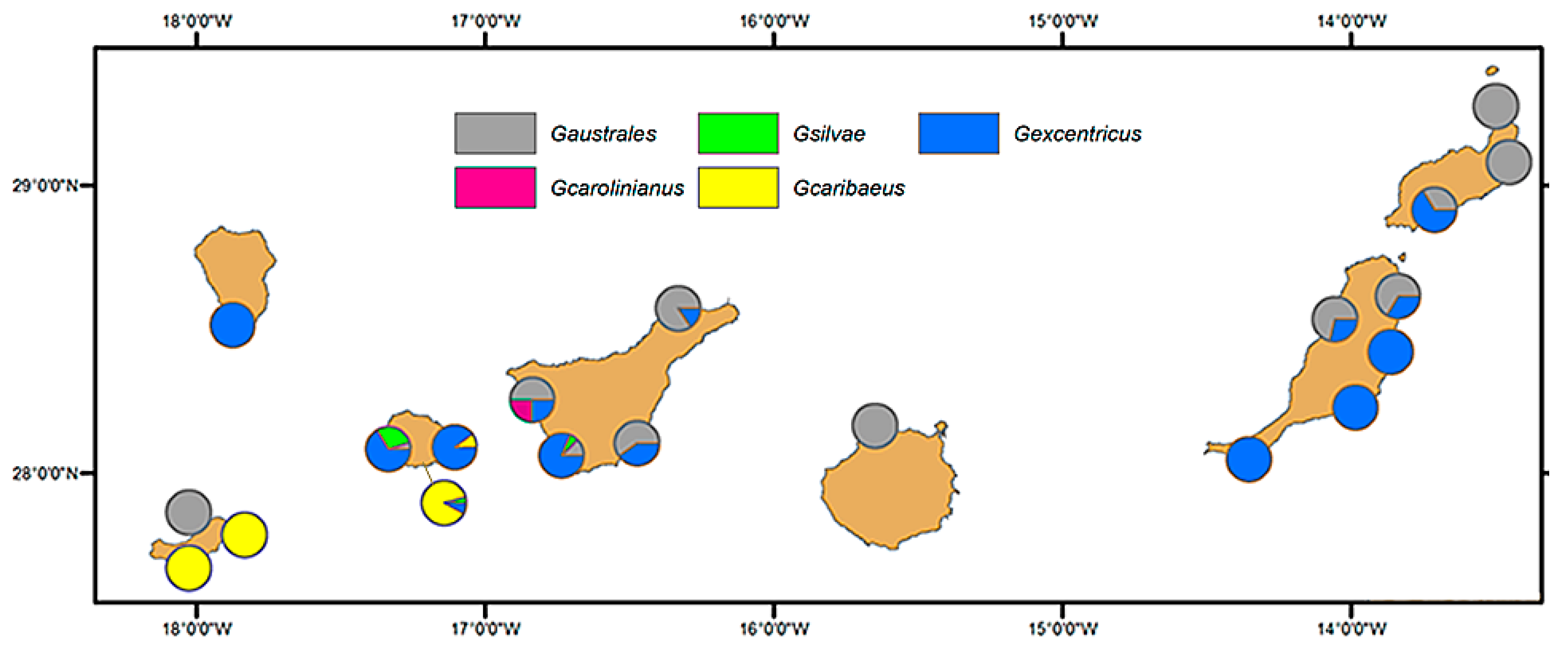
3.1. Geographical Distribution of the Species of Gambierdiscus Detected in the Archipelago of Canary Islands

Our results on the diversity of Gambierdiscus in La Gomera and La Palma complete the study of the diversity of this genus in the archipelago. The geographical distribution of the species recognized thus far is depicted in the map shown in Figure 6, which is the first presentation of the distribution of Gambierdiscus in the seven main islands of the Canary archipelago. The map is based on the results of [12] and those of the present study, and specifically on data of genetic sequences. These were mostly obtained from cultures established from cells isolated from field samples. However, the number of sequences corresponding to each island differed.
The dominant species of Gambierdiscus in the Canary Islands were G. excentricus and G. australes. Unlike their fairly coincidental distribution of these species in the archipelago, literature data on their worldwide geographical distribution show marked differences. Thus, G. australes was originally described from coral reef areas near Raivavae island (French Polynesia) and was considered to be limited to the Pacific Ocean [22], whereas G. excentricus was quite recently described from macrophytes in rock pools of the Canary Islands [17], and it had not been observed in well-studied areas as in the Pacific and until very recently in Caribbean Sea [10,25]. Nonetheless, during the last decade, an increasingly larger distribution of these two Gambierdiscus species has been reported. For example, G. australes was reported from the Indian Ocean, along the coast of Pakistan (Arabian Sea) [29], and was recently detected off the coasts of the Balearic Islands (Mediterranean Sea) [30]. To what extent this expansion reflects intensified studies of benthic dinoflagellates is unclear, but it may explain the detection of G. excentricus following its original identification in Canary Island samples [17]. G. excentricus was also found in the Atlantic Ocean, both along the island of Madeira [31,32] and, relatively close by, in Morocco—note that the species corresponding to Gambierdiscus spp. cited by [33] corresponds to G. excentricus according to [17]. Other reported detections of G. excentricus include the coastal waters of Brazil (Playa Tartaruga, Rio de Janeiro) [34] and Salalah, Oman (Arabian Sea) (María Saburova, personal communication).
The distributions of G. caribaeus, G. carolinianus, and G. silvae in the Canary Islands are apparently more limited than those of the two aforementioned species [12]. Nonetheless, in our study G. caribaeus and G. silvae were quite commonly detected off La Gomera. Thus far, there has been only one report of G. carolinianus in Tenerife [12]. The same authors reported G. caribaeus on the island of El Hierro, where it was associated with macroalgae and an artificial substrate. A high concentration of Gambierdiscus cells (>104 cells·g−1 wet weight of algae) was registered on the same island, at the port of La Restinga, and based on their morphology was identified as G. caribaeus [35]. We detected this species in the waters of La Gomera, associated with macroalgae and with sediment from an island harbor. Thus, compared to the other Gambierdiscus species, the distribution of G. caribaeus seems to be concentrated in the western islands. This may be due to a more tropical character of this species, given the warmer sea temperatures of La Gomera, as opposed to the cooler waters of Lanzarote and Fuerteventura. The worldwide distribution of G. caribaeus extends to throughout the Caribbean, including Florida (USA), but also to islands in the Pacific Ocean including Tahiti (French Polynesia) and Palau (Micronesia) [10]. In contrast, the worldwide distribution of G. silvae is thus far very restricted, as it is largely limited to the Atlantic Ocean. In addition to the Canary Islands, ribotype 1, from Belize [10], is coincident with G. sylvae, and it was also detected in the Cape Verde archipelago, as its morphological characteristics suggest that the species identified by [36] and cited as Goniodoma would be referred to today as G. silvae [16]. This species was also recently identified among Gambierdiscus specimens obtained in Greece (eastern Mediterranean) ([9]; Aligizaki K., personal communication).

3.2. Morphological Study

Cell depth and the shapes of the second apical (2′) and second antapical (2″″) plates have been described by several authors as differential morphological features of Gambierdiscus species [16,17,22,23,37]. The relatively central position of Po was a differential trait noted by [17] in their original description of G. excentricus. Morphological characterization of ten Gambierdiscus species by [23], including G. australes, G. carolinianus, and G. caribaeus, provided a dichotomous key in which those three species were separated based on the shapes of their 2′ and 2″″ plates. The present work applied those morphological capabilities to differentiate among the five Gambierdiscus species isolated from the Canary Islands. Moreover, by applying the knowledge obtained from cultures to field specimens, we obtained a more detailed morphological characterization of these dinoflagellates in nature and an approach to cell determinations that will be of value, at least until new quantitative molecular techniques are developed.
The cell sizes of the studied species were highly variable, and although the distributions of cell depth (D) and width (W) differed significantly, the large degree of overlap between the respective size ranges hinders the use of either (D) or (W) in species identification. Of the five studied species, only the smaller size (≤ 60 µm) of G. silvae can be considered as a useful trait for species differentiation. In the morphological study of field specimens, it allowed—along with the morphology of the thecal plates—the identification of G. silvae in samples from Playa de Santiago.
The high intraspecific variability of R1, R2, and R3, representing the shapes of 2′ and the 2″″ plates and the position of Po, demonstrated well the thecal shape variations within Gambierdiscus. This variability and the subtle differences between some species complicate a morphologically based discrimination. This problem has been repeatedly mentioned in the literature and explains why the first description of Gambierdiscus (G. toxicus [38]) included several similar species that could not be distinguished until molecular methodologies were used [22,23]. Morphological intraspecific variability has been attributed to the changes that occur during the division process, as even two recently divided daughter cells may differ in the cell shape [17]. In addition, the details observed in the present study suggest that morphological variability of the thecal plates occurs during cell division. For example, in recently divided cells of G. australes, the extent of 2″″plate elongation is far below the mean and is instead similar to that in G. excentricus (as it can be seen in the cell of G. australes showed in Figure 3k). In the cells of G. excentricus, the less dense upper end of the 2″″ plate suggests a progressive lengthening during cell growth, as indicated herein by R3 values well above the average for this species and contributing to the overlap with G. australes (as it can be seen in the cell of G. excentricus showed in Figure 3l).
Among the 16 species of Gambierdiscus described so far, it is obvious that limiting the number of species to those that appear in a certain area would facilitate the comparison and identification of them. It must be always in mind that the appearance of a new species in the Canary Islands should require new comparative studies. We found that the variability in the morphological features of Gambierdiscus was also a characteristic of the field specimens, but the consideration of two or more traits and even the general morphological appearance of the cell resulted in a classification. For example, two cells of G. cf. excentricus had very high R3 values (>1.8), within the range determined for G. cf. australes. Nonetheless, one of the cells could be unequivocally identified based on the eccentricity of Po and the shape of the second apical plate. In the other cell, the eccentricity of Po (R2 = 1.90) was within the ranges of G. cf. excentricus and G. cf. australes, but the similarity of the general cell morphology allowed an assignment to the former species.
Thus, despite the similarities among species from the Canary Islands, the parameters described and applied herein can be used in their discernment. While G. excentricus and G. silvae were easily discriminated among the five species, this may not be the case if other species are present in the samples. Accordingly, it will often be necessary to use more than one morphological trait to identify Gambierdiscus species. The parameters included in our study differed in their discriminatory power depending on the species. R2, representing the position of Po, had the best resolution, followed by R3 (elongation of the 2″″ plate). R1, indicating the shape of the 2′ plate, was also informative. Furthermore, we discovered that once the observer had become familiar with the traits of the different species, recognition of the general shape and configuration of the thecal plates could aid in the identification. In addition, the literature describes other potentially useful morphological characteristics of Gambierdiscus, for example, the symmetry of the 3″ plate, the dorsal end of the 2″″ plate, the cell surface pattern, and the shape of the apical pore complex (as examples see [11,16,23,34]).

3.3. Comparisons of the Results: Genetics vs. Morphology

The most important difference between the genetic and morphological analyses was the restriction of the detection of G. caribaeus to the genetic study, where it accounted for 18% of the obtained sequences. This difference demonstrates well the bias associated with each methodology. G. caribaeus occurred in just one sample (Playa Santiago), from which three other species were isolated (5 specimens of G. caribaeus, 2 of G. excentricus, and 1 of G. silvae). By contrast, all four specimens from the same sample that were studied morphologically could be assigned to G. cf. silvae. Consequently, either the cultures facilitated G. caribaeus detection or the morphological study was incorrect. This second possibility was ruled after we meticulously examined the morphological features of those four specimens and again classified them as G. silvae for the following reasons: (1) the small size of the specimens, (2) the hatchet-shaped 2′ plate, (3) the more centered position of Po, and (4) the short elongation of the 2″″ plate. Furthermore, the morphology of those specimens coincided with that of cells identified as G. cf. silvae by genetic analysis in other samples.
Amplification of LSUrDNA fragments by routine PCR does not provide quantitative data on the actual species distribution, given that the results are based on cultured cells or single individuals. Consequently, PCR analyses will be biased by the fact that specimens or species better adapted to laboratory conditions will be overrepresented such that the selected single cells are unlikely to be representative of the overall population. In our results, the smaller size of G. cf. silvae might cause this species to be overlooked when it is present together with the larger G. caribaeus cells isolated for cultures.
These observations point out the importance of semi-quantitative tools, as species-specific PCR assays [24], in estimating Gambierdiscus diversity. The results of those analyses in several geographical regions, such as the Gulf of Mexico and the Pacific [4,39,40], have revealed broad-ranging differences in species composition and in the relative proportions of the identified species. Similar studies in the Canary Islands will add to our knowledge of the spatio-temporal distribution and species composition of Gambierdiscus as well as to associate these trends with the prevailing environmental conditions and the detection of ciguatoxic fish.

4. Materials and Methods

4.1. Field Sampling and Processing

Fifty-three samples of macrophytes were collected from the littoral of the islands La Gomera and La Palma in Canary archipelago at six and three stations, respectively, between 4–9 October 2017 (Table 1, Figure 1). Two to eight samples were collected from each station gathering a representative range of material from each sampling location. In addition, 11 phytoplankton samples were obtained by net tows from 4 harbors located in the close vicinity of those stations (Table 1). The macrophyte samples were collected during low tide from tidal ponds and by snorkeling up to a maximum depth of 1.5 m. The phytoplankton samples were filtered through a nylon mesh with a mesh size of 300 µm and then concentrated by filtration through a 20 µm nylon mesh. One set of subsamples was kept for cell isolations and culture establishment, and another was formaldehyde-fixed in situ for identification and enumeration in the laboratory. The methodology used in the cell isolations and culture establishment of Gambierdiscus spp. was that described in [12]. Among the 61 culture isolates used in our phylogenetic analysis of Gambierdiscus, 58 were from La Gomera and 3 from La Palma.

4.2. Cell Enumeration and Light Microscopy

Formaldehyde-fixed epiphyte samples were stained with Fluorescent Brightner 28 (Sigma, St Louis, MO, USA) [41] for thecal identification and counted under UV light using an Axiovert 125 epifluorescence inverted microscope (Carl Zeiss AG, Germany). Quantitative data were obtained for the genus Gambierdiscus and for the other dominant dinoflagellates also present in the samples. Cell abundances were expressed as cells per gram wet weight of the host macrophyte (abbreviated as cells·g−1). The macrophytes were weighed after they had been drained of their water by manual squeezing. This is the most commonly used measurement method, and it allowed comparisons of our results with those of other studies, although many do not provide a detailed description of the procedure. In addition, because previous studies reported data from the Canary Islands as cells per gram blot dry weight, we also weighed the sampled macrophytes after blotting them overnight on absorbent laboratory paper [12]. The correction factor between the two procedures was estimated as the difference in the average of the percentages calculated from the wet and blot-dried measured weights.

4.3. DNA Extraction, PCR Amplification, and DNA Sequencing

Partial LSUrRNA gene sequences (D8-D10 region) were obtained from cells isolated from cultures as described by [12]. Briefly, single cells were transferred using a micropipette to an Eppendorf 5424R tube (Eppendorf AG, New York, USA), washed in three droplets of distilled water, frozen in liquid nitrogen, and stored at −20 °C until processing (on the same day). The cells were rinsed in 1 mL of distilled water, centrifuged again, and the supernatant was discarded. DNA was extracted using a modified Chelex procedure as described in [16] and eluted in TE buffer (25 mL). The quantity and purity of the DNA were analyzed in a Nanodrop Lite spectrophotometer (Thermo Scientific, Waltham, MA, USA).
The D8–D10 regions of the LSUrRNA gene were amplified using the primer pair FD8/RB [42]. The amplification reaction (20 μL) contained 0.75 pmol of each primer/µL and 2 μL of DNA extract in Horse-Power DNA polymerase master mix (Canvax, Spain), consisting of 2 × PCR buffer, 0.4 mM of each dNTP, 4 mM MgCl2, and 0.1 U Taq DNA polymerase/μL. The PCR conditions followed those reported in [12]. After their visualization by gel electrophoresis, the PCR products were purified using ExoSAP-IT (USB Corp., OH, USA), sequenced using the Big Dye Terminator v3.1 reaction cycle sequencing kit (Applied Biosystems, Foster City, CA, USA), and migrated in an AB 3130 sequencer (Applied Biosystems) at the CACTI sequencing facilities (Universidade de Vigo, Spain). The obtained partial gene sequences from the amplified LSUrRNA were deposited in GenBank. The accession numbers are provided in Table S3 in the online version of this article.

4.4. Morphological Study of Gambierdiscus

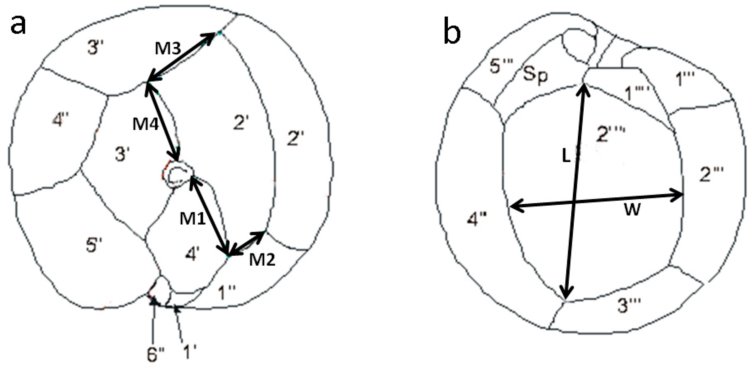
4.4.1. Morphometric Analysis

Cell morphology determinations were based on measurements of two thecal plates: the second apical (2′) plate, located on the epitheca, and the second antapical (2″″) plate, on the hypotheca (Figure 7). For the 2′ plate, the lengths of the 2′/4′ (M1 in Figure 7a), 2′/1″ (M2 in Figure 7a), 2′/3″ (M3 in Figure 7a), and 2′/3′ (M4 in Figure 7a) sutures were measured. For the 2″″ plate, the maximum length of the plate (L in Figure 7b) and the width measured from the middle of the left side to the middle of the right side of the plate (W in Figure 7b) were determined. Based on those measurements, the following three parameters were evaluated: (1) R1, defined as the ratio of M2/M3, was used in an assessment of the rectangular vs. the hatchet shape of the 2′ plate; (2) R2, defined as the ratio of M4/M1, represented the position of Po in the lateral edge of the 2′ plate and, therefore, the degree of eccentricity of Po in the cell; and (3) R3, defined as the ratio of L/W, as an indicator of the elongation of the 2″″ plate. Cell depth (D), corresponding to the dorso-ventral diameter, and cell width (W), corresponding to the transverse diameter, were also measured. All measurements were made on calcofluor-stained cells using digital imaging software (ZEN lite, ZEISS Microscopy) and an epifluorescence microscope (Leica DMLA, Wetzlar, Germany) equipped with a UV light source and an AxioCam HRc (Carl Zeiss, Jena, Germany) digital camera.
The thecal tabulation system [43] used in the present study was based on a modification of the Kofoid system as described by [44]. Thus, in our study, the first precingular plate (1′) was considered to be a small plate that does not contact Po (Figure 7a). This plate is regarded as the 1″ plate in other Gambierdiscus studies which consider, on other hand, the 4′ plate as the 1′ plate. The nomenclature used in our study has also been used in studies of others species, such as Gonyaulacales, and allows comparisons with species and genera sharing the same plate formula: Po, 4′, 6″, 5‴, and 2″″. Following the same criteria, although the sulcal posterior plate (Sp) in Gambierdiscus, Coolia, and Ostreopsis is outside the sulcus, it must be considered as Sp, such that the 1p plate described in previous reports of Gambierdiscus is herein denoted as the 2″″ plate (Figure 7b) [17,44].

4.4.2. Cultured Cells

The morphological study was carried out on cultured cells of Gambierdiscus species identified by sequencing of the D8–D10 regions of the LSUrRNA gene, as described in Section 4.3. All of the strains used in this study were obtained from the isolation of cells in samples collected from the Canary Islands. Measurements were performed on at least 62 cells of each species (1–3 strains according to the availability of strains of each species) (Table S4).

4.4.3. Field Samples

Morphological analyses were also performed on individual cells isolated from epiphytic samples taken from La Gomera (see Section 4.1). Measurements of the epitheca and hypotheca of the same specimen were made by placing individual cells between two coverslips, which allowed them to be observed and photographed from their apical and antapical views. The morphologies of a total of 42 cells obtained from seven samples of three stations, Santiago beach (4), Charco Condesa (32), and La Cueva beach (4) (Table 1), were determined.

4.5. Comparison of the Morphological versus Genetic Data

Gambierdiscus species identified morphologically and genetically were compared with respect to their abundances. Morphology results were obtained as described in Section 4.4.3 for 42 cells of Gambierdiscus and genetic data from 33 cultures of Gambierdiscus isolated from the same samples used in the morphological study: Santiago beach (8 isolates), Charco Condesa (14 isolates), and La Cueva beach (11 isolates). These cultures corresponded in part to those used to evaluate Gambierdiscus off La Gomera island (Section 4.1).

4.6. Statistical Analysis

A principal component analysis (PCA) was performed to analyze the data describing the composition of epiphytic dinoflagellates. It was conducted using logarithmically transformed cell concentrations and the statistical software package SPSS. The Kaiser–Meyer–Olkin measure of sampling adequacy was 0.69, and Bartlett’s test of sphericity, which tests for the presence of correlations among variables, was significant at p < 0.001.
Statistical analyses comparing the morphological parameters of the different Gambierdiscus species were performed using the statistical software package SPSS. A Shapiro–Wilks test (p > 0.05) showed that the variables were normally distributed. However, in a Levene’s test there was no homogeneity of variance among the variables, such that a non-parametric rank-based test (Kruskal–Wallis) was used in the species comparisons.

Supplementary Materials

The following are available online at https://www.mdpi.com/2072-6651/11/7/423/s1, Table S1: Cell concentrations (cells·mL−1) of epiphytic dinoflagellates on the islands of La Palma and La Gomera, Table S2: Mean rank values from Kruskal–Wallis test for cell size measurements and morphological parameters (see material and methods) of Gambierdiscus species. Table S3. Pairwise distances (773 nt alignment) between 54 Gambierdiscus sequences obtained in this study, calculated as the proportion (p) of nucleotide sites at which two sequences being compared are different. It was obtained by dividing the number of nucleotide differences by the total number of nucleotides compared, computed as d: Transitions + Transversions: proportion of nucleotide sites that are different (MEGA7 software). Reference sequences for G. silvae, G. excentricus, G. caribaeus, and G. australes were retrieved from Genbank (Acc.Nos. indicated below). Pairwise distances for 7 additional shorter sequences of G. excentricus and G. caribaeus obtained in this study are indicated below the identity matrix. Table S4: Culture strains of Gambierdiscus species used in the morphological study and their geographical origin.

Author Contributions

Conceptualization, I.B., F.R., and S.F.; formal morphological analysis, I.B. and I. R.; formal genetic analysis, F.R. and P.R.; software, I.B.; validation, I.B., F.R., and S.F.; resources, I.B., F.R, and S.F.; writing—original draft preparation, I.B.; writing—review and editing, I.B., F.R., and S.F.; funding acquisition, I.B., F.R., and S.F.

Funding

This work was funded by the Spanish national projects CICAN (CGL2013-40671-R) and DIANAS (CTM2017-86066-R), funded by Fecyt, and “Tropicalización y ciguatera en Canarias,” funded by Fundación Biodiversidad.

Acknowledgments

We thank Amelia Fernandez and Patricia Loures for culture maintenance.

Conflicts of Interest

The authors declare no conflict of interest.

References

  1. Yasumoto, T. Chemistry, etiology, and food chain dynamics of marine toxins. Proc. Jpn. Acad. Ser. B 2005, 81, 43–51. [Google Scholar] [CrossRef] [Green Version]
  2. Ledreux, A.; Brand, H.; Chinain, M.; Bottein, M.-Y.D.; Ramsdell, J.S. Dynamics of ciguatoxins from Gambierdiscus polynesiensis in the benthic herbivore Mugil cephalus: Trophic transfer implications. Harmful Algae 2014, 39, 165–174. [Google Scholar] [CrossRef]
  3. Stewart, I.; Eaglesham, G.K.; Poole, S.; Graham, G.; Paulo, C.; Wickramasinghe, W.; Sadler, R.; Shaw, G.R. Establishing a public health analytical service based on chemical methods for detecting and quantifying Pacific ciguatoxin in fish samples. Toxicon 2010, 56, 804–812. [Google Scholar] [CrossRef] [PubMed]
  4. Tester, P.A.; Vandersea, M.W.; Buckel, C.A.; Kibler, S.R.; Holland, W.C.; Davenport, E.D.; Clark, R.D.; Edwards, K.F.; Taylor, J.C.; Vander Pluym, J.L.; et al. Gambierdiscus (Dinophyceae) species diversity in the Flower Garden Banks National Marine Sanctuary, Northern Gulf of Mexico, USA. Harmful Algae 2013, 29, 1–9. [Google Scholar] [CrossRef]
  5. Lewis, R.J. The changing face of ciguatera. Toxicon 2001, 39, 97–106. [Google Scholar] [CrossRef]
  6. Skinner, M.P.; Brewer, T.D.; Johnstone, R.; Fleming, L.E.; Lewis, R.J. Ciguatera Fish Poisoning in the Pacific Islands (1998 to 2008). PLoS Negl. Trop. Dis. 2011, 5, e1416. [Google Scholar] [CrossRef] [PubMed]
  7. Boada, L.D.; Zumbado, M.; Luzardo, O.P.; Almeida-González, M.; Plakas, S.M.; Granade, H.R.; Abraham, A.; Jester, E.L.; Dickey, R.W. Ciguatera fish poisoning on the West Africa Coast: An emerging risk in the Canary Islands (Spain). Toxicon 2010, 56, 1516–1519. [Google Scholar] [CrossRef] [PubMed]
  8. Bravo, J.; Suárez, F.; Ramírez, A.; Acosta, F. Ciguatera, an Emerging Human Poisoning in Europe. J. Aquac. Mar. Boil. 2015, 3, 00053. [Google Scholar]
  9. Aligizaki, K.; Nikolaidis, G. Morphological identification of two tropical dinoflagellates of the genera Gambierdiscus and Sinophysis in the Mediterranean Sea. J. Biol. Res. Thessalon. 2008, 9, 75–82. [Google Scholar]
  10. Litaker, R.W.; Vandersea, M.W.; Faust, M.A.; Kibler, S.R.; Nau, A.W.; Holland, W.C.; Chinain, M.; Holmes, M.J.; Tester, P.A. Global distribution of ciguatera causing dinoflagellates in the genus Gambierdiscus. Toxicon 2010, 56, 711–730. [Google Scholar] [CrossRef] [PubMed]
  11. Nishimura, T.; Sato, S.; Tawong, W.; Sakanari, H.; Yamaguchi, H.; Adachi, M. Morphology of G ambierdiscus scabrosus sp. nov. (Gonyaulacales): A new epiphytic toxic dinoflagellate from coastal areas of Japan. J. Phycol. 2014, 50, 506–514. [Google Scholar] [CrossRef] [PubMed]
  12. Rodríguez, F.; Fraga, S.; Ramilo, I.; Rial, P.; Figueroa, R.I.; Riobó, P.; Bravo, I. Canary Islands (NE Atlantic) as a biodiversity ’hotspot’ of Gambierdiscus: Implications for future trends of ciguatera in the area. Harmful Algae 2017, 67, 131–143. [Google Scholar] [CrossRef] [PubMed]
  13. Villareal, T.; Hanson, S.; Qualia, S.; Jester, E.; Granade, H.; Dickey, R.; Villareal, T. Petroleum production platforms as sites for the expansion of ciguatera in the northwestern Gulf of Mexico. Harmful Algae 2007, 6, 253–259. [Google Scholar] [CrossRef]
  14. Tester, P.A.; Feldman, R.L.; Nau, A.W.; Kibler, S.R.; Litaker, R.W. Ciguatera fish poisoning and sea surface temperatures in the Caribbean Sea and the West Indies. Toxicon 2010, 56, 698–710. [Google Scholar] [CrossRef] [PubMed]
  15. Kibler, S.R.; Tester, P.A.; Kunkel, K.E.; Moore, S.K.; Litaker, R.W. Effects of ocean warming on growth and distribution of dinoflagellates associated with ciguatera fish poisoning in the Caribbean. Ecol. Model. 2015, 316, 194–210. [Google Scholar] [CrossRef] [Green Version]
  16. Fraga, S.; Rodríguez, F. Genus Gambierdiscus in the Canary Islands (NE Atlantic Ocean) with Description of Gambierdiscus silvae sp. nov., a New Potentially Toxic Epiphytic Benthic Dinoflagellate. Protist 2014, 165, 839–853. [Google Scholar] [CrossRef] [PubMed]
  17. Fraga, S.; Rodríguez, F.; Caillaud, A.; Diogène, J.; Raho, N.; Zapata, M. Gambierdiscus excentricus sp. nov. (Dinophyceae), a benthic toxic dinoflagellate from the Canary Islands (NE Atlantic Ocean). Harmful Algae 2011, 11, 10–22. [Google Scholar] [CrossRef] [Green Version]
  18. Pérez-Arellano, J.-L.; Luzardo, O.P.; Brito, A.P.; Cabrera, M.H.; Zumbado, M.; Carranza, C.; Angel-Moreno, A.; Dickey, R.W.; Boada, L.D. Ciguatera Fish Poisoning, Canary Islands. Emerg. Infect. Dis. 2005, 11, 1981–1982. [Google Scholar] [CrossRef] [Green Version]
  19. Costa, P.; Estevez, P.; Castro, D.; Solino, L.; Gouveia, N.; Santos, C.; Rodrigues, S.; Leao, J.; Gago-Martinez, A. New Insights into the Occurrence and Toxin Profile of Ciguatoxins in Selvagens Islands (Madeira, Portugal). Toxins 2018, 10, 524. [Google Scholar] [CrossRef]
  20. Estevez, P.; Castro, D.; Pequeño-Valtierra, A.; Leao, J.M.; Vilariño, O.; Diogène, J.; Gago-Martínez, A. An Attempt to Characterize the Ciguatoxin Profile in Seriola fasciata Causing Ciguatera Fish Poisoning in Macaronesia. Toxins 2019, 11, 221. [Google Scholar] [CrossRef]
  21. Estevez, P.; Castro, D.; Leao, J.M.; Yasumoto, T.; Dickey, R.; Gago-Martinez, A. Implementation of liquid chromatography tandem mass spectrometry for the analysis of ciguatera fish poisoning in contaminated fish samples from Atlantic coasts. Food Chem. 2019, 280, 8–14. [Google Scholar] [CrossRef] [PubMed]
  22. Chinain, M.; Faust, M.A.; Pauillac, S. Morphology and molecular analyses of three toxic species of Gambierdiscus (Dinophyceae): G. pacificus, sp. nov., G. australes, sp. nov., and G. polynesiensis, sp. nov. J. Phycol. 1999, 35, 1282–1296. [Google Scholar] [CrossRef]
  23. Litaker, R.W.; Vandersea, M.W.; Faust, M.A.; Kibler, S.R.; Chinain, M.; Holmes, M.J.; Holland, W.C.; Tester, P.A. Taxonomy of Gambierdiscus including four new species, Gambierdiscus caribaeus, Gambierdiscus carolinianus, Gambierdiscus carpenteri and Gambierdiscus ruetzleri (Gonyaulacales, Dinophyceae). Phycologia 2009, 48, 344–390. [Google Scholar] [CrossRef]
  24. Vandersea, M.W.; Kibler, S.R.; Holland, W.C.; Tester, P.A.; Schultz, T.F.; Faust, M.A.; Holmes, M.J.; Chinain, M.; Litaker, R.W. Development Of Semi-Quantitative Pcr Assays For The Detection And Enumeration Of Gambierdiscus Species (Gonyaulacales, Dinophyceae)(1). J. Phycol. 2012, 48, 902–915. [Google Scholar] [CrossRef] [PubMed]
  25. Litaker, R.W.; Holland, W.C.; Hardison, D.R.; Pisapia, F.; Hess, P.; Kibler, S.R.; Tester, P.A. Ciguatoxicity of Gambierdiscus and Fukuyoa species from the Caribbean and Gulf of Mexico. PLoS ONE 2017, 12, e0185776. [Google Scholar] [CrossRef] [PubMed]
  26. Pisapia, F.; Holland, W.C.; Hardison, D.R.; Litaker, R.W.; Fraga, S.; Nishimura, T.; Adachi, M.; Nguyen-Ngoc, L.; Séchet, V.; Amzil, Z.; et al. Toxicity screening of 13 Gambierdiscus strains using neuro-2a and erythrocyte lysis bioassays. Harmful Algae 2017, 63, 173–183. [Google Scholar] [CrossRef] [PubMed] [Green Version]
  27. Pisapia, F.; Sibat, M.; Herrenknecht, C.; Lhaute, K.; Gaiani, G.; Ferron, P.-J.; Fessard, V.; Fraga, S.; Nascimento, S.M.; Litaker, R.W.; et al. Maitotoxin-4, a Novel MTX Analog Produced by Gambierdiscus excentricus. Mar. Drugs 2017, 15, 220. [Google Scholar] [CrossRef] [PubMed]
  28. Tester, P.A.; Faust, M.A.; Vandersea, M.W.; Kibler, S.R.; Chinain, M.; Holmes, M.; Holland, C.; Litaker, R.W. Gambierdiscus toxicus: Taxonomic uncertainties concerning Gambierdiscus toxicus: Proposed epitype. In 12th International Conference on Harmful Algae; International Society for the Study of Harmful Algae and the Intergovernmental Oceanographic Commission of UNESCO: Copenhagen, Denmark, 2008; pp. 269–271. [Google Scholar]
  29. Munir, S.; Siddiqui, P.; Morton, S.L.; Munir, S. The occurrence of the ciguatera fish poisoning producing dinoflagellate genus Gambierdiscus in Pakistan waters. ALGAE 2011, 26, 317–325. [Google Scholar] [CrossRef]
  30. Tudó, A.; Toldrá, A.; Andree, K.; Rey, M.; Fernández-Tejedor, M.; Campás, M.; Diogène, J. First report of Gambierdiscus in the Western Mediterranean Sea (Balearic Islands). Harmful Algae News 2018, 59, 22–23. [Google Scholar]
  31. Kaufmann, M.; Böhm-Beck, M. Gambierdiscus and related benthic dinoflagellates from Madeira archipelago (NE Atlantic). Harmful Algae News 2013, 47, 18–19. [Google Scholar]
  32. Guiry, M.D.; Guiry, G.M. AlgaeBase; World-Wide Electronic Publication, National University of Ireland: Galway, Ireland, 2010; Available online: http://www.algaebase.org (accessed on 2 December 2010).
  33. Ennaffah, B.; Chaira, K. First report of Gambierdiscus in Moroccan Atlantic waters. Harmful Algae News 2015, 50, 20. [Google Scholar]
  34. Nascimento, S.M.; Melo, G.; Salgueiro, F.; Diniz, B.D.S.; Fraga, S. Morphology of Gambierdiscus excentricus (Dinophyceae) with emphasis on sulcal plates. Phycologia 2015, 54, 628–639. [Google Scholar] [CrossRef]
  35. Soler-Onís, E.; Fernandez-Zabala, J.; Ojeda-Rodriguez, A.; Amorin, A. Bloom of Gambierdicus caribaeus in the temperate-subtropical waters of El Hierro, Canary Islands (North East Atlantic). Harmful Algae News 2016, 55, 14–17. [Google Scholar]
  36. Silva, E.S. Contribution a L’étude du microplancton de Dakar et des régions maritimes voisines. Bull. Int. Law Soc. 1956, 18, 335–371. [Google Scholar]
  37. Rhodes, L.; Smith, K.F.; Verma, A.; Curley, B.G.; Harwood, D.T.; Murray, S.; Kohli, G.S.; Solomona, D.; Rongo, T.; Munday, R.; et al. A new species of Gambierdiscus (Dinophyceae) from the south-west Pacific: Gambierdiscus honu sp. nov. Harmful Algae 2017, 65, 61–70. [Google Scholar] [CrossRef]
  38. Adachi, R.; Fukuyo, Y. The thecal structure of the marine toxic dinoflagellate Gambierdiscus toxicus gen. et. sp. nov. collected in a ciguatera endemic area. Bull. Jpn. Soc. Sci. Fish. 1979, 45, 67–71. [Google Scholar] [CrossRef]
  39. Nishimura, T.; Hariganeya, N.; Tawong, W.; Sakanari, H.; Yamaguchi, H.; Adachi, M. Quantitative PCR assay for detection and enumeration of ciguatera-causing dinoflagellate Gambierdiscus spp. (Gonyaulacales) in coastal areas of Japan. Harmful Algae 2016, 52, 11–22. [Google Scholar] [CrossRef]
  40. Smith, K.F.; Biessy, L.; Argyle, P.A.; Trnski, T.; Halafihi, T.; Rhodes, L.L. Molecular Identification of Gambierdiscus and Fukuyoa (Dinophyceae) from Environmental Samples. Mar. Drugs 2017, 15, 243. [Google Scholar] [CrossRef]
  41. Fritz, L.; Triemer, R.E. A rapid simple technique utilizing calcofluor white M2R for the visualization of dinoflagellate thecal plates. J. Phycol. 1985, 21, 662–664. [Google Scholar] [CrossRef]
  42. Lenaers, G.; Maroteaux, L.; Michot, B.; Herzog, M. Dinoflagellates in evolution. A molecular phylogenetic analysis of large subunit ribosomal RNA. J. Mol. Evol. 1989, 29, 40–51. [Google Scholar] [CrossRef]
  43. Kofoid, C.A. On the Peridinium steini Jörgensen, with a note on the nomenclature of the skeleton of the Peridinidae. Arch. Protistenkd. 1909, 16, 25–47. [Google Scholar]
  44. Besada, E.G.; Loeblich, L.A.; Loeblich, A.R., III. Observations on tropical, benthic dinoflagellates form ciguatera-endemic areas: Coolia, Gambierdiscus, and Ostreopsis. Bull. Mar. Sci. 1982, 32, 723–735. [Google Scholar]
Figure 1. (a) Sampling localities in the coasts of La Palma and La Gomera (Canary Islands). Station numbers and their identification are provided in Table 1. (b) Average abundance of Gambierdiscus spp. (cells·g−1 macrophyte wet weight) in La Palma and La Gomera. (c) Number of sequences retrieved from individual Gambierdiscus species in molecular analyses of samples from each island. G. australes (blue), G. caribaeus (yellow), G. silvae (black), and G. excentricus (red).
Toxins 11 00423 g001
Figure 2. Box plots of cell width (a), cell depth (b), and parameters R1 (c), R2 (d), and R3 (e) used in the morphological analyses (see Section 4.4.1).
Toxins 11 00423 g002
Figure 3. Photographs of cultured cells of the five species of Gambierdiscus reported from the Canary Islands and including their thecal plates. Hypotheca (a) and epitheca (b) of G. australes. Epitheca (c) and hypotheca (d) of G. caribaeus. Epitheca (e) and hypotheca (f) of G. carolinianus. Epitheca (g) and hypotheca (h) of G. excentricus. Epitheca (i) and hypotheca (j) of G. silvae. For the hypothecae of G. australes (k), the difference in their staining intensity (asterisk) reveals that they derived from a recently divided cell. The hypotheca of G. excentricus (l) has a less dense upper end of the 2″″ plate (arrow). Scale bars = 10 µm.
Toxins 11 00423 g003
Figure 4. Scatterplots showing the relationships between D (cell depth) and R2 values (a) and between R1 and R3 values (b) in field cells of Gambierdiscus (black dots). The colored squares represent the ranges for standard deviation limits (dark colors) and maximum–minimum (light colors) of D vs. R2 (a) and R1 vs. R3 (b) as determined in the morphological study of cultured cells. The arrows indicate cells whose values are particularly mentioned in the text (Section 2.4).
Toxins 11 00423 g004
Figure 5. Abundance (%) of the Gambierdiscus species identified in a genetic analysis (a) and in a morphological study (b) from the La Gomera island samples.
Toxins 11 00423 g005
Figure 6. Geographical distribution of the different species of Gambierdiscus in the Canary Islands.
Toxins 11 00423 g006
Figure 7. Drawings of an epitheca (a) and hypotheca (b) of Gambierdiscus, showing the measurements used in the morphological study.
Toxins 11 00423 g007
Table 1. Location of sampling stations in La Palma and La Gomera Islands.
StationIslandLocalityCoordinatesType of Sample
1La PalmaCharco azul28.81089, −17.7642macrophyte
2La PalmaCancajos beach28.65232, −17.75951macrophyte
3La PalmaCaleta Ballena28.64465, −17.7573macrophyte
4La PalmaLa Zamora beach28.51485, −17.87486macrophyte
5La PalmaCharco verde28.57261, −17.90008macrophyte
6La PalmaTazacorte harbor28.6437, −17.94325net
7La PalmaTazacorte beach28.65157, −17.94887macrophyte
8La GomeraLa Cueva beach28.09081, −17.10565macrophyte
9La GomeraSan Sebastián harbor28.089, −17.10767net
10La GomeraSantiago beach28.02732, −17.1979macrophyte
11La GomeraSantiago harbor28.02629, −17.19755net
12La GomeraValle Rey harbor28.08069, −17.33259net
13La GomeraCharco Condesa28.08397, −17.33653macrophyte
Table 2. Rotated component matrix from the principal component analysis (PCA) for epiphytic dinoflagellates in the islands of La Gomera and La Palma.
Component
12
Prorocentrum0.568-
Coolia0.583-
Sinophysis0.709-
Ostreopsis-0.685
Vulcanodinium0.524-
Heterocapsa-0.792
Scripsiella-0.625
Gambierdiscus0.706-
Table 3. Mean, standard deviation, and maximum and minimum values of cell size measurements (µm) and morphological parameters (see material and methods) of Gambierdiscus species.
ParameterSpeciesMeanStdMaxMin
D (cell depth)G. australes816.39568
G. caribaeus767.29361
G. carolinianus734.68363
G. excentricus886.810373
G. silvae609.98141
W (cell width)G. australes787.59560
G. caribaeus746.78858
G. carolinianus714.98162
G. excentricus837.910170
G. silvae599.78440
R1G. australes0.720.131.020.47
G. caribaeus0.830.111.070.60
G. carolinianus0.500.080.710.33
G. excentricus0.670.110.960.43
G. silvae0.580.150.960.24
R2G. australes1.670.161.971.32
G. caribaeus1.340.221.920.82
G. carolinianus1.440.191.901.02
G. excentricus2.550.313.331.82
G. silvae1.020.161.360.63
R3G. australes1.910.202.421.50
G. caribaeus1.440.141.781.08
G. carolinianus1.310.121.561.07
G. excentricus1.710.152.091.37
G. silvae1.410.141.751.08

