Review

Impact of Altered Intestinal Microbiota on Chronic Kidney Disease Progression

by
Esmeralda Castillo-Rodriguez
1,
Raul Fernandez-Prado
1,
Raquel Esteras
1,
Maria Vanessa Perez-Gomez
1,
Carolina Gracia-Iguacel
1,
Beatriz Fernandez-Fernandez
1,
Mehmet Kanbay
2,
Alberto Tejedor
3,
Alberto Lazaro
3,
Marta Ruiz-Ortega
1,
Emilio Gonzalez-Parra
1,
Ana B. Sanz
1,
Alberto Ortiz
1 and
Maria Dolores Sanchez-Niño
1,*
1
Nephrology Department, IIS-Fundación Jiménez Díaz-Universidad Autónoma de Madrid, 28040 Madrid, Spain
2
Department of Internal Medicine, Koc University School of Medicine, Istanbul 34450, Turkey
3
Nefrología, IIS-Gregorio Marañón, Universidad Complutense de Madrid, 28007 Madrid, Spain
*
Author to whom correspondence should be addressed.
Toxins 2018, 10(7), 300; https://doi.org/10.3390/toxins10070300
Submission received: 15 June 2018 / Revised: 13 July 2018 / Accepted: 17 July 2018 / Published: 19 July 2018
(This article belongs to the Special Issue The Intestine and Uremia)

Abstract

:
In chronic kidney disease (CKD), accumulation of uremic toxins is associated with an increased risk of CKD progression. Some uremic toxins result from nutrient processing by gut microbiota, yielding precursors of uremic toxins or uremic toxins themselves, such as trimethylamine N-Oxide (TMAO), p-cresyl sulphate, indoxyl sulphate and indole-3 acetic acid. Increased intake of some nutrients may modify the gut microbiota, increasing the number of bacteria that process them to yield uremic toxins. Circulating levels of nutrient-derived uremic toxins are associated to increased risk of CKD progression. This offers the opportunity for therapeutic intervention by either modifying the diet, modifying the microbiota, decreasing uremic toxin production by microbiota, increasing toxin excretion or targeting specific uremic toxins. We now review the link between nutrients, microbiota and uremic toxin with CKD progression. Specific focus will be placed on the generation specific uremic toxins with nephrotoxic potential, the decreased availability of bacteria-derived metabolites with nephroprotective potential, such as vitamin K and butyrate and the cellular and molecular mechanisms linking these toxins and protective factors to kidney diseases. This information provides a conceptual framework that allows the development of novel therapeutic approaches.
Key Contribution: Altered gut microbiota may be a source of nephrotoxic uremic solutes that promote CKD progression. Some nutritional supplements may favour the development of a gut microbiota composition with unwanted properties. Conversely, gut microbiota may secrete molecules with nephroprotective properties. An in-depth understanding of this balance may help to develop novel therapeutic approaches for CKD.

1. CKD and CKD Progression

Around 10% of the adult population has chronic kidney disease (CKD), which is diagnosed when the estimated glomerular filtration rate (eGFR) falls below 60 mL/min/1.73 m2 or the urinary albumin:creatinine ratio (UACR) rises above 30 mg/g or there is other evidence of kidney injury, for more than 3 months, even in the presence of normal GFR [1].The thresholds for eGFR and albuminuria are not arbitrary: they signal the point when kidney injury results in both a higher risk of death and in a higher risk of CKD progression to end-stage kidney disease (ESKD) [2,3]. The drivers of CKD progression and accelerated aging when albuminuria is present but eGFR is preserved are unclear, although recently, albuminuria was reported to decrease the expression of the anti-aging and nephroprotective protein of kidney origin Klotho [4]. However, accumulation of uremic toxins has been considered a key contributor to the consequences of CKD when eGFR is <60 mL/min/1.73 m2 [5]. Despite this, their relative contribution to CKD progression is less well characterized than the role of other factors, such as persistence of the kidney insult, compensatory hyperfiltration, albuminuria and inflammation [6,7]. We have now focused on the current understanding of how microbiota-derived molecules may contribute to or protect from CKD progression.

2. The Intestinal Microbiota in CKD

There is experimental and clinical evidence derived from both genomic and non-genomic studies suggesting an abnormal microbiota composition in CKD (reviewed in [8,9,10]). Among factors potentially impacting on microbiota composition, we find diet changes, prescribed drugs, accumulation of toxins that may alter the gut microenvironment and the impaired immune response which may disturb the host:microbiota interaction and lead to repetitive antibiotic courses. Indeed, there are reports of an increase in bacterial species prone to proteolytic fermentation, such as Clostridium and Bacteroides and/or a decrease in bacteria that may be protective or release potentially nephroprotective molecules (e.g., short chain fatty acids), such as Lactobacillus. Understanding the specific changes in the gut microbiota in uraemia, and above all the factors driving these changes, may help to design novel therapeutic interventions aimed at restoring a more physiological or nephroprotective gut microbiota. In this regard, if the driving factors persist, any attempt to modulate the microbiota by prescribing “microbiota supplements” are likely to fail. Novel therapeutic approaches have become available that may provide a way around traditional stumbling blocks (e.g., well-tolerated potassium-lowering agents may replace a vegetable-poor diet for hyperkalaemia) [11,12]. Additionally, understanding the specific microbiota-derived molecules involved in driving CKD or in protecting from CKD may help design specific therapeutic interventions aimed at decreasing or boosting the production and/or activity of such molecules, or modify the microbiota.

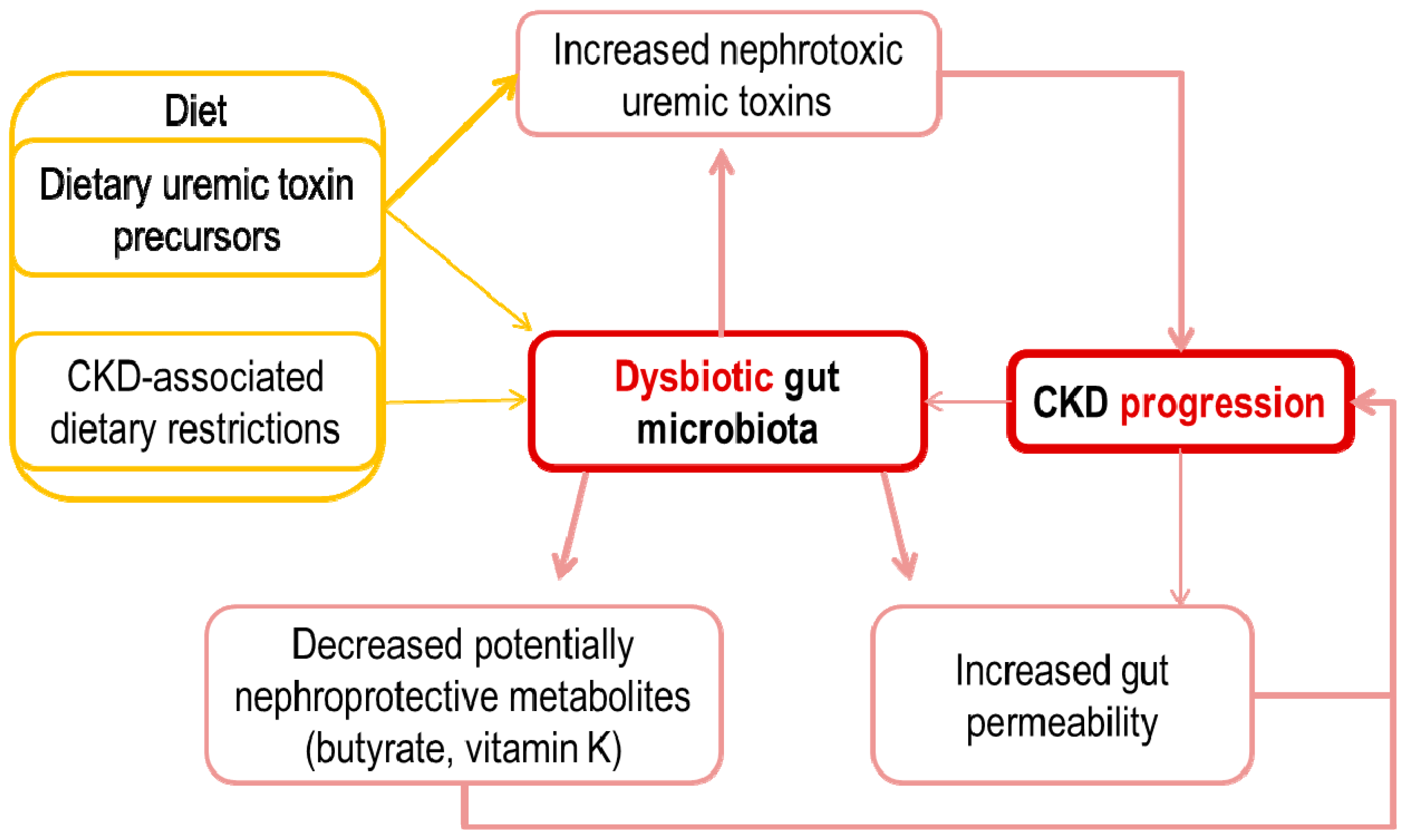
3. Potential Pathogenic Links between an Altered Intestinal Microbiota and CKD Progression

A number of pathogenic links between the intestinal microbiota, especially if this is altered and CKD progression have been described. These include an increased gut permeability, an overload of microbiota-generated potential nephrotoxins, such as p-cresyl sulphate (pCS), p-cresyl-glucuronide (pCG), indoxyl sulphate (IS), indole-3 acetic acid (IAA) and trimethylamine N-oxide (TMAO) [13] and decreased levels of certain microbiota-derived molecules that may have nephroprotective properties, such as butyrate and vitamin K (Table 1, Figure 1).

4. Increased Gut Permeability and Inflammation

CKD is associated to local and systemic inflammation and evidence of systemic inflammation has been associated with worse survival and kidney outcomes [38,39]. Drivers of inflammation include a decreased renal clearance of cytokines and inflammatory mediators as well as increased production of inflammatory mediators [40]. The gastrointestinal tract is a major source of chronic inflammation in CKD, both because of uremic toxins released by the microbiota, as discussed below and of an increased gut permeability that allows access of bacterial products to the circulation [41,42,43,44].
Circulating gut-derived bacterial DNA has been detected in patients with CKD and correlated with increased plasma C-reactive protein (CRP) and interleukin-6 (IL-6) [45,46,47]. Moreover, circulating levels of endotoxin, which is derived from the cell wall of Gram-negative bacteria, correlates with the severity of systemic inflammation in the absence of infection [48,49,50]. Although most studies have been performed in dialysis patients and circulating bacterial DNA is most frequent as CKD progresses, it was also found in non-dialysis CKD [50].
In cultured enterocytes, uremic plasma decreased transepithelial electrical resistance, denoting increased epithelial permeability [51]. This was accompanied by significant reductions in trans-cellular (i.e., claudin-1 and occluding) and intra-cellular (i.e., zonula occludens, ZO1) epithelial tight junction protein. Urea was identified as one of the potential drivers of these changes [52]. Additionally, oedema, hypervolemia or ischemia may disrupt the epithelial barrier. Finally, disruption of the physiological gut microbiota-enterocyte cross-talk as a result of an altered microbiota or defective innate immunity response because of uraemia, may also damage the gut epithelial barrier [53].

5. Metabolite Overload: Microbiota-Generated Nephrotoxins

Uremic toxins originating in the microbiota that have been linked in epidemiological studies and preclinical interventional studies to CKD progression include pCS and pCG, IS and IAA and TMAO.

5.1. pCS and pCG

p-cresol is synthesized by intestinal anaerobic bacteria from the dietary amino acids tyrosine and phenylalanine. Subsequent endogenous sulphate conjugation in the liver yields pCS and glucuronide conjugation in the enterocyte yields pCG, both normally excreted in urine through tubular secretion [54]. In CKD G1–G5 patients, total pCS and pCG accumulated to median levels of 50 μM (IQR, 21–104) and 0.22 μM (IQR, 0.08–0.60), respectively, reaching in individual G5 patients values up to 500 and 8 µM, respectively [5,14]. That is, pCS reaches levels around 200-fold higher than pCG [15]. With advancing CKD, the pCS to pCG ratio decreases and both higher total p-cresol (pCS+pCG) and a lower pCS to pCG ratio are independently associated to mortality [15]. However, most pCS and pCG is protein-bound and median free levels are 30- and 2-fold lower than total levels, respectively.
pCS has been associated with and may contribute to overall mortality, cardiovascular disease and progression of CKD [16,17]. While vascular effects may eventually negatively impact CKD progression, pCS also has direct effects on kidney cells, as recently reviewed [18]. Specifically, in kidney proximal tubular cells, pCS is cytotoxic and activates oxidative stress and proinflammatory and profibrotic responses, while decreasing nephroprotective factors such as Klotho [18,19,55]. Long-term (7 days) exposure to pCS induced apoptosis in cultured human proximal tubule cells in a concentration-dependent manner [19]. In addition, short-term (3 h) exposure to pCS promoted the expression of the TWEAK receptor Fn14, cooperated with TWEAK in promoting cell death and increased inflammatory gene expression. As expected, albumin alone was cytotoxic to proximal tubular cells [20]. Additionally, albumin also increased the inflammatory response to pCS concentrations found in the circulation of non-dialysis CKD patients. In contrast, no biological actions of pCG were observed on human proximal tubule cell inflammatory responses, either alone or in combination with pCS [19]. Probenecid decreased pCS-induced toxicity in proximal tubular cells, suggesting the requirement for active pCS transport into cells for the cytotoxic effects [55].
Both pCS and pCG decreased the function or the activity of proximal cell membrane transporters. pCS decreased the activity of multidrug resistance protein 4 (MRP4/ABCC4) and breast cancer resistance protein (BCRP/ABCG2), while pCG only decreased MRP4 [56]. Additionally, both pCS and pCG promoted evidence of epithelial-to-mesenchymal transition (EMT) and pCS promoted tubular damage and kidney fibrosis in vivo [55,56,57]. In fact, pCS activated the renal-angiotensin system in vivo and kidney fibrosis was decreased by losartan [57].

5.2. IS and IAA

IS is a small protein-bound molecule [58]. Bacterial tryptophanases in the colon converted dietary tryptophan to indole which is absorbed into the systemic circulation. Indole is further metabolized by the liver to form IS, which is then cleared by the kidneys through tubular secretion mediated by organic anion transporter OAT1 and OAT3. Total IS accumulates in CKD to a highest individual reported total value of 1100 µM and highest reported mean value of 211 µM, versus 2 µM in controls [18]. By contrast, free IS is undetectable in controls and maximum values in CKD are around 20 µM [18].
IS may contribute to vascular and renal disease progression. Although vascular effects may eventually negatively impact CKD progression, IS also has direct effects on kidney cells, that have been more intensively explored than the nephrotoxic effects of other toxins, as recently reviewed [18]. In cultured tubular cells, four key effects were observed: increased oxidative stress, increased inflammatory (e.g., chemokine secretion) and profibrotic (e.g., EMT, intrarenal renal-angiotensin system activation, increased TGF-β1 levels and activity) responses and decreased expression of nephroprotective factors (e.g., Klotho) [18,21,22]. Additionally, IS administration to subtotally nephrectomised rats accelerated fibrosis and CKD progression, as did its precursor indole [23,24,25,26]. Immunohistochemistry located IS to proximal and distal tubules, which express OAT1 (proximal tubules) and/or OAT3 (proximal and distal tubules), suggesting that IS accumulation in uraemia may facilitate tubular cell overload of the toxin [23]. In this regard, IS decreased the viability of cultured proximal tubular cells in an OAT1- and OAT3-dependent manner and this was inhibited by the OAT inhibitor probenecid [23]. Probenecid is routinely used in the clinic to reduce cidofovir nephrotoxicity by preventing entry into proximal tubular cells, an effect reproducible in cultured cells [59]. Interestingly, cilastatin also decrease tubular cell uptake of IS, although the effect on toxicity was not explored. Cilastatin protects cultured tubular cells and the kidneys in vivo from diverse nephrotoxic agents [60]. Cilastatin is also in clinical use to extend the half-life on certain β-lactam antibiotics (such as imipenem) by decreasing tubular uptake. IS also injures podocytes, perturbing the actin cytoskeleton, decreasing the expression of podocyte-specific genes and modulating the inflammatory response in culture and in vivo and causing albuminuria [61].
IAA is a protein-bound small molecule also derived from tryptophan metabolism and excreted in urine by tubular secretion through the OAT1 transporter [62]. Serum total IAA levels increase in patients with CKD as eGFR decreases; the mean uremic concentration is about 5 µM and the highest concentration is 50 µM [5]. In subtotally nephrectomised rats, the oral intake of 125 mg/kg/day IAA resulted in a higher excretion of N-acetyl-β-glucosiminadase (NAG), a marker of tubular injury, a lower GFR and higher glomerular sclerosis index, indicating that an excess of IAA on top of previously injured kidneys may accelerate CKD progression [28]. However, there is less information on actions of IAA on cultured renal cells. In porcine proximal tubular LLC-PK1, IAA at a concentration of 250 µM reduced viability mainly through induction of apoptosis [27]. However, this concentration is not found in vivo.
Both IS and IAA activate the transcription factor aryl hydrocarbon receptor (AhR), which regulates the cell response to environmental xenobiotics and promotes vascular inflammation and oxidative stress [63]. At concentrations found in the circulation of CKD patients, both molecules increased tissue factor expression in endothelial cells, vascular smooth muscle cells and peripheral blood mononuclear cells by increasing its stability through AhR signalling [64,65,66]. Tissue factor is an initiator of coagulation and AhR antagonists behave as antithrombotic agents in the CKD context [66]. Additional effects have been described in endothelial cells, including oxidative stress and dysfunction. In this regard, in cultured human endothelial cells, IAA activated an inflammatory non-genomic AhR/p38MAPK/NF-κB pathway that induced endothelial inflammation and oxidative stress [63]. Whether activation of AhR also account for adverse effects on tubular cells and podocytes is still unknown. At least some actions of IS on proximal tubular cells, such as downregulation of the Mas receptor, are indeed mediated by AhR [67]. Additional nuclear translocation of AhR was observed in podocytes, although whether this mediated IS-induced responses was not explored [61].

5.3. TMAO

TMAO is generated from trimethylamine (TMA)-containing nutrients present in seafood, or from TMA released by the gut microbiota. Dietary TMA precursors, such as choline, phosphatidylcholine (lecithin), or l-carnitine are metabolized by gut microbiota to generate TMA. Then, TMA is absorbed and metabolized in the liver by Flavin-containing monooxygenases (FMO1 and FMO3) to yield TMAO [68,69,70]. TMAO is eventually cleared mainly by the kidneys, excreted in urine in part through tubular cell secretion involving uptake by organic cation transporters (OCTs) OCT1 and OCT2 as well as transporters of the ATP-binding cassette (ABC) family, including ABCG2 (BCRP) and ABCB1 (MDR1) [71,72]. Multidrug and toxin extrusion protein 1 (MATE1) contributes to translocation of TMAO across the luminal membrane of proximal tubular cells [73]. Indeed, genetic ABCG2 variants modulate TMAO exposure in humans [72]. TMAO may also be metabolically retroconverted to TMA by certain gut bacteria, such as Enterobacteriaceae and this influences the microbiota composition [74]. Although not specifically studied, this process may be especially relevant in CKD, given the high availability of TMAO. Additionally, TMAO may also be excreted in sweat and exhaled air [13]. Haemodialysis efficiently removes TMAO [75]. The physiological role of TMAO in humans is unclear [76]. In some marine animals, it behaves as an osmolyte that maintains cell volume and counteracts the destabilizing effect of high urea concentration and hydrostatic pressure on proteins structures that inhibit ligand binding [77].
Circulating TMAO levels are elevated in subjects with impaired renal function. Plasma TMAO was higher among CKD G3–G5 patients (eGFR < 60 mL/min/1.73 m2) than in non-CKD patients (7.9; IQR 5.2–12.4 versus 3.4; IQR 2.3–5.3 µM, p < 0.001) [29]. In another study, median TMAO levels in CKD G4 was higher than in G3b patients (25.15; IQR 16.64–38.56 versus 14.32; IQR 9.14–22.69 µM) [30]. TMAO decrease following renal transplantation [78]. TMAO levels correlated with evidence of systemic inflammation and was an independent predictor of mortality and CVD in CKD patients [29,30,78,79]. TMAO may also contribute to the pathogenesis and progression of CKD [29,80]. In a metabolomic study on plasma from 1434 participants (Framingham Heart Study) with normal renal function at baseline, elevated choline and TMAO levels predicted the development of CKD [80].
The biological plausibility of cause-and-effect relationship between high TMAO levels and adverse kidney outcomes in human CKD has been addressed in experimental animals [29]. In mice fed a diet supplemented with choline (1.0%) or TMAO (0.12%) for 6 weeks, circulating TMAO levels increased as compared to a normal diet (0.08% choline) to levels (40, 100 and <5 µM, respectively) found in human CKD [29]. Mice fed the high choline or TMAO diets developed progressive renal tubulointerstitial fibrosis and dysfunction and TMAO levels correlated with kidney tubulointerstitial fibrosis and collagen deposition, increased phosphorylation of SMAD3, which signals the profibrotic activity of TGF-β1 and increased kidney injury molecule-1 (KIM-1) [29]. In an independent study, feeding mice a high fat diet increased TMAO levels as well as kidney tubulointerstitial fibrosis and collagen deposition, SMAD3 phosphorylation, KIM-1, plasma cystatin C and evidence of inflammation, such as NOX-4 and TNF-α. The inhibitor of TMA formation 3,3-Dimethyl-1-butanol (DMB) prevented these effects [81]. Thus, animal data are suggestive of a deleterious effect of TMAO on CKD. However, in the absence of cell culture studies, it is unclear whether this is a direct toxic effect of TMAO on renal cells or whether it is mediated by systemic effects of TMAO or the adaptive response to the diets used in the mice studies.

6. The Missing Ones: Microbiota-Generated Nephroprotective Factors

6.1. Vitamin K

Vitamin K refers to a group of fat-soluble vitamins required for the carboxylation of glutamate residues to form gamma-carboxyglutamate (Gla) residues, which are required for the function of, among others, proteins involved in coagulation and in the control of tissue calcification. Concomitant with carboxylation, reduced vitamin K (KH2) is oxidized to vitamin K epoxide (KO). KO must be recycled back to KH2 by the enzymes vitamin K epoxide reductase and vitamin K reductase in a pathway known as the vitamin K cycle [82]. An anti-inflammatory action has been proposed that is independent of its role as an enzyme co-factor [83]. There are two natural vitamers: vitamin K1 (phylloquinone), made by plants and found in green leafy vegetables and K2 (menaquinone) synthesized by bacteria in the gut and in fermented foods. Menaquinones have side chains of varying length and are generally denoted as MK-n, where n stands for the number of isoprene residues in the side chain, which differ in bioavailability, half-life and biodistribution [84]. The adequate intake level for vitamin K is estimated at 70 to 120 μg/day. However, it is thought that vegetable vitamin K1 is poorly bioavailable and contributes <10% of absorbed vitamin K, thus increasing the dependence on gut microbiota generated menaquinone. There is recent interest in subclinical vitamin K deficits, characterized by reduced carboxylation of Gla proteins that are mild enough not to prolong the prothrombin time.
In CKD, several defects related to vitamin K have been reported. In CKD G3–G5 patients not on dialysis with a mean age of 61 years, mean vitamin K1 levels were 2.1 ± 2.4 nM, which is in the range found in non-CKD patients (e.g., 1.54 ± 2.0 nM was reported for general population individuals with a mean age of 59 years) [85,86]. Despite this apparently normal vitamin K1 level, there was evidence of subclinical vitamin K deficiency, as defined by a percentage of uncarboxylated osteocalcin (ucOC) >20%, which is a more sensitive marker of vitamin K status. In CKD not on dialysis, ucOC was 27 ± 22% versus 16 ± 16% in healthy people [85,86]. In addition to potential gut microbiota derangements, that have been poorly studied for vitamin K, CKD patients appear to be prone to vitamin K deficiency for at least two reasons: a reduced dietary intake (diets low in potassium contain fewer leafy green vegetables; rich in K1 and diets low in phosphate contain fewer dairy products; rich in K2) and a reduced expression and activity of vitamin K 2,3-epoxide reductase (VKOR), the target of coumarins and other vitamin K-related enzymes [87].
Carboxylated matrix Gla protein (MGP) is a potent inhibitor of arterial calcification and needs vitamin K for synthesis [88]. Assessing levels of inactive dephospho-uncarboxylatedMGP (dp-ucMGP) provides insights into potential vitamin K deficiency [89]. In patients with diabetes, renal dysfunction, or macrovascular disease, dp-ucMGP behaves as a circulating biomarker associated with cardiovascular risk, more severe illness, or higher mortality [88]. Median plasma dp-ucMGP levels in CKD stages 3, 4 and 5 are 586 (452–888), 870 (594–1591) and 1050 (518–1298) pM, respectively [90]. In rat kidney failure, activity of the rate-limiting enzyme in the vitamin K cycle—gamma-glutamyl carboxylase (GGCX)—was reduced, resulting in increased serum ucMGP levels [91]. Vitamin K2 supplementation, better than K1, resulted in significantly lower calcium content in kidneys and aorta, decreasing medial elastocalcinosis [91,92]. In this regard, oral anticoagulants inhibiting the C1 subunit of the vitamin K epoxidereductase enzyme complex are major contributors to the severe clinical condition calciphylaxis [93]. Uraemia was also associated with a decrease in kidney vitamin K1 to menaquinone K-4 (MK-4) bioconversion enzyme, UBIAD1. Supplementation of vitamin K1 or the MK-4 form of vitamin K2 at pharmacological doses restored the abnormal vitamin K cycle activity and slowed the progression of vascular calcification [87]. In this regard, there is increasing concern about the potential negative effects of vitamin K targeting oral anticoagulants, such as warfarin and coumarin, on vascular calcification, given their frequent use in CKD patients with valvular calcification that required valve replacement or is complicated by atrial fibrillation and the increased sensitivity of these patients to vascular calcification [94].
More recently, a negative impact of warfarin on renal function has been described. Initial reports described haematuria-induced acute kidney injury (AKI) related to over coagulation [95]. Indeed, oral anticoagulation is a frequent driver of IgA nephropathy-associated AKI driven by macroscopic haematuria episodes [96]. Then, the association of transient over coagulation with increases in creatinine levels was observed, mainly in but not only on CKD patients [97]. More recently, more rapid CKD progression has been described in individuals anticoagulated with warfarin than in users of direct anticoagulants [98]. Thus, while macroscopic haematuria–associated AKI has been observed with any oral anticoagulant [99,100], drugs targeting vitamin K appear to increase the risk of CKD progression. Whether this is related to a nephroprotective action of vitamin K, the more frequent over coagulation episodes with warfarin or nephroprotective effects of direct anticoagulants related to factor Xa inhibition remains to be clarified. Surprisingly, Framingham Offspring study participants in the highest vitamin K1 quartile (≥1.78 nM) had an increased risk of CKD, whereas no association was observed with continuous vitamin K1 levels [92]. These data are weak as different results were obtained with different analyses and require validation. Additionally, assessing vitamin K2 may have been more informative, given that it comprises a higher portion of absorbed vitamin K.
The potential role of vitamin K disturbances in CKD progression and the consequences for dietary counselling, modulation of the microbiota, use of vitamin K supplements and choice of oral anticoagulants merits further studies.

6.2. Butyrate

Butyrate is a short-chain fatty acid produced by anaerobic human intestinal microbiota and is the preferred energy source for colon epithelial cells [101]. Butyrate is formed from two molecules of acetyl CoA which are converted, via the intermediates β-hydroxybutyryl CoA and crotonyl CoA, to butyryl CoA and finally to butyrate. The main bacteria butyrate-releasing are Faecalibacterium prausnitzii, Roseburia intestinalis and some Clostridium-related bacteria [33].
Butyrate concentrations in venous blood are very low. There are no estimates in CKD but in the general population it has been estimated at 2–4 µM or 35–65 µM by different authors [31,32]. A circadian rhythm has been described, with lower values after breakfast and transient increases after lunch and dinner, which have been ascribed to dietary fibre fermentation. In the intestinal lumen, butyrate concentrations are much higher, around 5–20 mM but this is not a good guide to production rates because of the very high colonic mucosa absorption [33]. Lower numbers of butyrate-producing bacteria were observed in human ESKD [102,103,104] but the concentration or uptake of butyrate in this population has not been measured yet.
Butyrate is a general inhibitor of histone deacetylases (HDAC) and protects against high-fat diet-induced metabolic changes [105], has anti-inflammatory properties [106], extends lifespan in experimental progeria mice [107] and protects from features of aging such as sarcopenia, which are prominent in CKD [108]. HDACs are key epigenetic regulators that have been linked to kidney injury and downregulation of Klotho expression [109,110]. Interestingly, the butyrate intermediary crotonyl CoA may be a source of crotonate, another epigenetic regulator which protects from kidney injury [6,111]. Additionally, like other short chain fatty acids, butyrate activates the GPR43 receptor [112].
In vivo, butyrate protects from kidney injury in animal models. The daily administration of 50 to 200 mg/kg sodium butyrate intraperitoneal for 8 days attenuated gentamicin-induced nephrotoxicity in rats in a dose-dependent manner [36]. This was ascribed to the inhibitory activity of butyrate on HDAC, decreased oxidative stress and increased expression of the chaperon prohibitin [36]. However, none of the experiments performed demonstrated that either the HDAC inhibitory activity or prohibitin expression were required for the nephroprotective effect. In experimental contrast-induced nephropathy in rats, administration of 500 mg/kg sodium butyrate intravenously decreased the severity of renal failure, assessed by serum creatinine and of systemic inflammation, assessed by serum IL-6 levels and NF-κB activation in the kidney [37].
The protective effect of butyrate has been confirmed in culture mouse glomerular mesangial cells, where it dampened the release of monocyte chemoattractant protein-1 (MCP-1), IL-1β and intercellular adhesion molecule-1 (ICAM-1) and decreased oxidative stress in response to lipopolysaccharide and high glucose [34]. However, the effective concentration, at 5 mM, is two to three orders of magnitude higher than butyrate concentrations in the circulation and unlikely to be reached by modulating butyrate production in the gut. In this regard, butyrate concentrations that are relevant for enterocytes may not be relevant for kidney cells. In human cultured renal cortical epithelial cells, 500 µM butyrate reduced MCP-1 mRNA in response to TNF-α by 18% and by 29% at 1000 µM [35]. Again, these concentrations are one or two orders of magnitude higher than in the circulation.

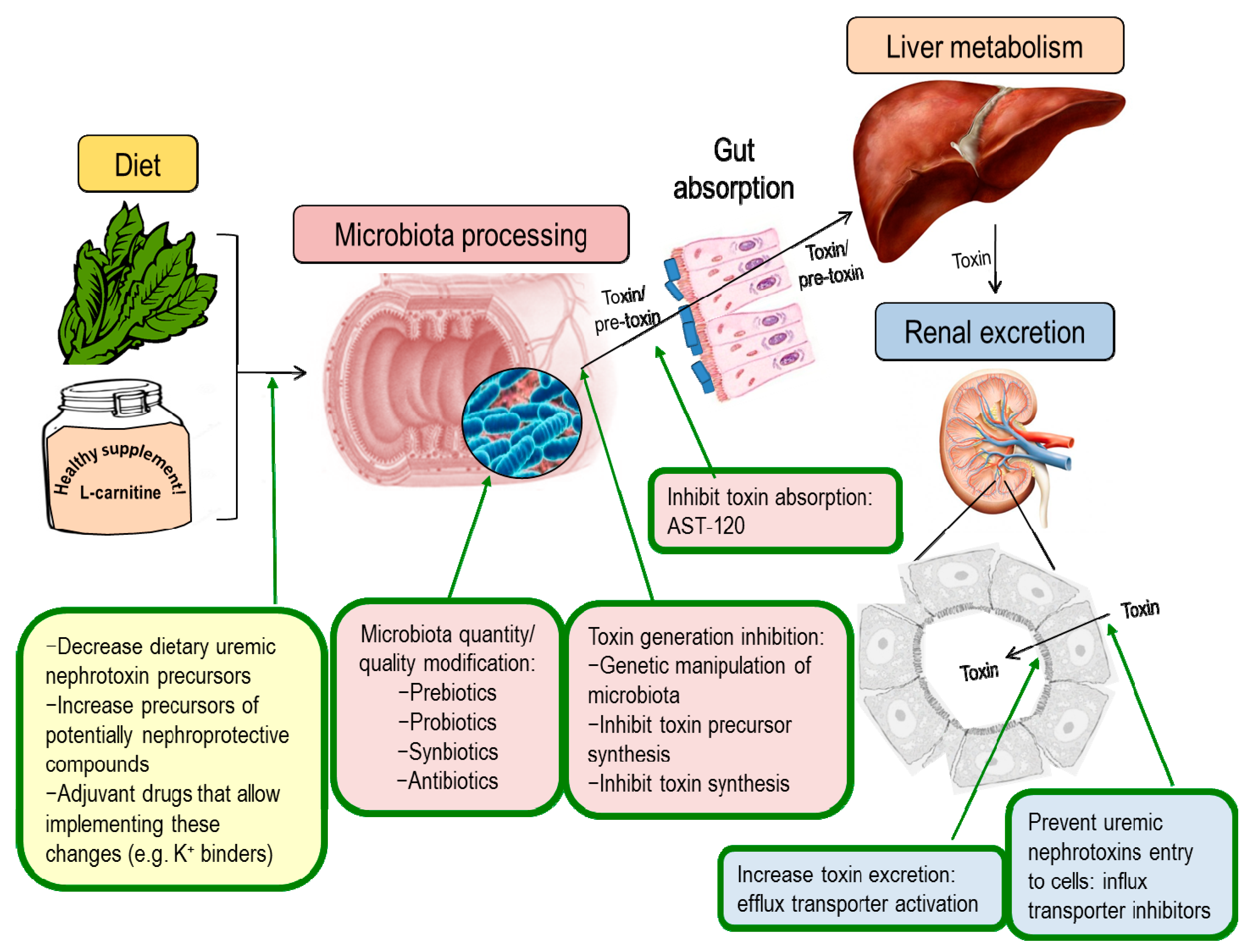
7. Nephroprotection Interventional Studies Targeting the Intestinal Microbiota or Microbiota-Derived Toxins

Several strategies have been tested in animal models to target microbiota-derived toxins and their nephrotoxicity, including reduction of uremic toxin synthesis, increasing the excretion by modulating the expression or activity of renal transporters, preventing gut absorption by using oral adsorbents and modulation of the microbiota composition (Figure 2). Of these, only the latter two have reached the clinical stage.

7.1. Modulation of Uremic Toxins Synthesis

A dietary restriction of uremic toxin precursors is a theoretical approach to limit uremic toxin synthesis. Despite the fact that superimposing additional restrictions on patients potentially suffering from anorexia and at high malnutrition risk seems impractical, some precursor of uremic toxins with nephrotoxin potential are sold as over-the-counter food supplements or prescription drugs and both patients and physicians should be aware of the potential dangers [113] (reviewed in [13], Table 2).
Genetic manipulation of bacteria may be used to reduce their ability to synthesize uremic toxin precursors. The overall safety of this strategy should be carefully explored, including the potential consequences for bacteria pathogenicity. In this regard, successful application of such a therapy would require that bacteria are able to transfer the genetic modification horizontally or that it creates a survival advantage over other non-genetically modified microbiota competitors. In this sense, BT1492 was identified as the more abundant tryptophanase in human gut microbiota and one mutation in this gene reduces indol synthesis and IS levels in mice [114]. Moreover, IS levels in mice were reduced by antibiotics that favour the growth of BT1492 negative bacteria [114].
Phytochemical polyphenols, such as quercetin and resveratrol, reduce synthesis of IS through inhibition of sulfotransferase (SULT), an enzyme implicates in IS synthesis in liver [115]. These drugs have proved nephroprotective in a variety of animal models but present pleiotropic actions and the contribution of IS synthesis inhibition to any nephroprotective effect remains unproven, even if reduced IS levels are observed, as is the case for cisplatin-induced AKI, since a better renal function would already be expected to decrease IS levels [115]. In addition, both drugs also protected from ischemia/reperfusion renal injury but in this case resveratrol did not reduce renal IS, questioning whether IS reduction contributes to nephroprotection [116].
Among a library of different drugs, meclofenamate, a non-steroidal anti-inflammatory drug, (NSAID) decreased IS synthesis. Meclofenamate reduced IS levels in serum, kidney and liver and protected from ischemia/reperfusion renal injury [117]. Given the adverse renal and cardiovascular safety profile of NSAIDs, their use to decrease uremic toxin synthesis seems unwarranted. However, the molecule may be modified to increase its efficiency in decreasing IS production while decreasing its NSAID profile. Even in this case, the safety of long-term suppression of IS production by the liver, itself a pathway for toxin metabolism and clearance, should be demonstrated.

7.2. Modulation of Renal Transporters

Proximal tubular cell transporters facilitate the entry of protein-bound uremic toxins into proximal tubular cells and their subsequent tubular secretion. Specifically, organic anion transporters of the OAT (SLC21) and OATP (SLC22/SLCO) families from the basolateral membrane and ATP-binding cassette (ABC) transporters such as MDR1, MRP2 and MRP4 in the luminal membrane have been implicated in the excretion of clinically relevant uremic toxins and may decrease when CKD progresses [118]. While in cultured cells, inhibition of these transporters with drugs such as probenecid decreased the cytotoxicity of uremic and non-uremic toxins [55,59], an increased activity of these transporters in vivo may increase kidney toxin clearance. The final result in vivo over kidney injury is not easy to predict. On one hand decreasing circulating levels of toxins through increased tubular secretion may decrease any adverse effects of these toxins over non-kidney cells. However, increasing transtubular traffic of the toxins may be detrimental for tubular cells. As an in vivo proof-of-concept, in transgenic rats, SLCO4C1 (SLC21A20) overexpression decreased kidney inflammation and hypertension following subtotal nephrectomy [118]. However, kidney function was not improved and tissue injury was not reported and was likely unchanged. Nephroprotection was associated to decreased plasma levels of uremic toxins such as guanidino succinate, asymmetric dimethylarginine and trans-aconitate but not of other better characterized nephrotoxic uremic toxins, such as IS [119]. Interestingly, SLCO4C1 expression is regulated by AhR: IS downregulates and statins increase SLCO4C1 expression through interaction with AhR [119,120]. Thus, while SLCO4C1 overexpression was not associated with clinically significant preservation of renal function in rat CKD, it provides insights into how to manipulate the activity of proximal tubular transporters to accelerate the clearance of uremic toxins that may accelerate CKD progression. In this regard, meclofenamate increased the expression of the organic anion transporters OAT1 and OAT3, favouring IS excretion in rats [117].

7.3. Adsorbents

AST-120 is an oral carbonic adsorbent which is used to decrease the absorption of uremic toxins, such as indols and retard CKD in Japan [121]. In experimental CKD, it reduces tissue accumulation of IS and pCS in diverse tissues, including the kidney and protected the kidneys in several animal models of AKI and CKD [122], However, two large multinational trials (EPPIC-1 and EPPIC-2), failed to show benefit on CKD progression [123]. However, a post-hoc, hypothesis-generating analysis observed that in the subgroup of high risk patients (urinary creatinine ratio ≥1.0 and haematuria) who were additionally treated with renin-angiotensin system blockade, AST-120 may have provided additional benefit in retarding CKD progression [124]. The large daily number of pills may negatively impact compliance and may be one of the underlying cause for the failure of clinical trials outside Japan.

7.4. Modulation of Microbiota Composition

Another approach to modulate uremic toxin production is the therapeutic manipulation of the composition of the gut microbiota. Several ongoing clinical trials are exploring the impact of changing the diet or using prebiotics (non-digestible food ingredients), probiotics (living microorganisms) or synbiotics (a mixture of both compounds) on microbiota composition [13]. In experimental animals, antibiotics decreased the production of uremic toxins [125]. It could be argued that they are frequently used in CKD patients. The potential adverse effects of antibiotic use for this purpose, both over the individual patient and on public health may outweigh the potential benefits. However, chronic non-absorbable antibiotics are clinically indicated for other conditions associated with microbiota-generated toxins, such as hepatic encephalopathy and clinical trials in CKD that provide information into risk:benefit balance may be worthy.
Hyperkalaemia is favoured by both progressive CKD and nephroprotective drugs such as renin-angiotensin system blockers. Besides limiting the use of these medications, it may trigger the prescription of low potassium diets. This usually entails limiting foods that promote a healthy microbiota such as fruits and vegetables. While the impact of low potassium diets as prescribed for CKD patients on the microbiota, uremic toxins levels and CKD progression has not been studied in detail, the recent availability of a new generation of drugs for hyperkalaemia, such as patiromer and zirconium cyclosilicate [11,12], offers the opportunity of designing clinical trials that address these issues.
The use of a low protein diet supplemented with essential amino acid and ketoacid supplements may offer some advantages in CKD patients but the impact on the gut microbiota and uremic toxin production is poorly characterized [126]. An ongoing trial (NCT02302287) is testing the impact of Mediterranean diet, ketoacid-supplemented low protein diet or a combination on the composition of the gut microbiota in CKD patients. Resistant starch, a class fermentable fibre, may be used as a prebiotic and has beneficial effects on obesity-related diabetes [127]. An ongoing clinical trial (NCT02706808) is exploring the impact of resistant starch on microbiota composition and uremic toxin levels in CKD patients.
Advanced glycation end-products (AGEs) may also promote kidney disease [128]. This is specially an issue in diabetic patients, in whom high glucose levels lead to increased levels of glucose-degradation products and in peritoneal dialysis patients, due to continuous glucose and glucose-degradation product absorption from the peritoneum [129]. Additionally, a number of processed foods are a source of dietary AGEs [130]. In this regard, a recent trial of dietary restriction of AGEs in peritoneal dialysis patients demonstrated a decrease in serum AGEs as well as a change in bacterial microbiota [129]. At baseline, peritoneal dialysis patients exhibited a lower relative abundance of Bacteroides and Alistipes genus and a higher abundance of Prevotella genus when compared to the published data of healthy population. Dietary AGE restriction decreased Prevotella copri and Bifidobacterium animalis relative abundance and increased Alistipes indistinctus, Clostridium citroniae, Clostridium hathewayi and Ruminococcus gauvreauii relative abundance [129].
Some studies have addressed the impact of oligosaccharide prebiotics on uremic toxin levels, with mixed success but none has addressed the impact on CKD progression. In haemodialysis patients, oligofructose inulin decreased p-cresol (pCS+pCG)) but not IS [13,131], while a synbiotic (Lactobacillus casei, Bifidobacterium breve and galacto-oligosaccharide) decreased pCS [13,132]. In CKD patients not on dialysis, arabinoxylan oligosaccharides did not modify the urinary excretion of pCS, pCG, IS or TMAO [131]. It is likely that the impact of any given prebiotic or synbiotic will depend on the baseline characteristics of the individual, including baseline dietary intake of oligosaccharides and uremic toxin precursors and microbiota composition. Additional clinical trials are addressing the impact of multispecies probiotic mixes on gut permeability and microbiota composition in CKD patients [133].

8. The Way Forward

Understanding the impact of an altered intestinal microbiota on CKD progression, be it through promotion of inflammation related to an increased gut permeability of through the generation of diet-derived nephrotoxic uremic toxins or their precursors or through decreased availability of nephroprotective molecules, has the potential to greatly impact the management of CKD and pave the way for novel therapeutic approaches to slow progression of CKD [3,8,41] (Figure 2). We should also remember that even in dialysis patients, slowing the progressive loss of residual renal function may have a substantial clinical impact, because residual renal function is inversely related to mortality and a benefit has been observed not only in peritoneal dialysis but also in haemodialysis [134,135]. The range of potential intervention spans from diet modification to population-level education efforts regarding the potential impact of food supplements to reducing uremic toxin generation rate and intestinal absorption, modifying the microbiome or design of interventions to limit toxin actions, such as targeting the AhR [9,136,137]. A detailed discussion of modulation of uremic toxins is provided by Koppe et al. in this issue [138]. In this regard, some drugs successfully tested preclinically in culture or in vivo, are already in clinical use, although sometimes for a different indication. For example, in cultured proximal tubular cells IS toxicity required toxin uptake by tubular transporters sensitive to probenecid and cilastatin and these two drugs are in clinical use and indeed, one of the clinical uses of probenecid is to prevent nephrotoxicity [59]. Additionally, a comprehensive understanding of the different factors involved, from diet to individual microbiota composition to a permissive genetic background to either generate higher toxin levels or facilitating toxin access to target renal cells or sensitizing to their toxicity, may facilitate a personalized approach, taking into account the multiple variables that impact on uremic toxins levels and, likely, the individual sensitivity to them. This may also help put into perspective the often-higher concentrations used in preclinical studied as compared to circulating levels in CKD patients. A systems biology approach, including microbiome and metabolomics analysis exploring individual characteristics regarding the microbiota and the uremic toxin profile should be explored in targeted populations in this regard. This information may yield a relatively low number of key data that may help individualize and monitor novel therapeutic approaches in a wider population.

Author Contributions

E.C.-R., R.F.-P., R.E., M.V.P.-G., C.G.-I., B.F.-F., M.K., A.T., A.L., M.R.-O., E.G.-P., A.B.S., A.O., M.D.S.-N. wrote the draft, M.D.S.-N. designed the structure and integrated the different parts. ECR made the figures. All authors read and approved the final version.

Funding

Research by the authors was funded by FIS ISCIII FEDER funds PI15/00298, PI16/02057, PI16/01298, PI16/01900, ISCIII-RETIC REDinREN RD16/0009, EUTOX, Comunidad de Madrid en Biomedicina B2017/BMD-3686 CIFRA2-CM, Fundación Renal Iñigo Alvarez de Toledo (FRIAT). Programa Intensificación Actividad Investigadora (ISCIII) to AO. Miguel Servet CP14/00133 to MDSN and MS12/03262 to ABS and Rio Hortega to MVPG.

Conflicts of Interest

The authors declare no conflict of interest.

References

  1. Sanchez-Niño, M.D.; Sanz, A.B.; Ramos, A.M.; Ruiz-Ortega, M.; Ortiz, A. Translational science in chronic kidney disease. Clin. Sci. 2017, 131, 1617–1629. [Google Scholar] [CrossRef] [PubMed]
  2. Ortiz, A.; Covic, A.; Fliser, D.; Fouque, D.; Goldsmith, D.; Kanbay, M.; Mallamaci, F.; Massy, Z.A.; Rossignol, P.; Vanholder, R.; et al. Epidemiology, contributors to and clinical trials of mortality risk in chronic kidney failure. Lancet 2014, 383, 1831–1843. [Google Scholar] [CrossRef]
  3. Vanholder, R.; Fouque, D.; Glorieux, G.; Heine, G.H.; Kanbay, M.; Mallamaci, F.; Massy, Z.A.; Ortiz, A.; Rossignol, P.; Wiecek, A.; et al. Clinical management of the uraemic syndrome in chronic kidney disease. Lancet Diabetes Endocrinol. 2016, 4, 360–373. [Google Scholar] [CrossRef]
  4. Fernandez-Fernandez, B.; Izquierdo, M.C.; Valiño-Rivas, L.; Nastou, D.; Sanz, A.B.; Ortiz, A.; Sanchez-Niño, M.D. Albumin downregulates Klotho in tubular cells. Nephrol. Dial. Transplant. 2018. [Google Scholar] [CrossRef] [PubMed]
  5. Duranton, F.; Cohen, G.; De Smet, R.; Rodriguez, M.; Jankowski, J.; Vanholder, R.; Argiles, A. European uremic toxin work group normal and pathologic concentrations of uremic toxins. J. Am. Soc. Nephrol. 2012, 23, 1258–1270. [Google Scholar] [CrossRef] [PubMed]
  6. Ruiz-Andres, O.; Sanchez-Niño, M.D.; Moreno, J.A.; Ruiz-Ortega, M.; Ramos, A.M.; Sanz, A.B.; Ortiz, A. Downregulation of kidney protective factors by inflammation: Role of transcription factors and epigenetic mechanisms. Am. J. Physiol. Renal Physiol. 2016, 311, F1329–F1340. [Google Scholar] [CrossRef] [PubMed]
  7. Zoccali, C.; Vanholder, R.; Massy, Z.A.; Ortiz, A.; Sarafidis, P.; Dekker, F.W.; Fliser, D.; Fouque, D.; Heine, G.H.; Jager, K.J.; et al. The systemic nature of CKD. Nat. Rev. Nephrol. 2017, 13, 344–358. [Google Scholar] [CrossRef] [PubMed]
  8. Vanholder, R.; Glorieux, G. The intestine and the kidneys: A bad marriage can be hazardous. Clin. Kidney J. 2015, 8, 168–179. [Google Scholar] [CrossRef] [PubMed]
  9. Ramezani, A.; Massy, Z.A.; Meijers, B.; Evenepoel, P.; Vanholder, R.; Raj, D.S. Role of the gut microbiome in uremia: A potential therapeutic target. Am. J. Kidney Dis. 2016, 67, 483–498. [Google Scholar] [CrossRef] [PubMed]
  10. Nallu, A.; Sharma, S.; Ramezani, A.; Muralidharan, J.; Raj, D. Gut microbiome in chronic kidney disease: Challenges and opportunities. Transl. Res. 2017, 179, 24–37. [Google Scholar] [CrossRef] [PubMed]
  11. Packham, D.K.; Rasmussen, H.S.; Lavin, P.T.; El-Shahawy, M.A.; Roger, S.D.; Block, G.; Qunibi, W.; Pergola, P.; Singh, B. Sodium zirconium cyclosilicate in hyperkalemia. N. Engl. J. Med. 2015, 372, 222–231. [Google Scholar] [CrossRef] [PubMed]
  12. Weir, M.R.; Bakris, G.L.; Bushinsky, D.A.; Mayo, M.R.; Garza, D.; Stasiv, Y.; Wittes, J.; Christ-Schmidt, H.; Berman, L.; Pitt, B.; et al. Patiromer in patients with kidney disease and hyperkalemia receiving RAAS inhibitors. N. Engl. J. Med. 2015, 372, 211–221. [Google Scholar] [CrossRef] [PubMed]
  13. Fernandez-Prado, R.; Esteras, R.; Perez-Gomez, M.V.; Gracia-Iguacel, C.; Gonzalez-Parra, E.; Sanz, A.B.; Ortiz, A.; Sanchez-Niño, M.D. Nutrients turned into toxins: Microbiota modulation of nutrient properties in chronic kidney disease. Nutrients 2017, 9, 489. [Google Scholar] [CrossRef] [PubMed]
  14. Meert, N.; Schepers, E.; Glorieux, G.; Van Landschoot, M.; Goeman, J.L.; Waterloos, M.-A.; Dhondt, A.; Van der Eycken, J.; Vanholder, R. Novel method for simultaneous determination of p-cresylsulphate and p-cresylglucuronide: Clinical data and pathophysiological implications. Nephrol. Dial. Transplant. 2012, 27, 2388–2396. [Google Scholar] [CrossRef] [PubMed]
  15. Poesen, R.; Evenepoel, P.; de Loor, H.; Kuypers, D.; Augustijns, P.; Meijers, B. Metabolism, protein binding and renal clearance of microbiota-derived p-cresol in patients with CKD. Clin. J. Am. Soc. Nephrol. 2016, 11, 1136–1144. [Google Scholar] [CrossRef] [PubMed]
  16. Wu, I.-W.; Hsu, K.-H.; Lee, C.-C.; Sun, C.-Y.; Hsu, H.-J.; Tsai, C.-J.; Tzen, C.-Y.; Wang, Y.-C.; Lin, C.-Y.; Wu, M.-S. p-Cresyl sulphate and indoxyl sulphate predict progression of chronic kidney disease. Nephrol. Dial. Transplant. 2011, 26, 938–947. [Google Scholar] [CrossRef] [PubMed]
  17. Lin, C.-J.; Wu, V.; Wu, P.-C.; Wu, C.-J. Meta-analysis of the associations of p-cresyl sulfate (PCS) and indoxyl sulfate (IS) with cardiovascular events and all-cause mortality in patients with chronic renal failure. PLoS ONE 2015, 10, e0132589. [Google Scholar] [CrossRef] [PubMed]
  18. Vanholder, R.; Schepers, E.; Pletinck, A.; Nagler, E.V.; Glorieux, G. The uremic toxicity of indoxyl sulfate and p-cresyl sulfate: a systematic review. J. Am. Soc. Nephrol. 2014, 25, 1897–1907. [Google Scholar] [CrossRef] [PubMed]
  19. Poveda, J.; Sanchez-Niño, M.D.; Glorieux, G.; Sanz, A.B.; Egido, J.; Vanholder, R.; Ortiz, A. p-Cresyl sulphate has pro-inflammatory and cytotoxic actions on human proximal tubular epithelial cells. Nephrol. Dial. Transplant. 2014, 29, 56–64. [Google Scholar] [CrossRef] [PubMed]
  20. Sanchez-Niño, M.D.; Fernandez-Fernandez, B.; Perez-Gomez, M.V.; Poveda, J.; Sanz, A.B.; Cannata-Ortiz, P.; Ruiz-Ortega, M.; Egido, J.; Selgas, R.; Ortiz, A. Albumin-induced apoptosis of tubular cells is modulated by BASP1. Cell Death Dis. 2015, 6, e1644. [Google Scholar] [CrossRef] [PubMed] [Green Version]
  21. Bolati, D.; Shimizu, H.; Higashiyama, Y.; Nishijima, F.; Niwa, T. Indoxyl sulfate induces epithelial-to-mesenchymal transition in rat kidneys and human proximal tubular cells. Am. J. Nephrol. 2011, 34, 318–323. [Google Scholar] [CrossRef] [PubMed]
  22. Sun, C.-Y.; Hsu, H.-H.; Wu, M.-S. p-Cresol sulfate and indoxyl sulfate induce similar cellular inflammatory gene expressions in cultured proximal renal tubular cells. Nephrol. Dial. Transplant. 2013, 28, 70–78. [Google Scholar] [CrossRef] [PubMed]
  23. Enomoto, A.; Takeda, M.; Tojo, A.; Sekine, T.; Cha, S.H.; Khamdang, S.; Takayama, F.; Aoyama, I.; Nakamura, S.; Endou, H.; et al. Role of organic anion transporters in the tubular transport of indoxyl sulfate and the induction of its nephrotoxicity. J. Am. Soc. Nephrol. 2002, 13, 1711–1720. [Google Scholar] [CrossRef] [PubMed]
  24. Niwa, T.; Ise, M. Indoxyl sulfate, a circulating uremic toxin, stimulates the progression of glomerular sclerosis. J. Lab. Clin. Med. 1994, 124, 96–104. [Google Scholar] [PubMed]
  25. Niwa, T.; Ise, M.; Miyazaki, T. Progression of glomerular sclerosis in experimental uremic rats by administration of indole, a precursor of indoxyl sulfate. Am. J. Nephrol. 1994, 14, 207–212. [Google Scholar] [CrossRef] [PubMed]
  26. Miyazaki, T.; Ise, M.; Seo, H.; Niwa, T. Indoxyl sulfate increases the gene expressions of TGF-β1, TIMP-1 and pro-α1(I) collagen in uremic rat kidneys. Kidney Int. Suppl. 1997, 62, S15–S22. [Google Scholar] [PubMed]
  27. Edamatsu, T.; Fujieda, A.; Ezawa, A.; Itoh, Y. Classification of five uremic solutes according to their effects on renal tubular cells. Int. J. Nephrol. 2014, 2014, 512178. [Google Scholar] [CrossRef] [PubMed]
  28. Satoh, M.; Hayashi, H.; Watanabe, M.; Ueda, K.; Yamato, H.; Yoshioka, T.; Motojima, M. Uremic toxins overload accelerates renal damage in a rat model of chronic renal failure. Nephron. Exp. Nephrol. 2003, 95, e111–e118. [Google Scholar] [CrossRef] [PubMed]
  29. Tang, W.H.W.; Wang, Z.; Kennedy, D.J.; Wu, Y.; Buffa, J.A.; Agatisa-Boyle, B.; Li, X.S.; Levison, B.S.; Hazen, S.L. Gut microbiota-dependent trimethylamine N-oxide (TMAO) pathway contributes to both development of renal insufficiency and mortality risk in chronic kidney disease. Circ. Res. 2015, 116, 448–455. [Google Scholar] [CrossRef] [PubMed]
  30. Kim, R.B.; Morse, B.L.; Djurdjev, O.; Tang, M.; Muirhead, N.; Barrett, B.; Holmes, D.T.; Madore, F.; Clase, C.M.; Rigatto, C.; et al. Advanced chronic kidney disease populations have elevated trimethylamine N-oxide levels associated with increased cardiovascular events. Kidney Int. 2016, 89, 1144–1152. [Google Scholar] [CrossRef] [PubMed]
  31. Muir, J.G.; Lu, Z.X.; Young, G.P.; Cameron-Smith, D.; Collier, G.R.; O’Dea, K. Resistant starch in the diet increases breath hydrogen and serum acetate in human subjects. Am. J. Clin. Nutr. 1995, 61, 792–799. [Google Scholar] [CrossRef] [PubMed]
  32. Wolever, T.M.; Josse, R.G.; Leiter, L.A.; Chiasson, J.L. Time of day and glucose tolerance status affect serum short-chain fatty acid concentrations in humans. Metabolism. 1997, 46, 805–811. [Google Scholar] [CrossRef]
  33. Pryde, S.E.; Duncan, S.H.; Hold, G.L.; Stewart, C.S.; Flint, H.J. The microbiology of butyrate formation in the human colon. FEMS Microbiol. Lett. 2002, 217, 133–139. [Google Scholar] [CrossRef] [PubMed] [Green Version]
  34. Huang, W.; Guo, H.-L.; Deng, X.; Zhu, T.-T.; Xiong, J.-F.; Xu, Y.-H.; Xu, Y. Short-chain fatty acids inhibit oxidative stress and inflammation in mesangial cells induced by high glucose and lipopolysaccharide. Exp. Clin. Endocrinol. Diabetes 2017, 125, 98–105. [Google Scholar] [CrossRef] [PubMed]
  35. Kobayashi, M.; Mikami, D.; Kimura, H.; Kamiyama, K.; Morikawa, Y.; Yokoi, S.; Kasuno, K.; Takahashi, N.; Taniguchi, T.; Iwano, M. Short-chain fatty acids, GPR41 and GPR43 ligands, inhibit TNF-α-induced MCP-1 expression by modulating p38 and JNK signaling pathways in human renal cortical epithelial cells. Biochem. Biophys. Res. Commun. 2017, 486, 499–505. [Google Scholar] [CrossRef] [PubMed]
  36. Sun, X.; Zhang, B.; Hong, X.; Zhang, X.; Kong, X. Histone deacetylase inhibitor, sodium butyrate, attenuates gentamicin-induced nephrotoxicity by increasing prohibitin protein expression in rats. Eur. J. Pharmacol. 2013, 707, 147–154. [Google Scholar] [CrossRef] [PubMed]
  37. Machado, R.A.; de Constantino, L.S.; Tomasi, C.D.; Rojas, H.A.; Vuolo, F.S.; Vitto, M.F.; Cesconetto, P.A.; de Souza, C.T.; Ritter, C.; Dal-Pizzol, F. Sodium butyrate decreases the activation of NF-κB reducing inflammation and oxidative damage in the kidney of rats subjected to contrast-induced nephropathy. Nephrol. Dial. Transplant. 2012, 27, 3136–3140. [Google Scholar] [CrossRef] [PubMed]
  38. Perez-Gomez, M.V.; Sanchez-Niño, M.D.; Sanz, A.B.; Zheng, B.; Martín-Cleary, C.; Ruiz-Ortega, M.; Ortiz, A.; Fernandez-Fernandez, B. Targeting inflammation in diabetic kidney disease: early clinical trials. Expert Opin. Inv. Drugs 2016, 25, 1045–1058. [Google Scholar] [CrossRef] [PubMed]
  39. Kooman, J.P.; Dekker, M.J.; Usvyat, L.A.; Kotanko, P.; van der Sande, F.M.; Schalkwijk, C.G.; Shiels, P.G.; Stenvinkel, P. Inflammation and premature aging in advanced chronic kidney disease. Am. J. Physiol. Renal Physiol. 2017, 313, F938–F950. [Google Scholar] [CrossRef] [PubMed]
  40. Castillo-Rodríguez, E.; Pizarro-Sánchez, S.; Sanz, A.B.; Ramos, A.M.; Sanchez-Niño, M.D.; Martin-Cleary, C.; Fernandez-Fernandez, B.; Ortiz, A. Inflammatory cytokines as uremic toxins: “Ni son todos los que estan, ni estan todos los que son”. Toxins 2017, 9, 114. [Google Scholar] [CrossRef] [PubMed]
  41. Mafra, D.; Fouque, D. Gut microbiota and inflammation in chronic kidney disease patients. Clin. Kidney J. 2015, 8, 332–334. [Google Scholar] [CrossRef] [PubMed] [Green Version]
  42. Lau, W.L.; Kalantar-Zadeh, K.; Vaziri, N.D. The gut as a source of inflammation in chronic kidney disease. Nephron 2015, 130, 92–98. [Google Scholar] [CrossRef] [PubMed]
  43. Sabatino, A.; Regolisti, G.; Brusasco, I.; Cabassi, A.; Morabito, S.; Fiaccadori, E. Alterations of intestinal barrier and microbiota in chronic kidney disease. Nephrol. Dial. Transplant. 2015, 30, 924–933. [Google Scholar] [CrossRef] [PubMed]
  44. Anders, H.-J.; Andersen, K.; Stecher, B. The intestinal microbiota, a leaky gut and abnormal immunity in kidney disease. Kidney Int. 2013, 83, 1010–1016. [Google Scholar] [CrossRef] [PubMed]
  45. Wang, F.; Jiang, H.; Shi, K.; Ren, Y.; Zhang, P.; Cheng, S. Gut bacterial translocation is associated with microinflammation in end-stage renal disease patients. Nephrol. 2012, 17, 733–738. [Google Scholar] [CrossRef] [PubMed]
  46. Shi, K.; Wang, F.; Jiang, H.; Liu, H.; Wei, M.; Wang, Z.; Xie, L. Gut bacterial translocation may aggravate microinflammation in hemodialysis patients. Dig. Dis. Sci. 2014, 59, 2109–2117. [Google Scholar] [CrossRef] [PubMed]
  47. Stenvinkel, P. Inflammation in end-stage renal disease—A fire that burns within. Contrib. Nephrol. 2005, 149, 185–199. [Google Scholar] [CrossRef] [PubMed]
  48. Feroze, U.; Kalantar-Zadeh, K.; Sterling, K.A.; Molnar, M.Z.; Noori, N.; Benner, D.; Shah, V.; Dwivedi, R.; Becker, K.; Kovesdy, C.P.; et al. Examining associations of circulating endotoxin with nutritional status, inflammation and mortality in hemodialysis patients. J. Renal Nutr. 2012, 22, 317–326. [Google Scholar] [CrossRef] [PubMed]
  49. Szeto, C.-C.; Kwan, B.C.-H.; Chow, K.-M.; Lai, K.-B.; Chung, K.-Y.; Leung, C.-B.; Li, P.K.-T. Endotoxemia is related to systemic inflammation and atherosclerosis in peritoneal dialysis patients. Clin. J. Am. Soc. Nephrol. 2008, 3, 431–436. [Google Scholar] [CrossRef] [PubMed]
  50. McIntyre, C.W.; Harrison, L.E.A.; Eldehni, M.T.; Jefferies, H.J.; Szeto, C.-C.; John, S.G.; Sigrist, M.K.; Burton, J.O.; Hothi, D.; Korsheed, S.; et al. Circulating endotoxemia: A novel factor in systemic inflammation and cardiovascular disease in chronic kidney disease. Clin. J. Am. Soc. Nephrol. 2011, 6, 133–141. [Google Scholar] [CrossRef] [PubMed]
  51. Vaziri, N.D.; Goshtasbi, N.; Yuan, J.; Jellbauer, S.; Moradi, H.; Raffatellu, M.; Kalantar-Zadeh, K. Uremic plasma impairs barrier function and depletes the tight junction protein constituents of intestinal epithelium. Am. J. Nephrol. 2012, 36, 438–443. [Google Scholar] [CrossRef] [PubMed]
  52. Vaziri, N.D.; Yuan, J.; Norris, K. Role of urea in intestinal barrier dysfunction and disruption of epithelial tight junction in chronic kidney disease. Am. J. Nephrol. 2013, 37, 1–6. [Google Scholar] [CrossRef] [PubMed]
  53. Zambetti, L.P.; Mortellaro, A. NLRPs, microbiota and gut homeostasis: Unravelling the connection. J. Pathol. 2014, 233, 321–330. [Google Scholar] [CrossRef] [PubMed]
  54. Gryp, T.; Vanholder, R.; Vaneechoutte, M.; Glorieux, G. p-Cresyl sulfate. Toxins 2017, 9, 52. [Google Scholar] [CrossRef] [PubMed]
  55. Watanabe, H.; Miyamoto, Y.; Honda, D.; Tanaka, H.; Wu, Q.; Endo, M.; Noguchi, T.; Kadowaki, D.; Ishima, Y.; Kotani, S.; et al. p-Cresyl sulfate causes renal tubular cell damage by inducing oxidative stress by activation of NADPH oxidase. Kidney Int. 2013, 83, 582–592. [Google Scholar] [CrossRef] [PubMed]
  56. Mutsaers, H.A.M.; Caetano-Pinto, P.; Seegers, A.E.M.; Dankers, A.C.A.; van den Broek, P.H.H.; Wetzels, J.F.M.; van den Brand, J.A.J.G.; van den Heuvel, L.P.; Hoenderop, J.G.; Wilmer, M.J.G.; et al. Proximal tubular efflux transporters involved in renal excretion of p-cresyl sulfate and p-cresyl glucuronide: Implications for chronic kidney disease pathophysiology. Toxicol. Vitr. 2015, 29, 1868–1877. [Google Scholar] [CrossRef] [PubMed]
  57. Sun, C.-Y.; Chang, S.-C.; Wu, M.-S. Uremic toxins induce kidney fibrosis by activating intrarenal renin-angiotensin-aldosterone system associated epithelial-to-mesenchymal transition. PLoS ONE 2012, 7, e34026. [Google Scholar] [CrossRef] [PubMed]
  58. Gulyassy, P.F.; Jarrard, E.; Stanfel, L.A. Contributions of hippurate, indoxyl sulfate and o-hydroxyhippurate to impaired ligand binding by plasma in azotemic humans. Biochem. Pharmacol. 1987, 36, 4215–4220. [Google Scholar] [CrossRef]
  59. Ortiz, A.; Justo, P.; Sanz, A.; Melero, R.; Caramelo, C.; Guerrero, M.F.; Strutz, F.; Müller, G.; Barat, A.; Egido, J. Tubular cell apoptosis and cidofovir-induced acute renal failure. Antivir. Ther. 2005, 10, 185–190. [Google Scholar] [PubMed]
  60. Humanes, B.; Lazaro, A.; Camano, S.; Moreno-Gordaliza, E.; Lazaro, J.A.; Blanco-Codesido, M.; Lara, J.M.; Ortiz, A.; Gomez-Gomez, M.M.; Martín-Vasallo, P.; et al. Cilastatin protects against cisplatin-induced nephrotoxicity without compromising its anticancer efficiency in rats. Kidney Int. 2012, 82, 652–663. [Google Scholar] [CrossRef] [PubMed]
  61. Ichii, O.; Otsuka-Kanazawa, S.; Nakamura, T.; Ueno, M.; Kon, Y.; Chen, W.; Rosenberg, A.Z.; Kopp, J.B. Podocyte injury caused by indoxyl sulfate, a uremic toxin and aryl-hydrocarbon receptor ligand. PLoS ONE 2014, 9, e108448. [Google Scholar] [CrossRef] [PubMed]
  62. Motojima, M.; Hosokawa, A.; Yamato, H.; Muraki, T.; Yoshioka, T. Uraemic toxins induce proximal tubular injury via organic anion transporter 1-mediated uptake. Br. J. Pharmacol. 2002, 135, 555–563. [Google Scholar] [CrossRef] [PubMed] [Green Version]
  63. Dou, L.; Sallée, M.; Cerini, C.; Poitevin, S.; Gondouin, B.; Jourde-Chiche, N.; Fallague, K.; Brunet, P.; Calaf, R.; Dussol, B.; et al. The cardiovascular effect of the uremic solute indole-3 acetic acid. J. Am. Soc. Nephrol. 2015, 26, 876–887. [Google Scholar] [CrossRef] [PubMed]
  64. Gondouin, B.; Cerini, C.; Dou, L.; Sallée, M.; Duval-Sabatier, A.; Pletinck, A.; Calaf, R.; Lacroix, R.; Jourde-Chiche, N.; Poitevin, S.; et al. Indolic uremic solutes increase tissue factor production in endothelial cells by the aryl hydrocarbon receptor pathway. Kidney Int. 2013, 84, 733–744. [Google Scholar] [CrossRef] [PubMed]
  65. Chitalia, V.C.; Shivanna, S.; Martorell, J.; Balcells, M.; Bosch, I.; Kolandaivelu, K.; Edelman, E.R. Uremic serum and solutes increase post-vascular interventional thrombotic risk through altered stability of smooth muscle cell tissue factor. Circulation 2013, 127, 365–376. [Google Scholar] [CrossRef] [PubMed]
  66. Shivanna, S.; Kolandaivelu, K.; Shashar, M.; Belghasim, M.; Al-Rabadi, L.; Balcells, M.; Zhang, A.; Weinberg, J.; Francis, J.; Pollastri, M.P.; et al. The aryl hydrocarbon receptor is a critical regulator of tissue factor stability and an antithrombotic target in uremia. J. Am. Soc. Nephrol. 2016, 27, 189–201. [Google Scholar] [CrossRef] [PubMed]
  67. Ng, H.-Y.; Yisireyili, M.; Saito, S.; Lee, C.-T.; Adelibieke, Y.; Nishijima, F.; Niwa, T. Indoxyl sulfate downregulates expression of Mas receptor via OAT3/AhR/Stat3 pathway in proximal tubular cells. PLoS ONE 2014, 9, e91517. [Google Scholar] [CrossRef] [PubMed]
  68. Moraes, C.; Fouque, D.; Amaral, A.C.F.; Mafra, D. Trimethylamine N-oxide from gut microbiota in chronic kidney disease patients: Focus on diet. J. Renal Nutr. 2015, 25, 459–465. [Google Scholar] [CrossRef] [PubMed]
  69. Schugar, R.C.; Willard, B.; Wang, Z.; Brown, J.M. Postprandial gut microbiota-driven choline metabolism links dietary cues to adipose tissue dysfunction. Adipocyte 2018, 7, 49–56. [Google Scholar] [CrossRef] [PubMed]
  70. Bennett, B.J.; de Aguiar Vallim, T.Q.; Wang, Z.; Shih, D.M.; Meng, Y.; Gregory, J.; Allayee, H.; Lee, R.; Graham, M.; Crooke, R.; et al. Trimethylamine-N-oxide, a metabolite associated with atherosclerosis, exhibits complex genetic and dietary regulation. Cell Metab. 2013, 17, 49–60. [Google Scholar] [CrossRef] [PubMed]
  71. Miyake, T.; Mizuno, T.; Mochizuki, T.; Kimura, M.; Matsuki, S.; Irie, S.; Ieiri, I.; Maeda, K.; Kusuhara, H. Involvement of organic cation transporters in the kinetics of trimethylamine N-oxide. J. Pharm. Sci. 2017, 106, 2542–2550. [Google Scholar] [CrossRef] [PubMed]
  72. Teft, W.A.; Morse, B.L.; Leake, B.F.; Wilson, A.; Mansell, S.E.; Hegele, R.A.; Ho, R.H.; Kim, R.B. Identification and characterization of trimethylamine-N-oxide uptake and efflux transporters. Mol. Pharm. 2017, 14, 310–318. [Google Scholar] [CrossRef] [PubMed]
  73. Gessner, A.; König, J.; Fromm, M.F. Contribution of multidrug and toxin extrusion protein 1 (MATE1) to renal secretion of trimethylamine-N-oxide (TMAO). Sci. Rep. 2018, 8, 6659. [Google Scholar] [CrossRef] [PubMed]
  74. Hoyles, L.; Jiménez-Pranteda, M.L.; Chilloux, J.; Brial, F.; Myridakis, A.; Aranias, T.; Magnan, C.; Gibson, G.R.; Sanderson, J.D.; Nicholson, J.K.; et al. Metabolic retroconversion of trimethylamine N-oxide and the gut microbiota. Microbiome 2018, 6, 73. [Google Scholar] [CrossRef] [PubMed] [Green Version]
  75. Bain, M.A.; Faull, R.; Fornasini, G.; Milne, R.W.; Evans, A.M. Accumulation of trimethylamine and trimethylamine-N-oxide in end-stage renal disease patients undergoing haemodialysis. Nephrol. Dial. Transplant. 2006, 21, 1300–1304. [Google Scholar] [CrossRef] [PubMed] [Green Version]
  76. Tomlinson, J.A.P.; Wheeler, D.C. The role of trimethylamine N-oxide as a mediator of cardiovascular complications in chronic kidney disease. Kidney Int. 2017, 92, 809–815. [Google Scholar] [CrossRef] [PubMed]
  77. Yancey, P.H. Organic osmolytes as compatible, metabolic and counteracting cytoprotectants in high osmolarity and other stresses. J. Exp. Biol. 2005, 208, 2819–2830. [Google Scholar] [CrossRef] [PubMed] [Green Version]
  78. Missailidis, C.; Hällqvist, J.; Qureshi, A.R.; Barany, P.; Heimbürger, O.; Lindholm, B.; Stenvinkel, P.; Bergman, P. Serum Trimethylamine-N-oxide is strongly related to renal function and predicts outcome in chronic kidney disease. PLoS ONE 2016, 11, e0141738. [Google Scholar] [CrossRef] [PubMed]
  79. Gruppen, E.G.; Garcia, E.; Connelly, M.A.; Jeyarajah, E.J.; Otvos, J.D.; Bakker, S.J.L.; Dullaart, R.P.F. TMAO is associated with mortality: Impact of modestly impaired renal function. Sci. Rep. 2017, 7, 13781. [Google Scholar] [CrossRef] [PubMed]
  80. Rhee, E.P.; Clish, C.B.; Ghorbani, A.; Larson, M.G.; Elmariah, S.; McCabe, E.; Yang, Q.; Cheng, S.; Pierce, K.; Deik, A.; et al. A combined epidemiologic and metabolomic approach improves CKD prediction. J. Am. Soc. Nephrol. 2013, 24, 1330–1338. [Google Scholar] [CrossRef] [PubMed]
  81. Sun, G.; Yin, Z.; Liu, N.; Bian, X.; Yu, R.; Su, X.; Zhang, B.; Wang, Y. Gut microbial metabolite TMAO contributes to renal dysfunction in a mouse model of diet-induced obesity. Biochem. Biophys. Res. Commun. 2017, 493, 964–970. [Google Scholar] [CrossRef] [PubMed]
  82. Tie, J.-K.; Stafford, D.W. Functional study of the vitamin K cycle enzymes in live cells. Methods Enzymol. 2017, 584, 349–394. [Google Scholar] [CrossRef] [PubMed]
  83. Harshman, S.G.; Shea, M.K. The role of vitamin K in chronic aging diseases: Inflammation, cardiovascular disease and osteoarthritis. Curr. Nutr. Rep. 2016, 5, 90–98. [Google Scholar] [CrossRef] [PubMed]
  84. Beulens, J.W.J.; Booth, S.L.; van den Heuvel, E.G.H.M.; Stoecklin, E.; Baka, A.; Vermeer, C. The role of menaquinones (vitamin K2) in human health. Br. J. Nutr. 2013, 110, 1357–1368. [Google Scholar] [CrossRef] [PubMed]
  85. Holden, R.M.; Morton, A.R.; Garland, J.S.; Pavlov, A.; Day, A.G.; Booth, S.L. Vitamins K and D status in stages 3–5 chronic kidney disease. Clin. J. Am. Soc. Nephrol. 2010, 5, 590–597. [Google Scholar] [CrossRef] [PubMed]
  86. Booth, S.L.; Broe, K.E.; Peterson, J.W.; Cheng, D.M.; Dawson-Hughes, B.; Gundberg, C.M.; Cupples, L.A.; Wilson, P.W.F.; Kiel, D.P. Associations between vitamin K biochemical measures and bone mineral density in men and women. J. Clin. Endocrinol. Metab. 2004, 89, 4904–4909. [Google Scholar] [CrossRef] [PubMed]
  87. Fusaro, M.; Plebani, M.; Iervasi, G.; Gallieni, M. Vitamin K deficiency in chronic kidney disease: Evidence is building up. Am. J. Nephrol. 2017, 45, 1–3. [Google Scholar] [CrossRef] [PubMed]
  88. Wei, F.-F.; Drummen, N.E.A.; Thijs, L.; Jacobs, L.; Herfs, M.; Van’t Hoofd, C.; Vermeer, C.; Staessen, J.A. Vitamin-K-dependent protection of the renal microvasculature: Histopathological studies in normal and diseased kidneys. Pulse 2016, 4, 85–91. [Google Scholar] [CrossRef] [PubMed]
  89. Van Ballegooijen, A.J.; Beulens, J.W. The role of vitamin K status in cardiovascular health: Evidence from observational and clinical studies. Curr. Nutr. Rep. 2017, 6, 197–205. [Google Scholar] [CrossRef] [PubMed]
  90. Thamratnopkoon, S.; Susantitaphong, P.; Tumkosit, M.; Katavetin, P.; Tiranathanagul, K.; Praditpornsilpa, K.; Eiam-Ong, S. Correlations of plasma desphosphorylated uncarboxylated matrix GLA protein with vascular calcification and vascular stiffness in chronic kidney disease. Nephron 2017, 135, 167–172. [Google Scholar] [CrossRef] [PubMed]
  91. Kaesler, N.; Magdeleyns, E.; Herfs, M.; Schettgen, T.; Brandenburg, V.; Fliser, D.; Vermeer, C.; Floege, J.; Schlieper, G.; Krüger, T. Impaired vitamin K recycling in uremia is rescued by vitamin K supplementation. Kidney Int. 2014, 86, 286–293. [Google Scholar] [CrossRef] [PubMed]
  92. O’Seaghdha, C.M.; Hwang, S.-J.; Holden, R.; Booth, S.L.; Fox, C.S. Phylloquinone and vitamin D status: associations with incident chronic kidney disease in the Framingham Offspring cohort. Am. J. Nephrol. 2012, 36, 68–77. [Google Scholar] [CrossRef] [PubMed]
  93. Nigwekar, S.U.; Thadhani, R.; Brandenburg, V.M. Calciphylaxis. N. Engl. J. Med. 2018, 378, 1704–1714. [Google Scholar] [CrossRef] [PubMed]
  94. Caluwé, R.; Pyfferoen, L.; De Boeck, K.; De Vriese, A.S. The effects of vitamin K supplementation and vitamin K antagonists on progression of vascular calcification: ongoing randomized controlled trials. Clin. Kidney J. 2016, 9, 273–279. [Google Scholar] [CrossRef] [PubMed]
  95. Brodsky, S.V.; Satoskar, A.; Chen, J.; Nadasdy, G.; Eagen, J.W.; Hamirani, M.; Hebert, L.; Calomeni, E.; Nadasdy, T. Acute kidney injury during warfarin therapy associated with obstructive tubular red blood cell casts: A report of 9 cases. Am. J. kidney Dis. 2009, 54, 1121–1126. [Google Scholar] [CrossRef] [PubMed]
  96. Moreno, J.A.; Martín-Cleary, C.; Gutiérrez, E.; Toldos, O.; Blanco-Colio, L.M.; Praga, M.; Ortiz, A.; Egido, J. AKI associated with macroscopic glomerular hematuria: Clinical and pathophysiologic consequences. Clin. J. Am. Soc. Nephrol. 2012, 7, 175–184. [Google Scholar] [CrossRef] [PubMed]
  97. Ware, K.; Brodsky, P.; Satoskar, A.A.; Nadasdy, T.; Nadasdy, G.; Wu, H.; Rovin, B.H.; Bhatt, U.; Von Visger, J.; Hebert, L.A.; et al. Warfarin-related nephropathy modeled by nephron reduction and excessive anticoagulation. J. Am. Soc. Nephrol. 2011, 22, 1856–1862. [Google Scholar] [CrossRef] [PubMed]
  98. Yao, X.; Tangri, N.; Gersh, B.J.; Sangaralingham, L.R.; Shah, N.D.; Nath, K.A.; Noseworthy, P.A. Renal outcomes in anticoagulated patients with atrial fibrillation. J. Am. Coll. Cardiol. 2017, 70, 2621–2632. [Google Scholar] [CrossRef] [PubMed]
  99. Golbin, L.; Vigneau, C.; Touchard, G.; Thervet, E.; Halimi, J.-M.; Sawadogo, T.; Lagoutte, N.; Siohan, P.; Zagdoun, E.; Hertig, A.; et al. Warfarin-related nephropathy induced by three different vitamin K antagonists: Analysis of 13 biopsy-proven cases. Clin. Kidney J. 2017, 10, 381–388. [Google Scholar] [CrossRef] [PubMed]
  100. Moeckel, G.W.; Luciano, R.L.; Brewster, U.C. Warfarin-related nephropathy in a patient with mild IgA nephropathy on dabigatran and aspirin. Clin. Kidney J. 2013, 6, 507–509. [Google Scholar] [CrossRef] [PubMed] [Green Version]
  101. Wächtershäuser, A.; Stein, J. Rationale for the luminal provision of butyrate in intestinal diseases. Eur. J. Nutr. 2000, 39, 164–171. [Google Scholar] [CrossRef] [PubMed]
  102. Jiang, S.; Xie, S.; Lv, D.; Wang, P.; He, H.; Zhang, T.; Zhou, Y.; Lin, Q.; Zhou, H.; Jiang, J.; et al. Alteration of the gut microbiota in Chinese population with chronic kidney disease. Sci. Rep. 2017, 7, 2870. [Google Scholar] [CrossRef] [PubMed]
  103. Jiang, S.; Xie, S.; Lv, D.; Zhang, Y.; Deng, J.; Zeng, L.; Chen, Y. A reduction in the butyrate producing species Roseburia spp. and Faecalibacterium prausnitzii is associated with chronic kidney disease progression. Antonie Van Leeuwenhoek 2016, 109, 1389–1396. [Google Scholar] [CrossRef] [PubMed]
  104. Wong, J.; Piceno, Y.M.; DeSantis, T.Z.; Pahl, M.; Andersen, G.L.; Vaziri, N.D. Expansion of urease- and uricase-containing, indole- and p-cresol-forming and contraction of short-chain fatty acid-producing intestinal microbiota in ESRD. Am. J. Nephrol. 2014, 39, 230–237. [Google Scholar] [CrossRef] [PubMed]
  105. Gao, Z.; Yin, J.; Zhang, J.; Ward, R.E.; Martin, R.J.; Lefevre, M.; Cefalu, W.T.; Ye, J. Butyrate improves insulin sensitivity and increases energy expenditure in mice. Diabetes 2009, 58, 1509–1517. [Google Scholar] [CrossRef] [PubMed]
  106. Maslowski, K.M.; Vieira, A.T.; Ng, A.; Kranich, J.; Sierro, F.; Yu, D.; Schilter, H.C.; Rolph, M.S.; Mackay, F.; Artis, D.; et al. Regulation of inflammatory responses by gut microbiota and chemoattractant receptor GPR43. Nature 2009, 461, 1282–1286. [Google Scholar] [CrossRef] [PubMed] [Green Version]
  107. Krishnan, V.; Chow, M.Z.Y.; Wang, Z.; Zhang, L.; Liu, B.; Liu, X.; Zhou, Z. Histone H4 lysine 16 hypoacetylation is associated with defective DNA repair and premature senescence in Zmpste24-deficient mice. Proc. Natl. Acad. Sci. USA 2011, 108, 12325–12330. [Google Scholar] [CrossRef] [PubMed] [Green Version]
  108. Walsh, M.E.; Bhattacharya, A.; Sataranatarajan, K.; Qaisar, R.; Sloane, L.; Rahman, M.M.; Kinter, M.; Van Remmen, H. The histone deacetylase inhibitor butyrate improves metabolism and reduces muscle atrophy during aging. Aging Cell 2015, 14, 957–970. [Google Scholar] [CrossRef] [PubMed] [Green Version]
  109. Fontecha-Barriuso, M.; Martin-Sanchez, D.; Ruiz-Andres, O.; Poveda, J.; Sanchez-Niño, M.D.; Valiño-Rivas, L.; Ruiz-Ortega, M.; Ortiz, A.; Sanz, A.B. Targeting epigenetic DNA and histone modifications to treat kidney disease. Nephrol. Dial. Transplant. 2018, 1–14. [Google Scholar] [CrossRef] [PubMed]
  110. Moreno, J.A.; Izquierdo, M.C.; Sanchez-Niño, M.D.; Suárez-Alvarez, B.; Lopez-Larrea, C.; Jakubowski, A.; Blanco, J.; Ramirez, R.; Selgas, R.; Ruiz-Ortega, M.; et al. The inflammatory cytokines TWEAK and TNFα reduce renal klotho expression through NFκB. J. Am. Soc. Nephrol. 2011, 22, 1315–1325. [Google Scholar] [CrossRef] [PubMed]
  111. Ruiz-Andres, O.; Sanchez-Niño, M.D.; Cannata-Ortiz, P.; Ruiz-Ortega, M.; Egido, J.; Ortiz, A.; Sanz, A.B. Histone lysine crotonylation during acute kidney injury in mice. Dis. Model. Mech. 2016, 9, 633–645. [Google Scholar] [CrossRef] [PubMed] [Green Version]
  112. Brown, A.J.; Goldsworthy, S.M.; Barnes, A.A.; Eilert, M.M.; Tcheang, L.; Daniels, D.; Muir, A.I.; Wigglesworth, M.J.; Kinghorn, I.; Fraser, N.J.; et al. The orphan G protein-coupled receptors GPR41 and GPR43 are activated by propionate and other short chain carboxylic acids. J. Biol. Chem. 2003, 278, 11312–13319. [Google Scholar] [CrossRef] [PubMed]
  113. Sanchez-Niño, M.D.; Ortiz, A. Differential effects of oral and intravenous l-carnitine on serum lipids: Is the microbiota the answer? Clin. Kidney J. 2014, 7, 437–441. [Google Scholar] [CrossRef] [PubMed]
  114. Devlin, A.S.; Marcobal, A.; Dodd, D.; Nayfach, S.; Plummer, N.; Meyer, T.; Pollard, K.S.; Sonnenburg, J.L.; Fischbach, M.A. Modulation of a circulating uremic solute via rational genetic manipulation of the gut microbiota. Cell Host Microbe 2016, 20, 709–715. [Google Scholar] [CrossRef] [PubMed]
  115. Kusumoto, M.; Kamobayashi, H.; Sato, D.; Komori, M.; Yoshimura, M.; Hamada, A.; Kohda, Y.; Tomita, K.; Saito, H. Alleviation of cisplatin-induced acute kidney injury using phytochemical polyphenols is accompanied by reduced accumulation of indoxyl sulfate in rats. Clin. Exp. Nephrol. 2011, 15, 820–830. [Google Scholar] [CrossRef] [PubMed]
  116. Saito, H.; Yoshimura, M.; Saigo, C.; Komori, M.; Nomura, Y.; Yamamoto, Y.; Sagata, M.; Wakida, A.; Chuman, E.; Nishi, K.; et al. Hepatic sulfotransferase as a nephropreventing target by suppression of the uremic toxin indoxyl sulfate accumulation in ischemic acute kidney injury. Toxicol. Sci. 2014, 141, 206–217. [Google Scholar] [CrossRef] [PubMed]
  117. Saigo, C.; Nomura, Y.; Yamamoto, Y.; Sagata, M.; Matsunaga, R.; Jono, H.; Nishi, K.; Saito, H. Meclofenamate elicits a nephropreventing effect in a rat model of ischemic acute kidney injury by suppressing indoxyl sulfate production and restoring renal organic anion transporters. Drug Des. Dev. Ther. 2014, 8, 1073–1082. [Google Scholar] [CrossRef]
  118. Suzuki, T.; Toyohara, T.; Akiyama, Y.; Takeuchi, Y.; Mishima, E.; Suzuki, C.; Ito, S.; Soga, T.; Abe, T. Transcriptional regulation of organic anion transporting polypeptide SLCO4C1 as a new therapeutic modality to prevent chronic kidney disease. J. Pharm. Sci. 2011, 100, 3696–3707. [Google Scholar] [CrossRef] [PubMed]
  119. Toyohara, T.; Suzuki, T.; Morimoto, R.; Akiyama, Y.; Souma, T.; Shiwaku, H.O.; Takeuchi, Y.; Mishima, E.; Abe, M.; Tanemoto, M.; et al. SLCO4C1 transporter eliminates uremic toxins and attenuates hypertension and renal inflammation. J. Am. Soc. Nephrol. 2009, 20, 2546–2555. [Google Scholar] [CrossRef] [PubMed]
  120. Akiyama, Y.; Kikuchi, K.; Saigusa, D.; Suzuki, T.; Takeuchi, Y.; Mishima, E.; Yamamoto, Y.; Ishida, A.; Sugawara, D.; Jinno, D.; et al. Indoxyl sulfate down-regulates SLCO4C1 transporter through up-regulation of GATA3. PLoS ONE 2013, 8, e66518. [Google Scholar] [CrossRef] [PubMed]
  121. Yamaguchi, J.; Tanaka, T.; Inagi, R. Effect of AST-120 in chronic kidney disease treatment: Still a controversy? Nephron 2017, 135, 201–206. [Google Scholar] [CrossRef] [PubMed]
  122. Sato, E.; Saigusa, D.; Mishima, E.; Uchida, T.; Miura, D.; Morikawa-Ichinose, T.; Kisu, K.; Sekimoto, A.; Saito, R.; Oe, Y.; et al. Impact of the oral adsorbent AST-120 on organ-specific accumulation of uremic toxins: LC-MS/MS and MS imaging techniques. Toxins 2017, 10, 19. [Google Scholar] [CrossRef] [PubMed]
  123. Schulman, G.; Berl, T.; Beck, G.J.; Remuzzi, G.; Ritz, E.; Arita, K.; Kato, A.; Shimizu, M. Randomized placebo-controlled EPPIC trials of AST-120 in CKD. J. Am. Soc. Nephrol. 2015, 26, 1732–1746. [Google Scholar] [CrossRef] [PubMed]
  124. Schulman, G.; Berl, T.; Beck, G.J.; Remuzzi, G.; Ritz, E.; Shimizu, M.; Kikuchi, M.; Shobu, Y. Risk factors for progression of chronic kidney disease in the EPPIC trials and the effect of AST-120. Clin. Exp. Nephrol. 2018, 22, 299–308. [Google Scholar] [CrossRef] [PubMed]
  125. Tang, W.H.W.; Wang, Z.; Levison, B.S.; Koeth, R.A.; Britt, E.B.; Fu, X.; Wu, Y.; Hazen, S.L. Intestinal microbial metabolism of phosphatidylcholine and cardiovascular risk. N. Engl. J. Med. 2013, 368, 1575–1584. [Google Scholar] [CrossRef] [PubMed]
  126. Cupisti, A.; Bolasco, P. Keto-analogues and essential aminoacids and other supplements in the conservative management of chronic kidney disease. Panminerva Med. 2017, 59, 149–156. [Google Scholar] [CrossRef] [PubMed]
  127. Koh, G.Y.; Rowling, M.J. Resistant starch as a novel dietary strategy to maintain kidney health in diabetes mellitus. Nutr. Rev. 2017, 75, 350–360. [Google Scholar] [CrossRef] [PubMed]
  128. Normand, G.; Lemoine, S.; Villien, M.; Le Bars, D.; Merida, I.; Irace, Z.; Troalen, T.; Costes, N.; Juillard, L. AGE content of a protein load is responsible for renal performances: A pilot study. Diabetes Care 2018, 41, 1292–1294. [Google Scholar] [CrossRef] [PubMed]
  129. Yacoub, R.; Nugent, M.; Cai, W.; Nadkarni, G.N.; Chaves, L.D.; Abyad, S.; Honan, A.M.; Thomas, S.A.; Zheng, W.; Valiyaparambil, S.A.; et al. Advanced glycation end products dietary restriction effects on bacterial gut microbiota in peritoneal dialysis patients; a randomized open label controlled trial. PLoS ONE 2017, 12, e0184789. [Google Scholar] [CrossRef] [PubMed]
  130. Goldberg, T.; Cai, W.; Peppa, M.; Dardaine, V.; Baliga, B.S.; Uribarri, J.; Vlassara, H. Advanced glycoxidation end products in commonly consumed foods. J. Am. Diet. Assoc. 2004, 104, 1287–1291. [Google Scholar] [CrossRef] [PubMed]
  131. Poesen, R.; Evenepoel, P.; de Loor, H.; Delcour, J.A.; Courtin, C.M.; Kuypers, D.; Augustijns, P.; Verbeke, K.; Meijers, B. The influence of prebiotic arabinoxylan oligosaccharides on microbiota derived uremic retention solutes in patients with chronic kidney disease: A randomized controlled trial. PLoS ONE 2016, 11, e0153893. [Google Scholar] [CrossRef] [PubMed]
  132. Tang, W.H.W.; Hazen, S.L. The contributory role of gut microbiota in cardiovascular disease. J. Clin. Invest. 2014, 124, 4204–4211. [Google Scholar] [CrossRef] [PubMed] [Green Version]
  133. Effectiveness of Probiotics to Treat End Stage Renal Disease. Available online: https://www.clinicaltrials.gov/ct2/show/NCT02929225 (accessed on 18 July 2018).
  134. Van der Wal, W.M.; Noordzij, M.; Dekker, F.W.; Boeschoten, E.W.; Krediet, R.T.; Korevaar, J.C.; Geskus, R.B. Full loss of residual renal function causes higher mortality in dialysis patients; findings from a marginal structural model. Nephrol. Dial. Transplant. 2011, 26, 2978–2983. [Google Scholar] [CrossRef] [PubMed] [Green Version]
  135. Marrón, B.; Remón, C.; Pérez-Fontán, M.; Quirós, P.; Ortíz, A. Benefits of preserving residual renal function in peritoneal dialysis. Kidney Int. Suppl. 2008, 73, S42–S51. [Google Scholar] [CrossRef] [PubMed]
  136. Glorieux, G.; Tattersall, J. Uraemic toxins and new methods to control their accumulation: game changers for the concept of dialysis adequacy. Clin. Kidney J. 2015, 8, 353–362. [Google Scholar] [CrossRef] [PubMed] [Green Version]
  137. Vanholder, R.C.; Eloot, S.; Glorieux, G.L.R.L. Future avenues to decrease uremic toxin concentration. Am. J. Kidney Dis. 2016, 67, 664–676. [Google Scholar] [CrossRef] [PubMed]
  138. Koppe, L.; Fouque, D.; Soulage, C.O. The role of gut microbiota and diet on uremic retention solutes production in the context of chronic kidney disease. Toxins 2018, 10, 155. [Google Scholar] [CrossRef] [PubMed]
Figure 1. Bidirectional relationship between gut microbiota and chronic kidney disease (CKD) progression that may result in acceleration of CKD progression.
Toxins 10 00300 g001
Figure 2. Potential therapeutic approaches on the gut microbiota-CKD progression axis. Only one of these approaches is used routinely in the clinic in some countries (AST-120). The rest are theoretical or have been tested only in preclinical cell culture or animal models.
Toxins 10 00300 g002
Table 1. Key uremic toxins of bacterial origin that may promote CKD progression and bacterial products with potential nephroprotective effects. Some of these toxins are protein-bound, mainly albumin-bound. Despite tubular cells not being physiologically exposed to high albumin concentrations, they actively uptake some of these compounds and intracellular concentrations may be higher than in other cell types.
CompoundTotal Plasma Concentration in CKD G4/G5 Non-Dialysis (µM)Lowest Concentration Active on Cultured Renal Cells (µM)Effects on Cultured Renal CellsEffects on Kidneys In VivoReferences
pCSMedian 50
Maximum 500
100Decreased viability, increased oxidative stress, increased inflammatory and profibrotic responses, decreased expression of nephroprotective factors and viability, decreased function of (MRP4/ABCC4 and BCRP/ABCG2) in tubular cells.Progression of CKD, kidney fibrosis
Promote epithelial-to-mesenchymal transition
Activate the renal-angiotensin system
[5,14,15,16,17,18,19,20]
pCGMedian 0.22
Maximum 8
25No actions observed on human proximal tubule cell inflammatory response
Decreased the function of proximal cell membrane transporters (MRP4)
Kidney fibrosis
Promotes epithelial-to-mesenchymal transition
[5,14,15]
ISMedian 211
Maximum 1100
1000Decreased viability, increased oxidative stress, increased inflammatory and profibrotic responses, decreased expression of nephroprotective factors and viability, nuclear AhR translocation in tubular cells. Podocyte injuryAccelerated fibrosis and CKD progression
Podocyte injury
[18,21,22,23,24,25,26]
IAAMedian 5
Maximum 50
250Reduced viability through induction of apoptosis in tubular cellsAccelerated CKD progression[27,28]
TMAOMedian 25
Upper quartile > 38
NDNo dataKidney tubulointerstitial fibrosis[29,30]
ButyrateNot data in CKD patients
2–65 in blood
5000–20,000 in gut lumen in non-CKD
500–1000Reduced inflammationProtects from gentamicin and contrast nephrotoxicity[31,32,33,34,35,36,37]
AhR: aryl hydrocarbon receptor; CKD: chronic kidney disease; IAA: indole-3 acetic acid, IS: indoxyl sulphate; pCS: p-cresyl sulphate; pCG: p-cresyl-glucuronide; TMAO: trimethylamine N-oxide.
Table 2. Over-the-counter food supplements or prescription drugs with the potential to generate uremic toxins with nephrotoxic effects. Information obtained from reference [13]. Putative benefit or indications refers to the reasons sellers use to peddle the supplement. It does not imply that we endorse these benefits or indications.
SupplementPutative Benefits or IndicationsResulting Uremic Toxins with Nephrotoxicity Potential
l-tyrosine (para-tyrosine, 4-hydroxyphenylalanine)Enhanced physical performance, enhanced cognitive performance,pCS, pCG
TryptophanAntidepressant, anxiolytic, sleep aidIS, IAA
Choline/phosphatidylcholine/lecithinLiver health, memory, Alzheimer disease, enhanced physical performance, pregnancyTMAO
l-carnitineEnhanced physical performance, haemodialysisTMAO
pCS: p-cresyl sulphate, pCG: p-cresyl-glucuronide, IS: indoxyl sulphate, IAA: indole-3 acetic acid, TMAO: trimethylamine N-oxide.

