Review

Relationships between the Toxicities of Radix Aconiti Lateralis Preparata (Fuzi) and the Toxicokinetics of Its Main Diester-Diterpenoid Alkaloids

School of Pharmacy, Faculty of Medicine, The Chinese University of Hong Kong, Shatin, New Territories, Hong Kong, China
*
Author to whom correspondence should be addressed.
Toxins 2018, 10(10), 391; https://doi.org/10.3390/toxins10100391
Submission received: 17 August 2018 / Revised: 5 September 2018 / Accepted: 14 September 2018 / Published: 26 September 2018
(This article belongs to the Special Issue Toxicity of Plant Toxins in Medical Herbs)

Abstract

:
The processed lateral root of Aconitum carmichaelii Deb (Aconiti Radix lateralis praeparata or Fuzi) is a potent traditional herbal medicine extensively used in treatment of cardiovascular diseases, rheumatism arthritis, and bronchitis in many Asian countries. Although Fuzi has promising therapeutic effects, its toxicities are frequently observed. Three main C19-diester-diterpenoid alkaloids (DDAs) are believed to be the principal toxins of the herb. Although toxicokinetic profiles of the toxic DDAs have already been examined in several studies, they have seldom been correlated with the toxicities of Fuzi. The current article aimed to investigate the relationship between the up-to-date toxicokinetic data of the toxic DDAs and the existing evidence of the toxic effects of Fuzi. Relationships between the cardiac toxicity and the plasma and heart concentration of DDAs in mice and rats were established. Based on our findings, clinical monitoring of the plasma concentrations of DDAs of Fuzi is recommended to prevent potential cardiac toxicities. Additionally, caution with respect to potential hepatic and renal toxicity induced by Fuzi should be exercised. In addition, further analyses focusing on the preclinical tissue distribution profile of DDAs and on the long-term toxicokinetic-toxicity correlation of DDAs are warranted for a better understanding of the toxic mechanisms and safer use of Fuzi.
Key Contribution: The current review investigates the relationship between the up-to-date toxicokinetic data of the toxic DDAs and the existing evidence of the toxic effects of Fuzi. Based on our findings, clinical monitoring of the plasma concentrations of DDAs of Fuzi is recommended to prevent potential cardiac toxicities. Additionally, caution with respect to potential hepatic and renal toxicity induced by Fuzi should be exercised.

1. Introduction

Aconitum carmichaelii Deb is a famous traditional Chinese medicinal herb. Its processed lateral root (Aconiti Radix lateralis praeparata or Fuzi) is extensively used in the treatments of cardiovascular diseases, rheumatism arthritis, bronchitis, pains, and hypothyroidism. In clinical practice in China, Fuzi is the constituent of more than 60 well-known traditional Chinese formulae frequently used [1]. A. carmichaeli belongs to the genus of Aconitum, which consists of over 300 species distributed in the temperate regions of the north hemisphere and 211 species in China. In total, 76 species of Aconitum have been used as herbal medicine or ethnomedicine in countries such as Indian, Vietnamese, Korean, Japanese, and China [1,2,3].
Although Fuzi has demonstrated promising therapeutic effects, its potential cardiotoxicity and neurotoxicity are frequently observed [2]. Thus, the clinical dosage of Fuzi is regulated in many Asian countries such as Japan, China, and Korea [4]. The clinical recommended daily dose of Fuzi is 3–15 g/person according to the Chinese Pharmacopoeia [5]. However, the actual clinical dose of Fuzi can sometimes be much higher, leading to numerous intoxication cases. From 2001 to 2010, there were about 5000 cases of aconite poisoning reported worldwide including China, Japan, Germany, and other countries [2,4,6,7,8,9,10,11,12,13]. Between 2012 and 2017 in Hong Kong, more than 41 aconite poisoning cases were reported. In Mainland China, at least 40 single or multi-person cases of fatal aconite poisoning were reported between 2003 and 2015, involving 53 victims [6]. It was found that aconite poisoning was mainly due to improper self-prescription, mistaken ingestion of A. carmichaeli for edible plants, and contamination of A. carmichaeli in other medicinal herbs [1,6,7,8]. In addition, aconite poisoning has been found in suicide and homicide cases [1,10,11,12,13].
The principal bioactive ingredients in aconite roots are the C19-diterpenoid alkaloids [1,14]. Three main diester-diterpenoid alkaloids (DDAs), namely, aconitine (AC), mesaconitine (MA), and hypaconitine (HA), are believed to be the major toxic components of the herb (Figure 1) [1,14,15,16]. The toxicity of AC has been elaborated since the 1980s [1,2,14,17], whilst the toxicokinetics of the three main toxic DDAs were largely unknown until the last decade. Since the absorption, distribution, metabolism, and excretion properties of these toxic ingredients are expected to determine the safe dose of the herb, understanding of the toxicity and toxicokinetic properties of the main toxic DDAs is essential for the dosage regimen and risk control of Fuzi. The pharmacokinetics of Fuzi and their biological mechanisms have been well-delineated and reviewed [18]. Based on the previous publications on toxicity and toxicokinetics of Fuzi and its DDAs, our current review aims to investigate the toxicity–toxicokinetic relationship of Fuzi and its main toxic DDAs, which may serve as references for further clinical safety assessment.
The following databases were searched to identify relevant literatures in both English and Chinese: Pubmed (from 1959 to August 2018) and China National Knowledge Infrastructure (from 1994 to Aug 2018). Both Latin and Chinese pinyin terms including “Fuzi”, “Lateral roots of Aconitum carmichaelii Debx”, “Aconite Lateralis Radix Preparata”, and “Aconite” were used as keywords to search the herb-related articles, and keywords including “Aconitum alkaloids”, “Diester-diterpenoid alkaloids”, “Aconitine”, “Mesaconitine”, and “Hypoconitine” were used for the search of compound-related articles. Any articles contained information involving the toxicity and/or toxicokinetics of DDAs in Fuzi were considered eligible for evaluation. Studies without determining the content of any DDAs in the tested Fuzi extract were excluded from the current review.

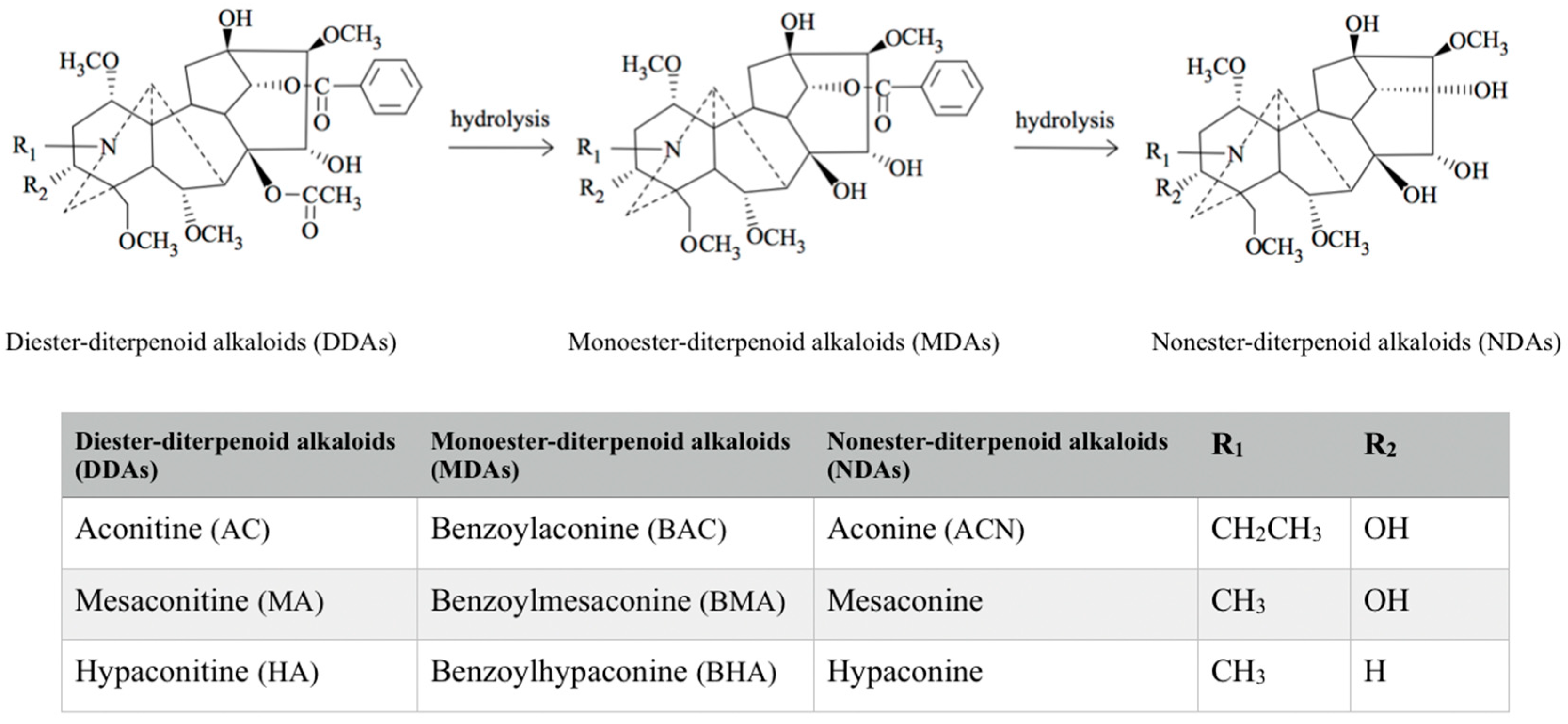
2. Classification of Aconitum Alkaloids

A. carmichaeli comprises chemicals that range from relatively non-toxic to deadly poisonous [1]. Over 122 chemical constituents, most of which are alkaloids, have been isolated and identified from A. carmichaelii [1]. Based on the difference between the number of carbon atoms and the type of alkaloid structure, they can be divided into three categories: C20-, C19-, and C18-diterpenoid alkaloids [1,16,19]. The major ones are C19-diterpenoid alkaloids, which only occur the Aconitum species [1] and can be further divided into diester-diterpenoid alkaloids (DDAs), monoester-diterpenoid alkaloids (MDAs), and non-esterified diterpenoid alkaloids (NDAs) (Figure 1) [1]. The C19-diterpenoid alkaloids are also believed to be responsible for some toxicities and certain pharmacological activities of A. carmichaelii [1,2,18]. Three main DDAs, AC, HA, and MA as shown in Figure 1, share a common C19-norditerpenoid skeleton, in which the C8 and C14 position can be occupied by an acetoxy, a benzoxy, or a hydroxyl group [1,2,18]. DDAs are not stable due to the acetyl groups at C8 position and benzoyl at the C14 position, which are easily hydrolyzed in the presence of water or heat [1,2,4,20]. Upon hydrolysis, DDAs will firstly lose one molecule of acetic acid and generate corresponding MDAs, reducing the toxicity to 1/200–1/500 of that of DDAs, and further lose benzoic acid molecules at C14 position to generate corresponding NDAs with toxicity of 1/2000–1/4000 of that of DDAs [1,3]. In the Chinese medicine preparation process, the traditional way of processing Fuzi by boiling in water can reduce its DDAs content due to their hydrolysis to less toxic MDAs and non-toxic NDAs [1].

3. Toxicities of the Main Diester-Diterpenoid Alkaloids in Fuzi

3.1. Clinical Toxicities of Fuzi

According to the Chinese Pharmacopoeia, the maximal therapeutic dose of Fuzi is 15 g/person, and the maximal total content of DDAs in Fuzi is 0.01% [5], suggesting that the highest clinical recommended dose of total DDAs would be 1.5 mg/person. However, the actual clinical dose of Fuzi can sometimes be much higher, leading to a high risk of intoxication. In clinical Fuzi-intoxication cases, a combination of neurological, gastrointestinal, and cardiovascular signs and symptoms could be seen [7]. The lowest oral dose of Fuzi to induce death in humans was recorded as 0.2 g/kg. The patients may present typical symptoms, such as nausea, vomiting, dizziness, palpitations, hypotension, arrhythmia, shock, and coma [1,3] with a mean latent period of 43.6 min [2]. Electrocardiography (ECG) may show ventricular tachycardia, ventricular fibrillation, premature ventricular contractions, multifocal ventricular ectopics, sinus tachycardia, and bradycardia [1,2,3]. Death may occur from ventricular tachyarrhythmia and heart arrest, which is most likely to happen within the first 24 h after intaking Aconitum [1,3]. Since there is no specific therapy, cardiovascular supportive treatment is usually applied for Aconitum poisoning [2,3,6]. The clinical presentation of Fuzi intoxication varies depending on the dosage of Fuzi and the infirmity of patient, and no specific dose–response relationship has been reported in clinical studies so far [2,3,6].

3.2. Toxicities of Fuzi and Its Main Diester-Diterpenoid Alkaloids in Pre-Clinical Models

Due to limited information on Fuzi toxicity mechanisms available from clinical poisoning cases, a large number of pre-clinical studies have been conducted to investigate the toxicities and their relevant mechanisms of both Fuzi and its DDAs.
The median lethal dose (LD50) was commonly used as the toxicity assessment of Fuzi and its DDAs in the early years. The LD50 of processed Fuzi for a single oral dose was reported as 100–145 g/kg in mice. The reported LD50 for a single oral dose of AC was 1.0–1.8 mg/kg in mice [3,21]. For a single oral dose of MA and HA in mice, the LD50 values were reported to be 1.9 and 5.8 mg/kg, respectively. In contrast to those of DDAs, the toxicity of the MDAs was around 1000-fold weaker, with the LD50 values (single dose, oral, mice) of benzoylaconine (BAC), benzoylmesaconine (BMA), and benzoylhypaconine (BHA) as 1.50, 0.81, and 0.83 g/kg, respectively [3].
Since measurement of lethal doses could only reflect the relative toxicities of different DDAs and Fuzi, advanced methodologies have recently been applied, such as ECG, serum biomarkers, histopathology, metabolomics, and lipodomic profile changes to better characterize the Fuzi poisoning [22,23,24]. In general, damage on the cardiovascular system was obvious in almost all published reports on Fuzi-intoxicated animal models after both bolus and long-term treatment of Fuzi. Significant increases in serum creatine kinases (CKs), lactate dehydrogenase (LDH), aspartate aminotransferase (AST), and B-type natriuretic peptide (BNP) have been observed. Arrhythmias could be observed at 30–120 min post-dosing [24]. Histological changes, including inflammatory infiltration, edema, and dilated blood vessels, were observed in cardiac tissues of rodents [1,24,25]. In addition, liver and kidney damage have been reported in several toxicity studies after the single or long-term oral administration of Fuzi extract in rodents [26]. Liver damage was evidenced by the elevated alanine aminotransferase (ALT) and AST in serum, as well as by the observation of edam and necrosis in the hepatic tissue [26]. Renal toxicity was manifested as increases of serum level of creatinine and blood urea nitrogen (BUN) and histological changes of scattered lymphocytes and atrophy in renal tissue [26].
Many studies attribute the toxicities of Fuzi to the DDAs [1,14,15,16]. The three main DDAs, namely AC, MA, and HA, share the same core structure and may share similar toxic effects and mechanisms [1,2,3]. AC-induced cardiac toxicity has manifested as elevated serum CK levels, necrosis in cardiac tissue, and arrhythmias in rodents, cats, rabbits, and dogs [1,2,3,27,28,29]. Mechanisms of the cardiac toxicity of AC have been thoroughly investigated. It was found that AC could bind to the sodium channel with high affinity [30], causing the channel to activate at more negative membrane potentials, prolonging the open state of the sodium channel, and favoring the entry of Na+ into cytosol [2,30]. More recently, AC was found to be able to promote Ca2+ overload in ventricular myocytes via perturbation of the Na+–Ca2+ exchange system [2,17] and the downregulation of the sarco/endoplasmic reticulum Ca2+-ATPase [25], triggering apoptosis in rats [25]. Other proposed arrhythmogenic mechanisms of AC mainly focused on its alteration of the intracellular Ca2+ concentration [31,32,33,34,35,36,37] and were only supported by a few in vitro experiments. In addition to AC, HA was also found to induce arrhythmias in dogs and was reported to be able to induce apoptosis on an in vitro model [38], whereas reports on MA toxicity remained rare.
In summary, the cardiac, hepatic, and renal toxicities of Fuzi have been widely reported. Although the cardiac toxicity of Fuzi derived from the toxic DDAs [1,2,3] has been demonstrated in various pre-clinical observations [17,39,40,41], no study has reported the toxicities of pure DDAs on liver, kidney, or brain. Therefore, the mechanisms for Fuzi-induced liver and kidney damage could not be delineated.

4. Toxicokinetic Characteristics of the Main Diester-Diterpenoid Alkaloids in Fuzi

Despite the large number of studies focusing on the toxicity mechanism of Fuzi since the 1980s, the toxicokinetic profiles of the main toxic DDAs in Fuzi were not well depicted until the last decade. In this section, the toxicokinetic properties of DDAs are discussed. The absorption, distribution, metabolism, and excretion characteristics of the DDAs discovered from both clinical and pre-clinical models are summarized.

4.1. Toxicokinetic Profiles of the Main Diester-Diterpenoid Alkaloids in Humans after Ingestion of Fuzi

Clinical studies in Aconitum poisoning cases depicted some toxicokinetic characteristics of the DDAs [10,12,13]. A clinical case report determined the plasma half-life of AC in a 21-year-old man as 3 h [10]. Toxicokinetic studies on five aconitine-poisoning cases with arrhythmia showed that the elimination half-lives of AC in serum ranged from 3.7 to 17.8 h and the half-lives of MA were around 2.8–5.8 h [12]. In all five patients, the serum concentrations of AC and MA became lower than 0.05 ng/mL after 35 h and 25 h, respectively [12]. The absorption phase of the blood DDA concentration vs. time curve has never been determined in patients, so no Tmax and Cmax can be obtained. DDAs preferentially distribute to the liver and kidney but not to the brain. In three autopsy cases of aconite poisoning, the DDAs levels were remarkably high in the liver and kidney, relatively low in the heart and blood, and only a trace amount of DDAs was recovered in the cerebrum [13]. The concentrations of DDAs in the liver, kidney, and heart were around 2.5–22 fold, 1.8–11.7 fold, and 1–3 fold of those in blood, while the concentrations of DDAs in the cerebrum were only 3–5% of those in blood [13]. A urine sample of a patient who accidentally ingested 10 g of A. carmichaeli and Aconitum kusnezoffii was analyzed. Apart from DDAs, MDAs, and NDAs, oxidation metabolites (16-O-demethylaconitine and 16-O-demethylhypaconitine) were also present in the urine [42]. It was also found from clinical samples that the concentrations of DDAs, MDAs, and NDAs in urine were much higher than those in blood, and were continually detectable up to 7 days after overdose [2]. In addition, a higher level of DDAs could be detected in the bile than that in the serum [18,43]. These clinical findings suggest that DDAs may be eliminated via both the kidney and the liver.

4.2. Toxicokinetic Characteristics of the Main Diester-Diterpenoid Alkaloids of Fuzi in Pre-Clinical Models

4.2.1. Absorption

Preclinical models provided more detailed toxicokinetic characteristics of the DDAs. After an oral dose of the pure compound and Fuzi extract, the bioavailability of AC was 8.24% and 4.72%, respectively [44]. Absorption of DDAs was rather fast. Tmax of AC after ingestion of pure compounds was around 25–131 min in rats and 15–35 in mice. The permeability (Papp AtoB) of AC, MA, and HA on the Caco-2 cell monolayer was 7.6 × 10⁻⁶, 8.2 × 10⁻⁶, and 21.5 × 10⁻⁶ cm/s respectively, while the permeability (Peff) of AC on a rat in-situ ileum perfusion model was around 0.5 × 10⁻⁵ cm/s. Both results from the Caco-2 cell and the in-situ intestinal perfusion model indicated medium to good absorbability of DDAs [45,46,47]. Transporters may involve and partially limit the absorption of DDAs across the intestinal epithelium. In the Caco-2 monolayer model, the efflux ratios of AC, MA, and HA were 34.6, 29.7, and 15.6, respectively, while those of the corresponding MDAs were approximately 4, and those of the corresponding NDAs equal to 1 [45,46,48,49]. Multidrug resistance protein 1 (MDR1) inhibitors, verapamil and cyclosporine A, can significantly decrease the efflux of AC on the Caco-2 cell model, and increase the intestinal permeability of AC on the rat in in-situ intestinal perfusion model [45,46,48,49]. In silico docking analyses also suggested that AC and verapamil possess similar MDR1 recognition mechanisms [46]. In addition, MK-571, an inhibitor of multidrug resistance-associated protein 2 (MRP2), exhibited inhibition on the efflux of AC but to a lesser extent than the MDR1 inhibitor on Caco-2 cells [45,48]. Taken together, MDR1 and MRP2 were involved in the transport of DDAs, partially hinder the absorbability of the toxic alkaloids.

4.2.2. Distribution

AC has low protein binding (23.9–31.9%), leading to its rapid distribution to various organs [44]. The liver and the kidney are the two major organs that AC is preferably distributed to, followed by the heart, blood, the spleen, and the lung [21,49]. In the liver, kidney, and heart, AC reached a peak concentration at around 10–240 min [21,49,50]. In one study, muscle was also found as a major tissue containing AC [50]. Due to the blood–brain barrier, only a trace amount of AC was found in brain tissue [49]. Such a distribution pattern in rodents is found to be very similar to that in humans, as described in Section 4.1.

4.2.3. Metabolism

It was previously hypothesized that DDAs may mainly be metabolized via hydrolysis. However, recent animal studies have revealed that the hydrolysis of DDAs to MDAs and NDAs may not be predominant in vivo [51,52,53,54]. In rats, the formation of hydrolysis metabolites (BAC and aconine) of AC was rather fast, evidenced by only a 15–30 min delay of their Tmax in comparison to that of AC. However, their plasma and tissue concentrations were much less than that of AC [21]. At 120 min, after oral administration of pure AC, the plasma concentrations of BAC were less than 1/10 of AC, and no ACN can be detected [21], and the heart concentrations of BAC and aconine (ACN) were only 1/20–1/10 and 1/300–1/200 of that of AC [21]. Only a small amount of the hydrolyzed products (BAC) can be recovered in rabbit stomach content 4 h after oral ingestion of AC (0.5 mg/kg) in vivo [55]. It had been hypothesized that the hydrolysis of DDAs to MDAs and NDAs may also be mediated by carboxylesterase [56], but supportive evidence is lacking. In addition to hydrolyzed metabolites, 16-O-demethylation has been found as a common metabolic pathway of the three main DDAs via Cytochrome P450 (CYP) in both liver and intestine microsome of rats and humans [51,52,56,57]. Both 16-O-demethylaconitine and 16-O-demethylhypaconitine were detected from rats and human plasma and urine samples after oral intake of A. carmichaeli [42,57]. Since demethylation pathways do not alter the structure of DDAs on C8 and C14, which is responsible for its arrhythmic toxicity, 16-O-demethylated DDAs may also exert certain toxicity. Moreover, other metabolic pathways of the DDAs including hydroxylation, deoxylation, demethylation, didemethylation/deethylation, dehydrogenation, and ester exchange were discovered in an in vitro model using liver and intestine microsome or intestinal bacteria [51,52,56,57]. Although Phase II metabolites was not found from in vitro incubation in intestine and liver microsome, hypo-mesaconitine glucuronic acid conjugate was found in the urine of rats dosed with MA [18,56].

4.2.4. Excretion

Urine is one of the major excretion routes for DDAs and their metabolites. After oral administration of AC, the urine concentration of AC peaked at 8 h post-dosing, and its major metabolite 16-O-demethylaconitine peaked at 6 h and remained detectable for up to 24 h post-dosing [2]. In addition to urine, AC can also be recovered from feces. However, the amounts of excreted DDAs and their metabolites in either urine or feces have never been compared.

4.2.5. Modulation of the Transporters and Enzymes

Since DDAs have been reported to regulate the expressions and activities of some efflux transporters and drug-metabolizing enzymes, they may be able to affect the toxicokinetic profiles of themselves or other co-treated xenobiotics. DDAs and their corresponding MDAs could increase MDR1, MRP2, and breast cancer resistance protein (BCRP) expressions in cell models and mice intestine, likely via activation of the nuclear factor E2-related factor-2 (Nrf2), nuclear receptors pregnane X receptor (PXR), and constitutive androstane receptor (CAR) [47,58]. A long-term low dose of AC increased the expressions and activities of the corresponding transporters that involved in the efflux of AC, and protected against further acute AC toxicity [47]. On the other hand, the effect of the DDAs and Fuzi extracts on CYP activities and expression levels has not been systematically investigated. Treatment of AC at 0.125 mg/kg for 7 days in rats did not affect CYP3A activity or protein levels [59,60]. Treatment of HA at 2.07 mg/kg for 7 days in rats significantly inhibited CYP3A activity and induced its mRNA level, and inhibited CYP2E activity without altering its mRNA level [61]. With downregulated protein and mRNA level, CYP3A activity was inhibited by seven-day oral administration of 600 mg/kg Fuzi extract in rats [62]. Further studies are needed to systematically elucidate the effect of DDAs and Fuzi extract on the metabolic enzymes.
In summary, the ADME characteristics of DDAs from animals are similar to those found in clinical observations. Additionally, more mechanisms of the ADME processes of DDAs have been derived from pre-clinical studies, including their medium to good absorbability in the intestine, limited in vivo hydrolysis, and their modulatory effect on the activities of MDR1, MRP2, BCRP transporters, and CYP enzymes.

5. Relationships between Toxicities of Fuzi Extract and the Toxicokinetic Profiles of Its Main Diester-Diterpenoid Alkaloids

5.1. Relationship between the Cardiac Toxicity and the Toxicokinetic Profiles of the Main Diester-Diterpenoid Alkaloids after Oral Intake of Their Pure Compounds

5.1.1. Relationship between the Acute Cardiac Toxicity and Toxicokinetic Profiles of the Main Diester-Diterpenoid Alkaloids after a Single Oral Dose

As shown in Table 1, the toxicokinetic and toxicity relationships on AC and HA have been studied after oral administration of their pure compounds in mice, rats, and dogs. Both the toxicity and toxicokinetic profile of AC were monitored at the same time after oral ingestion of pure AC, depicting a comprehensive profile of the dose-dependent toxic effect of AC in rats and mice. After bolus doses of 0.2 and 0.4 mg/kg AC in rats, cardiotoxic effects including decreases in heart rate and blood pressure were found to be dose-dependently aggravated along with the proportionally increased systemic exposure (AUC0–12 h) of AC, with no significant toxic effects observed on 0.1 mg/kg AC-treated rats [17]. Similar toxicity–toxicokinetic relationships can be found in mice. A bolus dose of 0.1 mg/kg AC in mice did not induce any observable toxicity, while 0.2 mg/kg AC induced abnormal precordial pulsation [49], and 1 mg/kg AC resulted in arrhythmia and death [21]. The cardiac toxicity of AC exacerbated as its peak concentration in heart tissue linearly increased dose-dependently. Based on these toxicokinetic data from mice and rats, the plasma concentration of AC at the lowest observable cardiac toxicity was found at around 7–13 ng/mL.
A few studies also investigated the toxicity and toxicokinetic profile of HA after bolus oral administration. After oral administration of 0.05, 0.15, and 0.45 mg/kg HA to beagle dogs, linearity of the peak plasma concentrations of HA was observed, and the cardiac toxicity (manifested as QT prolongation) was dose-dependently aggravated. Abnormality on ECG can be observed even at the lowest toxic dose of 0.05 mg/kg on beagle dogs, and the Cmax of HA was found to be 1.53 ng/mL at such a dose [40]. The toxicokinetic profile of HA was also investigated in rats receiving a single oral dose of 0.2 mg/kg HA. Compared with AC, HA demonstrated delayed Tmax, lower Cmax, and comparable T1/2 [63].

5.1.2. Relationship between the Sub-Chronic Toxicity and Toxicokinetic Profiles of the Aconitine after Multiple Oral Doses

Sub-chronic toxicity was induced after seven-day oral administration of 0.146 mg/kg AC to mice. Significant ventricular tachycardia, ventricular premature, and pathological changes in the myocardial tissues (including hyperchromatic nuclei and condensation of cytoplasm) were clearly observed [25]. It was also noticed that long-term exposure of a lower dose of AC might protect against the acute toxicity induced by a high bolus dose of AC. Long-term exposure to AC (0.6 mg/kg) for 14 days can reduce the mortality rate of the mice receiving a high bolus dose (1.8 mg/kg) of AC at Day 15 [47]. During a 22-day oral dose of 1 mg/kg AC in mice, the concentration of AC in blood, liver, kidney, and heart gradually decreased from Day 10 to Day 22 [21], while seven-day treatment of 0.5 mg/kg AC in rats did not alter its toxicokinetic profile significantly [44].
In summary, toxicity and toxicokinetic correlations on AC and HA after bolus doses have been established in animal models, while no such information regarding MA has ever been reported. The lowest toxic dose was found at 0.2 mg/kg for AC in mice and rats, and 0.05 mg/kg for HA in dogs, both of which are very close to the recommended clinical upper limit of the dose of DDAs in Fuzi (0.025 mg/kg in humans, equivalent to 0.15 mg/kg in rats or 0.045 mg/kg in dogs converted by the appropriate body surface area conversion factor [64]). The linear plasma kinetics of AC and HA were confirmed, and their systemic exposure levels (Cmax or AUC) at the corresponding lowest toxic bolus dose were determined. The plasma concentration of DDAs at the lowest observable cardiac toxicity dose was found as low as 1.5 ng/mL. Therefore, risk of cardiac toxicity should be revealed when the plasma concentration of DDAs reaches such a threshold. On the other hand, toxicity and toxicokinetic correlations after long-term treatment, which are critical for the study on chronic toxicity of these compounds, have never been reported.

5.2. Relationship between the Heart, Liver, and Kidney Toxicities of Fuzi and the Toxicokinetics of Its Main Diester-Diterpenoid Alkaloids after Oral Intake of Its Extract

In comparison to those from the oral administration of DDAs, the toxicokinetic profiles of DDAs after ingestion of Fuzi extract are more complicated. The toxicokinetic studies of the three major DDAs after oral administration of various Fuzi extracts are summarized and correlated with their toxicities as shown in Table 2. Since the three DDAs with similar toxicity are the major toxins in Fuzi, the total amount of them from different Fuzi extract preparations was used to represent the toxicity-related dose of Fuzi for comparison of toxicities among different studies.

5.2.1. Dose-Dependent Toxicities of Fuzi and the Toxicokinetic Profiles of Its Main Diester-Diterpenoid Alkaloids after Single Oral Administration

Besides the cardiac abnormity demonstrated by ECG, damage in organs including the heart, liver, and kidney were also found in animal models after a single oral dose of Fuzi extract. Only one study reported the dose-dependent toxicity of Fuzi extract with its DDA contents determined. In this study, the lowest toxic dose in rats was 2 g/kg Fuzi extract, which equals to 0.116, 0.367, and 0.586 mg/kg of AC, MA, and HA, or 1.1 mg/kg total DDAs. At such a dose, no elevated biomarker indicating heart, liver, or kidney dysfunction was observed, while mild edam was noticed in the liver and heart [26]. At a medium dose of 5 g/kg Fuzi (equivalent to 0.29, 1.168, and 1.466 mg/kg of AC, MA, and HA, or 2.9 mg/kg total DDAs), severer tissue damage to the heart, liver, and kidney was observed. At the high dose of 10 g/kg Fuzi (equivalent to 0.58, 2.336, and 2.932 mg/kg of AC, MA, and HA, or 5.8 mg/kg total DDAs), elevations of CK, LDH, AST, and urea levels were obvious, and were consistent with the remarkable damage in the heart, liver, and kidney [26]. Such toxicity investigation suggested dose-dependent damage in the heart, liver, and kidney induced by Fuzi.
On the other hand, toxicokinetic profiles of the three main DDAs after oral ingestion of Fuzi extracts have been studied in rats [68,69,70,71]. Generally, comparing the toxicokinetics after oral administration of the corresponding pure compounds, the Cmax and AUC0-last of AC and HA after administration of Fuzi extracts were significantly lower, while the T1/2 remained similar. The Tmax of AC in pure-AC-treated rats was also comparable to that in Fuzi extract-treated rats. The toxicokinetic characteristics of each DDA after ingestion of mild to severe toxic doses of Fuzi extracts are discussed individually in the following paragraphs.

AC-Related Toxicokinetic Profiles at the Toxic Level of Fuzi

Comparing with HA and MA, the doses of AC in the Fuzi extracts were relatively low (around 0.037–0.117 mg/kg AC). In a dose range of Fuzi (equivalent to 0.46–0.66 mg/kg total DDAs, or 0.06–0.118 mg/kg AC) that refers to mild toxicity, the AUC0-last and Cmax of AC was proportionally elevated as the dosage increased, and the T1/2 of AC was not significantly changed between different dosage groups [44,69,70], demonstrating a linear kinetic profile at the mild toxic level. However, at a high dose of Fuzi (equivalent to 4.9 mg/kg total DDAs or 0.177 mg/kg AC), and the dose-normalized AUC0-last (24.3 ± 8.9 min·kg/L) was significantly larger than those from the mild toxic dose (around 4.4 ± 0.5 to 8.3 ± 1.4 min kg/L) [71]. In addition, the lowest dose of Fuzi extract (equivalent to 0.356 mg/kg total DDAs, containing 0.037 mg/kg AC) resulted in significantly shorter T1/2 of AC, which may be due to the low and variable plasma concentrations [68].

HA-Related Toxicokinetic Profiles at the Toxic Level of Fuzi

The HA content in the Fuzi extracts was usually much higher than that of AC [72]. The doses of HA in Fuzi extracts were around 0.1–2.9 mg/kg in four different toxicokinetic studies [68,69,70,71]. Along with the increased doses, both Cmax and AUC0-last of HA were proportionally increased within the dose range from non-toxic (0.356 mg/kg total DDAs) to severely toxic (4.9 mg/kg total DDAs). The dose-normalized AUC0-last values of HA were around 4.9 ± 0.9 to 8.4 ± 0.3 min·kg/L [44,69,70]. The T1/2 of HA (ranged from 104 ± 2 to 559 ± 62 min) varied among different studies using Fuzi at an equivalent dose of 0.46–4.9 mg/kg total DDAs [68,69,70,71].

MA-Related Toxicokinetic Profiles at the Toxic Level of Fuzi

When Fuzi extracts were given to the rats at low toxic doses (equivalent to 0.356–0.66 mg/kg total DDAs or 0.138–0.3 mg/kg MA), linearity was observed in the AUC0-last and Cmax of MA, with dose-normalized AUC0-last of MA at around 2.8 ± 0.7 to 4.0 ± 0.8 min·kg/L, and the T1/2 (ranged from 251 ± 98 to 636 ± 210 min) was not significantly changed among different doses [68,69,71]. However, at a high toxic dose of Fuzi (equivalent to 4.9 mg/kg total DDAs or 1.805 mg/kg MA), the dose-normalized AUC0-last (6.1 ± 1.2 min·kg/L) was slightly larger than those from the low toxic dose [71]. Additionally, the lowest dose of MA (0.017 mg/kg in the Fuzi extract) led to a much higher dose-normalized AUC0-last (22.3 ± 1.8 min·kg/L), which may be due to the low and variable plasma concentrations [70].
Overall, the AUC0-last and Cmax of the three main DDAs were proportional to their doses with similar dose-normalized AUC0-last values at low toxic doses (0.356–0.66 mg/kg total DDAs) of the Fuzi extract. At a high toxic dose of Fuzi extract (4.9 mg/kg total DDAs), the dose-normalized AUC0-last values of AC and MA were significantly higher than those from the low dose. It is also noticed that, even under the same dose of AC and MA, their toxicokinetic parameters including AUC0-last and Tmax varied between the two groups of rats treated with different Fuzi extracts, suggesting that other co-existing components in different Fuzi extracts may affect the toxicokinetic profiles of AC and MA [69].

5.2.2. Dose-Dependent Toxicity of Fuzi and the Toxicokinetic Profile of Its Main Diester-Diterpenoid Alkaloids after Long-Term Oral Administration

In clinical practice, intake of Fuzi can be at lower dose level for sub-chronic treatment that usually lasts for 1–2 weeks. Table 3 demonstrates the toxicokinetics of DDAs and toxicities of Fuzi extracts after long-term oral administration in rats. The lowest long-term oral toxic dose of Fuzi extract in rats for 15 consecutive days was found to be at 1.28 g/kg (equivalent to 0.02, 0.06, and 0.03 mg/kg of AC, MA, and HA, or 0.11 total DDAs), which is much lower than the acute toxic dose (equivalent to 1.169 mg/kg total DDAs). At such a dose, serum biomarkers indicating heart, liver, and kidney dysfunction were all elevated [23]. At a higher dosage of Fuzi extract (equivalent to 0.22 total DDAs), together with the elevations of serum markers, histological damage related to edema, inflammation, necrosis, and vascular dilatation were noticed in the liver, kidney, and heart [23]. In addition, seven-day treatment of a Fuzi extract, which did not contain any AC (equivalent to 6.3 and 4.0 mg/kg of MA and HA) in rats also led to decreased body weights and severe renal toxicity [72]. In contrary to the toxicity findings, information on the toxicokinetic profiles of the toxic DDAs after long-term oral administration of Fuzi is limited. Only one study has reported the long-term toxicokinetic profile of AC after oral administration of Fuzi extract in rats. In this study, the AUC of AC was found to be significantly increased compared with that from the single-dose group, while the T1/2 and Cmax remained similar [44]. Such evidence suggests that the altered toxicokinetic profiles of the three DDAs after long-term administration of Fuzi may affect their toxicity. Further investigations are warranted to explore the relationships between long-term toxicokinetic profiles and the chronic toxicities of Fuzi and its DDAs.
In summary, a high dose of Fuzi extract usually leads to severe heart, liver, and kidney damage [26], which is evidenced by the high dose-normalized AUC0-last of AC and MA after a single oral dose of Fuzi extract. Correlation between the toxicokinetics of DDAs and the toxicity of Fuzi have been preliminarily studied but are still ambiguous. To further illustrate the quantitative relationship between the toxicokinetic profile and the toxicity of Fuzi extract, two issues need to be tackled: (1) Quality variability was observed among the Fuzi extracts used in different toxicity and toxicokinetic studies. The toxicokinetics of DDAs may be affected by other co-existing components in Fuzi [69], and additive effects between different DDAs and antagonistic effects between DDAs and other components of Fuzi have been reported to modulate the toxicity of Fuzi [73]. Such quality variability of Fuzi extract leads to inconsistent relationships between toxicities and toxicokinetic profiles from different studies. (2) Due to the lack of simultaneously determined toxicity and toxicokinetic profiles of Fuzi and its DDAs, and the lack of tissue distribution data of DDAs, it is difficult to delineate the correlation between organ damage and the toxicokinetics of DDAs after oral administration of Fuzi extracts on animal models.

6. Conclusions

This review is the first to summarize evidence of the relationships between DDA plasma kinetics and the toxicities of Fuzi. Such relationships have been well-identified in animals receiving pure DDA compounds and have been preliminarily investigated in animals receiving different Fuzi extracts, whereas the clinical evidence of such relationships are still lacking. Based on the pre-clinical evidence, it has been demonstrated that the narrow therapeutic window and large quality variability of Fuzi extracts may significantly affect its safe use in clinical practice. Based on our findings, standardized products are essential for the safe use of Fuzi. Although correlations between the exposure of DDAs and Fuzi-induced hepatic and renal toxicities are still ambiguous, dose-dependent hepatic and renal toxicities of Fuzi have been clearly observed in pre-clinical studies. Therefore, caution with respect to potential hepatic and renal toxicity induced by Fuzi should be exercised. Since the cardiac toxicity has been well-correlated with the plasma and heart concentrations of DDAs in mice and rats, with the plasma concentration of DDAs at the lowest observable cardiac toxicity being determined, clinical monitoring of the plasma concentrations of DDAs of Fuzi is recommended to prevent potential cardiac toxicities. Further analyses focusing on the tissue concentration profile of DDAs and on the long-term toxicokinetic-toxicity correlation of DDAs are warranted to better understand the toxic mechanisms and safer use of Fuzi.

Author Contributions

Conceptualization: Z.Z. and M.Y.; writing & review: Z.Z. and M.Y.; literature researching: M.Y. and X.J.

Funding

The current study is funded by Health and Medical Research Fund (15161541) by the Food and Health Bureau, Hong Kong SAR, China.

Conflicts of Interest

The authors declare no conflict of interest.

References

  1. Zhou, G.; Tang, L.; Zhou, X.; Wang, T.; Kou, Z.; Wang, Z. A review on phytochemistry and pharmacological activities of the processed lateral root of Aconitum carmichaelii Debeaux. J. Ethnopharmacol. 2015, 160, 173–193. [Google Scholar] [CrossRef] [PubMed]
  2. Chan, T.Y.K. Aconite poisoning. Clin. Toxicol. 2009, 47, 279–285. [Google Scholar] [CrossRef] [PubMed]
  3. Singhuber, J.; Zhu, M.; Prinz, S.; Kopp, B. Aconitum in Traditional Chinese Medicine—A valuable drug or an unpredictable risk? J. Ethnopharmacol. 2009, 126, 18–30. [Google Scholar] [CrossRef] [PubMed]
  4. Tai, C.J.; El-Shazly, M.; Wu, T.Y.; Lee, K.T.; Csupor, D.; Hohmann, J.; Chang, F.R.; Wu, Y.C. Clinical Aspects of Aconitum Preparations. Planta Med. 2015, 81, 1017–1028. [Google Scholar] [CrossRef] [PubMed] [Green Version]
  5. Chinese Pharmacopoeia Commission. Chinese Pharmacopoeia; China Medical Science Press: Beijing, China, 2015; Volume 1, pp. 191–193. [Google Scholar]
  6. Li, H.; Liu, L.; Zhu, S.; Liu, Q. Case reports of aconite poisoning in mainland China from 2004–2015: A retrospective analysis. J. Forensic Leg. Med. 2016, 42, 68–73. [Google Scholar] [CrossRef] [PubMed]
  7. Chan, T.Y.K. Aconitum Alkaloid Poisoning because of Contamination of Herbs by Aconite Roots. Phyther. Res. 2016, 30, 3–8. [Google Scholar] [CrossRef] [PubMed]
  8. Chan, T.Y.K. Contributory factors in herb-induced fatal aconite poisoning. Forensic Sci. Int. 2012, 223, 40–43. [Google Scholar] [CrossRef] [PubMed]
  9. Chan, T. Aconitum Alkaloid Poisoning Related to the Culinary Uses of Aconite Roots. Toxins 2014, 6, 2605–2611. [Google Scholar] [CrossRef] [PubMed] [Green Version]
  10. Moritz, F.; Compagnon, P.; Kaliszczak, I.G.; Kaliszczak, Y.; Caliskan, V.; Girault, C. Severe acute poisoning with homemade Aconitum napellus capsules: Toxicokinetic and clinical data. Clin. Toxicol. 2005, 43, 873–876. [Google Scholar] [CrossRef]
  11. Lin, C.C.; Phua, D.H.; Deng, J.F.; Yang, C.C. Aconitine intoxication mimicking acute myocardial infarction. Hum. Exp. Toxicol. 2011, 30, 782–785. [Google Scholar] [CrossRef] [PubMed]
  12. Fujita, Y.; Terui, K.; Fujita, M.; Kakizaki, A.; Sato, N.; Oikawa, K.; Aoki, H.; Takahashi, K.; Endo, S. Five cases of aconite poisoning: Toxicokinetics of aconitines. J. Anal. Toxicol. 2007, 31, 132–137. [Google Scholar] [CrossRef] [PubMed]
  13. Niitsu, H.; Fujita, Y.; Fujita, S.; Kumagai, R.; Takamiya, M.; Aoki, Y.; Dewa, K. Distribution of Aconitum alkaloids in autopsy cases of aconite poisoning. Forensic Sci. Int. 2013, 227, 111–117. [Google Scholar] [CrossRef] [PubMed]
  14. Nyirimigabo, E.; Xu, Y.; Li, Y.; Wang, Y.; Agyemang, K.; Zhang, Y. A review on phytochemistry, pharmacology and toxicology studies of Aconitum. J. Pharm. Pharmacol. 2015, 67, 1–19. [Google Scholar] [CrossRef] [PubMed]
  15. Jiang, B.; Lin, S.; Zhu, C.; Wang, S.; Wang, Y.; Chen, M.; Zhang, J.; Hu, J.; Chen, N.; Yang, Y.; et al. Diterpenoid alkaloids from the lateral root of Aconitum carmichaelii. J. Nat. Prod. 2012, 75, 1145–1159. [Google Scholar] [CrossRef] [PubMed]
  16. Bello-Ramírez, A.M.; Nava-Ocampo, A.A. A QSAR analysis of toxicity of Aconitum alkaloids. Fundam. Clin. Pharmacol. 2004, 18, 699–704. [Google Scholar] [CrossRef] [PubMed]
  17. Zhang, P.; Kong, D.; Du, Q.; Zhao, J.; Li, Q.; Zhang, J.; Li, T.; Ren, L. A conscious rat model involving bradycardia and hypotension after oral administration: A toxicokinetical study of aconitine. Xenobiotica 2017, 47, 515–525. [Google Scholar] [CrossRef] [PubMed]
  18. Wu, J.J.; Guo, Z.Z.; Zhu, Y.F.; Huang, Z.J.; Gong, X.; Li, Y.H.; Son, W.J.; Li, X.Y.; Lou, Y.M.; Zhu, L.J.; et al. A systematic review of pharmacokinetic studies on herbal drug Fuzi: Implications for Fuzi as personalized medicine. Phytomedicine 2018, 15, 187–203. [Google Scholar] [CrossRef] [PubMed]
  19. Pereira, F. Polypharmacology of Aconitum and Delphinium sp. Diterpene Alkaloids: Antiarrhythmic, Analgesic and Anti-Inflammatory Effects. Mini Rev. Org. Chem. 2017, 14. [Google Scholar] [CrossRef]
  20. Liu, S.; Li, F.; Li, Y.; Li, W.; Xu, J.; Du, H. A review of traditional and current methods used to potentially reduce toxicity of Aconitum roots in Traditional Chinese Medicine. J. Ethnopharmacol. 2017, 207, 237–250. [Google Scholar] [CrossRef] [PubMed]
  21. Wada, K.; Nihira, M.; Hayakawa, H.; Tomita, Y.; Hayashida, M.; Ohno, Y. Effects of long-term administrations of aconitine on electrocardiogram and tissue concentrations of aconitine and its metabolites in mice. Forensic Sci. Int. 2005, 148, 21–29. [Google Scholar] [CrossRef] [PubMed]
  22. Sun, B.; Zhang, M.; Zhang, Q.; Ma, K.; Li, H.; Li, F.; Dong, F.; Yan, X. Metabonomics study of the effects of pretreatment with glycyrrhetinic acid on mesaconitine-induced toxicity in rats. J. Ethnopharmacol. 2014, 154, 839–846. [Google Scholar] [CrossRef] [PubMed]
  23. Tan, Y.; Ko, J.; Liu, X.; Lu, C.; Li, J.; Xiao, C.; Li, L.; Niu, X.; Jiang, M.; He, X.; et al. Serum metabolomics reveals betaine and phosphatidylcholine as potential biomarkers for the toxic responses of processed Aconitum carmichaelii Debx. Mol. Biosyst. 2014, 10, 2305–2316. [Google Scholar] [CrossRef] [PubMed]
  24. Cai, Y.; Gao, Y.; Tan, G.; Wu, S.; Dong, X.; Lou, Z.; Zhu, Z.; Chai, Y. Myocardial lipidomics profiling delineate the toxicity of traditional Chinese medicine Aconiti Lateralis radix praeparata. J. Ethnopharmacol. 2013, 147, 349–356. [Google Scholar] [CrossRef] [PubMed]
  25. Sun, G.B.; Sun, H.; Meng, X.B.; Hu, J.; Zhang, Q.; Liu, B.; Wang, M.; Xu, H.B.; Sun, X.B. Aconitine-induced Ca2+overload causes arrhythmia and triggers apoptosis through p38 MAPK signaling pathway in rats. Toxicol. Appl. Pharmacol. 2014, 279, 8–22. [Google Scholar] [CrossRef] [PubMed]
  26. Zhou, H.; Zhang, P.; Hou, Z.; Xie, J.; Wang, Y.; Yang, B.; Xu, Y.; Li, Y. Research on the relationships between endogenous biomarkers and exogenous toxic substances of acute toxicity in Radix Aconiti. Molecules 2016, 21, 1623. [Google Scholar] [CrossRef] [PubMed]
  27. Matsuda, K.; Hoshi, T.; Kameyama, S. Effects of aconitine on the cardiac membrane potential of the dog. Jpn. J. Physiol. 1959, 9, 419–429. [Google Scholar] [CrossRef]
  28. Winslow, E. Hemodynamic and arrhythmogenic effects of aconitine applied to the left atria of anesthetized cats. Effects of amiodarone and atropine. J. Cardiovasc. Pharmacol. 1981, 3, 87–100. [Google Scholar] [CrossRef] [PubMed]
  29. Wang, T.; Huang, C.X.; Jiang, H.; Tang, Q.Z.; Yang, B.; Li, G.S. Effects of BmKIM on sodium current of isolated cardiomyocytes, transmembrane action potential and aconitine induced arrhythmia in vivo in rabbits. Zhonghua Xin Xue Guan Bing Za Zhi 2009, 37, 102–107. [Google Scholar] [PubMed]
  30. Ameri, A. The effects of Aconitum alkaloids on the central nervous system. Prog. Neurobiol. 1998, 56, 211–235. [Google Scholar] [CrossRef]
  31. Fu, M.; Wu, M.; Wang, J.F.; Qiao, Y.J.; Wang, Z. Disruption of the intracellular Ca2+ homeostasis in the cardiac excitation-contraction coupling is a crucial mechanism of arrhythmic toxicity in aconitine-induced cardiomyocytes. Biochem. Biophys. Res. Commun. 2007, 354, 929–936. [Google Scholar] [CrossRef] [PubMed]
  32. Fu, M.; Li, R.X.; Fan, L.; He, G.W.; Thornburg, K.L.; Wang, Z. Sarcoplasmic reticulum Ca2+ release channel ryanodine receptor (RyR2) plays a crucial role in aconitine-induced arrhythmias. Biochem. Pharmacol. 2008, 75, 2147–2156. [Google Scholar] [CrossRef] [PubMed]
  33. Peng, C.; Zheng, T.; Yang, F.; Li, Y.X.; Zhang, D.K. Study of neurotoxic effects and underlying mechanisms of aconitine on cerebral cortex neuron cells. Arch. Pharm. Res. 2009, 32, 1533–1543. [Google Scholar] [CrossRef] [PubMed]
  34. Peng, C.; Wang, L.; Wang, Y.H.; Li, Y.X.; Pan, Y. The toxicity of aconitine, emodin on ICC cell and the antagonist effect of the compatibility. Eur. J. Drug Metab. Pharmacokinet. 2009, 34, 213–220. [Google Scholar] [PubMed]
  35. Zhang, S.W.; Liu, Y.; Huang, G.Z.; Liu, L. Aconitine alters connexin43 phosphorylation status and [Ca2+] oscillation patterns in cultured ventricular myocytes of neonatal rats. Toxicol. In Vitro 2007, 21, 1476–1485. [Google Scholar] [CrossRef] [PubMed]
  36. Wu, J.; Wang, X.; Chung, Y.Y.; Koh, C.H.; Liu, Z.; Guo, H.; Yuan, Q.; Wang, C.; Su, S.; Wei, H. L-type calcium channel inhibition contributes to the proarrhythmic effects of aconitine in human cardiomyocytes. PLoS ONE 2017, 12, e0168435. [Google Scholar] [CrossRef] [PubMed]
  37. Zhou, Y.H.; Piao, X.M.; Liu, X.; Liang, H.H.; Wang, L.M.; Xiong, X.H.; Wang, L.; Lu, Y.J.; Shan, H.L. Arrhythmogenesis toxicity of aconitine is related to intracellular Ca2+ signals. Int. J. Med. Sci. 2013, 10, 1242–1249. [Google Scholar] [CrossRef] [PubMed]
  38. Bai, Y.; Du, S.; Li, F.; Huang, F.; Deng, R.; Zhou, J.; Chen, D. Histone deacetylase-high mobility group box-1 pathway targeted by hypaconitine suppresses the apoptosis of endothelial cells. Exp. Biol. Med. 2017, 242, 527–535. [Google Scholar] [CrossRef] [PubMed]
  39. Voss, L.J.; Voss, J.M.; McLeay, L.; Sleigh, J.W. Aconitine induces prolonged seizure-like events in rat neocortical brain slices. Eur. J. Pharmacol. 2008, 584, 291–296. [Google Scholar] [CrossRef] [PubMed]
  40. Xie, S.; Jia, Y.; Liu, A.; Dai, R.; Huang, L. Hypaconitine-induced QT prolongation mediated through inhibition of KCNH2 (hERG) potassium channels in conscious dogs. J. Ethnopharmacol. 2015, 166, 375–379. [Google Scholar] [CrossRef] [PubMed]
  41. Shu, H.; Yi-Ming, W.; Xu, L.P.; Miao, C.Y.; Su, D.F. Increased susceptibility of ventricular arrhythmias to aconitine in anaesthetized rats is attributed to the inhibition of baroreflex. Clin. Exp. Pharmacol. Physiol. 2004, 31, 249–253. [Google Scholar] [CrossRef] [PubMed]
  42. Zhang, H.G.; Sun, Y.; Duan, M.Y.; Chen, Y.J.; Zhong, D.F.; Zhang, H.Q. Separation and identification of Aconitum alkaloids and their metabolites in human urine. Toxicon 2005, 46, 500–506. [Google Scholar] [CrossRef] [PubMed]
  43. Ito, K.; Tanaka, S.; Funayama, M.; Mizugaki, M. Distribution of Aconitum alkaloids in body fluids and tissues in a suicidal case of aconite ingestion. J. Anal. Toxicol. 2000, 24, 348–353. [Google Scholar] [CrossRef] [PubMed]
  44. Tang, L.; Gong, Y.; Lv, C.; Ye, L.; Liu, L.; Liu, Z. Pharmacokinetics of aconitine as the targeted marker of Fuzi (Aconitum carmichaelii) following single and multiple oral administrations of Fuzi extracts in rat by UPLC/MS/MS. J. Ethnopharmacol. 2012, 141, 736–741. [Google Scholar] [CrossRef] [PubMed]
  45. Yang, C.; Li, Z.; Zhang, T.; Liu, F.; Ruan, J.; Zhang, Z. Transcellular transport of aconitine across human intestinal Caco-2 cells. Food Chem. Toxicol. 2013, 57, 195–200. [Google Scholar] [CrossRef] [PubMed]
  46. Yang, C.; Zhang, T.; Li, Z.; Xu, L.; Liu, F.; Ruan, J.; Liu, K.; Zhang, Z. P-glycoprotein is responsible for the poor intestinal absorption and low toxicity of oral aconitine: In vitro, in situ, in vivo and in silico studies. Toxicol. Appl. Pharmacol. 2013, 273, 561–568. [Google Scholar] [CrossRef] [PubMed]
  47. Wu, J.; Lin, N.; Li, F.; Zhang, G.; He, S.; Zhu, Y.; Ou, R.; Li, N.; Liu, S.; Feng, L.; et al. Induction of P-glycoprotein expression and activity by Aconitum alkaloids: Implication for clinical drug-drug interactions. Sci. Rep. 2016, 6, 25343. [Google Scholar] [CrossRef] [PubMed]
  48. Ye, L.; Yang, X.; Yang, Z.; Gao, S.; Yin, T.; Liu, W.; Wang, F.; Hu, M.; Liu, Z. The role of efflux transporters on the transport of highly toxic aconitine, mesaconitine, hypaconitine, and their hydrolysates, as determined in cultured Caco-2 and transfected MDCKII cells. Toxicol. Lett. 2013, 216, 86–99. [Google Scholar] [CrossRef] [PubMed]
  49. Zhu, L.; Wu, J.; Zhao, M.; Song, W.; Qi, X.; Wang, Y.; Lu, L.; Liu, Z. Mdr1a plays a crucial role in regulating the analgesic effect and toxicity of aconitine by altering its pharmacokinetic characteristics. Toxicol. Appl. Pharmacol. 2017, 320, 32–39. [Google Scholar] [CrossRef] [PubMed]
  50. Zhang, H.; Sun, S.; Zhang, W.; Xie, X.; Zhu, Z.; Chai, Y.; Zhang, G. Biological activities and pharmacokinetics of aconitine, benzoylaconine, and aconine after oral administration in rats. Drug Test. Anal. 2016, 8, 839–846. [Google Scholar] [CrossRef] [PubMed]
  51. Wang, Y.; Wang, S.; Liu, Y.; Yan, L.; Dou, G.; Gao, Y. Characterization of metabolites and cytochrome P450 isoforms involved in the microsomal metabolism of aconitine. J. Chromatogr. B Anal. Technol. Biomed. Life Sci. 2006, 844, 292–300. [Google Scholar] [CrossRef] [PubMed]
  52. Tang, L.; Ye, L.; Lv, C.; Zheng, Z.; Gong, Y.; Liu, Z. Involvement of CYP3A4/5 and CYP2D6 in the metabolism of aconitine using human liver microsomes and recombinant CYP450 enzymes. Toxicol. Lett. 2011, 202, 47–54. [Google Scholar] [CrossRef] [PubMed]
  53. Zhang, M.; Peng, C.S.; Li, X.B. Human intestine and liver microsomal metabolic differences between C19-diester and monoester diterpenoid alkaloids from the roots of Aconitum carmichaelii Debx. Toxicol. In Vitro 2017, 45, 318–333. [Google Scholar] [CrossRef] [PubMed]
  54. Ye, L.; Tang, L.; Gong, Y.; Lv, C.; Zheng, Z.; Jiang, Z.; Liu, Z. Characterization of metabolites and human P450 isoforms involved in the microsomal metabolism of mesaconitine. Xenobiotica 2011, 41, 46–58. [Google Scholar] [CrossRef] [PubMed]
  55. Sui, Z.; Li, N.; Liu, Z.; Yan, J.; Liu, Z. Metabolite profile analysis of aconitine in rabbit stomach after oral administration by liquid chromatography/electrospray ionization/multiple-stage tandem mass spectrometry. Xenobiotica 2013, 43, 628–635. [Google Scholar] [CrossRef] [PubMed]
  56. Zhang, M.; Peng, C.S.; Li, X.B. In vivo and in vitro metabolites from the main diester and monoester diterpenoid alkaloids in a traditional Chinese herb, the aconitum species. Evid.-Based Complement. Altern. Med. 2015, 2015, 252434. [Google Scholar] [CrossRef]
  57. He, H.; Yan, F. Relative quantification of the metabolite of aconitine in rat urine by LC-ESI-MS/MS and its application to pharmacokinetics. Anal. Sci. 2012, 28, 1203–1205. [Google Scholar] [CrossRef] [PubMed]
  58. Wu, J.J.; Zhu, Y.F.; Guo, Z.Z.; Lou, Y.M.; He, S.G.; Guan, Y.; Zhu, L.J.; Liu, Z.Q.; Lu, L.L.; Liu, L. Aconitum alkaloids, the major components of Aconitum species, affect expression of multidrug resistance-associated protein 2 and breast cancer resistance protein by activating the Nrf2-mediated signalling pathway. Phytomedicine 2017, 44, 87–97. [Google Scholar] [CrossRef] [PubMed]
  59. Zhu, L.; Yang, X.; Zhou, J.; Tang, L.; Xia, B.; Hu, M.; Zhou, F.; Liu, Z. The exposure of highly toxic aconitine does not significantly impact the activity and expression of cytochrome P450 3A in rats determined by a novel ultra performance liquid chromatography-tandem mass spectrometric method of a specific probe buspirone. Food Chem. Toxicol. 2013, 51, 396–403. [Google Scholar] [CrossRef] [PubMed]
  60. Zhu, L.; Lu, L.; Guo, E.; Wu, J.; Wang, Y.; Hu, M.; Liu, Z. The Influences of Aconitine, an Active/Toxic Alkaloid from Aconitum, on the Oral Pharmacokinetics of CYP3A Probe Drug Buspirone in Rats. Drug Metab. Lett. 2014, 8, 135–144. [Google Scholar] [CrossRef] [PubMed]
  61. Miao, X.; Wang, J.; Meng, M.; Chen, Y. Influence of hypaconitine and liquirtin used alone and together on mRNA expression and activity of CYP450 enzymes in rat liver. China J. Tradit. Chin. Med. Pharm. 2016, 31, 313–316. [Google Scholar]
  62. Wu, J.; Cheng, Z.; Zhu, L.; Lu, L.; Zhang, G.; Wang, Y.; Xu, Y.; Lin, N.; Liu, Z. Coadministration of Pinellia ternata can significantly reduce Aconitum carmichaelii to inhibit CYP3A activity in rats. Evid. Based Complement. Altern. Med. 2014, 2014. [Google Scholar] [CrossRef] [PubMed]
  63. Zhou, J. Pharmacokinetics and Cardiac Distribution of Hypaconitine in Rats. Master’s Thesis, Beijing University of Chinese Medicine, Beijing, China, 2010. [Google Scholar]
  64. U.S. Food and Drug Administration. Guidance for Industry Estimating the Maximum Safe Starting Dose in Initial Clinical Trials for Therapeutics in Adult Healthy Volunteers; U.S. Food and Drug Administration, Center for Drug Evaluation and Research (CDER): Rockville, MD, USA, 2005; p. 7.
  65. Wada, K.; Nihira, M.; Ohno, Y. Effects of chronic administrations of aconitine on body weight and rectal temperature in mice. J. Ethnopharmacol. 2006, 105, 89–94. [Google Scholar] [CrossRef] [PubMed]
  66. Fan, Y.F.; Xie, Y.; Liu, L.; Ho, H.M.; Wong, Y.F.; Liu, Z.Q.; Zhou, H. Paeoniflorin reduced acute toxicity of aconitine in rats is associated with the pharmacokinetic alteration of aconitine. J. Ethnopharmacol. 2012, 141, 701–708. [Google Scholar] [CrossRef] [PubMed]
  67. Wang, Z.H.; Wen, J.; He, Y. Research on pharmacokinetics of aconitine by LC-MS. J. Instrum. Anal. 2004, 23, 51–53. [Google Scholar]
  68. Wang, R. Quality Evaluation of Fuzi and Pharmacokinetic Study of Aconitine. Ph.D. Thesis, Beijing University of Chinese Medicine, Beijing, China, 2007. [Google Scholar]
  69. Liu, J.; Li, Q.; Yin, Y.; Liu, R.; Xu, H.; Bi, K. Ultra-fast LC-ESI-MS/MS method for the simultaneous determination of six highly toxic Aconitum alkaloids from Aconiti kusnezoffii radix in rat plasma and its application to a pharmacokinetic study. J. Sep. Sci. 2014, 37, 171–178. [Google Scholar] [CrossRef] [PubMed]
  70. Peng, W.W.; Li, W.; Li, J.S.; Cui, X.B.; Zhang, Y.X.; Yang, G.M.; Wen, H.M.; Cai, B.C. The effects of Rhizoma Zingiberis on pharmacokinetics of six Aconitum alkaloids in herb couple of Radix Aconiti Lateralis-Rhizoma Zingiberis. J. Ethnopharmacol. 2013, 148, 579–586. [Google Scholar] [CrossRef] [PubMed]
  71. Li, Y.; Li, Y.X.; Zhao, M.J.; Yuan, A.; Gong, X.H.; Zhao, M.J.; Peng, C. The Effects of Rheum palmatum L. on the Pharmacokinetic of Major Diterpene Alkaloids of Aconitum carmichaelii Debx. in Rats. Eur. J. Drug Metab. Pharmacokinet. 2017, 42, 441–451. [Google Scholar] [CrossRef] [PubMed]
  72. Li, L.; Sun, B.; Zhang, Q.; Fang, J.; Ma, K.; Li, Y.; Chen, H.; Dong, F.; Gao, Y.; Li, F.; et al. Metabonomic study on the toxicity of Hei-Shun-Pian, the processed lateral root of Aconitum carmichaelii Debx. (Ranunculaceae). J. Ethnopharmacol. 2008, 116, 561–568. [Google Scholar] [CrossRef] [PubMed]
  73. Zhou, Y.P.; Liu, W.H. Review and reappraise on researches of Fuzi’s effect on cardiovarscular system, part one. Pharmacol. Clin. Chin. Mater. Med. 2013, 2, 198–205. [Google Scholar]
  74. Xiao, R.P.; Lai, X.P.; Zhao, Y.; Yu, L.W.; Zhu, Y.L.; Li, G. Pharmacokinetic study of six aconitine alkaloids in aconiti lateralis radix praeparata in beagle dogs. J. Chin. Med. Mater. 2014, 37, 284–287. [Google Scholar]
Figure 1. Structure of the main C19-diterpenoid alkaloids of Fuzi.
Toxins 10 00391 g001
Table 1. Summary of toxicokinetics and toxicity of aconitine (AC) and hypaconitine (HA) after oral administrations in animal models.
CompoundSpeciesDuration/DoseToxicokinetic ParametersPharmacokinetics in HeartToxicity MeasurementReference
Tmax (min)Cmax (ng/mL)T1/2 (min)AUC0-last (min·ng/mL)Tmax (min)Cmax (ng/g)Serum BiomarkerHeart Histology ECGBehavior
ACMice7 days/0.146 mg/kgNRNRNRNRNRNR↑ CK, AST, LDHHyper-chromatic nuclei, condensed cytoplasmVentricular tachycardiaNR[25]
22 days/1 mg/kgNR↓compared to single doseNRNRNR↓compared to single doseNRNRNR↓ Body weight
↓ Rectal temperature
[65]
Single dose/0.1 mg/kg23.3 ± 2.16.5 ± 0.558.8 ± 5.5671.4 ± 63.01010.6 ± 2.7NS vs. control on CK, s-100 βNS vs. controlNRNS vs. control [49]
Single dose/0.2 mg/kg35.0 ± 6.013.0 ± 0.969.1 ± 8.21578.4 ± 118.83014.9 ± 2.6NRNRNRInactive, abnormal pulsation and breathing[49]
Single dose/1 mg/kg158.5 ± 0.4NRNR14.8571.3 ± 5.5NRNRArrhythmias14% Dead[21]
Rats7 days/0.504 mg/kg25.0 ± 4.410.1 ± 2.1211.8 ± 20.03329.4 ± 1199.0NRNRNRNRNRNR[44]
Single dose/0.1 mg/kg37.5 ± 3.06.9 ± 0.8NR1485.7 ± 144.3NRNRNRNRNS vs. control on heart rateNR[17]
Single dose/0.2 mg/kg52.2 ± 13.239.4 ± 3.2100.2 ± 12.69043.8 ± 725.4NRNRNRNRNRNR[46]
Single dose/0.2 mg/kg46.0 ± 15.09.7 ± 1.977.2 ± 8.41650.3 ± 359.2NRNRNRNRNRNR[66]
Single dose/0.2 mg/kg25.5 ± 5.87.5 ± 0.8NR1582.9 ± 125.1NRNRNRNR↓ Heart rate NR[17]
Single dose/0.4 mg/kg130.7 ± 10.07.7 ± 0.9NR2884.7 ± 135.9NRNRNRNR↓ Heart rate↓ Blood pressure[17]
Single dose/0.5 mg/kg30.1 ± 9.78.7 ± 5.3223.2 ± 53.02913.5 ± 981.1NRNRNRNRNRNR[44]
Single dose/0.5 mg/kg47.0 ± 8.444.3 ± 13.056.1 ± 15.610,092.0 ± 964.8NRNRNRNRNRNR[67]
HARatsSingle dose/2 mg/kg2402.73145.38834.03154.6 ± 5.9NRNRNR5% dead[63]
DogSingle dose/0.05 mg/kg601.53NRNRNRNRNRNRProlonged QT intervalNR[40]
Single dose/0.15 mg/kg605.74NRNRNRNRNRNRProlonged QT intervalNR[40]
Single dose/0.45 mg/kg6010.11NRNRNRNRNRNRProlonged QT intervalNR[40]
NR: not reported; NS: no statistically significant difference; ↓: decreased; ↑: increased.
Table 2. Summary of toxicokinetics of DDAs and toxicity of Aconitum L. extract after single oral administration in beagle dogs and rats.
SpeciesExtract DoseDose of Components (mg/kg)Targeted CompoundToxicokinetic ParametersToxicity MeasurementsReference
DDAsACHAMATmax (min)Cmax (ng/mL)T1/2 (min)AUC0-last (min·ng/mL)SerumHeartLiverKidney
Beagle dogFuzi extract 1.5 g/kg0.9400.1210.4060.413AC70 ± 914.1 ± 0.8273 ± 55514 ± 76NRNRNRNR[74]
HA70 ± 943.2 ± 1.5292 ± 1418,891 ± 455NRNRNRNR
MA70 ± 945.4 ± 1.8375 ± 621,638 ± 144NRNRNRNR
RatsFuzi extract 0.2 g/kg0.3560.0370.1810.138AC120 ± 601.0 ± 0.184 ± 16257 ± 45NRNRNRNR[68]
HA160 ± 925.3 ± 0.2104 ± 21485 ± 243NRNRNRNR
MA132 ± 961.9 ± 0.389 ± 5477 ± 175NRNRNRNR
Chuanwu extract NR0.460.060.100.30AC105 ± 161.3 ± 0.5294 ± 312353 ± 113NRNRNRNR[69]
HA105 ± 183.0 ± 0.6180 ± 81786 ± 180NRNRNRNR
MA105 ± 183.3 ± 1.0251 ± 198840 ± 204NRNRNRNR
0.660.060.300.30AC20 ± 82.1 ± 1.3462 ± 292498 ± 84NRNRNRNR
HA20 ± 117.5 ± 3.2310 ± 1022184 ± 708NRNRNRNR
MA25 ± 126.1 ± 3.8636 ± 2101200 ± 252NRNRNRNR
Fuzi extract 5.4 g/kg2.9510.0782.8560.017AC41 ± 140.9 ± 0.1220 ± 27340 ± 40NRNRNRNR[70]
HA71 ± 1431.7 ± 1.6253 ± 6713,910 ± 2504NRNRNRNR
MA56 ± 81.0 ± 0.1192 ± 49380 ± 31NRNRNRNR
Fuzi extract 0.0384 g/kg4.9000.1772.9181.805AC60 ± 010.2 ± 1.5644 ± 294297 ± 1570NRNRNRNR[71]
HA60 ± 060.2 ± 4.3559 ± 6224,635 ± 100NRNRNRNR
MA60 ± 024.8 ± 4.2617 ± 2310,988 ± 2192NRNRNRNR
Fuzi extract 4.5 g/kgNR0.118NRNRAC58 ± 223.2 ± 0.4218 ± 86640 ± 107NRNRNRNR[44]
Fuzi extract 2 g/kg1.1690.1160.5860.467NANRNRNRNRNS vs. control on CK, LDH, ALT, AST, and UreaNuclear varies in sizeMild edemaNS vs. control[26]
Fuzi extract 5 g/kg2.9240.2901.4661.168NANRNRNRNR↑ LDHDilated blood vesselsMild edemaScattered lymphocytes
Fuzi extract 10 g/kg5.8480.5802.9322.336NANRNRNRNR↑ CK, LDH, AST, UreaNuclear varies in sizeEdemaScattered atrophy
NA: not applicable; NR: not reported; NS: no statistically significant difference; ↑: increased.
Table 3. Summary of toxicokinetics of DDAs and toxicity of Fuzi extract after long-term oral administrations in rats.
DurationExtract DosageDose of Components (mg/kg)Targeted CompoundToxicokinetic ParametersToxicity MeasurementsReference
DDAsACHAMATmax (min)Cmax (ng/mL)T1/2 (min)AUC0-last (min·ng/mL)SerumHeartLiverKidney
7 daysFuzi 4.5 g/kg0.1180.118NRNRAC20 ± 92.6 ± 1.0384 ± 97989 ± 67NRNRNRNR[44]
Fuzi 17.6 g/kg2.0660.0000.8041.262NANRNRNRNRNS vs. control on BUNNRNRNR[72]
Fuzi 35.6 g/kg4.1800.0001.6272.553NANRNRNRNRNS vs. control on BUNNRNRNR
Fuzi 88.1 g/kg10.3430.0004.0266.317NANRNRNRNR↓ BUNNRNRNR
15 daysBaifupian 0.32 g/kg0.1750.0050.1620.008NANRNRNRNRNS vs. control on Creatine, BUN, ALT, CK, and LDHNRNRNR[23]
Baifupian 0.64 g/kg0.0550.0100.3240.016NANRNRNRNRNS vs. control on Creatine, BUN, ALT, CK and LDHNRNRNR
Baifupian 1.28 g/kg0.110.020.6470.032NANRNRNRNR↑ Creatine, BUN, ALT, CK, and LDHNRNRNR
Baifupian 2.56 g/kg0.220.041.2940.064NANRNRNRNR↑ Creatine, BUN, ALT, CK, and LDHInflammatory infiltration edemaNecrosis inflammationVascular dilatation
NA: not applicable; NR: not reported; NS: no statistically significant different; ↓: decrease; ↑: increase.

