Concept Paper

Tradeoff-in-the-Nephron: A Theory to Explain the Primacy of Phosphate in the Pathogenesis of Secondary Hyperparathyroidism

by
Kenneth R. Phelps
1,2
1
Research Service, Stratton Veterans’ Affairs Medical Center, Albany, NY 12208, USA
2
Department of Medicine, Division of Nephrology, Albany Medical College, Albany, NY 12208, USA
Nutrients 2017, 9(5), 427; https://doi.org/10.3390/nu9050427
Submission received: 27 February 2017 / Revised: 10 April 2017 / Accepted: 17 April 2017 / Published: 26 April 2017
(This article belongs to the Special Issue Nutrition and Chronic Kidney Disease)

Abstract

:
Chronic kidney disease (CKD) causes secondary hyperparathyroidism (SHPT). The cardinal features of SHPT are persistence of normocalcemia as CKD progresses and dependence of the parathyroid hormone concentration ([PTH]) on phosphate influx (IP). The tradeoff-in-the-nephron hypothesis integrates these features. It states that as the glomerular filtration rate (GFR) falls, the phosphate concentration ([P]CDN) rises in the cortical distal nephron, the calcium concentration ([Ca]CDN) in that segment falls, and [PTH] rises to maintain normal calcium reabsorption per volume of filtrate (TRCa/GFR). In a clinical study, we set GFR equal to creatinine clearance (Ccr) and IP equal to the urinary excretion rate of phosphorus (EP). We employed EP/Ccr as a surrogate for [P]CDN. We showed that TRCa/Ccr was high in patients with primary hyperparathyroidism (PHPT) and normal in those with SHPT despite comparably increased [PTH] in each group. In subjects with SHPT, we examined regressions of [PTH] on EP/Ccr before and after treatment with sevelamer carbonate or a placebo. All regressions were significant, and ∆[PTH] correlated with ∆EP/Ccr in each treatment cohort. We concluded that [P]CDN determines [PTH] in CKD. This inference explains the cardinal features of SHPT, much of the evidence on which other pathogenic theories are based, and many ancillary observations.

1. Introduction

Chronic kidney disease (CKD) causes the parathyroid hormone concentration ([PTH]) to rise to abnormally high values. This phenomenon, secondary hyperparathyroidism (SHPT), begins early in the course of CKD and increases in prevalence and severity as the glomerular filtration rate (GFR) falls [1,2,3,4,5]. A secondary skeletal lesion, osteitis fibrosa, evolves with SHPT and presumably contributes to the increased fracture risk of patients with CKD [6,7]. Excessive PTH may also play a role in extraskeletal manifestations of uremia [8,9].
SHPT exhibits two reproducible characteristics: the ionized calcium concentration ([Ca]i) is consistently physiologic until GFR is severely reduced [1,3], and [PTH] varies directly and substantially with phosphate influx (IP). In experimental CKD, [PTH] is elevated at customary IP but falls to normal if IP is reduced in proportion to GFR [10,11,12,13,14]. We have not found a reported exception to this rule.
The pathogenesis of SHPT is unresolved. In this paper we present a hypothesis, tradeoff-in-the-nephron, that integrates the primacy of IP with the paradox of normal [Ca]i and high [PTH]. The hypothesis is compatible with evidence on which other pathogenic theories are based, and it illuminates many ancillary observations. We suggest that resistance to the calcemic action of PTH arises in the cortical distal nephron (CDN), where PTH regulates calcium reabsorption [15]. An increased phosphate concentration at that site ([P]CDN) reduces the concentration of calcium ([Ca]CDN) through formation of complexes, and secondarily necessitates high [PTH] to maintain normal [Ca]i [16,17,18]. Since tradeoff-in-the-nephron depends entirely on inferred events in glomerular filtrate, we emphasize that the hypothesis pertains only to CKD that does not require dialysis. Abbreviations are defined at the end of the paper.

2. Explications of Secondary Hyperparathyroidism: A Chronology

2.1. The Primacy of Phosphate Influx

We define influx of phosphate (IP) as the net rate of phosphate flow from all sources into extracellular fluid. When plasma is in equilibrium with respect to phosphate, IP determines, equals, and is measurable as the urinary excretion rate, EP [19,20,21]. At any GFR, in animals or humans, [PTH] varies promptly and directly with oral or intravenous IP [22,23,24,25,26,27,28,29,30,31,32,33,34,35,36,37]. If a change in IP persists, the resulting change in [PTH] also persists [23,24,26,31,34,36,37].
[Ca]i or the total serum calcium concentration ([Ca]s) may vary inversely with IP [22,24,32], but IP affects [PTH] whether calcemia changes perceptibly or not [12,13,14,28,30,33,34,35,36,37]. The serum phosphorus concentration ([P]s) may vary directly with IP [17,18], but IP affects [PTH] whether [P]s changes or not [36,37]. SHPT is often associated with glandular hyperplasia, but reduction of IP normalizes [PTH] despite persistence of hyperplasia [28,29]. When the loss of GFR is modest, high [PTH] may coincide with low-normal [P]s at normal EP [36,38], and an oral bolus of phosphate may raise [PTH] even though [P]s falls [32]. In disorders characterized by impaired proximal tubular phosphate reabsorption, high IP induces SHPT even if low [P]s persists [39].
In the 1970s, Slatopolsky and colleagues reported that extreme limitation of IP prevented SHPT in 5/6 nephrectomized dogs, and subsequently showed that reduction of dietary phosphate in proportion to GFR produced an identical result [10,11]. In the same model, Kaplan and colleagues documented reversal of established SHPT with proportional phosphate restriction [12]; subsequently, other investigators duplicated or approximated this result in animals and humans [13,14,24,33,34,35,36,37,40]. Although a reduction in IP increased the concentration of 1,25-dihydroxyvitamin D (1,25D) in mild CKD [35,36], the same intervention lowered [PTH] without raising [1,25D] in more advanced disease [13,14,33,34,40,41].

2.2. The Original Tradeoff Hypothesis

Bricker proposed the following sequence of events to explain the role of phosphate in SHPT [42]: intake and gastrointestinal absorption of phosphate continue unabated as nephrons are lost; a temporary rise in plasma phosphate ([P]p) reduces [Ca]i through formation of complexes; parathyroid cells sense this reduction and raise [PTH] in response; increased [PTH] restores normal [Ca]i through actions on target organs and simultaneously corrects [P]p by reducing tubular phosphate reabsorption. A “tradeoff” thus occurs in which SHPT is the price paid for normal [Ca]i and [P]p.
Eventually, evidence appeared that was discordant with Bricker’s synthesis. Investigators identified patients with what would now be called Stage 3 CKD in whom [PTH] was increased despite low-normal [P]s [36,38], and oral phosphate raised [PTH] in such patients even though [P]s fell simultaneously [32]. In patients with hypophosphatemia due to impaired phosphate reabsorption, high IP raised [PTH] without correcting [P]s [39]. In vitro, modest increments in [P]p did not reduce [Ca]i [43].

2.3. Skeletal Resistance to PTH

As Slatopolsky, Kaplan, and their colleagues were linking SHPT to IP, others focused on the paradox of high [PTH] and normal [Ca]i. A source of calcium seemed resistant to PTH, and the skeleton was assumed to be that source. We have found no evidence that the CDN was considered.
Massry and colleagues measured effects of infused parathyroid extract (PTE) on the serum calcium concentration ([Ca]s) in humans. PTE raised [Ca]s by more than 1.0 mg/dL in subjects with normal GFR and by approximately 0.5 mg/dL in patients with mild, advanced, or end-stage renal disease [44]. Llach and colleagues examined responses to endogenous PTH by infusing the chelating agent ethylenediaminetetraacetic acid (EDTA); in comparison to control subjects, patients with mild CKD responded to EDTA with more severe hypocalcemia, much higher [PTH], and a more delayed recovery of [Ca]s [45].
Three hypotheses were offered to explain the blunted calcemic response in CKD: a deficiency of 1,25-dihydroxyvitamin D (1,25D) undermined the effect of PTH on osteolysis; circulating phosphate mediated skeletal resistance by an unknown mechanism; and chronically increased [PTH] down-regulated PTH receptors in bone. In dogs made uremic by ureteral ligation or nephrectomy, preliminary administration of 1,25D improved but did not normalize the calcemic response to PTE [46]. Somerville and Kaye found that 1,25D ameliorated PTH resistance in chronic but not acute renal failure [47]; in contrast, phosphate was the agent of resistance when uremia was created by intravenous infusion of urine from intact kidneys [48]. In an isolated rat-tail preparation, the same investigators demonstrated that phosphate could inhibit calcium release from bone [49].
In 5/6 nephrectomized dogs, Kaplan and colleagues observed that neither 1,25D nor phosphate restriction could normalize the calcemic response to PTH even though each intervention restored it partially [50]. Rodriguez and colleagues also achieved partial improvement with these interventions but found that parathyroidectomy restored the calcemic response completely [41,51]. It should be noted that parathyroidectomized animals were maintained with a high-calcium diet post-operatively and a low-phosphate diet during the PTH infusion [51].
We are reluctant to attribute SHPT to skeletal resistance to PTH. If kidneys are functional, and if GFR is assumed to equal creatinine clearance (Ccr), then the flux of calcium into plasma (ICa) equals the urinary excretion rate (ECa), and the impact of ICa on [Ca]i is measurable as calcium excreted per volume of filtrate (ECa/Ccr) [16]. If the skeletal resistance theory is correct, we should not see normal [PTH] when ECa/Ccr is low, or high [PTH] when ECa/Ccr is high. However, we found normal [PTH] despite minimal ECa/Ccr in some control subjects, and high [PTH] despite robust ECa/Ccr in some patients with CKD [16]. Low ICa did not provoke SHPT at normal GFR, and high ICa did not prevent it at reduced GFR. We doubt that the skeleton is the principal site of PTH resistance in SHPT.

2.4. Deficiency of 1,25-Dihyroxyvitamin D

The active metabolite of vitamin D, 1,25-dihydroxyvitamin D (1,25D), is synthesized throughout the nephron [52]. Its concentration falls as nephrons are lost, and SHPT is widely attributed to this phenomenon [1,2,4,5]. In theory, a reduction in [1,25D] could necessitate a rise in [PTH] by compromising intestinal absorption and tubular reabsorption of calcium [15,35,53], but the preferred explanation for SHPT at present is loss of the suppressive effect of 1,25D on PTH gene transcription [54,55]. This attribute of the metabolite is the basis for treatment of SHPT with vitamin D receptor activators (VDRAs) [56].
Despite the calcemic and genomic effects of 1,25D, evidence from multiple sources suggests that low [1,25D] does not cause high [PTH] in CKD. Levin and colleagues found normal [PTH] and low [1,25D] in 13% of a large sample with CKD [4]. Some investigators found inverse relationships between [PTH] and [1,25D] [1,2,4,5], but we did not [16,17]. Although 1,25D appeared to mediate the interaction between IP and [PTH] in mild CKD [35,36], high IP increased [PTH] in an animal model when [1,25D] did not fall [57], and low IP reduced [PTH] in advanced disease when [1,25D] did not rise [13,14,33,34,40,41]. If phosphate restriction can normalize [PTH] while [1,25D] remains suppressed, then deficiency of 1,25D cannot be the proximal cause of SHPT.

2.5. Direct Stimulation of PTH Secretion by Circulating Phosphate

In 1996, two groups showed that parathyroid tissue from normal rats secreted PTH in proportion to the phosphate concentration ([P]) in culture medium [57,58]. Two years later, the observation was repeated with hyperplastic tissue from patients with SHPT [59]. Whereas changes in [Ca]i altered [PTH] within one hour [58], changes in [P] did so over 3–5 h [57,58]. [P] did not affect PTH gene transcription [57,59]; observations by Moallem and colleagues suggested indirectly that cytosolic proteins stabilized PTH mRNA in response to high [P] [60].
Evidence of a direct relationship between [P]s and [PTH] was also found in vivo. Takahashi, Slatopolsky, and their colleagues demonstrated strong linear correlations between [PTH] and [P]s in rodents subjected to 5/6 nephrectomy [28,57]. Kates and colleagues confirmed a similar relationship in humans with CKD, but it was demonstrable only in subjects with serum creatinine ([cr]s) ≤3.0 mg/dL [61]. On some occasions our group also found significant linear regressions of [PTH] on [P]s [18].
We do not doubt that hyperphosphatemia increases PTH synthesis in CKD. However, when kidneys are functional, correlations between [P]s and [PTH] may reflect dependence of both concentrations on a third variable. If EP and TRP are rates of excretion and tubular reabsorption of phosphorus, [P]s equals the sum of EP/Ccr and TRP/Ccr [19]. EP/Ccr quantifies the contribution of IP to [P]s, but it also serves as a mathematical surrogate for [P]CDN, which we believe to be the principal determinant of [PTH] in CKD [17,18]. In patients with Stage 3 and 4 CKD, we found that [PTH] varied directly with EP/Ccr and [P]s before administration of sevelamer or a placebo, but with EP/Ccr alone after treatment [17]. We therefore attributed the correlation of [PTH] with [P]s to a dependence of both concentrations on EP/Ccr [18]. In our study and that of Kates and colleagues, most values of [P]s were in the normal range and were in fact lower than many fasting values of control subjects without SHPT [17,18,61,62]. Consequently, we suspect that [P]CDN, as represented by EP/Ccr, determined [PTH] in both studies. When kidneys are functional, the putative effect of [P]s on [PTH] cannot be separated from that of [P]CDN.

2.6. Impaired Suppression of the PTH Gene by Fibroblast Growth Factor 23 (FGF23)

FGF23 is a hormone made predominantly but not exclusively by osteocytes [63,64]. In CKD, its concentration is already increased when [PTH] begins to rise [32,65]. Its effects on parathyroid glands and renal tubules are initiated by simultaneous binding to a cognate receptor, FGFR1c, and a co-receptor, the membrane form of klotho [66]. When GFR is normal, FGF23 suppresses transcription of the PTH gene [67], but this action dissipates as GFR falls because FGFR1c and klotho recede in parathyroid tissue [68,69].
PTH and FGF23 reduce proximal tubular phosphate reabsorption by promoting removal of sodium-phosphate co-transporters from the brush border membrane [66], and both hormones increase calcium reabsorption in the distal convoluted tubule [70]. The actions of the two hormones are thought to be integrated at both sites, and both may be required to maintain normal [P]s and [Ca]i in CKD [70,71,72]. In theory, it is possible that the loss of the genomic effect of FGF23 in parathyroid tissue facilitates synthesis of PTH in CKD. It is also possible that the calcium-reabsorbing action of FGF23 promotes reversal of SHPT when IP is reduced in proportion to GFR [12,13,14,29].

2.7. Deficiency of 25-Hydroxyvitamin D (25D)

Although definitions of vitamin D insufficiency and deficiency are debated, [25D] ≥30 ng/mL (74.9 nmol/L) is generally accepted as evidence of full repletion [73,74,75,76]. Nevertheless, in CKD, use of vitamin D supplements to achieve [25D] of 30–40 ng/mL (99.8 nmol/L) has yielded marginal reductions of [PTH] [77,78,79]. To examine effects of higher [25D], Sprague and colleagues administered three doses of extended-release calcifediol [25D] to subjects with CKD [80]. A dose of 30 mcg/day achieved a mean [25D] of 37.3 ng/mL (93.1 nmol/L) and a 20.9% reduction in [PTH]; corresponding results of 60 and 90 mcg/day were [25D] of 66.9 and 84.8 ng/mL (167.0 and 211.7 nmol/L) and reductions in [PTH] of 32.8% and 39.3%, respectively. [1,25D] rose with the dose of 25D. The effect of [25D] between 30 and 40 ng/mL was again modest, and the response to higher doses was incomplete. A more protracted trial yielded qualitatively similar results [81]. Although ample doses of 25D induce partial reversal of SHPT, vitamin D insufficiency is not the primary cause of SHPT in CKD.

3. Tradeoff-in-the-Nephron

The ultrafilterable fraction of plasma calcium (Cauf) consists of Cai and a small amount bound to organic anions in complexes [82]. In normal health, [Ca]uf is maintained by influx from the gastrointestinal tract and by tubular reabsorption of filtered calcium. ICa determines and equals ECa [16].
The filtration rate of calcium, (GFR)[Ca]uf, is the sum of its excretion and reabsorption rates:
(1)
GFR[Ca]uf = ECa + TRCa. Division by GFR yields a formula for [Ca]uf:
(2)
[Ca]uf = ECa/GFR + TRCa/GFR. If creatinine clearance (Ccr) is assumed to equal GFR, then:
(3)
[Ca]uf = ECa/Ccr + TRCa/Ccr = [Ca]u[cr]s/[cr]u + TRCa/Ccr. It follows that:
(4)
TRCa/Ccr = [Ca]uf − ECa/Ccr = [Ca]uf − [Ca]u[cr]s/[cr]u [16].
At both normal and reduced GFR, [Ca]uf is on average 0.4–0.6 mg/dL greater than [Ca]i [16,82]. In our experience, mean [Ca]i of 5.0 mg/dL (1.25 mmol/L) was accompanied by mean [Ca]uf of 5.4 mg/dL. Since ICa and ECa fell in tandem with GFR, ECa/Ccr and TRCa/Ccr approximated 0.1 mg/dL and 5.3 mg/dL at any GFR [16].
We used Equation (4) to examine TRCa/Ccr as a function of [PTH] in seven patients with primary hyperparathyroidism (PHPT), 29 patients with CKD (mean MDRD estimated GFR of 29.5 mL/min/1.73 m2, range 14–49), and 28 controls with normocalcemia and estimated GFR >60 mL/min/1.73 m2 [16]. Because of wide dispersion around mean values, [PTH] was not significantly different in PHPT and SHPT even though the 11 highest values in the study were seen in the latter, but concentrations were significantly higher in both of these groups than in controls. Fasting ECa/Ccr, the measurable consequence of calcium influx, was comparable in all three groups. This finding led to the conclusion that increased TRCa/Ccr, not increased ICa, had caused hypercalcemia in PHPT [16]. Simultaneously, the results showed that [PTH] sufficient to increase TRCa/Ccr in PHPT had maintained normal TRCa/Ccr in SHPT (Figure 1). We therefore inferred that the CDN is partially resistant to the calcemic effect of PTH in CKD [16].
We reasoned that under conditions of reduced GFR and normal IP (measurable as EP), the concentration of phosphate in the CDN ([P]CDN) would be greater than normal, as Bank and colleagues had demonstrated by micropuncture [83]. We hypothesized that high [P]CDN would reduce the availability of Ca for reabsorption through the formation of soluble complexes or crystals, and would, thereby, necessitate increased [PTH] to maintain normal TRCa/Ccr, [Ca]uf, and [Ca]i. We believed that this hypothesis would elucidate the role of phosphate influx in the pathogenesis of SHPT and would explain the persistence of normocalcemia despite high [PTH] in CKD.
Supporting evidence for the hypothesis was available. Tiselius and colleagues had argued that distal tubular filtrate is normally supersaturated with calcium-phosphate compounds, and had shown with in vitro simulations that calcium-phosphate crystals would be the first to form in the CDN after addition of calcium [84,85]. In rats subjected to ¾ nephrectomy, Haut and colleagues had found that a high-phosphate diet promoted calcium deposition in lumens and cells of cortical nephrons, and had shown that kidney calcium content rose on this diet even if [P]s remained normal [86]. Biopsies had also revealed calcium deposition within CDNs of patients with phosphate-induced acute kidney injury [87]. Most importantly, treatment of SHPT with the calcimimetic agent cinacalcet had reduced [PTH], [Ca]s, and calcium reabsorption, but had not reduced ECa (or by inference, ICa) [71].
We published evidence for the tradeoff-in-the-nephron hypothesis in 2014. Our underlying assumptions were that glomerular filtration of phosphate is virtually complete [88]; IP determines and equals EP at any GFR [19,20,21,25,35]; [P]CDN rises at customary IP as GFR falls [83]; and increased [P]CDN promotes complexation of Ca as described above [84,85,86,87]. For simplicity, we also assumed that delivery of filtered phosphate to the CDN equals EP even though phosphate may be secreted into the distal nephron in CKD [83,89].
Twenty-nine patients with eGFR of 14–49 mL/min/1.73 m2 participated in a study designed to examine the tradeoff-in-the-nephron hypothesis [17]. They were seen in a research clinic on five occasions, each separated by four weeks. Informed consent was obtained at the first visit, and patients who were taking intestinal phosphate-binding agents discontinued them at that time. A course of cholecalciferol was prescribed at the second visit to minimize any possible contribution of vitamin D deficiency to SHPT. Patients were instructed in a phosphate-restricted diet at the third visit and were asked to continue the diet through the end of the study. At the fourth visit, subjects were randomly assigned to a course of sevelamer carbonate or placebo with meals. Metabolic studies obtained at this visit revealed that the dietary instruction had been ineffective. Results of the therapeutic trial were ascertained at the fifth visit.
We argued algebraically that EP/Ccr is proportional to [P]CDN and hypothesized that [PTH] would therefore vary directly with EP/Ccr [17]. The purpose of sevelamer carbonate administration was to reduce this ratio. ∆EP/Ccr was negative in all sevelamer recipients, and the mean change was −0.5 ± 0.1 mg/dL. In placebo recipients, ∆EP/Ccr was evenly distributed over a range of positive and negative values, and the mean change was 0.04 ± 0.12 mg/dL. We interpreted dispersion around this mean as evidence of random variation in phosphate intake.
In both groups, we found significant linear regressions of [PTH] on EP/Ccr and of ∆[PTH] on ∆EP/Ccr after treatment (Figure 2). Sevelamer recipients in whom ∆[PTH] did not vary with ∆EP/Ccr tended to have extremely low ECa/Ccr. The results supported the hypothesis that high [P]CDN necessitates high [PTH] to achieve normal TRCa/Ccr, and also suggested that sufficient [Ca]CDN is essential to the salutary effect of reduced IP on [PTH] [17].

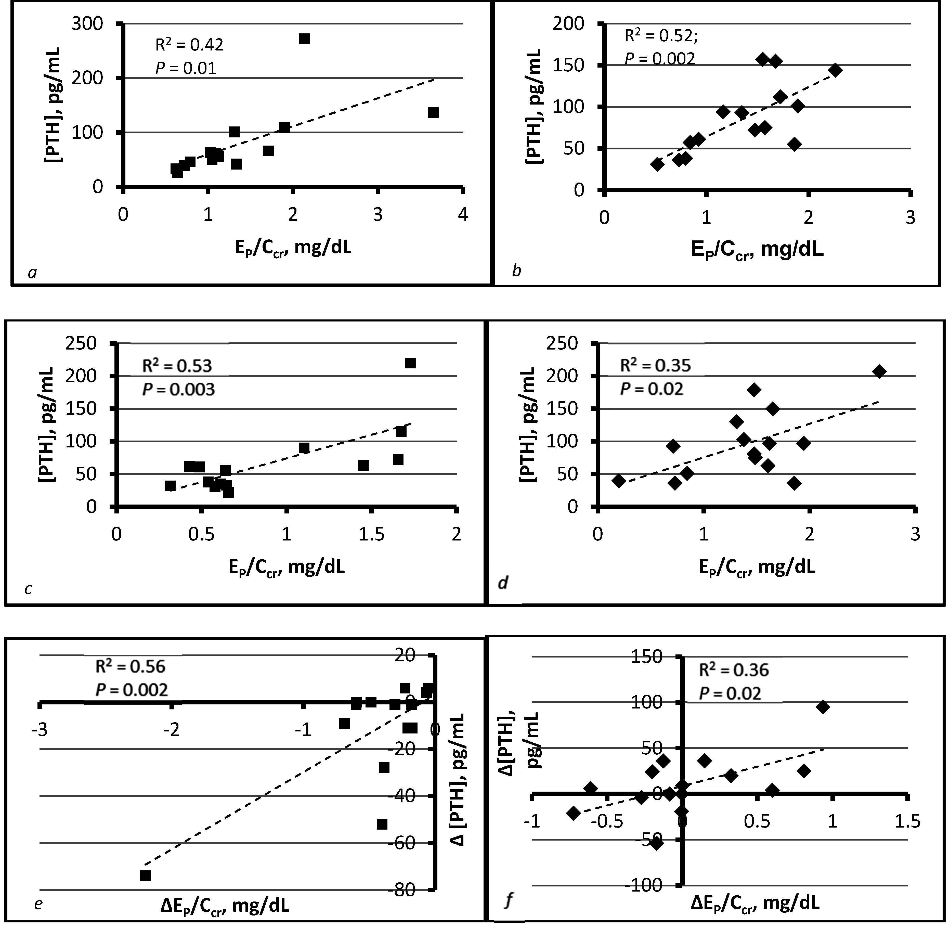
4. Compatibility of Tradeoff-in-the-Nephron with Existing Data

Tradeoff-in-the-nephron is a straightforward hypothesis. It states that high [P]CDN reduces [Ca]CDN by complexation and thus necessitates high [PTH] to maintain normal calcium reabsorption. [P]CDN may rise as a consequence of high IP at a normal GFR or normal IP at a reduced GFR. The hypothesis implies that [PTH] rises in either circumstance, and this implication has been confirmed repeatedly [10,11,12,13,14,22,23,24,25,26,27,28,29,30,31,32,33,34,35,36,37,40,41,50]. Tradeoff-in-the-nephron explains the tight relationship of [PTH] to IP in CKD and accounts for the requirement of high [PTH] to maintain normal TRCa/Ccr and [Ca]i. If EP/Ccr is proportional to [P]CDN, it follows that [PTH] should be a recognizable function of EP/Ccr. Our work has supported this inference [17,18].
In theory, calcium, 1,25D, or phosphate could affect the synthesis and release of PTH in CKD. Of these, only calcium regulates immediate secretion of stored hormone through its interaction with the membrane calcium receptor [90]. If IP affects [PTH] by determining calcium availability for reabsorption, then changes in IP should alter [PTH] quickly. In vivo and in vitro studies have confirmed this expectation [24,30,31,58].
Tradeoff-in-the-nephron explains why [PTH] was high as long as EP/Ccr was high [23,26] and low as long as EP/Ccr was low [34]. The hypothesis explains why [PTH] fell with IP while hyperphosphatemia persisted [27,40]. It accounts for the chronicity of SHPT in CKD, in which [P]CDN is continuously increased at normal IP [83]. The hypothesis explains why [PTH] correlated with EP but not [P]s in early CKD [91], and with EP/Ccr but not [P]s after administration of sevelamer or placebo [17]. It accounts for increased calciuria despite high [PTH] after an oral bolus of phosphate [32]. It explains why [PTH] was elevated in patients with mild CKD, normal IP and low-normal [P]s [35,36], and why [PTH] rose after a bolus of phosphate even though [P]s fell simultaneously [32]. The hypothesis provides a mechanism for high [PTH] in response to high IP despite persistent hypophosphatemia [39]. Most importantly, it predicts normalization of [P]CDN, EP/Ccr, and [PTH] when IP is reduced in proportion to GFR [10,11,12,13,14,17,24,28,29,30,50].
The principal alternatives to tradeoff-in-the-nephron involve skeletal resistance to the calcemic action of PTH, the effect of 1,25D to suppress transcription of the PTH gene, and direct stimulation of PTH synthesis and secretion by circulating phosphate. Much of the evidence for these theories is compatible with our hypothesis. In subjects with functioning kidneys, 1,25D could have enhanced the calcemic response to PTH through its independent effect on calcium reabsorption in the CDN [15]. In addition to limiting calcium egress from bone [41,49], phosphate could have introduced resistance to PTH in the CDN by the mechanism implied in our hypothesis. Instead of making bone more sensitive to PTH, parathyroidectomy could have necessitated a diet that ensured maximal calcium reabsorption from the CDN in response to the hormone [51].
Recurrent themes emerge from studies of the calcemic response to PTH. Typically, the magnitude of the response was less at reduced than at normal GFR, and preparatory phosphate restriction or 1,25D administration mitigated but did not eliminate this difference [38,41,44,45,46,47,50,51]. A notable exception occurred when IP was brought to zero in a model of uremia that left kidneys intact; in that instance, the calcemic response was restored completely [48]. These observations make sense if PTH acted on the CDN as well as the skeleton to raise [Ca]s. When filtrate contained no phosphate, a full complement of nephrons permitted a normal response to PTH even though experimental animals were uremic [48]. In other studies, a deficit of nephrons imposed a limit on the response to PTH that neither phosphate restriction nor 1,25D could overcome [38,41,44,45,46,47,50,51].
The premise that the CDN is the site of PTH resistance is also supported by effects of the calcimimetic agent cinacalcet. In patients with Stage 3 and 4 CKD, the drug reduced [PTH] by 43.1%, but simultaneously kept mean [Ca]s between 8.5 and 9.0 mg/dL even though ECa rose or remained unchanged [71]. Since ICa determined ECa, and since ICa and TRCa maintain [Ca]uf at a given GFR [16], it follows that reduction of [PTH] with cinacalcet led to reduction of TRCa/GFR. High [PTH] was apparently required for reabsorption sufficient to maintain normocalcemia [71].
The capacity of VDRAs to suppress PTH gene transcription can be exploited before ESRD is reached [77,92], but efficacy of the intervention does not confirm reversal of pathogenesis. If deficiency of 1,25D were the cause of SHPT, then normal [PTH] would be incompatible with low [1,25D] in CKD. Numerous investigators have documented this combination after sufficient reduction of IP [13,14,33,34,40,41], and tradeoff-in-the-nephron explains why the combination is possible.
EP/Ccr is a determinant of [P]s, and [P]s is a linear function of EP/Ccr in CKD [18,20,28,57,61]. At the same time, EP/Ccr is approximately proportional to [P]CDN [17,18]. If [PTH] varies directly with [P]s in vivo, the reason may be that [PTH] also varies directly with EP/Ccr. We suggest that this confounding association is responsible for correlations between [PTH] and [P]s in Stage 3 and 4 CKD [18,61].

5. Therapeutic Implications of Tradeoff-in-the-Nephron

Tradeoff-in-the-nephron implies that [PTH] is normal if [P]CDN is normal. EP/Ccr is our surrogate for [P]CDN. Since IP determines EP, a reduction of IP in proportion to GFR yields normal EP/Ccr. Proportional reduction of IP was precisely the intervention that prevented and reversed SHPT in animal models of CKD [10,11,12,13,14,50]. It follows that normalization of EP/Ccr should do the same for patients with SHPT.
In the 1980s and 1990s, European investigators employed severe dietary phosphate restriction to reduce [PTH] in patients with CKD [33,34,40]. Today, in the United States, a similar result requires a drastic revision of eating habits, including avoidance of phosphate preservatives [93,94]. This effort is necessary because to date, the most successful human studies of intestinal phosphate binders have reduced EP by 25%–50% and [PTH] by 13%–35% [37,95,96,97,98]. Our theory and many animal studies suggest that EP/Ccr must be reduced to normal to reverse SHPT completely; if GFR has been reduced by 80%, EP must be reduced by 80%. In addition to diet and binders, blockade of sodium-hydrogen exchanger 3 (NHE3) and inhibition of the intestinal sodium-phosphate 2b co-transporter may ultimately be required to lower IP sufficiently [99,100]. Our experience suggests that normal ECa/Ccr must also be established [17,91]. Attainment of [25D] >30 ng/mL may reduce [PTH] modestly, but we endorse it for other reasons [74]. We presume that normalization of [PTH] is desirable, but concede that the point is debatable [101].

6. Conclusions

Discordant empiric observations undermine each of the major theories concerning the pathogenesis of SHPT. We have sought a unifying explanation for the two most consistent features of the syndrome, which are dependence of [PTH] on IP and persistence of normal [Ca]i until CKD is far advanced. Tradeoff-in-the-nephron accounts for these features. The hypothesis also provides alternate explanations for much of the evidence on which other theories are based, and it sheds light on numerous ancillary observations. It traces SHPT to high [P]CDN and predicts normal [PTH] at normal EP/Ccr. An abundance of evidence is consistent with this prediction. The veracity of tradeoff-in-the-nephron is testable in patients by rigorous but feasible interventions.

Acknowledgments

Work performed by the author and described in this paper was supported with resources of and made possible by facilities at the Stratton Veterans’ Affairs Medical Center, Albany, NY, USA. Genzyme Corporation (now a subsidiary of Sanofi) provided a research grant and study medications. The trial described herein is registered with ClinicalTrials.gov (Identifier NCT01191762).

Conflicts of Interest

The author declares no conflict of interest.

Abbreviations

CKDChronic kidney disease
GFRGlomerular filtration rate (volume/time)
CcrCreatinine clearance (volume/time)
PTHParathyroid hormone
PTEParathyroid extract
PHPTPrimary hyperparathyroidism
SHPTSecondary hyperparathyroidism
CDNCortical distal nephron
[Ca]sSerum calcium concentration, mass/volume
[Ca]iSerum ionized calcium concentration, mass/volume
[Ca]ufSerum ultrafilterable calcium concentration, mass/volume
[Ca]uUrine calcium concentration, mass/volume
[Ca]CDNCalcium concentration in the cortical distal nephron, mass/volume
ICaInflux of calcium (into extracellular fluid or plasma), mass/time
ECaUrinary excretion rate of calcium, mass/time
TRCaRate of tubular reabsorption of calcium, mass/time
ECa/CcrAmount of calcium excreted per volume of filtrate, mass/volume
TRCa/CcrAmount of calcium reabsorbed per volume of filtrate, mass/volume
[P]sSerum phosphorus concentration, mass/volume
[P]pPlasma phosphorus concentration, mass/volume
[P]uUrine phosphorus concentration, mass/volume
[P]CDNPhosphorus concentration in the cortical distal nephron, mass/volume
IPInflux of phosphorus into extracellular fluid or plasma, mass/time
EPUrinary excretion rate of phosphorus, mass/time
TRPTubular reabsorption rate of phosphorus, mass/time
EP/CcrAmount of phosphorus excreted per volume of filtrate, mass/volume
TRP/CcrAmount of phosphorus reabsorbed per volume of filtrate, mass/volume
25D25-hydroxyvitamin D
1,25D1,25-dihydroxyvitamin D
EDTAEthylenediaminetetraacetic acid
VDRAVitamin D receptor activator
mRNAMessenger RNA
FGF23Fibroblast growth factor 23
NHE3Sodium-hydrogen exchanger 3

References

  1. Pitts, T.O.; Piraino, B.H.; Mitro, R.; Chen, T.C.; Segre, G.V.; Greenberg, A.; Puschett, J.B. Hyperparathyroidism and 1,25-dihydroxyvitamin D deficiency in mild, moderate, and severe renal failure. J. Clin. Endocrinol. Metab. 1988, 67, 876–881. [Google Scholar] [CrossRef] [PubMed]
  2. Reichel, H.; Deibert, B.; Schmidt-Gayk, H.; Ritz, E. Calcium metabolism in early chronic renal failure: Implications for the pathogenesis of hyperparathyroidism. Nephrol. Dial. Transplant. 1991, 6, 162–169. [Google Scholar] [CrossRef] [PubMed]
  3. Martinez, I.; Saracho, R.; Montenegro, J.; Llach, F. A deficit of calcitriol synthesis may not be the initial factor in the pathogenesis of secondary hyperparathyroidism. Nephrol. Dial. Transplant. 1996, 11, 22–28. [Google Scholar] [CrossRef] [PubMed]
  4. Levin, A.; Bakris, G.L.; Molitch, M.; Smulders, M.; Tian, J.; Williams, L.A.; Andress, D.L. Prevalence of abnormal serum vitamin D, PTH, calcium and phosphorus in patients with chronic kidney disease: Results of the study to evaluate early kidney disease. Kidney Int. 2007, 71, 31–38. [Google Scholar] [CrossRef] [PubMed]
  5. Craver, L.; Marco, M.P.; Martinez, I.; Rue, M.; Borras, M.; Martin, M.L.; Sarro, F.; Valdivielso, J.M.; Fernandez, E. Mineral metabolism parameters throughout chronic kidney disease stages 1–5—Achievement of K/DOQI target ranges. Nephrol. Dial. Transplant. 2007, 22, 1171–1176. [Google Scholar] [CrossRef] [PubMed]
  6. Malluche, H.H.; Ritz, E.; Lange, H.P.; Kutschera, J.; Hodgson, M.; Seiffert, U.; Schoeppe, W. Bone histology in incipient and advanced renal failure. Kidney Int. 1976, 9, 355–362. [Google Scholar] [CrossRef] [PubMed]
  7. Kim, S.M.; Long, J.; Montez-Rath, M.; Leonard, M.; Chertow, G.M. Hip fracture in patients with non-dialysis-requiring chronic kidney disease. J. Bone Miner. Res. 2016, 31, 1803–1809. [Google Scholar] [CrossRef] [PubMed]
  8. Ritz, E.; Stefanski, A.; Rambausek, M. The role of the parathyroid glands in the uremic syndrome. Am. J. Kidney Dis. 1995, 26, 808–813. [Google Scholar] [CrossRef]
  9. Goodman, W.G. The consequences of uncontrolled secondary hyperparathyroidism and its treatment in chronic kidney disease. Semin. Dial. 2004, 17, 209–216. [Google Scholar] [CrossRef] [PubMed]
  10. Slatopolsky, E.; Caglar, S.; Pennell, J.P.; Taggart, D.D.; Canterbury, J.M.; Reiss, E.; Bricker, N.S. On the pathogenesis of hyperparathyroidism in chronic experimental renal insufficiency in the dog. J. Clin. Investig. 1971, 50, 492–499. [Google Scholar] [CrossRef] [PubMed]
  11. Slatopolsky, E.; Caglar, S.; Gradowska, L.; Canterbury, J.; Reiss, E.; Bricker, N.S. On the prevention of secondary hyperparathyroidism in experimental chronic renal disease using “proportional reduction” of dietary phosphorus intake. Kidney Int. 1972, 2, 147–151. [Google Scholar] [CrossRef] [PubMed]
  12. Kaplan, M.A.; Canterbury, J.M.; Bourgoignie, J.J.; Veliz, G.; Gavellas, G.; Reiss, E.; Bricker, N.S. Reversal of hyperparathyroidism in response to dietary phosphorus restriction in the uremic dog. Kidney Int. 1979, 15, 43–48. [Google Scholar] [CrossRef] [PubMed]
  13. Lopez-Hilker, S.; Dusso, A.S.; Rapp, N.S.; Martin, K.J.; Slatopolsky, E. Phosphorus restriction reverses hyperparathyroidism in uremia independent of changes in calcium and calcitriol. Am. J. Physiol. 1990, 259, F432–F437. [Google Scholar] [PubMed]
  14. Denda, M.; Finch, J.; Slatopolsky, E. Phosphorus accelerates the development of parathyroid hyperplasia and secondary hyperparathyroidism in rats with renal failure. Am. J. Kidney Dis. 1996, 28, 596–602. [Google Scholar] [CrossRef]
  15. Boros, S.; Bindels, R.J.M.; Hoenderop, J.G.J. Active Ca2+ reabsorption in the connecting tubule. Pflug. Arch. 2009, 458, 99–109. [Google Scholar] [CrossRef] [PubMed]
  16. Phelps, K.R.; Stote, K.S.; Mason, D. Tubular calcium reabsorption and other aspects of calcium homeostasis in primary and secondary hyperparathyroidism. Clin. Nephrol. 2014, 82, 83–91. [Google Scholar] [CrossRef] [PubMed]
  17. Phelps, K.R.; Stote, K.S.; Mason, D. Use of sevelamer to examine the role of intraluminal phosphate in the pathogenesis of secondary hyperparathyroidism. Clin. Nephrol. 2014, 82, 191–201. [Google Scholar] [CrossRef] [PubMed]
  18. Phelps, K.R.; Mason, D.L.; Stote, K.S. Phosphate homeostasis, parathyroid hormone, and fibroblast growth factor 23 in stages 3 and 4 chronic kidney disease. Clin. Nephrol. 2016, 85, 251–259. [Google Scholar] [CrossRef] [PubMed]
  19. Phelps, K.R.; Mason, D.L. Parameters of phosphorus homeostasis at normal and reduced GFR: Theoretical considerations. Clin. Nephrol. 2015, 83, 167–176. [Google Scholar] [CrossRef] [PubMed]
  20. Phelps, K.R.; Mason, D.L.; Stote, K.S. Parameters of phosphorus homeostasis at normal and reduced GFR: Empiric observations. Clin. Nephrol. 2015, 83, 208–217. [Google Scholar] [CrossRef] [PubMed]
  21. Slatopolsky, E.; Robson, A.M.; Elkan, I.; Bricker, N.S. Control of phosphate excretion in uremic man. J. Clin. Investig. 1968, 47, 1865–1874. [Google Scholar] [CrossRef] [PubMed]
  22. Reiss, E.; Canterbury, J.M.; Bercovitz, M.A.; Kaplan, E.L. The role of phosphate in the secretion of parathyroid hormone in man. J. Clin. Investig. 1970, 49, 2146–2149. [Google Scholar] [CrossRef] [PubMed]
  23. Jowsey, J.; Reiss, E.; Canterbury, J.M. Long-term effects of high phosphate intake on parathyroid hormone levels and bone metabolism. Acta Orthop. Scand. 1974, 45, 801–808. [Google Scholar] [CrossRef] [PubMed]
  24. Kaplan, M.A.; Canterbury, J.M.; Gavellas, G.; Jaffe, D.; Bourgoignie, J.J.; Reiss, E.; Bricker, N.S. Interrelations between phosphorus, calcium, parathyroid hormone and renal phosphate excretion in response to an oral phosphorus load in normal and uremic dogs. Kidney Int. 1978, 14, 207–214. [Google Scholar] [CrossRef] [PubMed]
  25. Bover, J.; Rodriguez, M.; Trinidad, P.; Jara, A.; Martinez, M.E.; Machado, L.; Llach, F.; Felsenfeld, A.J. Factors in the development of secondary hyperparathyroidism during graded renal failure in the rat. Kidney Int. 1994, 45, 953–961. [Google Scholar] [CrossRef] [PubMed]
  26. Krapf, R.; Glatz, M.; Hulter, H.N. Neutral phosphate administration generates and maintains renal metabolic alkalosis and hyperparathyroidism. Am. J. Physiol. 1995, 268, F802–F807. [Google Scholar] [PubMed]
  27. Estepa, J.C.; Aguilera-Tejero, E.; Lopez, I.; Almaden, Y.; Rodriguez, M.; Felsenfeld, A.J. Effect of phosphate on parathyroid hormone secretion in vivo. J. Bone Miner. Res. 1999, 14, 1848–1854. [Google Scholar] [CrossRef] [PubMed]
  28. Takahashi, F.; Denda, M.; Finch, J.L.; Brown, A.L.; Slatopolsky, E. Hyperplasia of the parathyroid gland with secondary hyperparathyroidism. Kidney Int. 2002, 61, 1332–1338. [Google Scholar] [CrossRef] [PubMed]
  29. Ritter, C.S.; Martin, D.R.; Lu, Y.; Slatopolsky, E.; Brown, A.J. Reversal of secondary hyperparathyroidism by phosphate restriction restores parathyroid calcium-sensing receptor expression and function. J. Bone Miner. Res. 2002, 17, 2206–2213. [Google Scholar] [CrossRef] [PubMed]
  30. Martin, D.R.; Ritter, C.S.; Slatopolsky, E.; Brown, A.J. Acute regulation of parathyroid hormone by dietary phosphate. Am. J. Physiol. 2005, 289, E729–E734. [Google Scholar] [CrossRef] [PubMed]
  31. Nagano, N.; Miyata, S.; Abe, M.; Kobayashi, N.; Wakita, S.; Yamashita, T.; Wada, M. Effect of manipulating serum phosphorus with phosphate binder on circulating PTH and FGF23 in renal failure rats. Kidney Int. 2006, 69, 531–537. [Google Scholar] [CrossRef] [PubMed]
  32. Isakova, T.; Gutierrez, O.; Shah, A.; Castaldo, L.; Holmes, J.; Lee, H.; Wolf, M. Post-prandial mineral metabolism and secondary hyperparathyroidism in early chronic kidney disease. J. Am. Soc. Nephrol. 2008, 19, 615–623. [Google Scholar] [CrossRef] [PubMed]
  33. Combe, C.; Aparicio, M. Phosphorus and protein restriction and parathyroid function in chronic renal failure. Kidney Int. 1994, 46, 1381–1386. [Google Scholar] [CrossRef] [PubMed]
  34. Combe, C.; Morel, D.; de Precigout, V.; Blanchetier, V.; Bouchet, J.L.; Potaux, L.; Fournier, A.; Aparicio, M. Long-term control of hyperparathyroidism in advanced renal failure by low-phosphorus low-protein diet supplemented with calcium (without changes in plasma calcitriol). Nephron 1995, 70, 287–295. [Google Scholar] [CrossRef] [PubMed]
  35. Portale, A.A.; Booth, B.E.; Halloran, B.P.; Morris, R.C., Jr. Effect of dietary phosphorus on circulating cconcentrations of 1,25-dihydroxyvitamin D and immunoreactive parathyroid hormone in children with moderate renal insufficiency. J. Clin. Investig. 1984, 73, 1580–1589. [Google Scholar] [CrossRef] [PubMed]
  36. Llach, F.; Massry, S.G. On the mechanism of secondary hyperparathyroidism in moderate renal insufficiency. J. Clin. Endocrinol. Metab. 1985, 61, 601–606. [Google Scholar] [CrossRef] [PubMed]
  37. Oliveira, R.B.; Cancela, A.L.E.; Graciolli, F.G.; Dos Reis, L.M.; Draibe, S.A.; Cuppari, L.; Carvalho, A.B.; Jorgetti, V.; Canziani, M.E.; Moyses, R.M.A. Early control of PTH and FGF23 in normophosphatemic CKD patients: A new target in CKD-MBD therapy? Clin. J. Am. Soc. Nephrol. 2010, 5, 286–291. [Google Scholar] [CrossRef] [PubMed]
  38. Wilson, L.; Felsenfeld, A.; Drezner, M.K.; Llach, F. Altered divalent metabolism in early renal failure. Role of 1,25(OH)2D. Kidney Int. 1985, 27, 565–573. [Google Scholar] [CrossRef] [PubMed]
  39. Rivkees, S.A.; El-Hajj-Fuleihan, G.; Brown, E.M.; Crawford, J.D. Tertiary hyperparathyroidism during high phosphate therapy of familial hypophosphatemic rickets. J. Clin. Endocrinol. Metab. 1992, 75, 1514–1518. [Google Scholar] [PubMed]
  40. Lucas, P.A.; Brown, R.C.; Woodhead, J.S.; Coles, G.A. 1,25-dihydroxycholecalciferol and parathyroid hormone in advanced chronic renal failure: Effects of simultaneous protein and phosphorus restriction. Clin. Nephrol. 1986, 25, 7–10. [Google Scholar] [PubMed]
  41. Rodriguez, M.; Martin-Malo, A.; Martinez, M.E.; Torres, A.; Felsenfeld, A.J.; Llach, F. Calcemic response to parathyroid hormone in renal failure: Role of phosphorus and its effect on calcitriol. Kidney Int. 1991, 40, 1055–1062. [Google Scholar] [CrossRef] [PubMed]
  42. Bricker, N.S. On the pathogenesis of the uremic state: An exposition of the “trade-off hypothesis”. N. Engl. J. Med. 1972, 286, 1093–1099. [Google Scholar] [PubMed]
  43. Adler, A.J.; Ferran, N.; Berlyne, G.M. Effect of inorganic phosphate on serum ionized calcium concentration: A reassessment of the “trade-off hypothesis”. Kidney Int. 1985, 28, 932–935. [Google Scholar] [CrossRef] [PubMed]
  44. Massry, S.G.; Coburn, J.W.; Lee, D.B.N.; Jowsey, J.; Kleeman, C.R. Skeletal resistance to parathyroid hormone in renal failure. Studies in 105 human subjects. Ann. Int. Med. 1973, 78, 357–364. [Google Scholar] [CrossRef] [PubMed]
  45. Llach, F.; Massry, S.G.; Singer, F.R.; Kurokawa, K.; Kaye, J.H.; Coburn, J.W. Skeletal resistance to endogenous parathyroid hormone in patients with early renal failure. A possible cause for secondary hyperparathyroidism. J. Clin. Endocrinol. Metab. 1975, 41, 339–345. [Google Scholar] [CrossRef] [PubMed]
  46. Massry, S.G.; Stein, R.; Garty, J.; Arieff, A.I.; Coburn, J.W.; Norman, A.W.; Friedler, R.M. Skeletal resistance to the calcemic action of parathyroid hormone in uremia: Role of 1,25(OH)2D. Kidney Int. 1976, 9, 467–474. [Google Scholar] [CrossRef] [PubMed]
  47. Somerville, P.J.; Kaye, M. Resistance to parathyroid hormone in renal failure: Role of vitamin D metabolites. Kidney Int. 1978, 14, 245–254. [Google Scholar] [CrossRef] [PubMed]
  48. Somerville, P.J.; Kaye, M. Evidence that resistance to the calcemic action of parathyroid hormone in rats with acute uremia is caused by phosphate retention. Kidney Int. 1979, 16, 552–560. [Google Scholar] [CrossRef] [PubMed]
  49. Somerville, P.J.; Kaye, M. Action of phosphorus on calcium release in isolated perfused rat tails. Kidney Int. 1982, 22, 348–354. [Google Scholar] [CrossRef] [PubMed]
  50. Kaplan, M.A.; Canterbury, J.M.; Gavellas, G.; Jaffe, D.; Bourgoignie, J.J.; Reiss, E.; Bricker, N.S. The calcemic and phosphaturic effects of parathyroid hormone in the normal and uremic dog. Metabolism 1978, 27, 1785–1792. [Google Scholar] [CrossRef]
  51. Rodriguez, M.; Felsenfeld, A.J.; Llach, F. Calcemic response to parathyroid hormone in renal failure: Role of calcitriol and the effect of parathyroidectomy. Kidney Int. 1991, 40, 1063–1068. [Google Scholar] [CrossRef] [PubMed]
  52. Zehnder, D.; Bland, R.; Walker, E.A.; Bradwell, A.R.; Howie, A.J.; Hewison, M.; Stewart, P.M. Expression of 25-hydroxyvitamin D3-1α hydroxylase in the human kidney. J. Am. Soc. Nephrol. 1999, 10, 2465–2473. [Google Scholar] [PubMed]
  53. Walling, M.W. Intestinal calcium and phosphate transport: Differential responses to vitamin D3 metabolites. Am. J. Physiol. 1977, 233, E488–E494. [Google Scholar] [PubMed]
  54. Silver, J.; Russell, J.; Sherwood, L.M. Regulation by vitamin D metabolites of messenger ribonucleic acid for preproparathyroid hormone in isolated bovine parathyroid cells. Proc. Natl. Acad. Sci. USA 1985, 82, 4270–4273. [Google Scholar] [CrossRef] [PubMed]
  55. Silver, J.; Naveh-Many, T.; Mayer, H.; Schmelzer, H.J.; Popovtzer, M.M. Regulation by vitamin D metabolites of parathyroid hormone gene transcription in vivo in the rat. J. Clin. Investig. 1986, 78, 1296–1301. [Google Scholar] [CrossRef] [PubMed]
  56. Sprague, S.M.; Coyne, D. Control of secondary hyperparathyroidism by vitamin D receptor agonists in chronic kidney disease. Clin. J. Am. Soc. Nephrol. 2010, 5, 512–518. [Google Scholar] [CrossRef] [PubMed]
  57. Slatopolsky, E.; Finch, J.; Denda, M.; Ritter, C.; Zhong, M.; Dusso, A.; MacDonald, P.N.; Brown, A.J. Phosphorus restriction prevents parathyroid gland growth. High phosphorus directly stimulates PTH secretion in vitro. J. Clin. Investig. 1996, 97, 2534–2540. [Google Scholar] [CrossRef] [PubMed]
  58. Almaden, Y.; Canalejo, A.; Hernandez, A.; Ballesteros, E.; Garcia-Navarro, S.; Torres, A.; Rodriguez, M. Direct effect of phosphorus on PTH secretion from whole rat parathyroid glands in vitro. J. Bone Miner. Res. 1996, 11, 970–976. [Google Scholar] [CrossRef] [PubMed]
  59. Almaden, Y.; Hernandez, A.; Torregrosa, V.; Canalejo, A.; Sabate, L.; Fernandez, C.L.; Campistol, J.M.; Torres, A.; Rodriguez, M. High phosphate level directly stimulates parathyroid hormone secretion and synthesis by human parathyroid hormone tissue in vitro. J. Am. Soc. Nephrol. 1998, 9, 1845–1852. [Google Scholar] [PubMed]
  60. Moallem, E.; Kilav, R.; Silver, J.; Naveh-Many, T. RNA-protein binding and post-transcriptional regulation of parathyroid hormone gene expression by calcium and phosphate. J. Biol. Chem. 1998, 273, 5253–5259. [Google Scholar] [CrossRef] [PubMed]
  61. Kates, D.M.; Sherrard, D.J.; Andress, D.L. Evidence that serum phosphate is independently associated with serum PTH in patients with chronic renal failure. Am. J. Kidney Dis. 1997, 30, 809–813. [Google Scholar] [CrossRef]
  62. Phelps, K.R.; Mason, D.L. Evidence that TmP/GFR can be estimated with the Walton-Bijvoet nomogram in chronic kidney disease. Clin. Nephrol. 2017, in press. [Google Scholar] [CrossRef] [PubMed]
  63. Liu, S.; Zhou, J.; Tang, W.; Jiang, X.; Rowe, D.W.; Quarles, L.D. Pathogenic role of FGF23 in Hyp mice. Am. J. Physiol. 2006, 291, E38–E49. [Google Scholar] [CrossRef] [PubMed]
  64. Zanchi, C.; Locatelli, M.; Benigni, A.; Corna, D.; Tomasoni, S.; Rottoli, D.; Gaspari, F.; Remuzzi, G.; Zoja, C. Renal expression of FGF23 in progressive renal disease of diabetes and the effect of ACE inhibitor. PLoS ONE 2013, 8, e70775. [Google Scholar] [CrossRef] [PubMed]
  65. Isakova, T.; Wahl, P.; Vargas, G.S.; Gutierrez, O.M.; Scialla, J.; Xie, H.; Appleby, D.; Nessel, L.; Bellovich, K.; Chen, J.; et al. Fibroblast growth factor 23 is elevated before parathyroid hormone and phosphate in chronic kidney disease. Kidney Int. 2011, 79, 1370–1378. [Google Scholar] [CrossRef] [PubMed]
  66. Komaba, H.; Fukagawa, M. FGF23-parathyroid interaction: Implications in chronic kidney disease. Kidney Int. 2010, 77, 292–298. [Google Scholar] [CrossRef] [PubMed]
  67. Ben-Dov, I.Z.; Galitzer, H.; Lavi-Moshayoff, V.; Goetz, R.; Kuro-o, M.; Mohammadi, M.; Sirkis, R.; Naveh-Many, T.; Silver, J. The parathyroid is a target organ for FGF23 in rats. J. Clin. Investig. 2007, 117, 4003–4008. [Google Scholar] [CrossRef] [PubMed]
  68. Galitzer, H.; Ben-Dov, I.Z.; Silver, J.; Naveh-Many, T. Parathyroid cell resistance to fibroblast growth factor 23 in secondary hyperparathyroidism of chronic kidney disease. Kidney Int. 2010, 77, 211–218. [Google Scholar] [CrossRef] [PubMed]
  69. Canalejo, R.; Canalejo, A.; Martinez-Moreno, J.M.; Rodriguez-Ortiz, M.E.; Estepa, J.C.; Mendoza, F.J.; Munoz-Castaneda, J.R.; Shalhoub, V.; Almaden, Y.; Rodriguez, M. FGF23 fails to inhibit uremic parathyroid glands. J. Am. Soc. Nephrol. 2010, 21, 1125–1135. [Google Scholar] [CrossRef] [PubMed]
  70. Andrukhova, O.; Streicher, C.; Zeitz, U.; Erben, R.G. Fgf23 and parathyroid hormone signaling interact in kidney and bone. Mol. Cell. Endocrinol. 2016, 436, 224–239. [Google Scholar] [CrossRef] [PubMed]
  71. Chonchol, M.; Locatelli, F.; Abboud, H.E.; Charytan, C.; de Francisco, A.L.M.; Jolly, S.; Kaplan, M.; Roger, S.D.; Sarkar, S.; Albizem, M.B.; et al. A randomized, double-blind, placebo-controlled study to assess the efficacy and safety of cinacalcet HCl in participants with CKD not receiving dialysis. Am. J. Kidney Dis. 2009, 53, 197–207. [Google Scholar] [CrossRef] [PubMed]
  72. Hasegawa, H.; Nagano, N.; Urakawa, I.; Yamazaki, Y.; Iijima, K.; Fujita, T.; Yamashita, T.; Fukumoto, S.; Shimada, T. Direct evidence for a causative role of FGF23 in the abnormal renal phosphate handling and vitamin D metabolism in rats with early-stage chronic kidney disease. Kidney Int. 2010, 78, 975–980. [Google Scholar] [CrossRef] [PubMed]
  73. Rosen, C.J. Vitamin D insufficiency. N. Engl. J. Med. 2011, 364, 248–254. [Google Scholar] [CrossRef] [PubMed]
  74. Holick, M.F. Vitamin D deficiency. N. Engl. J. Med. 2007, 357, 266–281. [Google Scholar] [CrossRef] [PubMed]
  75. Manson, J.E.; Brannon, P.M.; Rosen, C.J.; Taylor, C.L. Vitamin D deficiency—Is there really a pandemic? N. Engl. J. Med. 2016, 375, 1817–1820. [Google Scholar] [CrossRef] [PubMed]
  76. Hollis, B.W.; Wagner, C.L. Normal serum vitamin D levels. N. Engl. J. Med. 2005, 352, 515. [Google Scholar] [PubMed]
  77. Moe, S.M.; Saifullah, A.; LaClair, R.E.; Usman, S.A.; Yu, Z. A randomized trial of cholecalciferol versus doxercalciferol for lowering parathyroid hormone in chronic kidney disease. Clin. J. Am. Soc. Nephrol. 2010, 5, 299–306. [Google Scholar] [CrossRef] [PubMed]
  78. Alvarez, J.A.; Law, J.; Coakley, K.E.; Zughaier, S.M.; Hao, L.; Salles, K.S.; Wasse, H.; Gutierrez, O.M.; Ziegler, T.R.; Tangpricha, V. High-dose cholecalciferol reduces parathyroid hormone in patients with early chronic kidney disease: A pilot, randomized, double-blind, placebo-controlled trial. Am. J. Clin. Nutr. 2012, 96, 672–679. [Google Scholar] [CrossRef] [PubMed]
  79. Nigwekar, S.U.; Bhan, I.; Thadhani, R. Ergocalciferol and cholecalciferol in CK.D. Am. J. Kidney Dis. 2012, 60, 139–156. [Google Scholar] [CrossRef] [PubMed]
  80. Sprague, S.M.; Silva, A.L.; Al-Saghir, F.; Damle, R.; Tabash, S.P.; Petkovich, M.; Messner, E.J.; White, J.A.; Melnick, J.Z.; Bishop, C.W. Modified-release calcifediol effectively controls secondary hyperparathyroidism associated with vitamin D insufficiency in chronic kidney disease. Am. J. Nephrol. 2014, 40, 535–545. [Google Scholar] [CrossRef] [PubMed]
  81. Sprague, S.M.; Crawford, P.W.; Melnick, J.Z.; Strugnell, S.A.; Ali, S.; Mangoo-Karim, R.; Lee, S.; Petkovich, P.M.; Bishop, C.W. Use of extended-release calcifediol to treat secondary hyperparathyroidism in stages 3 and 4 chronic kidney disease. Am. J. Nephrol. 2016, 44, 316–325. [Google Scholar] [CrossRef] [PubMed]
  82. Wills, M.R.; Lewin, M.R. Plasma calcium fractions and the protein-binding of calcium in normal subjects and in patients with hypercalcaemia and hypocalcaemia. J. Clin. Pathol. 1971, 24, 856–866. [Google Scholar] [CrossRef] [PubMed]
  83. Bank, N.; Su, W.S.; Aynedjian, H.S. A micropuncture study of renal phosphate transport in rats with chronic renal failure and secondary hyperparathyroidism. J. Clin. Investig. 1978, 61, 884–894. [Google Scholar] [CrossRef] [PubMed]
  84. Tiselius, H.G. Estimated levels of supersaturation with calcium phosphate and calcium oxalate in the distal tubule. Urol. Res. 1997, 25, 153–159. [Google Scholar] [CrossRef] [PubMed]
  85. Luptak, J.; Bek-Jensen, H.; Fornander, A.-M.; Hojgaard, I.; Nilsson, M.-A.; Tiselius, H.-G. Crystallization of calcium oxalate and calcium phosphate at supersaturation levels corresponding to those in different parts of the nephron. Scanning Microsp. 1994, 8, 47–62. [Google Scholar]
  86. Haut, L.L.; Alfrey, A.C.; Guggenheim, S.; Buddington, B.; Schrier, N. Renal toxicity of phosphate in rats. Kidney Int. 1980, 17, 722–731. [Google Scholar] [CrossRef] [PubMed]
  87. Markowitz, G.S.; Nasr, S.H.; Klein, P.; Anderson, H.; Stack, J.I.; Alterman, L.; Price, B.; Radhakrishnan, J.; D’Agati, V.D. Renal failure due to acute nephrocalcinosis following oral sodium phosphate bowel cleansing. Hum. Pathol. 2004, 35, 675–684. [Google Scholar] [CrossRef] [PubMed]
  88. Walser, M. Ion association. VI. Interactions between calcium, magnesium, inorganic phosphate, citrate, and protein in normal human plasma. J. Clin. Investig. 1961, 40, 723–730. [Google Scholar] [CrossRef] [PubMed]
  89. Knox, F.G.; Osswald, H.; Marchand, G.R.; Spielman, W.S.; Haas, J.A.; Berndt, T.; Youngberg, S.P. Phosphate transport along the nephron. Am. J. Physiol. 1977, 233, F261–F268. [Google Scholar] [PubMed]
  90. Goodman, W.G.; Quarles, L.D. Development and progression of secondary hyperparathyroidism in chronic kidney disease: Lessons from molecular genetics. Kidney Int. 2008, 74, 276–288. [Google Scholar] [CrossRef] [PubMed]
  91. Martinez, I.; Saracho, R.; Montenegro, J.; Llach, F. The importance of dietary calcium and phosphorus in the secondary hyperparathyroidism of patients with early renal failure. Am. J. Kidney Dis. 1997, 29, 496–502. [Google Scholar] [CrossRef]
  92. Kovesdy, C.P.; Lu, J.L.; Malakauskas, S.M.; Andress, D.L.; Kalantar-Zadeh, K.; Ahmadzadeh, S. Paricalcitol versus ergocalciferol for secondary hyperparathyroidism in CKD stages 3 and 4: A randomized controlled trial. Am. J. Kidney Dis. 2012, 59, 58–66. [Google Scholar] [CrossRef] [PubMed]
  93. Uribarri, J.; Calvo, M.S. Hidden sources of phosphorus in the typical American diet: Does it matter in nephrology? Semin. Dial. 2003, 16, 186–188. [Google Scholar] [CrossRef] [PubMed]
  94. Sullivan, C.; Sayre, S.S.; Leon, J.B.; Machekano, R.; Love, T.E.; Porter, D.; Marbury, M.; Sehgal, A. Effect of food additives on hyperphosphatemia among patients with end-stage renal disease. A randomized controlled trial. JAMA 2009, 301, 629–635. [Google Scholar] [CrossRef] [PubMed]
  95. Sprague, S.M.; Abboud, H.; Qiu, P.; Dauphin, M.; Zhang, P.; Finn, W. Lanthanum carbonate reduces phosphorus burden in patients with CKD stages 3 and 4: A randomized trial. Clin. J. Am. Soc. Nephrol. 2009, 4, 178–185. [Google Scholar] [CrossRef] [PubMed]
  96. Block, G.A.; Wheeler, D.C.; Persky, M.S.; Kestenbaum, B.; Ketteler, M.; Spiegel, D.M.; Allison, M.A.; Asplin, J.; Smits, G.; Hoofnagle, A.N.; et al. Effects of phosphate binders in moderate CKD. J. Am. Soc. Nephrol. 2012, 23, 1407–1415. [Google Scholar] [CrossRef] [PubMed]
  97. Yokoyama, K.; Hirakata, H.; Akiba, T.; Fukagawa, M.; Nakayama, M.; Sawada, K.; Kumagai, Y.; Block, G.A. Ferric citrate hydrate for the treatment of hyperphosphatemia in nondialysis-dependent CKD. Clin. J. Am. Soc. Nephrol. 2014, 9, 543–552. [Google Scholar] [CrossRef] [PubMed]
  98. Block, G.A.; Fishbane, S.; Rodriguez, M.; Smits, G.; Shemesh, S.; Pergola, P.E.; Wolf, M.; Chertow, G.M. A 12-week, double-blind, placebo-controlled trial of ferric citrate for the treatment of iron deficiency anemia and reduction of serum phosphate in patients with CKD stages 3–5. Am. J. Kidney Dis. 2015, 65, 728–736. [Google Scholar] [CrossRef] [PubMed]
  99. Labonte, E.D.; Carreras, C.W.; Leadbetter, M.R.; Kozuka, K.; Kohler, J.; Koo-McCoy, S.; He, L.; Dy, E.; Black, D.; Zhong, Z.; et al. Gastrointestinal inhibition of sodium-hydrogen exchanger 3 reduces phosphorus absorption and protects against vascular calcification in CKD. J. Am. Soc. Nephrol. 2015, 26, 1138–1149. [Google Scholar] [CrossRef] [PubMed]
  100. Rao, M.; Steffes, M.; Bostom, A.; Ix, J.H. Effect of niacin on FGF23 concentration in chronic kidney disease. Am. J. Nephrol. 2014, 39, 484–490. [Google Scholar] [CrossRef] [PubMed]
  101. Kidney Disease: Improving Global Outcomes (KDIGO) CKD-MBD Work Group. Clinical practice guidelines for the management of CKD-MBD. Kidney Int. 2009, 76, S1–S130. [Google Scholar]
Figure 1. Plots of ECa/Ccr and TRCa/Ccr against [PTH] in control subjects and patients with primary and secondary hyperparathyroidism (PHPT and SHPT). All data are derived from morning fasting specimens of urine and serum or plasma. Circles represent normal controls. Triangles and diamonds represent patients with PHPT and SHPT (CKD), respectively. Frame (a) shows that the lowest recorded values of ECa/Ccr in controls were compatible with normal [PTH]. It also shows that a minority of patients with CKD exhibited high ECa/Ccr and high [PTH] simultaneously. Frame (b) shows that [PTH] capable of causing high TRCa/Ccr in patients with PHPT maintained normal TRCa/Ccr in patients with CKD. Reproduced from [16] with permission of the publisher (Dustri-Verlag). ECa, Urinary excretion rate of calcium, mass/time; Ccr, Creatinine clearance (volume/time); TRCa, Rate of tubular reabsorption of calcium, mass/time; PTH, Parathyroid hormone; CKD, Chronic kidney disease.
Nutrients 09 00427 g001
Figure 2. Relationship of [PTH] to EP/Ccr in sevelamer and placebo recipients. Squares pertain to the sevelamer group and diamonds to the placebo group. Graphs (a) and (c) show regressions of [PTH] on EP/Ccr before and after administration of sevelamer carbonate for four weeks. Graphs (b) and (d) show the same regressions before and after administration of a placebo for four weeks. Graphs (e) and (f) show regressions of ∆[PTH] on ∆EP/Ccr in the sevelamer and placebo groups, respectively, where “∆” = change during treatment. All regressions are statistically significant. Adapted from [17] with permission of the publisher (Dustri-Verlag). EP, urinary excretion rate of phosphorus, mass/time; Ccr, creatinine clearance, volume/time.
Nutrients 09 00427 g002

