Perspective

The “Jekyll Side” of the S100B Protein: Its Trophic Action in the Diet

by
Fabrizio Michetti
and
Vincenzo Romano Spica
*
Department of Movement, Human, and Health Sciences, University of Rome “Foro Italico”, 00135 Rome, Italy
*
Author to whom correspondence should be addressed.
Nutrients 2025, 17(5), 881; https://doi.org/10.3390/nu17050881
Submission received: 16 January 2025 / Revised: 26 February 2025 / Accepted: 27 February 2025 / Published: 28 February 2025
(This article belongs to the Section Nutrition and Public Health)

Abstract

:
The calcium-binding S100B protein is concentrated in glial cells (including enteroglial cells) in the nervous system. Its conformation and amino acid composition are significantly conserved in different species; this characteristic suggests conserved biological role(s) for the protein. The biological activity is concentration-dependent: low physiological concentrations exert a neurotrophic effect, while high concentrations exert a proinflammatory/toxic role. The proinflammatory/toxic role of S100B currently attracts the scientific community’s primary attention, while the protein’s physiological action remains unraveled—yet remarkably interesting. This is now a topical issue due to the recently consolidated notion that S100B is a natural trophic nutrient available in breast milk and/or other aliments, possibly interacting with other body districts through its impact on microbiota. These recent data may offer novel clues to understanding the role of this challenging protein.

Graphical Abstract

1. Introduction

The S100B protein is a calcium-binding protein concentrated in glial cells (mainly astrocytes) in the nervous system. The protein is also characterized by its solubility in a 100% saturated solution with ammonium sulfate. This peculiarity was at the basis of its denomination, which was originally the S100 protein. Interestingly, the conformation and amino acid composition of S100B have been shown to be significantly conserved in different species, suggesting that it may have a crucially conserved biological role and, possibly, a functional specificity [1]. This protein is part of the S100 protein family, which has over 20 calcium-binding proteins. They exhibit structural similarities and modulate the activity of a large number of targets operating as calcium-activated switches in different tissues. S100 proteins constitute a subgroup within the EF-hand protein superfamily, which is characterized by a calcium-binding loop forming a conserved pentagonal arrangement around the calcium ion (the EF-hand motif). Each S100 protein is characterized by the presence of two Ca2+-modulated motifs of the EF-hand type interconnected by an intermediate region, which is often referred to as the hinge region, resulting in a helix–loop–helix arrangement. S100B is an acidic homodimer (2 beta subunits) of 9–14 kDa per monomer that constituted the bulk of the protein fraction originally isolated from brain extracts in the early 1960s. Using chromatographic and electrophoretic methods, the protein originally was detectable in brain extracts but not in non-neural tissues; consequently, it was regarded as specific to the nervous system. In the nervous system, S100B has been shown to be concentrated in astrocytes, as above indicated, and other glial cell types, namely enteric glial cells, and it has also been reportedly located in some neuron subpopulations. The protein was later identified in definite extra-neural cell types, such as chondrocytes, melanocytes, Langerhans cells, dendritic cells of lymphoid organs, some lymphocyte cell types, adrenal medulla satellite cells, skeletal muscle satellite cells, tubular kidney cells, Leydig cells, non-nervous structures of the eye, and adipocytes. Additionally, S100B-like immunoreactivity has been detected in invertebrates, such as the planarian (Dugesia gonocephala), the silkworm (Bombyx mori), the earthworm (Pheretima communissima), the cockroach (Periplaneta americana) [2,3], and, most importantly, in spinach (Spinacia oleracea) and other plants [4,5]. These latter findings, together with the detection of S100B in milk [6,7], are of particularly relevant interest considering the novel view indicating that this protein is a natural component of different aliments, with a potential role as a dietary driver.
To date, the activity of S100B has been studied in different tissues and biological fluids. It has appeared to be closely associated with its concentration. In particular, low (nanomolar) concentrations of the protein, which are regarded as physiologic and were the original focus of research, have been found to exert a trophic effect (the “Jekyll side” of S100B), while high (micromolar) concentrations of the protein appear to exert a toxic/proinflammatory role (the “Hyde side” of S100B), behaving as a danger/damage-associated molecular pattern (DAMP) protein [1,8,9]. In either case, extracellular S100B is regarded as interacting with surrounding cell types mainly through the Receptor for Advanced Glication Endproducts (RAGE), a ubiquitous, transmembrane, immunoglobulin-like receptor that binds to a diverse range of extracellular ligands and intracellular effectors [10]. At present, the “Hyde side” of S100B appears to attract the primary interest of the scientific community, especially regarding the use of the protein levels as a biomarker of different disorders, such as Alzheimer’s disease (global prevalence of dementia: 0.4‰) and multiple sclerosis (global prevalence: 0.04‰), but also as a pathogenic factor and, as a consequence, as a putative therapeutic target. Among other possibilities, some members of the S100 protein family (S100A9) have been attributed roles in neurogenerative diseases due to their fibrillization under the effect of calcium divalent ions [11]. At present, S100B is also regarded as a biomarker in assessing a tumor’s load, stage, and prognosis [12,13].
This interest, which undeniably deserves to be intensely cultivated, has originated from the direct application of the related findings to pathological conditions, which may include extreme or urgent clinical conditions. However, the physiological role of this protein (the “Jekyll side”), which likely constitutes the main biological (trophic) function of S100B and is far from being understood in depth, may offer information that could form the foundation for new insights and potential applications. The physiological role of the protein was intensively investigated soon after its discovery, and it is now particularly salient in light of the recent discovery that the protein is also present in foods and represents a nutritional driver that could display a trophic effect. In fact, the protein is present in milk from different mammalian species and in widely diffused aliments such as dairy products, vegetables, and fruits [5,6]. In this respect, an effect on the biodiversity of the gut microbiota [14] has been shown [15,16]. This Perspective paper intends to draw attention to the “Jekyll side” of S100B, emphasizing its presence in the diet, and stimulate research in this novel direction.

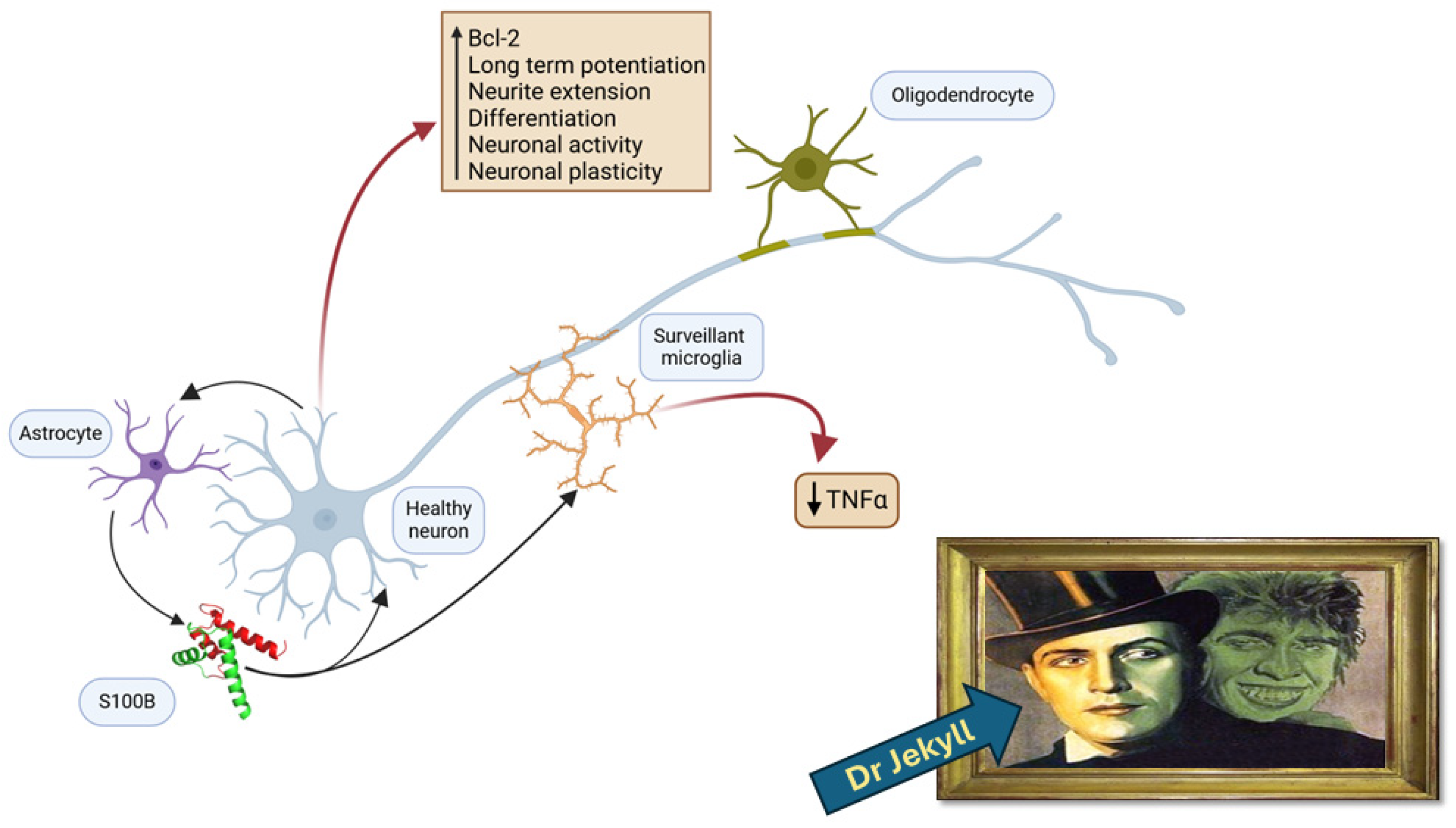
2. The S100B Protein as a Protective and Trophic Factor

Many studies that are reported in this section date back several years, but they deserve now to be reconsidered. The earlier observational findings, published soon after the discovery of the protein, identified S100B (at that time known as S100) as a molecule playing a key, possibly trophic, role in biological organisms. In particular, the protein was shown to conserve its immunological features during phylogenesis, as indicated above [17,18,19]. Interestingly, during ontogenesis, the pattern of its accumulation was shown to parallel the functional maturation of regions of the central nervous system in different species, including humans. In particular, S100B levels appeared to be elevated during the period of glial cell proliferation and neuronal differentiation [20,21,22,23,24,25]. More recently, S100B regulation has been reported to affect the differentiation during neurodevelopment [26]. Several mechanistic studies, in many cases in vitro, were then performed in order to investigate the processes accompanying the physiological/trophic role proposed for the S100B protein (Figure 1). Thus, S100B was shown to stimulate neurite outgrowth [27,28,29] and enhance cell survival [29,30] in cultures of neurons and glial cells. The extracellular trophic activities of S100B were considered to require a disulfide-linked, dimeric form of the protein [31], while the antisense inhibition of glial production of the S100B constitutive monomer (S100b) resulted in alterations in cell morphology, cytoskeletal organization, and cell proliferation [32]. A series of papers, distributed over approximately two decades, proposed a role for the nanomolar concentrations (regarded as physiological) of S100B in adult neurogenesis [33]. An intraventricular S100B infusion (50 ng) was shown to induce neurogenesis within the hippocampus, which could be associated with enhanced cognitive functioning, following an experimental traumatic brain injury (lateral fluid percussion) in rats [34]. In mice subjected to an experimental brain injury (unilateral parietal cryolesion), the intraperitoneal treatment with a nanomolar concentration of S100B significantly enhanced the early progenitor cell proliferation in the hippocampal subgranular zone (SGZ), the germinal area of the hippocampus, as well as the cell survival and migration to the granule cell layer (GCL) and promoted neuronal differentiation [35]. Likewise, the treatment with a nanomolar concentration of S100B for 7 days using osmotic micropumps augmented the hippocampal neurogenesis and synaptogenesis in rats after a traumatic brain injury (lateral fluid percussion) [36]. Moreover, long-term increased nanomolar S100B levels have also been shown to promote progenitor cell proliferation in the SGZ in juvenile, adult, and one-year-old S100B transgenic mice, as well as migration to the GCL in older S100B transgenic mice, suggesting a neurotrophic role in neuroplasticity, neurogenesis, and neuroregenerative medicine [37]. A protective role for the protein has also been proposed in Alzheimer’s disease (AD) alongside findings that indicate its pathogenic role at high concentrations and its role as a biomarker in biological fluids [1]. Thus, S100B has been shown to suppress the Aβ42 and Tau aggregation and toxicity by acting as a chaperone and metal ion buffering protein [38,39,40,41,42]. S100B was also shown to reduce the levels of inflammatory cytokines, such as Il-17 and IFN-a2, overexpressed after treatment with Aβ42, in cultured astrocytes, suggesting that the susceptibility of S100B to oxidation influences its protective activities [43]. Despite the reported findings that support a physiological role for S100B, a univocal function has not yet been established. Presently, the extracellular trophic activities have generated increased interest in light of the novel view interpreting S100B as a nutritional driver.

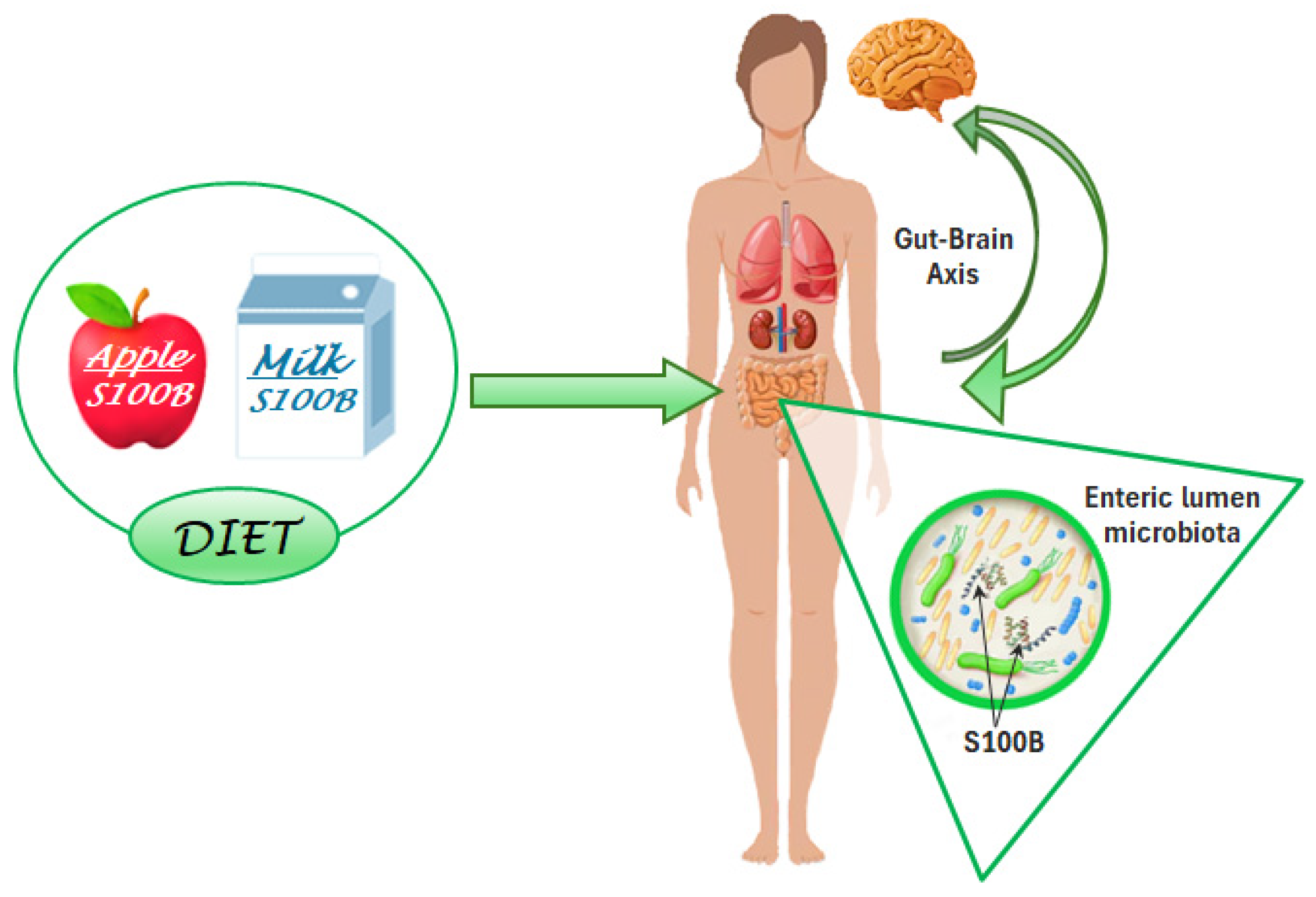
3. The S100B Protein as a Nutrient in Different Aliments

3.1. Milk

A relevant finding that supports a trophic role for S100B involves its detection in human breast milk [6,7] since the nutrient composition of human milk is considered to be essential for the growth, development, and health of infants, and breastfeeding for newborns is recognized as providing the infant with an optimal start in life [44,45]. Thus, S100B, as a putative trophic factor, might be considered among the mechanisms responsible for the beneficial effects of breastfeeding [46]. The natural role of S100B in milk appears not to be restricted to humans, since the protein has been identified in the milk produced by other mammalian species, namely donkeys, sheep, goats, and cows, although at a lower concentration than in humans [47]. It should be noted that S100B was shown to be essentially lost in milk formulae and donor milk subjected to Holder pasteurization, likely due to the preparation procedures [48,49].

3.2. Edible Plants

In addition, after an earlier study indicating the immunochemical and immunohistochemical detection of S100B-like immunoreactivity in Spinacia oleracea tissues [4], this protein was identified in diffused edible plants both in silico and in vivo. In particular, the S100B motif was expected in silico in the genome of some species belonging to the Viridiplantae clade, including various widely diffused edible plants such as Malus domestica, Durio zibethinus, and Artocarpus heterophyllus. S100B-like immunoreactive material was also detected in samples from fruits (e.g., apples, durian, jackfruit, and their derivatives) or leaves of the same species [5]. These recent findings corroborate the concept that S100B is naturally present in several foods. As a consequence, the possibility that S100B naturally exerts a nutritional and trophic function in the diet appears reasonable (Table 1) [50].

3.3. Gut Microbiota Interactions

The question remains how the S100B protein, when consumed with milk or other aliments, may interact with the tissues of the host that assumed it, since data regarding its gut absorption are not available. A possible explanation comes from recent findings indicating a novel role for the protein within the intestinal lumen, as S100B has been shown to affect gut microbiota biodiversity. Gut microbiota are known to consist of millions of microorganisms present in the human intestinal apparatus, playing a key role in food digestion, immune control, and antitumor responses, as well as in the synthesis of beneficial compounds and the control of neural regulation [14]. Based on the microbiota composition, proteins putatively interacting with S100B domains were identified in silico, both in healthy subjects and in patients with inflammatory bowel disease. Interestingly, differences in the interacting domain occurrences were exhibited between the two groups [15]. These results led to the experimental investigation of the role of S100B as a candidate signaling molecule in the gut microbiota communication machinery. Thus, the in silico data were experimentally confirmed in mice, in which the S100B levels correlated with the microbiota biodiversity. The correlation was significantly reduced after treatment with the S100B inhibitor pentamidine [51], indicating that the correlation was likely influenced by the modulation of S100B activity [16]. Taken together, these data furnish the missing link in the chain: microbiota-dependent processes might mediate the S100B influences on tissues of the host, including the nervous system through the gut–brain axis (Figure 2).
It should also be noted that, as an additional or alternative source of S100B for interactions with microbiota, S100B is known to be a constituent of enteroglial cells, corresponding to astrocytes in the enteric nervous system [52]. Once released by the enteroglial cells (S100B is known to be released by astrocytes in the central nervous system), S100B might trigger phenomena that are dependent on microbiota activation. In this way, as a hypothesis, an S100B-dependent influence mediated by microbiota might occur in tissues even when the protein is not ingested through aliments.

4. Conclusions

The novel perspective interpreting the S100B protein as a nutrient possibly displaying trophic effects valorizes the physiological role of the protein. This side of the protein (the “Jekyll side”) has been underestimated in recent times, probably since urgent needs based on pathological conditions have been more compelling (the “Hyde side”). In light of the presence of S100B in aliments, one might even consider the possibility that S100B in nature plays a role as a nutritional driver in living beings, including humans, from the neonatal stage, when it is ingested through milk, through the adult stages if a healthy diet rich in vegetables, fruits, and dairy products is followed. The interaction of the protein with gut microbiota offers a possible explanation for the role of enteric S100B, ingested with aliments and/or secreted from enteroglial cells. This novel perspective may constitute a fruitful line of research, potentially opening unexpected views of the physiological role(s) of this challenging protein.

Author Contributions

F.M. and V.R.S. were involved in all steps, including conceptualization; investigation; writing—original draft preparation; writing—review and editing. All authors have read and agreed to the published version of the manuscript.

Funding

This research received no external funding.

Acknowledgments

The authors are grateful to Tatsiana Pobat, Serena Platania, and Gabriele di Sante for their support in the graphics and editing. The Department of Movement, Human and Health Sciences as well as the Nando and Elsa Pedretti Foundation are also acknowledged for supporting the literature review. Special thanks to Elisabetta Clementi and Elena Scaramucci for their useful discussions and to Federica Valeriani for revising text and supporting an optimal editing.

Conflicts of Interest

The authors are collaborating with a research startup holding a patent entitled “Protein or its composition for use in treatment of the microbiota” n. IT102021000021146.

References

  1. Michetti, F.; Clementi, M.E.; Di Liddo, R.; Valeriani, F.; Ria, F.; Rende, M.; Di Sante, G.; Romano Spica, V. The S100B Protein: A Multifaceted Pathogenic Factor More Than a Biomarker. Int. J. Mol. Sci. 2023, 24, 9605. [Google Scholar] [CrossRef] [PubMed]
  2. Michetti, F.; Cocchia, D. S-100-like immunoreactivity in a planarian. An immunochemical and immunocytochemical study. Cell Tissue Res. 1982, 223, 575–582. [Google Scholar] [CrossRef] [PubMed]
  3. Endo, Y.; Endo, T. Immunohistochemical demonstration of S-100 protein in the brain neurosecretory cells of invertebrates (insects and earthworms). Neurosci. Lett. 1988, 90, 11–14. [Google Scholar] [CrossRef] [PubMed]
  4. Michetti, F.; Grilli Caiola, M.; Botti, F.; Bertini, G.; Cocchia, D. Immunochemical and immunohistochemical detection of S-100-like immunoreactivity in spinach tissues. J. Histochem. Cytochem. 1992, 40, 839–843. [Google Scholar] [CrossRef]
  5. Romano Spica, V.; Volpini, V.; Valeriani, F.; Carotenuto, G.; Arcieri, M.; Platania, S.; Castrignanò, T.; Clementi, M.E.; Michetti, F. In Silico Predicting the Presence of the S100B Motif in Edible Plants and Detecting Its Immunoreactive Materials: Perspectives for Functional Foods, Dietary Supplements and Phytotherapies. Int. J. Mol. Sci. 2024, 25, 9813. [Google Scholar] [CrossRef]
  6. Gazzolo, D.; Monego, G.; Corvino, V.; Bruschettini, M.; Bruschettini, P.; Zelano, G.; Michetti, F. Human. milk contains S100B protein. Biochim. Biophys. Acta 2003, 1619, 209–212. [Google Scholar] [CrossRef]
  7. Aşkan, Ö.Ö.; Karpuzoğlu, F.H.; Kayı, A.B.; Özden, T.A.; Gökçay, G.; Keskindemirci, G. Unique content of breastmilk: Neurotrophic growth factors in breastmilk at 2 years and beyo. Eur. J. Pediatr. 2024, 183, 4729–4734. [Google Scholar] [CrossRef]
  8. Roh, J.S.; Sohn, D.H. Damage-Associated Molecular Patterns in Inflammatory Diseases. Immune Netw. 2018, 18, e27. [Google Scholar] [CrossRef]
  9. Kaur, J.; Singh, H.; Naqvi, S. Intracellular DAMPs in Neurodegeneration and Their Role in Clinical Therapeutics. Mol. Neurobiol. 2023, 60, 3600–3616. [Google Scholar] [CrossRef]
  10. Huttunen, H.J.; Kuja-Panula, J.; Sorci, G.; Agneletti, A.L.; Donato, R.; Rauvala, H. Co-regulation of neurite outgrowth and cell survival by amphoterin and S100 proteins through receptor for advanced glycation end products (RAGE) activation. J. Biol. Chem. 2000, 275, 40096–40105. [Google Scholar] [CrossRef]
  11. Carapeto, A.P.; Marcuello, C.; Faísca, P.F.N.; Rodrigues, M.S. Morphological and Biophysical Study of S100A9 Protein Fibrils by Atomic Force Microscopy Imaging and Nanomechanical Analysis. Biomolecules 2024, 14, 1091. [Google Scholar] [CrossRef]
  12. Gambichler, T.; Brown, V.; Steuke, A.K.; Schmitz, L.; Stockfleth, E.; Susok, L. Baseline laboratory parameters predicting clinical outcome in melanoma patients treated with ipilimumab: A single-centre analysis. J. Eur. Acad. Dermatol. Venereol. 2017, 32, 972–977. [Google Scholar] [CrossRef] [PubMed]
  13. Salzmann, M.; Enk, A.H.; Hassel, J.C. S100 as Serum Tumor Marker in Advanced Uveal Melanoma. Biomolecules 2023, 13, 529. [Google Scholar] [CrossRef]
  14. Loh, J.S.; Mak, W.Q.; Tan, L.K.S.; Ng, C.X.; Chan, H.H.; Yeow, S.H.; Foo, J.B.; Ong, Y.S.; How, C.W.; Khaw, K.Y. Microbiota-gut-brain axis and its therapeutic applications in neurodegenerative diseases. Signal Transduct. Target. Ther. 2024, 9, 1–53. [Google Scholar] [CrossRef]
  15. Orsini, M.; Di Liddo, R.; Valeriani, F.; Mancin, M.; D’Incà, R.; Castagnetti, A.; Aceti, A.; Parnigotto, P.P.; Romano Spica, V.; Michetti, F. In Silico Evaluation of Putative S100B Interacting Proteins in Healthy and IBD Gut Microbiota. Cells 2020, 9, 1697. [Google Scholar] [CrossRef]
  16. Romano Spica, V.; Valeriani, F.; Orsini, M.; Clementi, M.E.; Seguella, L.; Gianfranceschi, G.; Di Liddo, R.; Di Sante, G.; Ubaldi, F.; Ria, F.; et al. S100B Affects Gut Microbiota Biodiversity. Int. J. Mol. Sci. 2023, 24, 2248. [Google Scholar] [CrossRef] [PubMed]
  17. Levine, L.; Moore, B.W. Structural relatedness of a vertebrate brain acidic protein as measured immunochemically. Neurosci. Res. Prog. Bull 1965, 3, 18–23. [Google Scholar]
  18. Moore, B.W.; Perez, V.J.; Gehring, M. Assay and regional distribution of a soluble protein characteristic of the nervous system. J. Neurochem. 1968, 15, 265–272. [Google Scholar] [CrossRef]
  19. Kessler, D.; Levine, L.; Fasman, G.D. Conformational and immunological properties of a bovine brain acidic protein (S-100) Biochemistry. Easton 1968, 7, 758–764. [Google Scholar] [CrossRef]
  20. Zuckerman, J.E.; Herschman, H.R.; Levine, L. Appearance of a brain specific antigen (th S-100 protein) during human foetal development. J. Neurochem. 1970, 17, 247–251. [Google Scholar] [CrossRef]
  21. Wenger, B.; Friedman, H. S-100 Protein in Embryonic Chick Retinae. Nature 1970, 228, 1214. [Google Scholar] [CrossRef]
  22. Cicero, T.J.; Cowan, W.M.; Moore, B.W. Changes in the concentrations of the two brain specific proteins, s-100 and 14-3-2, during the development of the avian optic tectum. Brain Res. 1970, 24, 1–10. [Google Scholar] [CrossRef] [PubMed]
  23. Herschman, H.R.; Levine, L.; De Vellis, J. Appearance of a brain-specific antigen (S-100 protein) in the developing rat brain. J. Neurochem. 1971, 18, 629–633. [Google Scholar] [CrossRef]
  24. Cicero, T.J.; Provine, R.R. The levels of the brain-specific proteins, S-100 and 14-3-2, in the developing chick spinal cord 1972. Brain Res. 1972, 44, 294–298. [Google Scholar] [CrossRef] [PubMed]
  25. Van Hartesveldt, C.; Moore, B.; Hartman, B.K. Transient midline raphe glial structure in the developing rat. J. Comp. Neurol. 1986, 253, 174–184. [Google Scholar] [CrossRef] [PubMed]
  26. Hernández-Ortega, K.; Canul-Euan, A.A.; Solis-Paredes, J.M.; Borboa-Olivares, H.; Reyes-Muñoz, E.; Estrada-Gutierrez, G.; Camacho-Arroyo, I.F. S100B actions on glial and neuronal cells in the developing brain: An overview. Front. Neurosci. 2024, 18, 1425525. [Google Scholar] [CrossRef]
  27. Kligman, D.; Marshak, D.R. Purification and characterization of a neurite extension factor from bovine brain. Proc. Natl. Acad. Sci. USA 1985, 82, 7136–7139. [Google Scholar] [CrossRef]
  28. Kligman, D.; Hsieh, L.J. Neurite extension factor induces rapid morphological differentiation of mouse neuroblastoma cells in defined medium. Dev. Brain Res. 1987, 33, 296–300. [Google Scholar] [CrossRef]
  29. Winningham-Major, F.; Staecker, J.L.; Barger, S.W.; Coats, S.; Van Eldik, L.J. Neurite extension and neuronal survival activities of recombinant S100/3 proteins that differ in the content and position of cysteine residues. J. Cell Biol. 1989, 109, 3063–3071. [Google Scholar] [CrossRef]
  30. Selinfreund, R.H.; Barger, S.W.; Pledger, W.J.; Van Eldik, L.J. Neurotrophic protein S100 beta stimulates glial cell proliferation. Proc. Natl. Acad. Sci. USA 1991, 88, 3554–3558. [Google Scholar] [CrossRef] [PubMed]
  31. Barger, S.W.; Wolchok, S.R.; Van Eldik, L.J. Disulfide-linked S100 beta dimers and signal transduction. Biochim. Biophys. Acta 1992, 1160, 105–112. [Google Scholar] [CrossRef] [PubMed]
  32. Selinfreund, R.H.; Barger, S.W.; Welsh, M.J.; Van Eldik, L.J. Antisense inhibition of glial S100 beta production results in alterations in cell morphology, cytoskeletal organization, and cell proliferation. J. Cell Biol. 1990, 111 Pt 1, 2021–2028. [Google Scholar] [CrossRef] [PubMed]
  33. Shi, J.; Wang, Z.; Wang, Z.; Shao, G.; Li, X. Epigenetic regulation in adult neural stem cells. Front. Cell Dev. Biol. 2024, 12, 1331074. [Google Scholar] [CrossRef] [PubMed]
  34. Kleindienst, A.; McGinn, M.J.; Harvey, H.B.; Colello, R.J.; Hamm, R.J.; Bullock, M.R. Enhanced hippocampal neurogenesis by intraventricular S100B infusion is associated with improved cognitive recovery after traumatic brain injury. J. Neurotrauma 2005, 22, 645–655. [Google Scholar] [CrossRef]
  35. Kleindienst, A.; Grünbeck, F.; Buslei, R.; Emtmann, I.; Buchfelder, M. Intraperitoneal treatment with S100B enhances hippocampal neurogenesis in juvenile mice and after experimental brain injury. Acta Neurochir. 2013, 155, 1351–1360. [Google Scholar] [CrossRef]
  36. Baecker, J.; Wartchow, K.; Sehm, T.; Ghoochani, A.; Buchfelder, M.; Kleindienst, A. Treatment with the Neurotrophic Protein S100B Increases Synaptogenesis after Traumatic Brain Injury. J. Neurotrauma 2020, 37, 1097–1107. [Google Scholar] [CrossRef] [PubMed]
  37. Rodrigues, L.; Wartchow, K.M.; Buchfelder, M.; Souza, D.O.; Gonçalves, C.A.; Kleindienst, A. Longterm Increased S100B Enhances Hippocampal Progenitor Cell Proliferation in a Transgenic Mouse Model. Int. J. Mol. Sci. 2022, 23, 9600. [Google Scholar] [CrossRef]
  38. Cristóvão Joana, S.; Morris Vanessa, K.; Cardoso, I.; Leal Sónia, S.; Martínez, J.; Botelho Hugo, M. The neuronal S100B protein is a calcium-tuned suppressor of amyloid-β aggregation. Sci. Adv. 2018, 4, eaaq1702. [Google Scholar] [CrossRef]
  39. Cristovao, J.S.; Figueira, A.J.; Carapeto, A.P.; Rodrigues, M.S.; Cardoso, I.; Gomes, C.M. The S100B Alarmin Is a Dual-Function Chaperone Suppressing Amyloid-beta Oligomerization through Combined Zinc Chelation and Inhibition of Protein Aggregation ACS Chem. Neuro sci. 2020, 11, 2753–2760. [Google Scholar]
  40. Cristóvão, J.S.; Moreira, G.G.; Rodrigues, F.E.P.; Carapeto, A.P.; Rodrigues, M.S.; Cardoso, I. Cu(2+)-binding to S100B triggers polymerization of disulfide cross-linked tetramers with enhanced chaperone activity against amyloid-β aggregation. Chem. Commun. 2021, 57, 379–382. [Google Scholar] [CrossRef]
  41. Moreira, G.G.; Cantrelle, F.X.; Quezada, A.; Carvalho, F.S.; Cristóvão, J.S.; Sengupta, U. Dynamic interactions and Ca2+-binding modulate the holdase-type chaperone activity of S100B preventing tau aggregation and seeding. Nat. Commun. 2021, 12, 6292. [Google Scholar] [CrossRef]
  42. Figueira, A.J.; Moreira, G.G.; Saavedra, J.; Cardoso, I.; Gomes, C.M. Tetramerization of the S100B chaperone spawns a Ca2+ independent regulatory surface that enhances anti-aggregation activity and client specificity. J. Mol. Biol. 2022, 434, 167791. [Google Scholar] [CrossRef] [PubMed]
  43. Coelho, R.; De Benedictis, C.A.; Sauer, A.K.; Figueira, A.J.; Faustino, H.; Grabrucker, A.M.; Gomes, C.M. Secondary Modification of S100B Influences Anti Amyloid-β Aggregation Activity and Alzheimer’s Disease Pathology. Int. J. Mol. Sci. 2024, 25, 1787. [Google Scholar] [CrossRef]
  44. World Health Organization. Global Strategy for Infant and Young Child Feeding; World Health Organization: Geneva, Switzerland, 2003. [Google Scholar]
  45. Petersohn, I.; Hellinga, A.H.; Van Lee, L.; Keukens, N.; Bont, L.; Hettinga, K.A.; Feskens, E.J.E.M. Maternal diet and human milk composition: An updated systematic review. Front. Nutr. Jan. 2024, 23, 1320560. [Google Scholar] [CrossRef] [PubMed]
  46. Li, R.; Xia, W.; Zhang, Z.; Wu, K. S100B protein, brain-derived neurotrophic factor, and glial cell line-derived neurotrophic factor in human milk. PLoS ONE 2011, 6, e21663. [Google Scholar] [CrossRef]
  47. Galvano, F.; Frigiola, A.; Gagliardi, L.; Ciotti, S.; Bognanno, M.; Iacopino, A.M.; Nigro, F.; Tina, G.L.; Cavallaro, D.; Mussap, M.; et al. S100B milk concentration in mammalian species. Front. Biosci. 2009, 1, 542–546. [Google Scholar] [CrossRef]
  48. Nigro, F.; Gagliardi, L.; Ciotti, S.; Galvano, F.; Pietri, A.; Tina, G.L.; Cavallaro, D.; La Fauci, L.; Iacopino, L.; Bognanno, M.; et al. S100B Protein concentration in milk-formulas for preterm and term infants. Correlation with industrial preparation procedures. Mol. Nutr. Food Res. 2008, 52, 609–613. [Google Scholar] [CrossRef]
  49. Peila, C.; Coscia, A.; Bertino, E.; Li Volti, G.; Galvano, F.; Visser, G.H.A.; Gazzolo, D.; Holder, J. Pasteurization affects S100B concentrations in human milk. Matern. Fetal Neonatal Med. 2018, 31, 513–517. [Google Scholar] [CrossRef]
  50. Ghaffar, T.; Volpini, V.; Platania, S.; Glogowski, P.A.; Gianfranceschi, G.; Vassioukovitch, O.; Valeriani, F.; Michetti, F.; Romano Spica, V. A novel role for S100B in diet and gut-microbiota regulation. In Proceedings of the Gut Microbiota for Health World Summit 2025, Washington, DC, USA, 15–16 March 2025; Available online: https://agau.gastro.org/cw/course-details?entryId=17324988#nav-home (accessed on 16 January 2024).
  51. Charpentier, T.H.; Wilder, P.T.; Liriano, M.A.; Varney, K.M.; Pozharski, E.; MacKerell, A.D.; Coop, A.; Toth, E.A.; Weber, D.J. Divalent metal ion complexes of S100B in the absence and presence of pentamidine. J. Mol. Biol. 2008, 382, 56–73. [Google Scholar] [CrossRef]
  52. Ferri, G.L.; Probert, L.; Cocchia, D.; Michetti, F.; Marangos, P.J.; Polak, J.M. Evidence for the presence of S-100 protein in the glial component of the human enteric nervous system. Nature 1982, 297, 409–410. [Google Scholar] [CrossRef]
Figure 1. The “Jekyll side” of S100B. The trophic function of S100B in the nervous system involves astrocytes as the main source of the protein, oligodendrocytes for myelination processes, microglia for surveillance processes and TNFα regulation, and neurons as the final targets. The functional pathways include the antiapoptotic Bcl-2 increase, long-term potentiation, neurite extension, stimulation of neuronal activity, plasticity, and differentiation. (Created with BioRender.com).
Nutrients 17 00881 g001
Figure 2. S100B is present in the diet and exerts positive effects. Common aliments, including apples and milk, as well as various edible plants and dairy products, contain S100B. The positive Jekyll effects are mediated by an action on gut microbiota biodiversity. When ingested, S100B may influence the gut–brain axis—and possibly other axes regulating different body districts. (Created with BioRender.com).
Nutrients 17 00881 g002
Table 1. Concentration of S100B protein in various food sources.
Food SourceS100B Concentration
Farm animal milk0.03–180 µg/L
Cheese–Dairy0.01–0.4 µg/Kg
Fruit–Vegetables0.04–180 µg/Kg
Disclaimer/Publisher’s Note: The statements, opinions and data contained in all publications are solely those of the individual author(s) and contributor(s) and not of MDPI and/or the editor(s). MDPI and/or the editor(s) disclaim responsibility for any injury to people or property resulting from any ideas, methods, instructions or products referred to in the content.

