


In the original publication [1], there was a mistake in Figures 1E and 3G. This data error occurred due to an unforeseen mistake during the data organization process. The correct Figure 1 and Figure 3 appear below. The authors state that the scientific conclusions are unaffected. This correction was approved by the Academic Editor. The original publication has also been updated.
Figure 1.
CSP32 induced morphological changes and phagocytosis of macrophages. Cells were treated with the indicated concentrations of CSP32 and LPS for 24 h. (A) Cell viability was assessed by MTT assay. (B) Representative microscopy images of morphological changes. (C,D) Representative flow cytometric histogram and quantitative analysis of the phagocytosis capacity using fluorescent FITC-IgG latex beads. Data are expressed as the mean ± SD (n = 4). ** p <0.01 compared with the control. (E) The phagocytic cells were visualized by fluorescence microscopy. The nuclei were stained with DAPI. Scale bar: 200 μm.
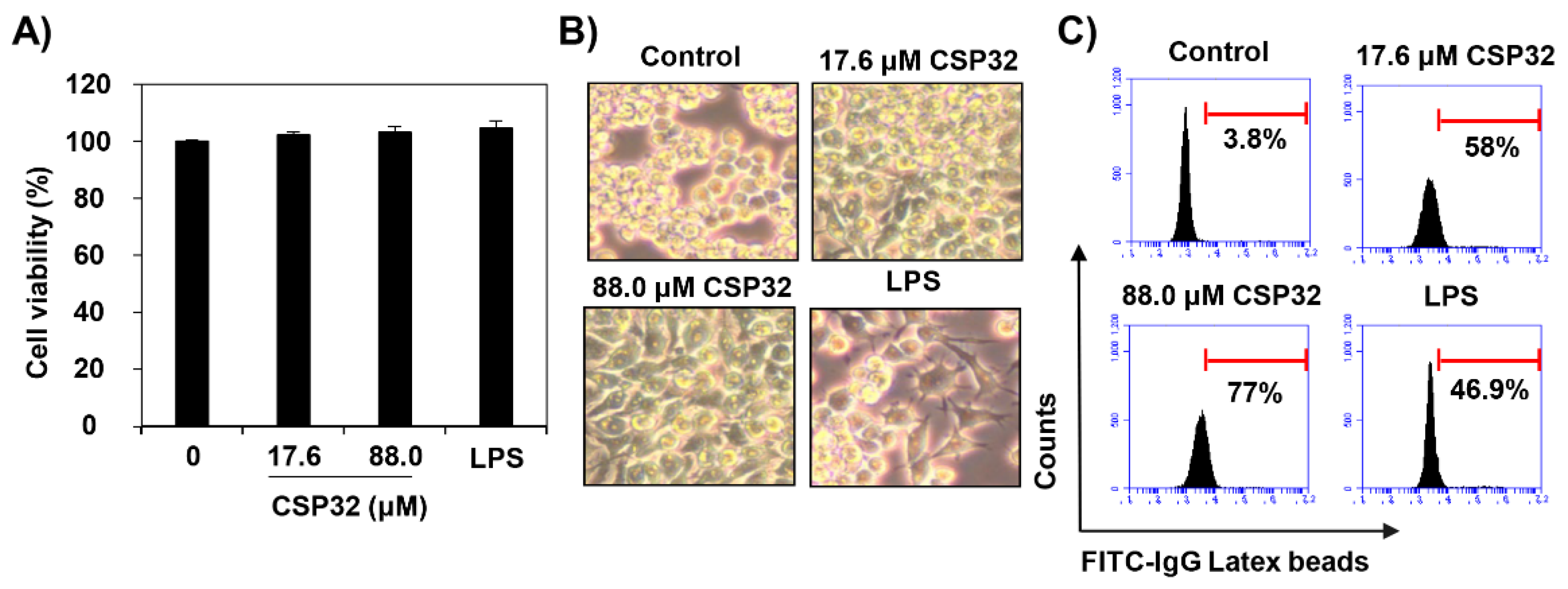
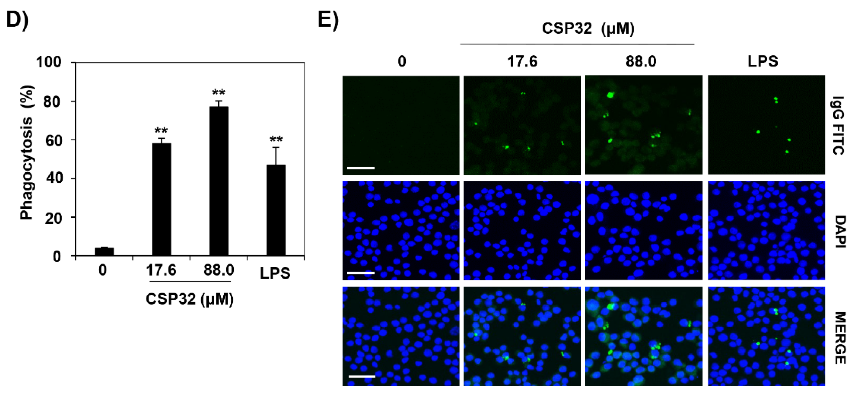
Figure 3.
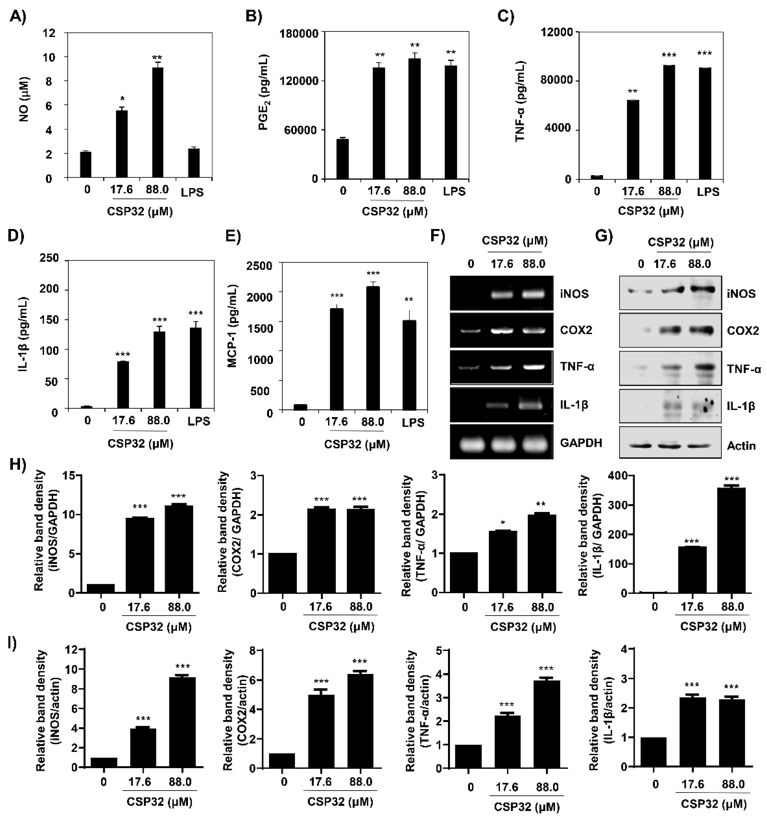
CSP32 increased the levels of markers for M1 macrophages. Cells were treated with the indicated concentrations of CSP32 and LPS for 24 h. (A) The amount of NO in the cell supernatant was measured using Griess reagents. The levels of PGE2 (B), TNF-α (C), IL-1β (D), and MCP-1 (E) in the culture supernatants were measured by ELISA kits. Data are expressed as the mean ± SD (n = 4). * p < 0.05, ** p < 0.01, and *** p < 0.001 compared with the control. mRNA (F) and protein (G) expression of markers of M1 macrophages, including iNOS, COX-2, TNF-α, and IL-1β. GAPDH and β-actin were used as internal controls for RT-PCR and Western blotting. Quantitative analysis of mRNA (H) and protein (I) expression. The expression of each protein was indicated as a fold change relative to the control. Data are expressed as the mean ± SD (n = 3). * p < 0.05, ** p < 0.01, and *** p < 0.001 compared with the control.
Reference
- Ji, S.Y.; Lee, H.; Hwangbo, H.; Hong, S.-H.; Cha, H.-J.; Park, C.; Kim, D.-H.; Kim, G.-Y.; Kim, S.; Kim, H.-S.; et al. A Novel Peptide Oligomer of Bacitracin Induces M1 Macrophage Polarization by Facilitating Ca2+ Influx. Nutrients 2020, 12, 1603. [Google Scholar] [CrossRef] [Scilit] [PubMed]
Disclaimer/Publisher’s Note: The statements, opinions and data contained in all publications are solely those of the individual author(s) and contributor(s) and not of MDPI and/or the editor(s). MDPI and/or the editor(s) disclaim responsibility for any injury to people or property resulting from any ideas, methods, instructions or products referred to in the content. |
© 2025 by the authors. Licensee MDPI, Basel, Switzerland. This article is an open access article distributed under the terms and conditions of the Creative Commons Attribution (CC BY) license (https://creativecommons.org/licenses/by/4.0/).