Bioactive Nutritional Components Within the Planetary Health Diet for Preventing Sarcopenic Obesity and Diabetic Sarcopenia: A Systematic Review
 , , , , and
Abstract
1. Introduction
2. Materials and Methods
2.1. Protocol and Registration
2.2. Eligibility Criteria
2.3. Information Sources and Search Strategy
2.4. Data Extraction and Items
2.5. Assessment of Study Quality
2.6. Data Synthesis and Analysis
2.7. Ethical Considerations
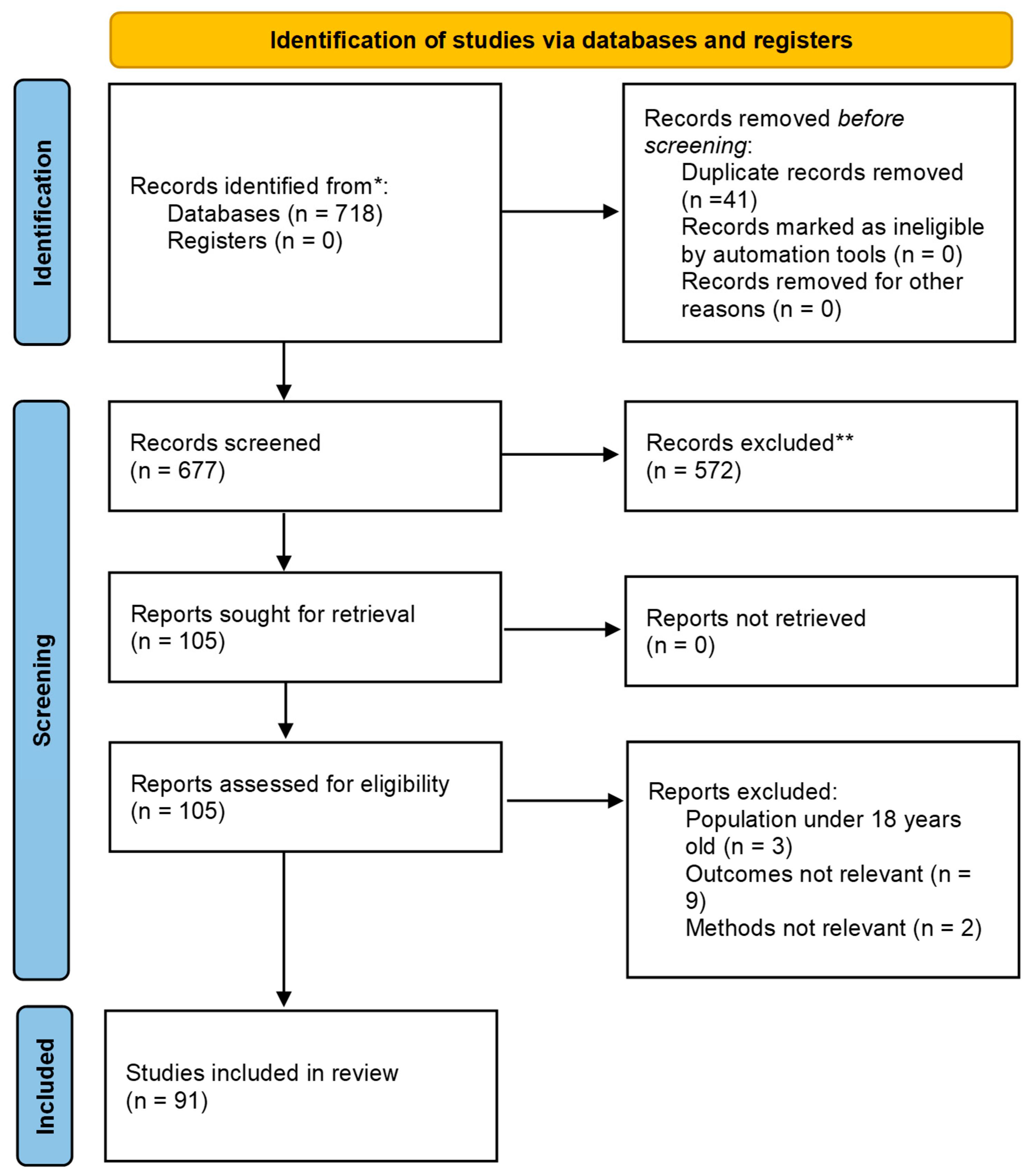
3. Results
4. Discussion
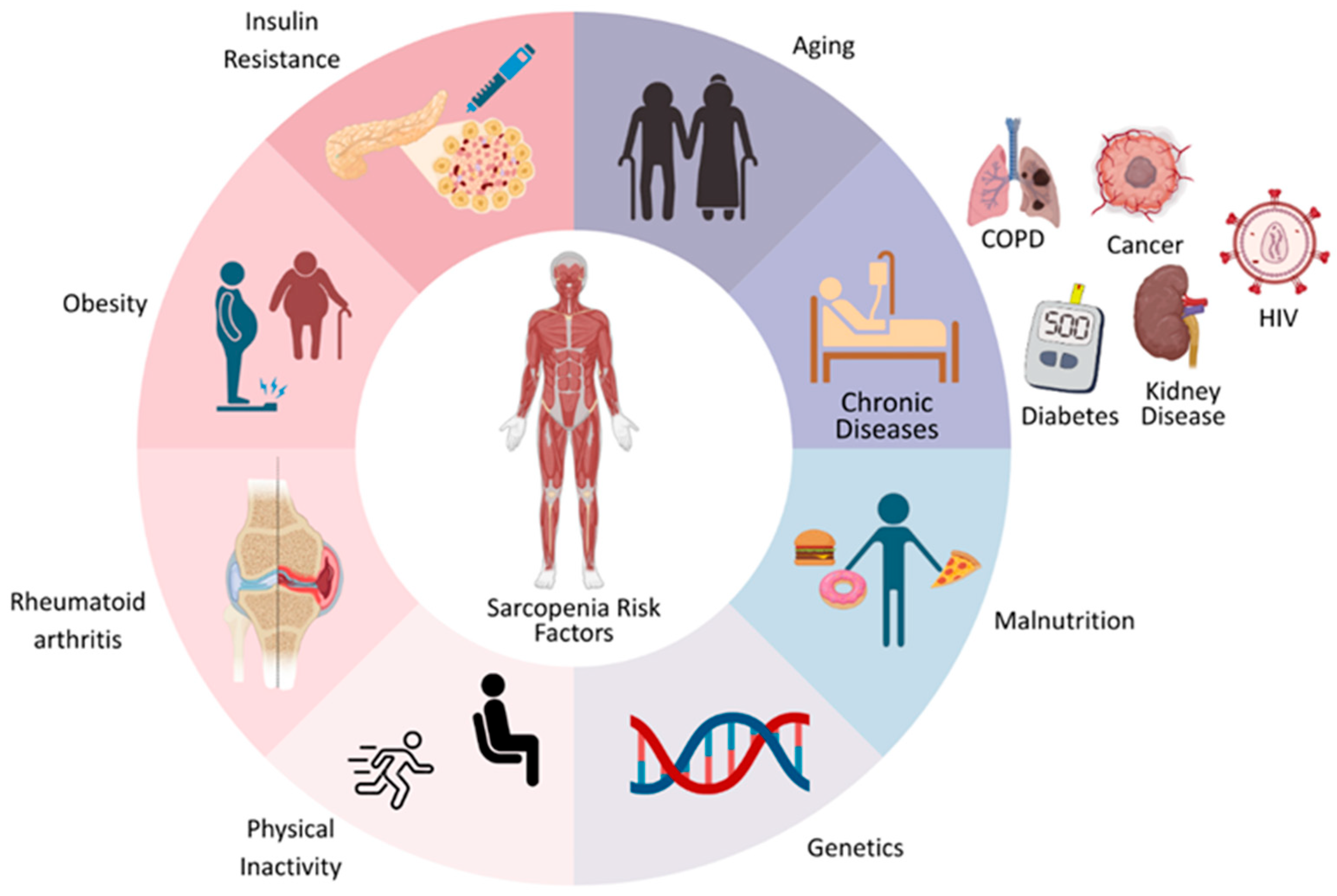
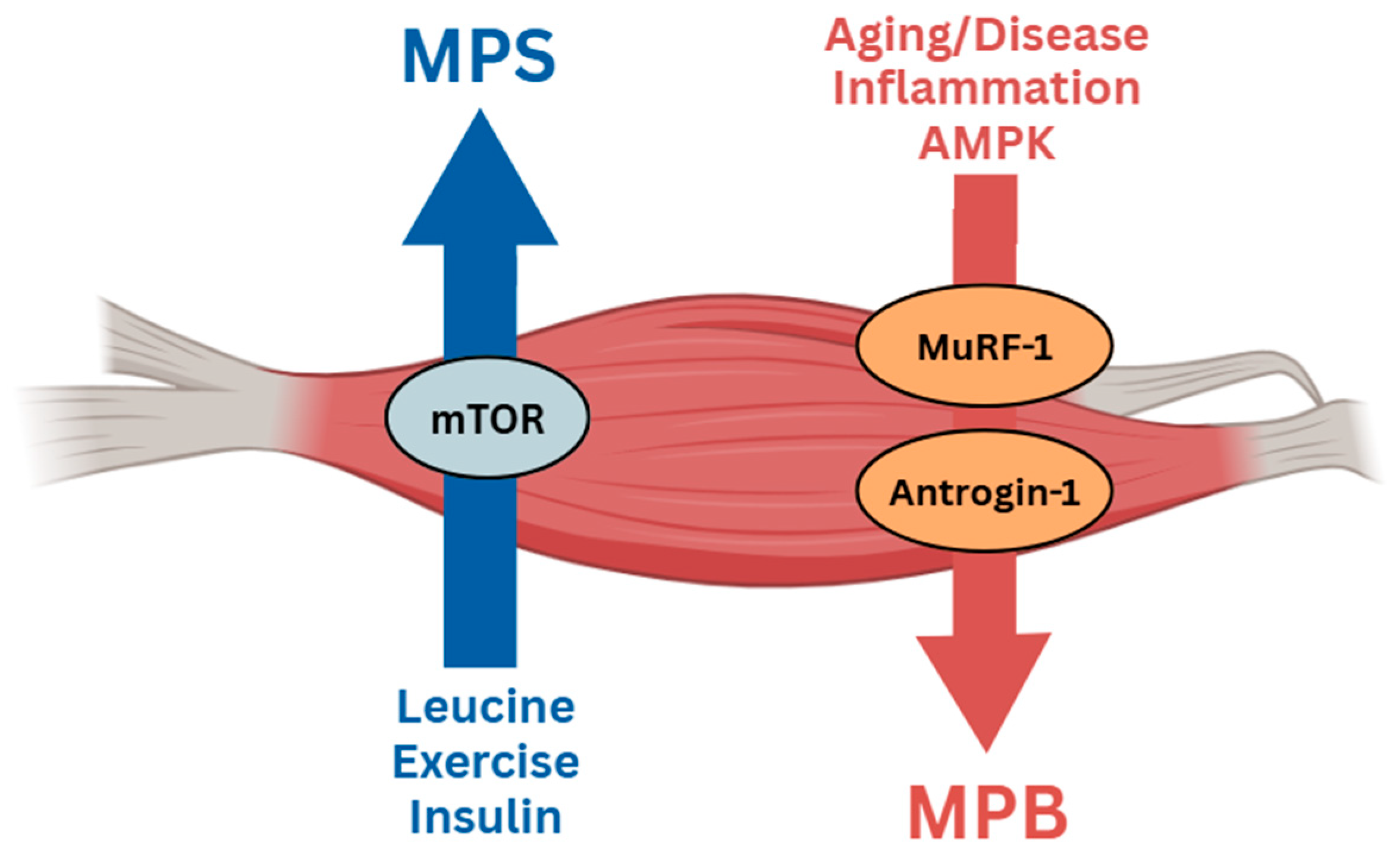
4.1. Pathophysiology and Metabolic Dysregulation in Sarcopenia Risk Factors
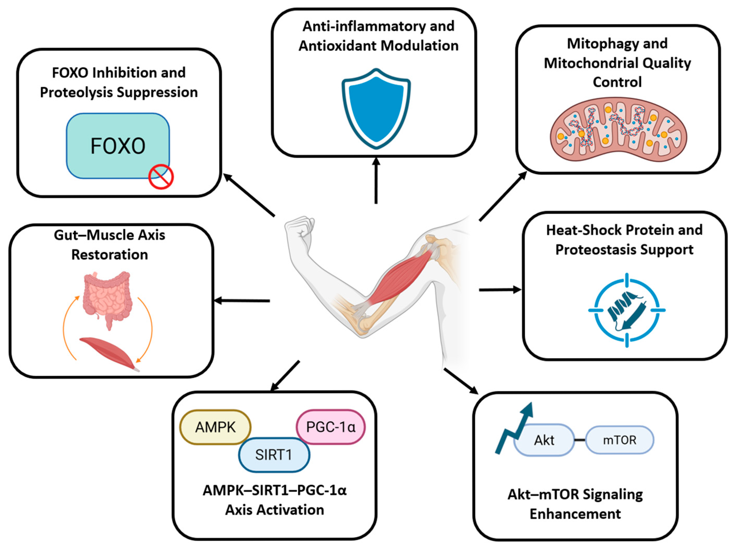
4.2. Findings and Evidence Trends from Reviewed Studies
4.2.1. AMPK–SIRT1–PGC-1α Axis Activation
4.2.2. Akt–mTOR Signaling Enhancement
4.2.3. FOXO Inhibition and Proteolysis Suppression
4.2.4. Mitophagy and Mitochondrial Quality Control
4.2.5. Anti-Inflammatory and Antioxidant Modulation
4.2.6. Gut–Muscle Axis Restoration
4.2.7. Heat Shock Protein and Proteostasis Support
4.3. Whole-Diet and Nutritional Approaches in Managing SO and DS
4.3.1. Legumes and Soy-Derived Foods
4.3.2. Vegetables, Herbs, and Polyphenol-Rich Plants
4.3.3. Fruits and Fermented Plant Products
4.3.4. Animal- and Marine-Based Protein Sources
4.3.5. Fermented and Microbiota-Active Foods
4.3.6. Practical Applications and Limitation
5. Conclusions
Author Contributions
Funding
Data Availability Statement
Conflicts of Interest
References
- Gianfredi, V.; Nucci, D.; Pennisi, F.; Maggi, S.; Veronese, N.; Soysal, P. Aging, longevity, and healthy aging: The public health approach. Aging Clin. Exp. Res. 2025, 37, 125. [Google Scholar] [CrossRef]
- World Health Organization. Ageing and Health. Available online: https://www.who.int/news-room/fact-sheets/detail/ageing-and-health (accessed on 1 October 2025).
- Shur, N.F.; Creedon, L.; Skirrow, S.; Atherton, P.J.; MacDonald, I.A.; Lund, J.; Greenhaff, P.L. Age-related changes in muscle architecture and metabolism in humans: The likely contribution of physical inactivity to age-related functional decline. Ageing Res. Rev. 2021, 68, 101344. [Google Scholar] [CrossRef]
- Ikeda, T.; Katayama, S.; Kitagawa, T. The combined intervention of neuromuscular electrical stimulation and nutrition therapy: A scoping review. Clin. Nutr. ESPEN 2023, 54, 239–250. [Google Scholar] [CrossRef]
- Chen, L.-K.; Woo, J.; Assantachai, P.; Auyeung, T.-W.; Chou, M.-Y.; Iijima, K.; Jang, H.C.; Kang, L.; Kim, M.; Kim, S.; et al. Asian Working Group for Sarcopenia: 2019 Consensus Update on Sarcopenia Diagnosis and Treatment. J. Am. Med. Dir. Assoc. 2020, 21, 300–307.e302. [Google Scholar] [CrossRef] [PubMed]
- Rosenberg, I.H. Sarcopenia: Origins and Clinical Relevance1. J. Nutr. 1997, 127, 990S–991S. [Google Scholar] [CrossRef] [PubMed]
- Papadopoulou, S.K. Sarcopenia: A Contemporary Health Problem among Older Adult Populations. Nutrients 2020, 12, 1293. [Google Scholar] [CrossRef]
- Xie, G.; Jin, H.; Mikhail, H.; Pavel, V.; Yang, G.; Ji, B.; Lu, B.; Li, Y. Autophagy in sarcopenia: Possible mechanisms and novel therapies. Biomed. Pharmacother. 2023, 165, 115147. [Google Scholar] [CrossRef]
- Lim, H.S.; Park, Y.H.; Suh, K.; Yoo, M.H.; Park, H.K.; Kim, H.J.; Lee, J.H.; Byun, D.W. Association between Sarcopenia, Sarcopenic Obesity, and Chronic Disease in Korean Elderly. J. Bone Metab. 2018, 25, 187–193. [Google Scholar] [CrossRef]
- Wang, M.; Tan, Y.; Shi, Y.; Wang, X.; Liao, Z.; Wei, P. Diabetes and Sarcopenic Obesity: Pathogenesis, Diagnosis, and Treatments. Front. Endocrinol. 2020, 11, 568. [Google Scholar] [CrossRef] [PubMed]
- Borba, V.Z.C.; Costa, T. Sarcopenic obesity: A review. Arch. Endocrinol. Metab. 2025, 68, e240084. [Google Scholar] [CrossRef]
- Cho, Y.K.; Jung, H.N.; Kim, E.H.; Lee, M.J.; Park, J.Y.; Lee, W.J.; Kim, H.K.; Jung, C.H. Association between sarcopenic obesity and poor muscle quality based on muscle quality map and abdominal computed tomography. Obesity 2023, 31, 1547–1557. [Google Scholar] [CrossRef] [PubMed]
- Ellulu, M.S.; Patimah, I.; Khaza’ai, H.; Rahmat, A.; Abed, Y. Obesity and inflammation: The linking mechanism and the complications. Arch. Med. Sci. 2017, 13, 851–863. [Google Scholar] [CrossRef] [PubMed]
- Purnamasari, D.; Tetrasiwi, E.N.; Kartiko, G.J.; Astrella, C.; Husam, K.; Laksmi, P.W. Sarcopenia and Chronic Complications of Type 2 Diabetes Mellitus. Rev. Diabet. Stud. 2022, 18, 157–165. [Google Scholar] [CrossRef]
- Sadeghi, A.; Niknam, M.; Momeni-Moghaddam, M.A.; Shabani, M.; Aria, H.; Bastin, A.; Teimouri, M.; Meshkani, R.; Akbari, H. Crosstalk between autophagy and insulin resistance: Evidence from different tissues. Eur. J. Med. Res. 2023, 28, 456. [Google Scholar] [CrossRef] [PubMed]
- Tessari, P. Stepwise Discovery of Insulin Effects on Amino Acid and Protein Metabolism. Nutrients 2024, 16, 119. [Google Scholar] [CrossRef]
- Ostler, J.E.; Maurya, S.K.; Dials, J.; Roof, S.R.; Devor, S.T.; Ziolo, M.T.; Periasamy, M. Effects of insulin resistance on skeletal muscle growth and exercise capacity in type 2 diabetic mouse models. Am. J. Physiol. Endocrinol. Metab. 2014, 306, E592–E605. [Google Scholar] [CrossRef]
- Xie, Z.; Li, Y.; Li, X.; Zhang, J. A review of mitochondrial dysfunction in diabetic sarcopenia: Mechanisms, diagnosis, and treatment approaches. J. Int. Med. Res. 2025, 53, 3000605251355996. [Google Scholar] [CrossRef]
- Wei, S.; Nguyen, T.T.; Zhang, Y.; Ryu, D.; Gariani, K. Sarcopenic obesity: Epidemiology, pathophysiology, cardiovascular disease, mortality, and management. Front. Endocrinol. 2023, 14, 1185221. [Google Scholar] [CrossRef]
- Montalvo, R.N.; Hardee, J.P.; VanderVeen, B.N.; Carson, J.A. Resistance Exercise’s Ability to Reverse Cancer-Induced Anabolic Resistance. Exerc. Sport. Sci. Rev. 2018, 46, 247–253. [Google Scholar] [CrossRef]
- Chen, X.; Ji, Y.; Liu, R.; Zhu, X.; Wang, K.; Yang, X.; Liu, B.; Gao, Z.; Huang, Y.; Shen, Y.; et al. Mitochondrial dysfunction: Roles in skeletal muscle atrophy. J. Transl. Med. 2023, 21, 503. [Google Scholar] [CrossRef]
- Shukla, M.; Narayan, M. Proteostasis and Its Role in Disease Development. Cell Biochem. Biophys. 2025, 83, 1725–1741. [Google Scholar] [CrossRef] [PubMed]
- Mo, Q.; Kulyar, M.F.-e.-A.; Ding, Y.; Zhang, Y.; Pan, H.; Li, J. Thiram induces myocardial oxidative damage and apoptosis in broilers via interfering their cardiac metabolism. Ecotoxicol. Environ. Saf. 2022, 247, 114225. [Google Scholar] [CrossRef]
- Wiedmer, P.; Jung, T.; Castro, J.P.; Pomatto, L.C.D.; Sun, P.Y.; Davies, K.J.A.; Grune, T. Sarcopenia—Molecular mechanisms and open questions. Ageing Res. Rev. 2021, 65, 101200. [Google Scholar] [CrossRef]
- Calvani, R.; Picca, A.; Coelho-Júnior, H.J.; Tosato, M.; Marzetti, E.; Landi, F. Diet for the prevention and management of sarcopenia. Metabolism 2023, 146, 155637. [Google Scholar] [CrossRef]
- Nazri, N.S.M.; Vanoh, D.; Soo, K.L. Natural Food for Sarcopenia: A Narrative Review. Malays. J. Med. Sci. 2022, 29, 28–42. [Google Scholar] [CrossRef]
- Abdi Dezfouli, R.; Zargar Balajam, N.; Heshmat, R.; Shafiee, G. The efficacy of Sarcomeal® oral supplementation plus vitamin D3 on muscle parameters, metabolic factors, and quality of life in diabetic sarcopenia: A randomized controlled clinical trial. Aging Clin. Exp. Res. 2025, 37, 81. [Google Scholar] [CrossRef]
- Huang, Y.; Zhu, X.; Chen, K.; Lang, H.; Zhang, Y.; Hou, P.; Ran, L.; Zhou, M.; Zheng, J.; Yi, L.; et al. Resveratrol prevents sarcopenic obesity by reversing mitochondrial dysfunction and oxidative stress via the PKA/LKB1/AMPK pathway. Aging 2019, 11, 2217–2240. [Google Scholar] [CrossRef]
- Okamura, T.; Hamaguchi, M.; Bamba, R.; Nakajima, H.; Yoshimura, Y.; Kimura, T.; Hashimoto, Y.; Majima, S.; Senmaru, T.; Ushigome, E.; et al. Brazilian green propolis improves gut microbiota dysbiosis and protects against sarcopenic obesity. J. Cachexia Sarcopenia Muscle 2022, 13, 3028–3047. [Google Scholar] [CrossRef] [PubMed]
- Okamura, T.; Hasegawa, Y.; Hamaguchi, M.; Sasano, R.; Fukui, M. The role of chicken eggs in modulating sarcopenic obesity and gut microbiota in db/db mice. Front. Microbiol. 2023, 14, 1281217. [Google Scholar] [CrossRef] [PubMed]
- Chen, H.; Wang, Z.; Lee, C.; Liu, R.; Song, J.; Zhang, J.; Du, N.; Kang, C. Linking the Planetary Health Diet Index to sarcopenia: The mediating effect of the non-high-density lipoprotein cholesterol to high-density lipoprotein cholesterol ratio (NHHR). Front. Nutr. 2025, 12, 1504037. [Google Scholar] [CrossRef] [PubMed]
- Willett, W.; Rockström, J.; Loken, B.; Springmann, M.; Lang, T.; Vermeulen, S.; Garnett, T.; Tilman, D.; DeClerck, F.; Wood, A.; et al. Food in the Anthropocene: The EAT–Lancet Commission on Healthy Diets from Sustainable Food Systems. Lancet 2019, 393, 447–492. [Google Scholar] [CrossRef] [PubMed]
- Commission, E.A.-L. Healthy Diets From Sustainable Food Systems: Summary Report of the EAT-Lancet Commission; EAT: Oslo, Norway, 2019. [Google Scholar]
- Zhao, W.; Zhao, B.; Meng, X.; Li, B.; Wang, Y.; Yu, F.; Fu, C.; Yu, X.; Li, X.; Dai, C.; et al. The regulation of MFG-E8 on the mitophagy in diabetic sarcopenia via the HSPA1L-Parkin pathway and the effect of D-pinitol. J. Cachexia Sarcopenia Muscle 2024, 15, 934–948. [Google Scholar] [CrossRef] [PubMed]
- Yu, X.; Li, P.; Li, B.; Yu, F.; Zhao, W.; Wang, X.; Wang, Y.; Gao, H.; Cheng, M.; Li, X. d-Pinitol Improves Diabetic Sarcopenia by Regulation of the Gut Microbiome, Metabolome, and Proteome in STZ-Induced SAMP8 Mice. J. Agric. Food Chem. 2024, 72, 14466–14478. [Google Scholar] [CrossRef] [PubMed]
- Kim, D.Y.; Kang, Y.H.; Kang, M.K. Umbelliferone attenuates diabetic sarcopenia by modulating mitochondrial quality and the ubiquitin-proteasome system. Phytomedicine 2025, 144, 156930. [Google Scholar] [CrossRef]
- Lee, H.; Kim, S.Y.; Lim, Y. Lespedeza bicolor extract supplementation reduced hyperglycemia-induced skeletal muscle damage by regulation of AMPK/SIRT/PGC1α–related energy metabolism in type 2 diabetic mice. Nutr. Res. 2023, 110, 1–13. [Google Scholar] [CrossRef]
- Han, Y.; Jeong, Y.; Lee, J.H.; Pack, S.P. Transcriptional regulation of multi-tissue mRNA expression by L-arabinose mitigates metabolic disorders in leptin receptor-deficient mice. Food Biosci. 2024, 61, 104881. [Google Scholar] [CrossRef]
- Jin, H.; Oh, H.J.; Lee, B.Y. GABA Prevents Age-Related Sarcopenic Obesity in Mice with High-Fat-Diet-Induced Obesity. Cells 2023, 12, 2146. [Google Scholar] [CrossRef]
- Jin, H.; Oh, H.J.; Nah, S.Y.; Lee, B.Y. Gintonin-enriched fraction protects against sarcopenic obesity by promoting energy expenditure and attenuating skeletal muscle atrophy in high-fat diet-fed mice. J. Ginseng Res. 2022, 46, 454–463. [Google Scholar] [CrossRef]
- Shin, J.E.; Jeon, S.H.; Lee, S.J.; Choung, S.Y. The Administration of Panax Ginseng Berry Extract Attenuates High-Fat-Diet-Induced Sarcopenic Obesity in C57BL/6 Mice. Nutrients 2022, 14, 1747. [Google Scholar] [CrossRef]
- Han, M.J.; Choung, S.Y. Codonopsis lanceolata ameliorates sarcopenic obesity via recovering PI3K/Akt pathway and lipid metabolism in skeletal muscle. Phytomedicine 2022, 96, 153877. [Google Scholar] [CrossRef]
- Kwon, E.Y.; Kim, J.W.; Choi, J.Y.; Hwang, K.; Han, Y. Setaria viridis Ethanol Extract Attenuates Muscle Loss and Body Fat Reduction in Sarcopenic Obesity by Regulating AMPK in High-Fat Diet-Induced Obese Mice. Food Sci. Nutr. 2025, 13, e70655. [Google Scholar] [CrossRef] [PubMed]
- Mo, X.; Shen, L.; Wang, X.; Ni, W.; Li, L.; Xia, L.; Liu, H.; Cheng, R.; Wen, L.; Xu, J.; et al. Melatonin Mitigates Sarcopenic Obesity via Microbiota and Short-Chain Fatty Acids: Evidence From Epidemiologic and In Vivo Studies. J. Cachexia Sarcopenia Muscle 2025, 16, e13869. [Google Scholar] [CrossRef]
- Long, Y.; Wu, Y.; Zhong, Y.; Wu, Y.; Ye, H.; Luo, Y.; Xiao, L.; Ma, Y.; Wang, M. Resveratrol as a potential therapeutic agent for sarcopenic obesity: Insights from in vivoperiments. Biomed. Pharmacother. 2024, 179, 117396. [Google Scholar] [CrossRef]
- Chae, J.; Lee, E.; Oh, S.M.; Ryu, H.W.; Kim, S.; Nam, J.O. Aged black garlic (Allium sativum L.) and aged black elephant garlic (Allium ampeloprasum L.) alleviate obesity and attenuate obesity-induced muscle atrophy in diet-induced obese C57BL/6 mice. Biomed. Pharmacother. 2023, 163, 114810. [Google Scholar] [CrossRef]
- Ye, H.; Long, Y.; Yang, J.-M.; Wu, Y.-L.; Dong, L.-Y.; Zhong, Y.-B.; Luo, Y.; Wang, M.-Y. Curcumin regulates autophagy through SIRT3-SOD2-ROS signaling pathway to improve quadriceps femoris muscle atrophy in KOA rat model. Sci. Rep. 2024, 14, 8176. [Google Scholar] [CrossRef]
- Chang, S.S.; Chen, L.H.; Huang, K.C.; Huang, S.W.; Chang, C.C.; Liao, K.W.; Hu, E.C.; Chen, Y.P.; Chen, Y.W.; Hsu, P.C.; et al. Plant-based polyphenol rich protein supplementation attenuated skeletal muscle loss and lowered the LDL level via gut microbiota remodeling in Taiwan’s community-dwelling elderly. Food Funct. 2023, 14, 9407–9418. [Google Scholar] [CrossRef] [PubMed]
- Bai, Y.; Zhang, X.; Ouyang, F.; Wu, Y.; Wang, Y.; Zhang, J.; Meng, F.; Ren, S. Coix seed oil alleviates collagen-induced arthritis-associated muscle atrophy and functional decline by modulating gut microbiota dysbiosis. Phytomedicine 2025, 146, 157134. [Google Scholar] [CrossRef]
- Guo, Y.; Wang, Q.; Lv, Y.; Xia, F.; Chen, X.; Mao, Y.; Wang, X.; Ding, G.; Yu, J. Serum metabolome and gut microbiome alterations are associated with low handgrip strength in older adults. Aging 2024, 16, 2638–2656. [Google Scholar] [CrossRef]
- McKendry, J.; Lowisz, C.V.; Nanthakumar, A.; MacDonald, M.; Lim, C.; Currier, B.S.; Phillips, S.M. The effects of whey, pea, and collagen protein supplementation beyond the recommended dietary allowance on integrated myofibrillar protein synthetic rates in older males: A randomized controlled trial. Am. J. Clin. Nutr. 2024, 120, 34–46. [Google Scholar] [CrossRef] [PubMed]
- Lim, G.; Lim, Y. Effects of Whey Peptide Supplementation on Sarcopenic Obesity in High-Fat Diet-Fed Mice. Nutrients 2022, 14, 4402. [Google Scholar] [CrossRef]
- Camajani, E.; Persichetti, A.; Watanabe, M.; Contini, S.; Vari, M.; Di Bernardo, S.; Faro, M.; Lubrano, C.; Gnessi, L.; Caprio, M.; et al. Whey Protein, L-Leucine and Vitamin D Supplementation for Preserving Lean Mass during a Low-Calorie Diet in Sarcopenic Obese Women. Nutrients 2022, 14, 1884. [Google Scholar] [CrossRef]
- Singlár, Z.; Szentesi, P.; Fodor, J.; Angyal, Á.; Csernoch, L.; Sztretye, M. Assessing the Potential of Nutraceuticals as Geroprotectors on Muscle Performance and Cognition in Aging Mice. Antioxidants 2021, 10, 1415. [Google Scholar] [CrossRef]
- Lu, L.; Li, X.; Lv, L.; Xu, Y.; Wu, B.; Huang, C. Associations between omega-3 fatty acids and insulin resistance and body composition in women with polycystic ovary syndrome. Front. Nutr. 2022, 9, 1016943. [Google Scholar] [CrossRef] [PubMed]
- Kim, K.H.; Chung, Y.; Huh, J.-W.; Park, D.J.; Cho, Y.; Oh, Y.; Jeong, H.; Yoon, J.; Kang, J.-H.; Shin, H.-S.; et al. Gut microbiota of the young ameliorates physical fitness of the aged in mice. Microbiome 2022, 10, 238. [Google Scholar] [CrossRef] [PubMed]
- Baek, J.-S.; Shin, Y.-J.; Ma, X.; Park, H.-S.; Hwang, Y.-H.; Kim, D.-H. Bifidobacterium bifidum and Lactobacillus paracasei alleviate sarcopenia and cognitive impairment in aged mice by regulating gut microbiota-mediated AKT, NF-κB, and FOXO3a signaling pathways. Immun. Ageing 2023, 20, 56. [Google Scholar] [CrossRef]
- Grahnemo, L.; Nethander, M.; Coward, E.; Gabrielsen, M.E.; Sree, S.; Billod, J.M.; Sjögren, K.; Engstrand, L.; Dekkers, K.F.; Fall, T.; et al. Identification of three bacterial species associated with increased appendicular lean mass: The HUNT study. Nat. Commun. 2023, 14, 2250. [Google Scholar] [CrossRef] [PubMed]
- Kang, L.; Li, P.; Wang, D.; Wang, T.; Hao, D.; Qu, X. Alterations in intestinal microbiota diversity, composition, and function in patients with sarcopenia. Sci. Rep. 2021, 11, 4628. [Google Scholar] [CrossRef]
- Kang, M.; Kang, M.; Yoo, J.; Lee, J.; Lee, S.; Yun, B.; Song, M.; Kim, J.-M.; Kim, H.W.; Yang, J.; et al. Dietary supplementation with Lacticaseibacillus rhamnosus IDCC3201 alleviates sarcopenia by modulating the gut microbiota and metabolites in dexamethasone-induced models. Food Funct. 2024, 15, 4936–4953. [Google Scholar] [CrossRef]
- Kang, M.; Yun, B.; Mun, D.; Kim, S.; Jeong, K.C.; Kim, Y.; Oh, S. Bovine colostrum-derived exosomes alleviate muscle degeneration by modulating gut microbiota and metabolic homeostasis in atrophy models. J. Anim. Sci. Technol. 2025, 67, 892–908. [Google Scholar] [CrossRef]
- Szyller, J.; Bil-Lula, I. Heat Shock Proteins in Oxidative Stress and Ischemia/Reperfusion Injury and Benefits from Physical Exercises: A Review to the Current Knowledge. Oxid. Med. Cell Longev. 2021, 2021, 6678457. [Google Scholar] [CrossRef]
- KHasti, H.; Emadi, L.; Azizi, S.; Mohammadi, E.; Tavakkoli, H. Effects of Thermal Manipulation and Serotonin Modulation on Brain HSP70 and HSP90 Gene Expression in Late Embryogenesis of Broilers. Vet. Med. Sci. 2025, 11, e70195. [Google Scholar] [CrossRef]
- Mankhong, S.; Kim, S.; Moon, S.; Lee, J.S.; Cho, E.J.; Kwak, H.B.; Park, D.H.; Ryu, J.K.; Kang, J.H. Melatonin and Exercise Counteract Sarcopenic Obesity through Preservation of Satellite Cell Function. Int. J. Mol. Sci. 2023, 24, 6097. [Google Scholar] [CrossRef]
- Liu, D.; Wang, S.; Liu, S.; Wang, Q.; Che, X.; Wu, G. Frontiers in sarcopenia: Advancements in diagnostics, molecular mechanisms, and therapeutic strategies. Mol. Asp. Med. 2024, 97, 101270. [Google Scholar] [CrossRef]
- Sharma, A.; Chatterjee, S.; Lee, Y.-H.; Lee, S.-S. Targeting Crosstalk of Signaling Pathways among Muscles-Bone-Adipose Tissue: A Promising Therapeutic Approach for Sarcopenia. Aging Dis. 2023, 15, 1619. [Google Scholar] [CrossRef]
- Zhang, C.; Wang, D.; Wu, X.; Zhang, Q.; Chen, L.; Lang, J.; Wu, C. Effect of sarcopenia combined with Metabolic Syndrome (MS) on the prognosis of intertrochanteric fractures: A retrospective clinical study. BMC Musculoskelet. Disord. 2025, 26, 728. [Google Scholar] [CrossRef]
- Park, J.; Hong, S.-M.; Kim, J.; Kim, S. trans-Resveratrol and Hesperidin Supplementation with Treadmill Exercise Alleviates Methylglyoxal-Induced Skeletal Muscle Dysfunction. Biomol. Ther. 2025, 33, 890–900. [Google Scholar] [CrossRef] [PubMed]
- Wang, J.; Huang, Y.; Wang, Z.; Liu, J.; Liu, Z.; Yang, J.; He, Z. The mTOR Signaling Pathway: Key Regulator and Therapeutic Target for Heart Disease. Biomedicines 2025, 13, 397. [Google Scholar] [CrossRef] [PubMed]
- Nunes-Pinto, M.; Bandeira de Mello, R.G.; Pinto, M.N.; Moro, C.; Vellas, B.; Martinez, L.O.; Rolland, Y.; de Souto Barreto, P. Sarcopenia and the biological determinants of aging: A narrative review from a geroscience perspective. Ageing Res. Rev. 2025, 103, 102587. [Google Scholar] [CrossRef]
- Ren, Q.; Chen, S.; Chen, X.; Niu, S.; Yue, L.; Pan, X.; Li, Z.; Chen, X. An Effective Glucagon-Like Peptide-1 Receptor Agonists, Semaglutide, Improves Sarcopenic Obesity in Obese Mice by Modulating Skeletal Muscle Metabolism. Drug Des. Devel Ther. 2022, 16, 3723–3735. [Google Scholar] [CrossRef]
- Mann, V.; Sundaresan, A.; Shishodia, S. Overnutrition and Lipotoxicity: Impaired Efferocytosis and Chronic Inflammation as Precursors to Multifaceted Disease Pathogenesis. Biology 2024, 13, 241. [Google Scholar] [CrossRef]
- Pacifici, F.; Della-Morte, D.; Piermarini, F.; Arriga, R.; Scioli, M.G.; Capuani, B.; Pastore, D.; Coppola, A.; Rea, S.; Donadel, G.; et al. Prdx6 Plays a Main Role in the Crosstalk Between Aging and Metabolic Sarcopenia. Antioxidants 2020, 9, 329. [Google Scholar] [CrossRef]
- Egerman, M.A.; Glass, D.J. Signaling pathways controlling skeletal muscle mass. Crit. Rev. Biochem. Mol. Biol. 2014, 49, 59–68. [Google Scholar] [CrossRef]
- Glaviano, A.; Foo, A.S.C.; Lam, H.Y.; Yap, K.C.H.; Jacot, W.; Jones, R.H.; Eng, H.; Nair, M.G.; Makvandi, P.; Geoerger, B.; et al. PI3K/AKT/mTOR signaling transduction pathway and targeted therapies in cancer. Mol. Cancer 2023, 22, 138. [Google Scholar] [CrossRef]
- Ye, L.; Fu, X.; Li, Q. Mitochondrial Quality Control in Health and Disease. MedComm 2025, 6, e70319. [Google Scholar] [CrossRef]
- Long, Y.-F.; Chow, S.K.-H.; Cui, C.; Wong, R.M.Y.; Qin, L.; Law, S.-W.; Cheung, W.-H. Regulation of mitochondrial dynamic equilibrium by physical exercise in sarcopenia: A systematic review. J. Orthop. Transl. 2022, 35, 37–52. [Google Scholar] [CrossRef] [PubMed]
- Pickrell, A.M.; Youle, R.J. The roles of PINK1, parkin, and mitochondrial fidelity in Parkinson’s disease. Neuron 2015, 85, 257–273. [Google Scholar] [CrossRef]
- Tang, X.; Zou, Y.; Yang, S.; Chen, Z.; Zhou, Z.; Peng, X.; Tang, C. Impaired Mitophagy Contributes to Pyroptosis in Sarcopenic Obesity Zebrafish Skeletal Muscle. Nutrients 2025, 17, 1711. [Google Scholar] [CrossRef] [PubMed]
- Dantas, W.S.; Zunica, E.R.M.; Heintz, E.C.; Vandanmagsar, B.; Floyd, Z.E.; Yu, Y.; Fujioka, H.; Hoppel, C.L.; Belmont, K.P.; Axelrod, C.L.; et al. Mitochondrial uncoupling attenuates sarcopenic obesity by enhancing skeletal muscle mitophagy and quality control. J. Cachexia Sarcopenia Muscle 2022, 13, 1821–1836. [Google Scholar] [CrossRef]
- Lei, Y.; Gan, M.; Qiu, Y.; Chen, Q.; Wang, X.; Liao, T.; Zhao, M.; Chen, L.; Zhang, S.; Zhao, Y.; et al. The role of mitochondrial dynamics and mitophagy in skeletal muscle atrophy: From molecular mechanisms to therapeutic insights. Cell Mol. Biol. Lett. 2024, 29, 59. [Google Scholar] [CrossRef] [PubMed]
- Khan, M.S.H.; Hegde, V. Obesity and Diabetes Mediated Chronic Inflammation: A Potential Biomarker in Alzheimer’s Disease. J. Pers. Med. 2020, 10, 42. [Google Scholar] [CrossRef]
- Li, G.; Jin, B.; Fan, Z. Mechanisms Involved in Gut Microbiota Regulation of Skeletal Muscle. Oxid. Med. Cell Longev. 2022, 2022, 2151191. [Google Scholar] [CrossRef] [PubMed]
- Wang, L.; Wang, S.; Zhang, Q.; He, C.; Fu, C.; Wei, Q. The role of the gut microbiota in health and cardiovascular diseases. Mol. Biomed. 2022, 3, 30. [Google Scholar] [CrossRef]
- Hu, C.; Yang, J.; Qi, Z.; Wu, H.; Wang, B.; Zou, F.; Mei, H.; Liu, J.; Wang, W.; Liu, Q. Heat shock proteins: Biological functions, pathological roles, and therapeutic opportunities. MedComm 2022, 3, e161. [Google Scholar] [CrossRef]
- Choudhary, D.; Mediani, L.; Carra, S.; Cecconi, C. Studying heat shock proteins through single-molecule mechanical manipulation. Cell Stress. Chaperones 2020, 25, 615–628. [Google Scholar] [CrossRef]
- Jung, U.J. Sarcopenic Obesity: Involvement of Oxidative Stress and Beneficial Role of Antioxidant Flavonoids. Antioxidants 2023, 12, 1063. [Google Scholar] [CrossRef]
- Su, Y.; Zheng, X. HSP70-Mediated Autophagy-Apoptosis-Inflammation Network and Neuroprotection Induced by Heat Acclimatization. Biology 2025, 14, 774. [Google Scholar] [CrossRef]
- Li, X.; Yu, Y.; Gorshkov, B.; Haigh, S.; Bordan, Z.; Weintraub, D.; Rudic, R.D.; Chakraborty, T.; Barman, S.A.; Verin, A.D.; et al. Hsp70 Suppresses Mitochondrial Reactive Oxygen Species and Preserves Pulmonary Microvascular Barrier Integrity Following Exposure to Bacterial Toxins. Front. Immunol. 2018, 9, 1309. [Google Scholar] [CrossRef]
- Chew, W.; Lim, Y.P.; Lim, W.S.; Chambers, E.S.; Frost, G.; Wong, S.H.; Ali, Y. Gut-muscle crosstalk. A perspective on influence of microbes on muscle function. Front. Med. 2022, 9, 1065365. [Google Scholar] [CrossRef] [PubMed]
- Liang, L.; Saunders, C.; Sanossian, N. Food, gut barrier dysfunction, and related diseases: A new target for future individualized disease prevention and management. Food Sci. Nutr. 2023, 11, 1671–1704. [Google Scholar] [CrossRef] [PubMed]



| Intervention | Source | Study Type | Population/Model | Dose and Duration | Muscle Outcomes | Metabolic Outcomes | Mechanism | Ref. |
|---|---|---|---|---|---|---|---|---|
| D-pinitol targeting MFG-E8 | Soybean seeds | Animal and Cell | Mice: Older diabetic mice with muscle loss. Groups: (1) diabetic mice untreated, (2) diabetic mice given D-pinitol Cells: C2C12 muscle cells stressed with advanced glycation end-products (AGEs) or D-galactose. Groups: (1) stressed cells untreated, (2) stressed cells treated with D-pinitol. | Mice: Given D-pinitol 150 mg/kg/day by mouth for 6 weeks. Cells: Treated with D-pinitol 160 µM for 48 h. | Muscle strength: ↑ grip strength, ↑ endurance Muscle mass: ↑ lean mass, ↑ muscle weight, ↑ fiber size Muscle function: improved mitochondrial structure; in cells: ↑ survival, ↓ damage, restored mitophagy | Diabetes: ↓ blood glucose | Mitochondrial QC/Mitophagy: ↑ PINK1, ↑ Parkin, ↑ LC3B, ↓ P62 (restored mitophagy). Proteostasis/HSP: MFG-E8 suppression → release of HSPA1L–Parkin axis. | [34] |
| D-pinitol | Vigna sinensis, soybean, and pine plants. | Animal | STZ-induced SAMP8 diabetic aging mice (n = 10–12/group) | 150 mg/kg/day orally for 8 weeks | Muscle strength: ↑ grip strength Muscle mass: ↑ lean mass, ↑ gastrocnemius weight, ↑ muscle fiber size, ↑ bone mineral density Muscle function: improved muscle fiber morphology | Diabetes: ↓ fasting blood glucose Other: improved gut microbiota profile; metabolite and protein normalization | Gut–muscle axis: Beneficial microbial shifts (↑ Lachnospiraceae; ↓ Parabacteroides, Akkermansia). Proteostasis/HSP: Protein expression normalization (↑ Trim54; ↓ Arl6ip5, SNX6). Other: Normalized 44 metabolites; regulated β-alanine/histidine metabolism, ABC transporters, Ca2+ signaling. | [35] |
| Umbelliferone | Natural coumarin in carrots, parsley, celery, cumin, and fennel | Animal and Cell | Mice: db/db diabetic mice vs. non-diabetic controls. Cells: C2C12 muscle cells exposed to high glucose (50 mM) | Mice: 10 mg/kg/day orally for 8 weeks. Cells: 10–20 μM for 4 days | Muscle strength: ↑ grip strength Muscle mass: ↑ lean mass Muscle function: restored Type I fibers; ↑ myotube formation; ↑ myogenic markers (MyoD, Myogenin, Myh2); improved muscle morphology | Diabetes: ↓ HbA1c Obesity: ↓ fat mass | AMPK–SIRT1–PGC-1α: ↑ AMPK, ↑ SIRT1, ↑ PGC-1α → enhanced mitochondrial biogenesis. Mitochondrial QC/dynamics: ↑ MFN1, ↑ OPA1; ↓ DRP1, ↓ FIS1 (restored fusion–fission balance). FOXO/UPS: ↓ MuRF1, ↓ Atrogin-1, ↓ FoxO3a (reduced proteolysis). Akt–mTOR/Myogenesis: ↑ MyoD, ↑ Myogenin, ↑ Myh2 (promoted differentiation). | [36] |
| Lespedeza bicolor extract (LBE) | Lespedeza bicolor | Animal | Male C57BL/6J mice; type 2 diabetes induced by high-fat diet + 2 low-dose STZ injections | Oral gavage: 100 mg/kg/day (low) or 250 mg/kg/day (high), 12 weeks | Muscle mass: ↑ muscle weight, ↑ muscle fiber size | Diabetes: ↓ fasting glucose, ↓ HbA1c, ↑ glucose tolerance, ↑ insulin signaling | AMPK–SIRT1–PGC-1α: Activated pathway → ↑ mitochondrial biogenesis, ↑ energy metabolism. Akt–mTOR/GLUT4 pathway: ↑ GLUT4 translocation → enhanced insulin signaling. Inflammation/Oxidative stress: ↓ TNF-α, ↓ IL-6, ↓ MCP-1, ↓ 4-HNE. FOXO/UPS: ↓ FoxO3a, ↓ Atrogin-1, ↓ MuRF1 → reduced muscle proteolysis. | [37] |
| L-arabinose | Plant sugar (from hemicellulose in plant cell walls) | Animal | db/db mice (leptin receptor-deficient, obese, type 2 diabetes with muscle atrophy tendency) | Diet with 5% L-arabinose for 16 weeks | Muscle mass: ↑ gastrocnemius weight Muscle function: ↑ myogenesis (via ↑ IRS1/GLUT4) | Diabetes: ↓ fasting glucose, ↓ HOMA-IR, ↑ glucose tolerance, ↑ insulin sensitivity Obesity: ↓ body weight, ↓ fat mass | AMPK–SIRT1–PGC-1α: ↑ PGC-1α and ↑ CPT1b → enhanced fatty acid oxidation. Akt–mTOR/Insulin signaling: ↑ GLUT4, ↑ IRS1 → improved muscle glucose uptake and myogenesis. Inflammation: ↓ TNF-α, ↓ IL-6, ↓ IL-1β, ↓ NF-κB. Other: ↓ gluconeogenesis (↓ PEPCK, ↓ G6Pase), ↑ glycolysis (↑ GK). | [38] |
| Brazilian green propolis | Baccharis dracunculifolia extract (Yamada Bee Company, Japan) | Animal and Cell | Mice: Db/m (control) vs. Db/Db (sarcopenic obesity model) ± propolis. Cells: C2C12 myotubes exposed to palmitic acid ± propolis, artepillin C, or kaempferide | Mice: 0.08%, 0.4%, 2% propolis in chow, 8 weeks Cells: Propolis 100 µg/mL; Artepillin C 10.6 µg/mL; Kaempferide 1.89 µg/mL, 96 h | Muscle strength: ↑ grip strength Muscle mass: ↑ soleus mass, ↑ plantaris mass Muscle function: ↓ muscle atrophy genes; protection from PA-induced atrophy; ↑ ATP production; ↑ mitochondrial respiration | Diabetes: improved glucose tolerance Obesity: ↓ visceral fat Other: ↓ hepatic enzymes; ↓ liver fibrosis | Gut–muscle axis: Improved microbiota (↑ Bacteroidetes/Firmicutes ratio, ↑ Butyricicoccus, ↑ Acetivibrio); ↑ SCFAs. Inflammation/Immune modulation: ↓ inflammation; ↓ CD36; ↑ fatty acid excretion; macrophage shift to M2; ↑ ILC2. Mitochondrial function: Artepillin C and kaempferide protected mitochondria from palmitate, ↑ ATP, ↑ respiration. Other: CD36 suppression → reduced lipid uptake stress. | [29] |
| γ-Aminobutyric acid (GABA) | Four-carbon non-proteinogenic amino acid (abundant in vegetables, fruits, fermented foods) | Animal | Male C57BL/6J mice, young (3 mo) and aged (20 mo); HFD-induced obesity ± GABA | Oral gavage, 10 or 30 mg/kg/day, 8 weeks | Muscle strength: ↑ grip strength Muscle mass: ↑ lean mass, ↑ muscle fiber CSA Muscle function: ↓ Atrogin-1, ↓ MuRF1, ↓ myostatin; ↑ myogenic proteins (MyoD, Myogenin, Myf5/6) | Diabetes: ↓ fasting glucose, ↓ insulin resistance Obesity: ↓ visceral adipose tissue (VAT), ↓ adipocyte size Lipids: ↓ TG, ↓ TC, ↓ LDL; ↑ HDL Other: ↑ mitochondrial markers in muscle; ↑ thermogenic markers in fat; ↑ testosterone (aged mice) | Akt–mTOR anabolic signaling (PI3K/Akt activation, mTOR/4EBP1 ↑ → protein synthesis). FOXO/UPS proteolysis suppression (↓ FoxO3a, ↓ Atrogin-1, ↓ MuRF1). AMPK–SIRT1–PGC-1α activation (↑ PGC-1α, ↑ NRF1, ↑ TFAM, ↑ UCP3). Anti-inflammatory and antioxidant pathways (↓ TNF-α, ↓ IL-6, ↓ IL-1β). Other: Enhanced lipolysis/thermogenesis (p-PKA, ATGL, HSL, MGL; UCP1 in VAT) | [39] |
| Gintonin-enriched fraction (GEF) | Non-saponin glyco-lipoprotein fraction from Korean ginseng (Panax ginseng Meyer) | Animal | Male ICR mice (4 wk), divided into ND, HFD, HFD + GEF 50 mg/kg/day, HFD + GEF 150 mg/kg/day | Oral gavage, 50 mg/kg/day or 150 mg/kg/day for 6 weeks | Muscle strength: ↑ grip strength Muscle mass: ↑ skeletal muscle mass, ↑ muscle fiber CSA Muscle function: ↑ myogenic proteins (MyoD, Myogenin, MEF-2, MYH7); ↓ Atrogin-1, ↓ MuRF1 | Obesity: ↓ body mass gain; ↓ visceral and subcutaneous WAT; ↓ adipocyte size Other: ↑ rectal temperature; ↑ mitochondrial biogenesis markers in muscle; WAT browning/thermogenesis | AMPK–SIRT1–PGC-1α activation (↑ PGC-1α, ↑ NRF1, ↑ TFAM, ↑ UCP3). Akt–mTOR anabolic signaling (↑ MyoD, ↑ Myogenin, ↑ MEF-2, ↑ MYH7 → myogenesis). FOXO/UPS proteolysis suppression (↓ Atrogin-1, ↓ MuRF1). Mitochondrial quality control/mitophagy (↑ mitochondrial biogenesis and energy expenditure—best fit under mitochondrial QC). Other: WAT browning and thermogenesis via AMPK → ↑ PKA/ATGL/HSL/MGL, ↑ PRDM16, ↑ UCP1 | [40] |
| Panax ginseng berry extract (GBE) | Ginseng berries (Panax ginseng Meyer), standardized to 5% ginsenoside Re | Animal | Male C57BL/6 mice, HFD 9 weeks to induce sarcopenic obesity; then GBE 50, 100, 200 mg/kg/day groups | Oral gavage, 4 weeks with continued HFD | Muscle strength: ↑ grip strength Muscle mass: ↑ quadriceps mass, ↑ gastrocnemius mass, ↑ soleus mass, ↑ muscle fiber CSA | Diabetes: ↓ insulin Lipids: ↓ TG, ↓ TC, ↓ LDL; ↑ HDL/TC ratio Obesity: ↓ adipose tissue mass, ↓ adipocyte size; ↓ food efficiency ratio | Akt–mTOR anabolic signaling (restored IRS1–PI3K–Akt; ↑ mTOR, ↑ S6K1, ↑ 4E-BP1 → ↑ protein synthesis). FOXO/UPS proteolysis suppression (↓ FoxO3a, ↓ Atrogin-1, ↓ MuRF1). Anti-inflammatory and antioxidant pathways (↓ TNF-α, ↓ IL-6, ↓ IL-1β). Other: Inactivation of PKCθ/PKCζ | [41] |
| Codonopsis lanceolata (CL) extract | Perennial medicinal plant (roots; Jeonju, Korea). | Animal and Cell | Mice: Male C57BL/6, HFD 9 wks → obesity, then CL 6 wks with HFD. Groups: Normal, Control, CL50, CL100, CL200. Cells: C2C12 myotubes + palmitic acid ± TS | Mice: CL oral gavage 50, 100, 200 mg/kg/day for 6 weeks. Cells: TS 0.3 or 0.9 µg/mL, 24 h | Muscle strength: ↑ grip strength Muscle mass: ↑ gastrocnemius mass, ↑ quadriceps mass, ↑ soleus mass, ↑ muscle fiber CSA Muscle function: TS prevented PA-induced atrophy in C2C12 myotubes | Obesity/Diabetes/Other metabolic outcomes Obesity: ↓ body weight, ↓ fat mass, ↓ adipocyte CSA Lipids: ↓ TC, ↓ LDL, ↑ HDL/TC ratio Diabetes: ↓ intramyocellular TGs; improved insulin sensitivity (↓ HOMA-IR) Other: — | Akt–mTOR anabolic signaling (restored PI3K/Akt; ↑ p-S6K1, ↑ p-4EBP1 → ↑ protein synthesis). FOXO/UPS proteolysis suppression (↓ MuRF1, ↓ Atrogin-1 via ↑ p-FoxO3a). Other: Improved lipid metabolism in muscle (↓ SREBP-1c, DGAT2, SCD1; ↑ CPT1, ACOX1, UCP3) | [42] |
| Setaria viridis (SV) ethanol extract | Common annual grass native to Eurasia and North Africa (Poaceae family) | Animal | Male C57BL/6J mice (4 wk), 3 groups: ND (5% kcal fat), HFD (60% fat), HFD + SV (0.3% SV extract) | 0.3% SV extract in HFD diet (~420 mg/kg/day), 20 weeks | Muscle mass: ↑ gastrocnemius (GAS) mass, ↑ quadriceps (QUA) mass, ↑ tibialis anterior (TA) mass, ↑ muscle fiber CSA, ↑ leg thickness Muscle function: ↑ IGF-1, ↓ myostatin | Obesity: ↓ body weight, ↓ WAT, ↓ intramuscular fat Lipids: ↓ TG, ↓ TC, ↓ non-HDL, ↑ HDL/TC ratio, ↓ ApoB/ApoA1 Diabetes: improved liver enzymes (ALT/AST) Other: ↓ inflammation, ↓ oxidative stress (↓ TBARS; ↑ GSH/GR/GPx), ↓ fibrosis | AMPK–SIRT1–PGC-1α activation (↑ AMPK, ↑ SIRT1, ↑ PGC-1α; docking: luteolin-7-O-glucoside binds AMPK ATP site → direct activator). Akt–mTOR anabolic signaling (↑ mTOR, ↑ p-S6K1, ↑ 4EBP1). FOXO/UPS proteolysis suppression (↓ Atrogin-1, ↓ MuRF1, ↓ FoxO3a). Mitochondrial quality control/mitophagy (↑ mitochondrial biogenesis). Anti-inflammatory and antioxidant pathways (↓ TNF-α, IL-6, IL-1β; ↓ TBARS; ↑ GSH/GR/GPx). Other: Improved lipid metabolism and fibrosis reduction | [43] |
| Lonicera caerulea | Honeysuckle berry, HB extract | Animal | Male C57BL/6 mice (6 wk) fed HFD (45% kcal fat); 6 groups: ND, HFD, HFD + Orlistat (20 mg/kg), HFD with different concentrations of HB extract | Oral gavage, 100, 200, and 400 mg/kg/day, 8 weeks | Muscle strength: ↑ grip strength Muscle mass: ↑ hindlimb muscle volume, ↑ muscle fiber CSA Muscle function: ↓ Atrogin-1, ↓ MuRF1; ↑ SIRT1, ↑ PGC-1α; ↑ antioxidant enzymes | Obesity: ↓ body weight gain, ↓ abdominal fat, ↓ subcutaneous fat (micro-CT) Lipids: ↓ serum TG, ↓ TC Other: ↓ leptin, ↑ adiponectin | AMPK–SIRT1–PGC-1α activation (↑ PGC-1α, ↑ SIRT1). FOXO/UPS proteolysis suppression (↓ Atrogin-1, ↓ MuRF1). Anti-inflammatory and antioxidant pathways (↑ SOD, ↑ GPx, ↑ CAT—antioxidant defense). | [44] |
| Resveratrol (RSV) | Natural polyphenol (grapes, berries, peanuts) | Animal and Cell | Rats: Young (3 mo) vs. aged (18 mo) Sprague–Dawley; HFD-fed aged rats ± RSV. Cells: L6 myotubes ± palmitate (0.75 mM) ± RSV (1–25 μM) | Rats: RSV 0.4% diet (~400 mg/kg/day), 10–20 wks. Cells: RSV 25 μM, 24 h | Muscle strength: ↑ grip strength Muscle mass: ↑ gastrocnemius (GA) mass, ↑ tibialis anterior (TA) mass, ↑ muscle fiber CSA Muscle function: Protected C2C12 myotubes from PA-induced atrophy (prevented ↓ myotube diameter, ↓ MHC); ↓ ROS; ↓ intracellular TG accumulation. | Obesity: ↓ body fat, ↓ intramuscular TG Lipids: ↓ serum TG, ↓ TC, ↓ LDL-C; ↑ HDL-C Other: ↑ mitochondrial function | AMPK–SIRT1–PGC-1α activation (PKA/LKB1 → AMPK activation; ↑ PGC-1α, ↑ TFAM, ↑ mtDNA). Mitochondrial quality control/mitophagy (improved mitochondrial biogenesis; ↑ MFN2, ↓ DRP1; ↑ ATP; ↓ ROS). Akt–mTOR anabolic signaling (↑ p-mTOR, ↑ p-S6K → restored protein synthesis). FOXO/UPS proteolysis suppression (↓ FoxO3a, ↓ Atrogin-1, ↓ MuRF1). Anti-inflammatory and antioxidant pathways (↓ ROS in PA-treated myotubes). Other: Upstream activation via PKA/LKB1 | [28] |
| Resveratrol (RSV) | Natural polyphenol (grape, berries, peanuts) | Animal + in silico | Male C57BL/6 mice, 18 mo (aged). Groups: Control diet, HFD (sarcopenic obesity), HFD + RSV | 0.4% RSV mixed in HFD (~400 mg/kg/day), 20 weeks | Muscle strength: ↑ grip strength Muscle mass: ↑ TA, GAS, QF muscle mass; ↑ muscle fiber CSA Muscle function: Improved muscle morphology; ↓ intramuscular lipid droplets; ↓ Atrogin-1 and MuRF1 | Obesity: ↓ body weight; ↓ visceral and subcutaneous fat Lipids: ↓ TG, ↓ TC, ↓ LDL; ↑ HDL Diabetes: ↓ fasting glucose, ↓ insulin, ↓ HOMA-IR | FOXO/UPS proteolysis suppression (↓ Atrogin-1, ↓ MuRF1). Anti-inflammatory and antioxidant pathways (↓ IL-6, ↓ TNF-α, ↓ CRP). Other: Improved lipid and glucose metabolism (better insulin sensitivity). | [45] |
| Aged black garlic (ABG) and aged black elephant garlic (ABEG) | Allium sativum L., Allium ampeloprasum L. (aged extracts) | Animal and Cells | Male C57BL/6 mice, HFD-induced obesity (10 wks). Groups: ND, HFD, HFD + ABG, HFD + ABEG. Cells: C2C12 myotubes ± palmitate ± ABG/ABEG; 3T3-L1 adipocytes ± ABG/ABEG | Mice: oral gavage, 100 mg/kg/day for 10 weeks. Cells: ABG/ABEG for 48 h (C2C12) or 4 days (3T3-L1) | Muscle mass: ↑ muscle mass/body weight; ↑ MyHC; protection against PA-induced atrophy (C2C12) Muscle function: ↑ PGC-1α; restored mitochondrial markers (NRF1, TFAM); ↓ Atrogin-1 and MuRF1; ↑ myogenic factors (MyoD, Myogenin, MRF4) | Obesity: ↓ body weight gain; ↓ WAT mass; ↓ adipocyte size Lipids: ↓ TG, ↓ TC, ↓ LDL Other: ↓ ALT, ↓ AST | ↑Akt–mTOR anabolic signaling ↑ protein synthesis) FOXO/UPS proteolysis suppression (↓ Atrogin-1, ↓ MuRF1). Mitochondrial quality control/mitophagy (↑ NRF1, ↑ TFAM, improved mitochondrial integrity). Akt–mTOR myogenesis (↑ MyoD, ↑ Myogenin, ↑ MRF4). Other: WAT browning/thermogenesis (↑ UCP1, ↑ PGC-1α); identification of S-methyl-L-cysteine and L-proline as bioactives. | [46] |
| Curcumin | Natural polyphenol | Animal (in vivo) | Rats: Male Sprague–Dawley, 5 weeks old. Groups: (1) Control, (2) Sham, (3) KOA model via Hulth surgery, (4) KOA + Curcumin | 150 mg/kg/day orally (0.5% CMC vehicle) for 5 weeks | Muscle strength: Improved gait performance (↑ contact area, ↑ intensity, ↑ stance time) Muscle mass: ↑ quadriceps fiber CSA Muscle function: ↓ Atrogin-1 and MuRF1 (reduced proteolysis) | Other: ↓ ROS, ↑ SOD2 activity, ↓ excessive autophagy; improved cartilage integrity (↓ OARSI score) | Anti-inflammatory and antioxidant pathways (SIRT3–SOD2 activation →↓ROS). FOXO/UPS proteolysis suppression (↓ Atrogin-1, ↓ MuRF1). Proteostasis/HSP pathways (restored proteostasis; reduced excessive autophagy). | [47] |
| Plant-based polyphenol-rich protein (PRP) | Extracted from plant sources (rich in flavonoids and phenolic amino acids) | Animal | Aged mice (20 months old, sarcopenic model) | 0.5% or 1% PRP in diet for 8 weeks | Muscle strength: ↑ grip strength Muscle mass: ↑ muscle fiber CSA, ↑ lean mass | Other: ↓ IL-6, ↓ TNF-α; ↑ mitochondrial activity | ↑AMPK–SIRT1–PGC-1α activation ↑Akt–mTOR anabolic signaling → enhanced anabolic drive Anti-inflammatory and antioxidant pathways (↓ IL-6, ↓ TNF-α). Gut–muscle axis/microbiota–SCFA signaling (↑ SCFA-producing bacteria: Akkermansia, Lactobacillus) | [48] |
| Coix Seed Oil (CSO) | Coix lacryma-jobi L. (plant oil rich in linoleic acid and indirubin) | Animal and cell | Collagen-induced arthritis rats (RS model); C2C12 myotubes (Leptin-induced atrophy) | Rats: 2.1 or 8.4 g/kg oral for 28 days; Cells: 1000–2000 µg/mL 48 h | Muscle strength: ↑ grip strength Muscle mass: ↑ total muscle mass, ↑ muscle fiber CSA Muscle function: ↑ myogenic markers (MyoD, MyoG), ↓ atrophy markers (Atrogin-1, MuRF1) | Other: ↓ IL-6, ↓ TNF-α, ↓ leptin | FOXO/UPS proteolysis suppression (↓ Atrogin-1, ↓ MuRF1). Anti-inflammatory pathways (↓ IL-6, ↓ TNF-α, ↓ leptin). Gut–muscle axis/microbiota–SCFA signaling (↑ Lactobacillus, ↓ Bacteroides). Other: Suppressed leptin–JAK2–STAT3 axis; enhanced myogenesis and mitochondrial function. | [49] |
| Cinnamic acid derivatives (cinnamoylglycine, 4-methoxycinnamic acid, 3,4,5-trimethoxycinnamic acid, sinapinic acid) | Plant polyphenol metabolites produced via gut microbiota | Human clinical (cross-sectional metabolomics) | 15 older adults (77–90 y), low vs. normal HGS | N/A: measured serum and fecal levels | Muscle strength: ↓ handgrip strength associated with ↓ circulating cinnamic acid metabolites | Other: ↓ fecal polyphenol metabolites linked with gut dysbiosis and impaired microbial metabolism | Anti-inflammatory and antioxidant pathways: Lower metabolites associated with ↑ inflammation. Gut–muscle axis/microbiota–SCFA signaling: Gut dysbiosis reduces microbial polyphenol metabolism → reduced beneficial cinnamic derivatives → poorer muscle function. Other: Decreased metabolites suggest ↓ mitochondrial efficiency and impaired cellular energetics. | [50] |
| Pea protein isolate supplementation | Pea protein isolate (NUTRALYS S85 Plus N; Roquette Frères) | Randomized, double-blind, controlled human trial | Healthy older males (72 ± 4 y); n = 31 assigned to whey (n = 10), pea (n = 11), collagen (n = 10) | Phase 1 (7 days): controlled diet providing protein at the RDA (0.8 g/kg BW/day). Phase 2 (next 7 days): same diet + pea protein supplement (25 g at breakfast + 25 g at lunch; total = 50 g/day). | Muscle mass: ↑ myofibrillar protein synthesis (~9%) Muscle function: ↑ mTORC1 and rpS6 activation; ↑ post-meal aminoacidemia (especially leucine); no change in physical activity. | Other: Pea protein matched whey in stimulating MPS; collagen showed no improvement | Akt–mTOR anabolic signaling (↑ mTORC1, ↑ rpS6 → ↑ MPS). Proteostasis/HSP pathways: (enhanced daily MPS overcoming anabolic resistance). Other: Improved amino acid distribution at low-protein meals enhanced total daily anabolic response. | [51] |
| Intervention | Source | Study Type | Population/Model | Dose and Duration | Muscle Outcomes | Metabolic Outcomes | Mechanism | Ref. |
|---|---|---|---|---|---|---|---|---|
| Dried whole egg; FMT from egg-fed donors | Whole chicken egg (dried) | Animal | Male db/db mice; Egg− vs. Egg+. Antibiotic-depleted mice for FMT(Egg-fed) vs. FMT(Control) | Egg: 1% egg in chow for 8 weeks FMT: Antibiotic pretreatment (age 6–8 wks), followed by FMT twice weekly until 16 weeks of age | Muscle strength: ↑ grip strength Muscle mass: ↑ soleus mass, ↑ plantaris mass, ↑ muscle fiber CSA Muscle function: ↑ differentiation markers (MyoD, Myogenin, MHC) | Obesity: ↓ visceral fat, ↑ spontaneous activity Diabetes: Improved glucose tolerance, ↑ insulin sensitivity Other: FMT recipients reproduced muscle and metabolic benefits → gut-mediated mechanism | Akt–mTOR anabolic signaling (↑ p70S6K, ↑ 4EBP1 → ↑ protein synthesis). FOXO/UPS proteolysis suppression (↓ AMPKα, ↓ FoxO1, ↓ MuRF1 → ↓ protein breakdown). Gut–muscle axis/microbiota–SCFA signaling (microbiota shift ↑ Vampirovibrio; FMT reproduced muscle and metabolic benefits). Other: ↑ intestinal amino acid transporters (Slc6a18/6a19/38a6) → ↑ circulating BCAAs and lysine; enhanced muscle amino acid availability. | [30] |
| Whey peptide (WP) ± resistance exercise (RE) | Hydrolyzed whey protein | Animal | Male C57BL/6J mice (8 mo), HFD (60% fat, 8 wks) → sarcopenic obesity. Groups: CON (normal diet), OB (HFD), RE (HFD + exercise), WP (HFD + WP), WPE (HFD + WP + exercise). | oral gavage, 1500 mg/kg/day, 8 weeks ladder climbing, 5×/week, 8–10 reps, 10–20% body weight load, 8 weeks. | Muscle strength: ↑ (with RE; strongest in WPE) Muscle mass: WP preserved lean mass; ↑ fiber CSA; WPE ↑ gastrocnemius weight Muscle function: WPE improved muscle morphology. | Obesity: WP ↓ fat gain, ↓ adipocyte size; WPE strongest ↓ body fat and improved composition Other: Synergistic effect when WP combined with resistance exercise | AMPK–SIRT1–PGC-1α activation (in adipose: ↑ AMPK/PGC-1α → improved energy metabolism). Akt–mTOR anabolic signaling (↑ Akt/mTOR in muscle → ↑ protein synthesis). FOXO/UPS proteolysis suppression (↓ Atrogin-1, ↓ MuRF1). Anti-apoptotic/antioxidant (↓ Bax; not a main category → placed under Other). Other: ↓ adipogenesis (↓ PPARγ, ↓ C/EBPα). | [52] |
| Whey protein + L-leucine + vitamin D | Whey protein supplement (18 g protein, 4.1 g leucine, 200 IU vit D3) | Human clinical trial (open, uncontrolled, pilot) | 16 post-menopausal obese women (age 50–70 y, BMI 31.7–44.1, HOMA-IR ≥ 2.5, sarcopenic obesity, sedentary) | 45 days, LCD 1000 kcal/day + daily supplement (total protein ~1.38 g/kg/day, vit D ~600 IU/day) | Muscle strength: ↑ handgrip strength (15.3 → 20.1 kg) Muscle mass: Lean mass preserved (55%) Muscle function: ↑ physical function (SPPB 7.5 → 8.9) | Obesity: ↓ BMI (37.6 → 35.7), ↓ waist circumference (107 → 102 cm) Diabetes: ↓ insulin (17.4 → 10.4 µIU/mL), ↓ HOMA-IR (4.8 → 2.3) Other: Mild ↑ BUN; overall well tolerated | Akt–mTOR anabolic signaling (leucine + whey → ↑ mTORC1 → ↑ muscle protein synthesis). Proteostasis/HSP pathways (preserved MPS during caloric restriction = maintained proteostasis). Other: Vitamin D support for muscle and bone; high-quality whey amino acids improving overall anabolic response. | [53] |
| Sarcomeal® sachet (whey protein, creatine, glutamine, BCAAs, HMB) + vitamin D3 | Commercial supplement (Karen Pharm., Iran) | Human RCT | 60 adults with type 2 diabetes and sarcopenia (age 50–75 y, Tehran) | 1 sachet/day (20 g protein, 2 g glutamine, 1.5 g creatine, 2 g HMB, 2 g BCAAs) + 1000 IU vit D daily; 12 weeks | Muscle strength: ↑ grip strength (+1.3 kg) Muscle mass: ↑ lean mass (+1.70 kg), ↑ lean mass index (LMI), ↑ skeletal muscle index (SMI) Muscle function: Preserved functional quality of life (QoL) vs. decline in controls | Obesity: Body weight maintained (no excess fat gain) | Akt–mTOR anabolic signaling (whey protein + leucine/BCAAs → ↑ MPS). FOXO/UPS proteolysis suppression (HMB + glutamine → ↓ muscle breakdown). Other: Creatine → ↑ muscle energy; vitamin D → supported muscle and bone function. | [27] |
| Krill oil supplementation | Antarctic krill oil | Animal (aging mice) | C57BL/6 mice (aging model) | 25 g krill oil per kg chow (≈100–300 mg/kg body weight EPA/DHA) for 4 weeks | Muscle strength: ↑ grip strength; ↑ twitch force; ↑ tetanic force (EDL muscle) Muscle mass: No loss of muscle mass Muscle function: Preserved mitochondrial Ca2+ uptake; maintained excitation–contraction coupling | Other: Improved mitochondrial integrity and muscle contractile function | Akt–mTOR anabolic signaling (EPA/DHA → ↑ mTOR–p70S6K → ↑ muscle protein synthesis). Mitochondrial quality control/mitophagy (↑ Mfn2; improved mitochondrial dynamics & Ca2+ handling). Anti-inflammatory and antioxidant pathways (EPA/DHA reducing oxidative stress → preserved proteostasis). Other: ↑ MCU expression → improved mitochondrial Ca2+ homeostasis; cognitive improvements. | [54] |
| Omega-3 polyunsaturated fatty acids (PUFAs) | Dietary fish and seafood (EPA, DHA, DPA) + serum phospholipid omega-3 levels | Human | 185 women with polycystic ovary syndrome (PCOS) (mean age 29 ± 6 y, BMI ≈ 21 kg/m2)) | Dietary intake ≈ 1.05 ± 0.42 g/day total n-3 PUFAs; long-chain ≈ 44.3 ± 17.1 mg/day (EPA ≈ 19.8 mg, DHA ≈ 18.1 mg). Serum total n-3 ≈ 4.84 ± 1.96% of total phospholipid FAs | Muscle mass: ↑ muscle mass (DXA/BIA) | Diabetes: Lower HOMA-IR (β −0.09 to −0.18) Obesity: ↓ fat mass; ↓ body-fat % | Anti-inflammatory and antioxidant pathways (↓ NF-κB activity). Mitochondrial quality control/lipid oxidation (↑ mitochondrial β-oxidation). Other: Improved insulin signaling (↑ GLUT4, ↑ IRS-1); ↓ ER stress; activation of GPR120 pathway. | [55] |
| Intervention | Source | Study Type | Population/Model | Dose and Duration | Muscle Outcomes | Metabolic Outcomes | Mechanism | Ref. |
|---|---|---|---|---|---|---|---|---|
| Young-donor gut microbiota (FMT) | Restored gut microbial community (youth-type microbiota) | Animal study | Aged C57BL/6J mice (22 months) | Oral FMT 3× per week × 8 weeks | Muscle strength: ↑ grip strength Muscle mass: ↑ lean mass Muscle function: ↑ mitochondrial biogenesis; ↓ muscle atrophy genes (MuRF1, Atrogin-1) | Other: ↓ inflammatory markers; rejuvenated gut microbial profile | AMPK–SIRT1–PGC-1α activation (↑ mitochondrial biogenesis). FOXO/UPS proteolysis suppression (↓ MuRF1, ↓ Atrogin-1). Gut–muscle axis/microbiota–SCFA signaling (↑ SCFA-producing bacteria: Lachnospiraceae, Akkermansia). Anti-inflammatory pathways (reduced systemic and muscle inflammation). | [56] |
| Lactobacillus paracasei P62, Bifidobacterium bifidum P61 (alone or combined) | Human gut isolates; probiotic strains | Animal + cell | C57. BL/6 aged mice (18 mo); C2C12 cells. | 1 × 10⁹ CFU/mouse/day × 8 weeks 1 × 10⁴ CFU/mL in vitro | Muscle strength: ↑ grip strength Muscle mass: ↑ muscle mass Muscle function: ↑ running endurance; ↑ mitochondrial biogenesis markers (PGC-1α, SIRT1); ↑ MyHC; ↓ MuRF1, ↓ FOXO3a | Other: ↓ systemic IL-6, ↓ NF-κB (improved metabolic inflammation); restored gut microbiota composition (↑ Akkermansiaceae, ↓ Deferribacteraceae) | Akt–mTOR anabolic signaling (↑ Akt → ↑ protein synthesis). FOXO/UPS proteolysis suppression (↓ FOXO3a, ↓ MuRF1, ↓ Atrogin-1/MAFbx). Mitochondrial quality control/biogenesis (↑ PGC-1α, ↑ SIRT1). Anti-inflammatory pathways (↓ NF-κB, ↓ IL-6). Gut–muscle axis/microbiota–SCFA signaling (restored beneficial taxa such as Akkermansiaceae). | [57] |
| Bifidobacterium pseudolongum, Turicibacter sanguinis, Clostridium cocleatum | Gut-derived bacterial isolates | Animal + Cell study | Aged C57BL/6 mice (18–24 mo); C2C12 myotubes | Oral gavage 1 × 10⁹ CFU/day × 6 weeks; metabolites 48 h in vitro | Muscle strength: ↑ grip strength Muscle mass: ↑ muscle fiber CSA Muscle function: ↑ mitochondrial biogenesis markers (PGC-1α, SIRT1) | Diabetes: ↑ insulin sensitivity Other: ↓ IL-6, ↓ TNF-α; improved systemic metabolic inflammation and gut ecosystem | AMPK–SIRT1–PGC-1α activation (↑ mitochondrial biogenesis). FOXO/UPS proteolysis suppression (suppressed FOXO3a). Anti-inflammatory pathways (↓ NF-κB, ↓ IL-6, ↓ TNF-α). Gut–muscle axis/microbiota–SCFA signaling (bacteria-derived metabolites improved insulin sensitivity and muscle metabolism). | [58] |
| Gut microbiota composition and loss of short-chain fatty acid–producing bacteria | Human gut microbiota | Clinical observational | Older adults ≥ 60 y (27 sarcopenic/possibly sarcopenic vs. 60 controls) | Single-time fecal sampling analyzed by 16S rRNA sequencing | Muscle strength: ↓ grip strength Muscle mass: ↓ muscle mass | Diabetes: Insulin resistance associated with ↑ LPS biosynthesis Other: ↓ microbial diversity; ↓ butyrate-producing genera (Lachnospira, Fusicantenibacter, Roseburia, Eubacterium, Lachnoclostridium); ↑ Lactobacillus; ↑ systemic inflammation | Gut–muscle axis/microbiota–SCFA signaling (↓ SCFA producers → ↓ butyrate → impaired muscle mitochondrial energy metabolism). Anti-inflammatory pathways (↑ LPS biosynthesis → systemic inflammation). Other: ↓ amino acid biosynthesis pathways; ↓ transporters/cytoskeletal proteins contributing to muscle degradation. | [59] |
| Lacticaseibacillus paracasei PS23 (Lactica™) | Probiotic strain (fermented food bacterium) | Animal and cell study | 20-month-old C57BL/6J mice; C2C12 myotubes | 1 × 10⁹ CFU/day orally × 12 weeks; metabolites 48 h | Muscle strength: ↑ grip strength Muscle mass: ↑ muscle fiber CSA Muscle function: ↑ PGC-1α expression; ↑ AMPK phosphorylation | Diabetes: ↑ glucose tolerance; improved insulin sensitivity Other: ↓ systemic inflammatory cytokines (IL-6, TNF-α); improved gut microbiota composition | AMPK–SIRT1–PGC-1α activation (↑ AMPK phosphorylation, ↑ PGC-1α → enhanced mitochondrial biogenesis). Anti-inflammatory pathways (↓ IL-6, ↓ TNF-α). Gut–muscle axis/microbiota–SCFA signaling (improved gut microbial profile contributing to metabolic benefits). | [60] |
| Bovine colostrum-derived exosomes (BCEs) | Milk exosome fraction from colostrum | Animal and cell | C2C12 cells; C. elegans PD4251; DEX-treated C57BL/6J mice | C2C12 cells; C. elegans PD4251; DEX-treated C57BL/6J mice | Muscle strength: ↑ grip strength Muscle mass: ↑ myotube diameter Muscle function: ↑ MyoD, ↑ Myogenin (enhanced myogenesis); ↓ MuRF1 & ↓ Atrogin-1 (reduced proteolysis) | Other: ↓ TNF-α (reduced metabolic inflammation); ↑ Lachnospiraceae; restored L-alanine and succinic acid → improved gut–muscle metabolic axis | FOXO/UPS proteolysis suppression (↓ FoxO3a activity; ↓ MuRF1/Atrogin-1). Anti-inflammatory pathways (↓ TNF-α). Gut–muscle axis/microbiota–SCFA signaling (↑ Lachnospiraceae; restored gut-derived metabolites). | [61] |
| Intervention | Source | Study Type | Population/Model | Dose and Duration | Muscle Outcomes | Metabolic Outcomes | Mechanism | Ref. |
|---|---|---|---|---|---|---|---|---|
| Exercise training (aerobic, resistance) | - | Animal and Human | Diabetic rodents; obese/sarcopenic elderly | 5–14 weeks of training | Muscle function: ↑ antioxidant enzymes (SOD, CAT); ↓ MDA; improved mitochondrial function; cardioprotection | Other: Improved systemic redox status and mitochondrial resilience | Mitochondrial quality control/mitophagy: Enhanced CASA/autophagy, improved mitochondrial turnover. Proteostasis/HSP pathways: ↑ HSP70, HSP27, CRYAB → improved protein folding and aggregation control. Anti-inflammatory and antioxidant: ↑ SOD/CAT; ↓ lipid peroxidation. Other: Restores global muscle proteostasis and resilience. | [62] |
| Thermal manipulation (TM) during incubation | Environmental incubator temperature | Animal | Fertilized Ross 308 broiler eggs | 39.5 °C, relative humidity 65%, applied from ED13–ED17; exposure increased 2 → 10 h/day; embryos collected ED18 | - | Other: ↓ HSP70/HSP90 expression in embryonic brain; presence of histopathological lesions (gliosis, edema) | Proteostasis/HSP pathways: Suppressed heat shock response (↓ HSP70/HSP90) during embryogenesis. Other: Indicates impaired cellular stress resilience and developmental adaptation under TM. | [63] |
| Melatonin and Exercise | Melatonin (Sigma-Aldrich, drinking water) and treadmill training | Animal and Cell | Mice: Male SAMP8, 24 weeks old, HFD to induce sarcopenic obesity. Groups: ND, HFD-control, HFD + melatonin, HFD + exercise, HFD + melatonin + exercise. Cells: Primary myoblasts from satellite cells and C2C12 myoblasts exposed to H2O2 ± melatonin | Mice: Melatonin ~10 mg/kg/day in drinking water, 8 weeks. Exercise: treadmill 30 min/day, 5 days/week, 8 weeks. Cells: Melatonin treatment during senescence assays | Muscle strength: ↑ grip strength Muscle mass: ↑ gastrocnemius mass, ↑ tibialis anterior mass, preserved soleus CSA Muscle function: ↓ extramyocyte space, preserved Pax7+ satellite cell pool, improved regenerative capacity Muscle function (cell level): ↓ SA-β-gal+ senescent cells, ↑ BrdU+ proliferation, ↑ MyoD+ commitment, ↑ fusion index, ↑ myotube diameter | Diabetes: ↓ fasting glucose | Mitochondrial quality control: ↑ mitochondrial calcium retention; protection from oxidative damage. Proteostasis/anti-senescence: ↓ p16Ink → reduced senescence burden; normalization of cell cycle regulators. Akt–mTOR/myogenesis: ↑ MyoD, ↑ proliferation and differentiation, ↑ myotube fusion index. Anti-inflammatory and antioxidant: Melatonin reduced oxidative stress-induced senescence in vitro (H2O2 model). Other mechanisms: Maintenance of satellite cell function → enhanced muscle regeneration. | [64] |
Disclaimer/Publisher’s Note: The statements, opinions and data contained in all publications are solely those of the individual author(s) and contributor(s) and not of MDPI and/or the editor(s). MDPI and/or the editor(s) disclaim responsibility for any injury to people or property resulting from any ideas, methods, instructions or products referred to in the content. |
© 2025 by the authors. Licensee MDPI, Basel, Switzerland. This article is an open access article distributed under the terms and conditions of the Creative Commons Attribution (CC BY) license (https://creativecommons.org/licenses/by/4.0/).
Share and Cite
Elvina, L.; Chen, C.-M.; Nguyen, D.H.N.; Wei, C.-C.; Su, C.-T.; Fang, T.-C.; Sutanto, F.; Li, S.-C. Bioactive Nutritional Components Within the Planetary Health Diet for Preventing Sarcopenic Obesity and Diabetic Sarcopenia: A Systematic Review. Nutrients 2025, 17, 3656. https://doi.org/10.3390/nu17233656
Elvina L, Chen C-M, Nguyen DHN, Wei C-C, Su C-T, Fang T-C, Sutanto F, Li S-C. Bioactive Nutritional Components Within the Planetary Health Diet for Preventing Sarcopenic Obesity and Diabetic Sarcopenia: A Systematic Review. Nutrients. 2025; 17(23):3656. https://doi.org/10.3390/nu17233656
Chicago/Turabian StyleElvina, Lia, Chiao-Ming Chen, Dang Hien Ngan Nguyen, Chun-Che Wei, Chien-Tien Su, Te-Chao Fang, Fandi Sutanto, and Sing-Chung Li. 2025. "Bioactive Nutritional Components Within the Planetary Health Diet for Preventing Sarcopenic Obesity and Diabetic Sarcopenia: A Systematic Review" Nutrients 17, no. 23: 3656. https://doi.org/10.3390/nu17233656
APA StyleElvina, L., Chen, C.-M., Nguyen, D. H. N., Wei, C.-C., Su, C.-T., Fang, T.-C., Sutanto, F., & Li, S.-C. (2025). Bioactive Nutritional Components Within the Planetary Health Diet for Preventing Sarcopenic Obesity and Diabetic Sarcopenia: A Systematic Review. Nutrients, 17(23), 3656. https://doi.org/10.3390/nu17233656