Article

Effects of Donor Human Milk and Formula Supplementation on Bone Metabolism and Clinical Outcomes in Preterm Infants Receiving Mother’s Own Milk

1
Department of Neonatology, Dana Dwek Children’s Hospital, Tel Aviv Sourasky Medical Center, Tel Aviv 6423906, Israel
2
Gray Faculty of Medical & Health Sciences, Tel Aviv University, Tel Aviv 6423906, Israel
*
Author to whom correspondence should be addressed.
Nutrients 2025, 17(20), 3263; https://doi.org/10.3390/nu17203263
Submission received: 19 September 2025 / Revised: 29 September 2025 / Accepted: 15 October 2025 / Published: 17 October 2025

Abstract

Background: Human milk (HM) is the optimal nutrition for preterm infants, but supplementation is often required to meet their unique nutritional needs. Donor human milk (DHM) and preterm formula are commonly used alternatives, yet their impacts on bone metabolism and clinical outcomes remain incompletely defined. Objective: To compare the effects of exclusive mother’s own milk (MOM), MOM supplemented with DHM, and MOM supplemented with preterm formula on bone metabolism markers, growth milestones, and clinical outcomes in very preterm and very low birth weight (VLBW) infants. Methods: We conducted a retrospective review of medical records for infants born at <32 weeks’ gestation or <1500 g birth weight between January 2018 and June 2023. Feeding groups included exclusive MOM (N = 135), MOM + DHM (N = 74), and MOM + Formula (N = 54). Biochemical markers were assessed at baseline and on days 7, 14, and 28. Multivariate regression analyses evaluated predictors of growth and clinical outcomes. Results: Infants in the MOM group had significantly lower gestational age and birth weight, with higher rates of respiratory morbidity. Time to full enteral feeding and duration of parenteral nutrition were longer in the MOM group, but feeding regimen was not an independent predictor of these outcomes. By day 14, the MOM group had higher alkaline phosphatase levels and lower phosphorus levels compared to mix feeding groups, but these differences resolved by day 28. Calcium levels varied between groups but remained within normal ranges. Necrotizing Enterocolitis (NEC) incidence did not differ significantly across feeding regimens and was primarily associated with longer parenteral nutrition duration. Conclusions: Supplementation of MOM with either DHM or preterm formula supported adequate growth and bone metabolism without increasing NEC risk. Feeding regimen did not independently influence time to full enteral feeding or length of hospitalization, which were driven primarily by infant maturity and clinical status. Both DHM and preterm formula are viable supplements to MOM, ensuring nutritional adequacy without adverse bone health effects. Prospective studies are needed to evaluate long-term outcomes of these feeding strategies.

1. Introduction

Human milk is widely regarded as the optimal source of nutrition for preterm infants, providing vital immune protection while supporting growth and development [1,2,3,4]. It is composed of approximately 87% water, 1% protein, 4% lipids, and 7% carbohydrates (including 1–2.4% oligosaccharides), alongside essential minerals and vitamins. However, to meet the unique nutritional requirements of preterm infants and ensure adequate growth and bone mineralization, fortification of human milk is necessary [4,5,6]. Bone mineral deficiency remains a frequent issue in preterm neonates, with prevalence 23–50% for VLBW/ELBW infants and substantial burden carried into childhood [7]. The primary goal is to achieve intrauterine growth rates equivalent to fetuses of the same gestational age and to support optimal neurodevelopment [6,8,9]. Despite advances in neonatal nutrition, achieving optimal postnatal growth in very low birth weight (VLBW) infants remains challenging. Reviews emphasize the importance of early, adequate, and individualized nutrition to minimize extrauterine growth restriction, prioritizing fortified human milk and using specialized formulas when necessary [10,11]. When mother’s own milk (MOM) is unavailable or insufficient, donor human milk (DHM) is the preferred alternative, followed by preterm formula [2,4]. However, DHM differs from MOM due to variations in donor lactation stages and the effects of pasteurization, which reduce its nutritional, immunologic, and bioactive content [12,13]. Formula feeding has been associated with greater weight gain and linear growth compared to DHM but is also linked to an increased risk of necrotizing enterocolitis (NEC) [14,15,16]. NEC remains the most severe neonatal gastrointestinal emergency, associated with significant morbidity, mortality, and adverse neurodevelopmental outcomes, particularly among preterm infants [17].
In addition to gastrointestinal complications, preterm and VLBW infants are at increased risk for reduced bone mineral content (BMC) and metabolic bone disease due to the interruption of third-trimester mineral accretion [18,19]. Adequate calcium, phosphorus, and vitamin D intake is essential to prevent osteopenia and support bone health in this population. Because human milk alone does not provide sufficient minerals for preterm infants, fortification or supplementation with preterm formula is recommended [20,21]. Although DHM offers biological advantages over formula, its use is limited by high costs and the need for specialized infrastructure. Globally, approximately 750 human milk banks exist, predominantly in high-income countries. The World Health Organization recommends DHM as the first alternative when MOM is unavailable or insufficient, particularly for preterm infants born before 32 weeks gestation or with a birth weight below 1500 g [22]. In Israel, the first human milk bank was established in 2019, and our Neonatal Intensive Care Unit (NICU) was among the first to implement routine DHM use in August 2020. Given these considerations, this study aimed to compare the effects of exclusive MOM feeding, MOM supplemented with DHM, and MOM supplemented with preterm formula on bone metabolism markers, growth, and clinical outcomes in very preterm and VLBW infants.

2. Methods

2.1. Participants

We conducted a retrospective review of medical records for very preterm infants (gestational age [GA] <32 weeks) and VLBW infants (<1500 g) born between 1 January 2018 and 30 June 2023. According to institutional guidelines, these infants were eligible to receive DHM supplementation when indicated, starting from August 2020. Infants included in the analysis were those fed exclusively with MOM, MOM supplemented with DHM, or MOM supplemented with preterm formula (before DHM availability). Both MOM and DHM feeds were fortified in accordance with institutional protocols. Similac Special Care 24 formula was administered during the initial days of life until full enteral feeding of 140 mL/kg/day was achieved, after which infants were transitioned to Similac Neosure formula.
Exclusion criteria were the presence of chromosomal abnormalities, death prior to hospital discharge, or feeding regimens that did not fit the three defined study groups. Biochemical markers of bone metabolism, including alkaline phosphatase (ALKP), phosphorus, and calcium were obtained from the initial blood chemistry panel and subsequently on days 7, 14, and 28 of life. Biochemical markers levels were evaluated using the Siemens ADVIA 2400 Chemistry System based on photometry. This study was approved by the local Institutional Review Board (0172-22-TLV), which granted a waiver of informed consent due to its retrospective design. All procedures were conducted in accordance with Good Clinical Practice guidelines and the Declaration of Helsinki.

2.2. Data Collection

Demographic and clinical data collected included GA, sex, birth weight (BW), mode of delivery, use of surfactant, length of hospital stay, and neonatal morbidities (hypoglycemia, respiratory distress syndrome [RDS], NEC, spontaneous intestinal perforation [SIP], bronchopulmonary dysplasia [BPD], feeding intolerance [FI], and bloodstream infection [BSI]). Newborn size classifications—appropriate (AGA), small (SGA), and large for gestational age (LGA)—were defined according to Dollberg’s chart [23]. Growth milestones assessed included the duration of parenteral nutrition (PN), age at initiation of enteral feeding, day of regaining birth weight, and time to achieve full enteral feeding.
NEC was defined as Bell’s stage II or higher. Feeding intolerance was defined as gastric residuals ≥ 20%. Full enteral feeding was defined as achieving an enteral intake ≥ 140 mL/kg/day. Out-of-range bone metabolism markers were defined as ALKP > 500 IU/L, calcium < 7 mg/dL, or phosphorus < 4.8 mg/dL [18,24,25].

2.3. Statistical Analysis

Categorical variables were summarized as frequencies and percentages. Continuous variables were assessed for normality using the Shapiro–Wilk test and reported as means ± standard deviations or medians with interquartile ranges, as appropriate. Comparisons of continuous variables between groups were performed using Mann–Whitney U, Kruskal–Wallis, or one-way ANOVA tests. Post hoc analyses were conducted with Bonferroni correction for multiple comparisons, and adjusted p-values are reported, as appropriate. Categorical variables were compared using Chi-square or Fisher’s exact tests. Boxplots were used to visually represent the distribution of continuous variables. Multiple linear regression analyses were performed to assess the impact of potential confounders on time to achieve full enteral feeding and length of hospital stay. Logistic regression analyses were conducted to evaluate predictors of NEC and BSI. All analyses were performed using IBM SPSS Statistics for Windows, version 29. A p-value < 0.05 was considered statistically significant.

3. Results

3.1. Study Population

Of 375 eligible neonates during the six-year study period, 263 were included in the analysis: 135 received exclusive MOM, 54 received MOM supplemented with formula (MOM + Formula), and 74 received MOM supplemented with donor human milk (MOM + DHM) (Figure 1).

3.2. Demographic and Feeding Characteristics

Table 1 summarizes demographic and clinical characteristics across feeding groups. Infants in the MOM group had significantly lower GA and BW compared to both mixed feeding groups (p < 0.001). Time to initiation of enteral feeding and time to regain BW did not differ significantly among groups.
However, time to achieve full enteral feeding was longer in the MOM group (median 15 days) compared to MOM + Formula (10 days, p < 0.001) and MOM + DHM (9 days, p < 0.001). Similarly, duration of PN and length of hospital stay were significantly longer in the MOM group. Median hospital stay was 68 days in MOM, compared to 39 days in MOM + Formula (p < 0.001) and 43 days in MOM + DHM (p = 0.001); MOM + Formula had a significantly shorter stay than MOM + DHM (p = 0.020). In mixed feeding groups, MOM constituted the majority of feeds. The median MOM intake percentage was lower in MOM + Formula compared to MOM + DHM (79.5% vs. 90%, p = 0.013) (Table 1). Consequently, formula contributed ~20% of total feeds, and DHM ~10%.

3.3. Clinical Outcomes

Infants in the MOM group were generally more clinically unstable, with higher rates of RDS and BPD (p < 0.001 for both), and greater surfactant use (p = 0.002). Hypoglycemia rates were similar across groups (~20%). NEC incidence was higher in MOM group (18.5%) compared to MOM + DHM (12.2%) and MOM + Formula (5.6%), though this did not reach significance (p = 0.059). Logistic regression showed only PN duration was independently associated with NEC risk (OR = 1.092 per day, p < 0.001), while BW, GA, and feeding regimen were not. BSI rates were higher in the MOM group (20%) compared to MOM + Formula (5.6%) and MOM + DHM (8.1%) (p = 0.009). In logistic regression analysis, lower GA (OR = 0.765 per week increase, p = 0.014) and longer PN duration (OR = 1.045 per additional day, p < 0.001) were significantly associated with increased BSI risk. Feeding regimen was not an independent predictor.

3.4. Predictors of Growth and Clinical Outcomes

Duration of PN was longer with lower GA (B = −1.903, p = 0.003), lower BW (B = −0.013, p = 0.001), FI (B = 6.902, p = 0.002), and surfactant use (B = 6.106, p = 0.004). Feeding regimen was not predictive (Adjusted R2 = 0.320, p < 0.001). Time to full enteral feeding was delayed by FI (B = 9.494, p < 0.001) and surfactant use (B = 5.842, p = 0.003), and reduced with higher BW (B = −0.011 per g, p = 0.002) (Adjusted R2 = 0.306, p < 0.001). Length of hospitalization was shorter with higher GA (B = −6.854, p < 0.001) and BW (B = −0.021 per g, p < 0.001), but longer with extended PN duration (B = 0.700, p < 0.001) and surfactant use (B = 6.682, p = 0.006). Feeding regimen showed a non-significant trend (B = 1.606, p = 0.059). This model explained 77% of variance (Adjusted R2 = 0.768, p < 0.001).

3.5. Bone Metabolism Markers

Table 2 presents serum bone metabolism markers. ALKP levels were highest in MOM + DHM group on day 7 (median 303 IU/L; MOM [median 261 IU/L] vs. MOM + DHM, adjusted p = 0.024). By day 14, MOM had higher ALKP than MOM + Formula (397 vs. 343 IU/L, p = 0.005). The proportion with ALKP >500 IU/L was highest in MOM (26.9%) versus MOM + Formula (5.7%) and MOM + DHM (9.7%), p < 0.001. Phosphorus levels were lower in MOM on day 14 compared to MOM + Formula (6.1 vs. 6.8 mg/dL, p < 0.001) and MOM + DHM (6.1 vs. 6.6 mg/dL, p = 0.006), with no difference between the mixed feeding groups. No phosphorus <4.8 mg/dL was observed in MOM + Formula group (p = 0.046). Calcium levels varied significantly among groups at all-time points, with consistently lower levels in MOM, though these differences were not clinically significant. No cases of calcium < 7 mg/dL were observed, except one in MOM + DHM at day 14. By day 28, ALKP, phosphorus, and calcium levels were similar across feeding groups.

4. Discussion

This study evaluated the impact of three feeding regimens, MOM, MOM supplemented with DHM, and MOM supplemented with formula, on bone metabolism markers, growth milestones, and clinical outcomes in very preterm and VLBW infants. The findings provide important insights into optimal nutritional strategies for this vulnerable population. The composition of MOM evolves with infant growth, rendering it well suited to meet developmental needs [1,2]. DHM has been associated with reduced NEC prevalence; however, its nutritional profile is less optimal due to lower macronutrient levels and nutrient losses during pasteurization [12,13,16]. Gates et al. have shown that DHM has significantly lower levels of protein, sodium, chloride, potassium, and zinc compared to both early and mature preterm human milk [12]. The protein content of DHM may be as little as half the amount found in early preterm milk and substantially below recommended protein intakes for VLBW infants, highlighting the importance of fortification when using DHM to meet nutritional needs [12]. On the other hand, calorie, carbohydrate, calcium, phosphorus, magnesium, and vitamin D content were not statistically different between DHM and preterm milk forms [12]. In contrast, formula provides consistent macronutrient and micronutrient content but lacks critical immunological and cellular components [1,2,15].
Infants in the MOM group had significantly lower GA and BW, reflecting greater illness severity, as evidenced by higher rates of RDS, BPD, and surfactant administration. While time to initiation of enteral feeding and time to regain BW did not differ significantly between groups, the MOM group required a longer time to achieve full enteral feeding and longer durations of PN, resulting in extended hospital stays. Multiple regression analyses confirmed that lower GA and BW, FI, and surfactant use were the primary determinants of prolonged PN duration, delayed full enteral feeding, and extended hospitalization. Feeding regimen itself was not an independent predictor, underscoring that infant maturity and clinical status are more influential than feeding type alone in determining these outcomes.
These observations align with established evidence that preterm birth is the leading cause of neonatal morbidity and mortality worldwide, with risks increasing as GA and BW decrease [26,27,28]. Lower GA and BW are associated with higher rates of respiratory complications and prolonged hospitalizations, emphasizing the need for individualized medical and nutritional management in this population.
Our data also revealed significant differences in bone metabolism markers during the first two weeks of life. By day 14, ALKP levels were highest in the MOM group, with a greater proportion exceeding 500 IU/L, a threshold associated with increased risk of metabolic bone disease [18]. Phosphorus levels were lowest in the MOM group at the same time point, and there was a trend suggesting that MOM + Formula feeding reduced the risk of hypophosphatemia (<4.8 mg/dL). Although calcium levels varied significantly across groups, differences were not clinically meaningful, as almost no cases of hypocalcemia (<7 mg/dL) were observed. Importantly, by day 28, differences in bone metabolism markers across feeding regimens had resolved.
Notably, infants in the mixed feeding groups received predominantly MOM with appropriate fortification, and supplementation with either DHM or formula resulted in bone metabolism marker levels comparable to exclusive MOM feeding at one month of age. These findings align with a prospective cohort study by Körnmann et al., which found no significant differences in bone mineralization or growth at term-corrected age among very preterm infants fed fortified MOM, DHM, or preterm formula, highlighting that adequate fortification is more critical than the milk source itself [29]. Furthermore, our findings showed that predominantly MOM-fed infants supplemented with preterm formula to achieve adequate volume did not exhibit an increased risk of NEC. In our cohort, NEC risk was primarily associated with longer PN duration rather than feeding regimen. Existing evidence suggests that it is the absence of human milk, with its unique immunological and trophic factors, rather than formula exposure per se, that increases NEC risk [30,31,32].
Strengths of this study include its relatively large sample size, detailed biochemical assessments at multiple time points, and rigorous multivariate analyses accounting for key clinical confounders. However, limitations include its retrospective design, which precludes causal inference, and differences in baseline clinical status between groups, which may confound associations despite statistical adjustments.

5. Conclusions

In conclusion, supplementation of MOM with either DHM or formula did not delay achievement of full enteral feeding, as delays were primarily attributable to lower BW and greater illness severity in exclusively MOM-fed infants. Additionally, DHM supplementation to predominantly MOM-fed infants resulted in bone metabolism marker levels comparable to those seen with formula supplementation and exclusive MOM feeding by day 28. Formula supplementation, which constituted a median of one-fifth of total intake, was not associated with an increased risk of NEC. These findings suggest that both DHM and preterm formula are viable supplements to MOM when needed, supporting nutritional adequacy without adverse effects on bone health. Future prospective studies are warranted to evaluate the long-term impacts of these feeding strategies on growth, bone health, and neurodevelopmental outcomes in very preterm infants.

Author Contributions

Conceptualization J.H., D.M. and L.M., Methodology and data acquisition, J.H., B.F. and L.M., Validation, S.M. and A.H., Data analysis, L.M., Original draft preparation, J.H. and L.M., Review and editing, D.M., S.M. and A.H. All authors have read and agreed to the published version of the manuscript.

Funding

This research received no external funding.

Institutional Review Board Statement

This study was approved by the local Institutional Review Board (0172-22-TLV, 8 April 2022). All procedures were conducted in accordance with Good Clinical Practice guidelines and the Declaration of Helsinki.

Informed Consent Statement

Informed consent was waived due to retrospective design of this study.

Data Availability Statement

The data presented in this study are available on request from the corresponding author due to privacy.

Conflicts of Interest

The authors declare no conflicts of interest.

References

  1. Section on Breastfeeding; Eidelman, A.I.; Schanler, R.J.; Johnston, M.; Landers, S.; Noble, L.; Szucs, K.; Viehmann, L. Breastfeeding and the use of human milk. Pediatrics 2012, 129, e827–e841. [Google Scholar] [CrossRef] [PubMed]
  2. Underwood, M.A. Human milk for the premature infant. Pediatr. Clin. N. Am. 2013, 60, 189–207. [Google Scholar] [CrossRef] [PubMed]
  3. Lapidaire, W.; Lucas, A.; Clayden, J.D.; Clark, C.; Fewtrell, M.S. Human milk feeding and cognitive outcome in preterm infants: The role of infection and NEC reduction. Pediatr. Res. 2022, 91, 1207–1214. [Google Scholar] [CrossRef]
  4. Moro, G.E.; Arslanoglu, S. Editorial: Human Milk in the Feeding of Preterm Infants: Established and Debated Aspects. Front. Pediatr. 2020, 8, 378. [Google Scholar] [CrossRef]
  5. Embleton, N.D.M.; Jennifer Moltu, S.; Lapillonne, A.; van den Akker, C.H.; Carnielli, V.; Fusch, C.; Gerasimidis, K.; van Goudoever, J.B.; Haiden, N.M.; Iacobelli, S.; et al. Enteral Nutrition in Preterm Infants (2022): A Position Paper from the ESPGHAN Committee on Nutrition and Invited Experts. J. Pediatr. Gastroenterol. Nutr. 2023, 76, 248–268. [Google Scholar] [CrossRef]
  6. Arslanoglu, S.; Boquien, C.-Y.; King, C.; Lamireau, D.; Tonetto, P.; Barnett, D.; Bertino, E.; Gaya, A.; Gebauer, C.; Grovslien, A.; et al. Fortification of Human Milk for Preterm Infants: Update and Recommendations of the European Milk Bank Association (EMBA) Working Group on Human Milk Fortification. Front. Pediatr. 2019, 7, 76. [Google Scholar] [CrossRef]
  7. Kavurt, S.; Demirel, N.; Yücel, H.; Unal, S.; Yıldız, Y.T.; Bas, A.Y. Evaluation of radiologic evidence of metabolic bone disease in very low birth weight infants at fourth week of life. J. Perinatol. 2021, 41, 2668–2673. [Google Scholar] [CrossRef]
  8. Yang, J.; Tang, Q.; Zhou, P. Narrative review of methodological advances in human milk fortification: For better preterm infant growth. Front. Pediatr. 2024, 12, 1466528. [Google Scholar] [CrossRef]
  9. Silveira, R.C.; Procianoy, R.S. Effects of Early Nutrition on Premature Infants. Nutrients 2025, 17, 1648. [Google Scholar] [CrossRef]
  10. De Curtis, M.; Rigo, J. The nutrition of preterm infants. Early Hum. Dev. 2012, 88 (Suppl. S1), S5–S7. [Google Scholar] [CrossRef] [PubMed]
  11. Suganuma, M.; Rumbold, A.R.; Miller, J.; Chong, Y.F.; Collins, C.T. A Systematic Review and Meta-Analysis of Human Milk Feeding and Short-Term Growth in Preterm and Very Low Birth Weight Infants. Nutrients 2021, 13, 2089. [Google Scholar] [CrossRef]
  12. Gates, A.; Hair, A.B.; Salas, A.A.; Thompson, A.B.; Stansfield, B.K. Nutrient Composition of Donor Human Milk and Comparisons to Preterm Human Milk. J. Nutr. 2023, 153, 2622–2630. [Google Scholar] [CrossRef]
  13. Parker, L.A.; Koernere, R.; Fordham, K.; Bubshait, H.; Eugene, A.; Gefre, A.; Bendixen, M. Mother’s Own Milk Versus Donor Human Milk: What’s the Difference? Crit. Care Nurs. Clin. N. Am. 2024, 36, 119–133. [Google Scholar] [CrossRef] [PubMed]
  14. Lu, A.S.; Harrison, C.M. Formula feeding results in better growth and weight gain compared to donor breast milk in preterm and low birthweight infants, with a greater risk in necrotising enterocolitis. Arch. Dis. Child.-Educ. Pract. 2020, 105, 381–382. [Google Scholar] [CrossRef] [PubMed]
  15. Muts, J.; van Keulen, B.J.; van Goudoever, J.B.; van den Akker, C.H. Formula protein versus human milk protein and the effects on growth in preterm born infants. Curr. Opin. Clin. Nutr. Metab. Care 2025, 28, 33–38. [Google Scholar] [CrossRef]
  16. Quigley, M.; Embleton, N.D.; Meader, N.; McGuire, W. Donor human milk for preventing necrotising enterocolitis in very preterm or very low-birthweight infants. Cochrane Database Syst. Rev. 2024, CD002971. [Google Scholar] [CrossRef] [PubMed]
  17. Neu, J. Necrotizing enterocolitis. World Rev. Nutr. Diet. 2014, 110, 253–263. [Google Scholar] [CrossRef]
  18. Faienza, M.F.; D’AMato, E.; Natale, M.P.; Grano, M.; Chiarito, M.; Brunetti, G.; D’AMato, G. Metabolic Bone Disease of Prematurity: Diagnosis and Management. Front. Pediatr. 2019, 7, 143. [Google Scholar] [CrossRef]
  19. Faerk, J.; Petersen, S.; Peitersen, B.; Michaelsen, K.F. Diet and bone mineral content at term in premature infants. Pediatr. Res. 2000, 47, 148–156. [Google Scholar] [CrossRef]
  20. Mimouni, F.B.; Mandel, D.; Lubetzky, R.; Senterre, T. Calcium, phosphorus, magnesium and vitamin D requirements of the preterm infant. World Rev. Nutr. Diet. 2014, 110, 140–151. [Google Scholar] [CrossRef]
  21. Czech-Kowalska, J. Mineral and nutritional requirements of preterm infant. Semin. Fetal Neonatal Med. 2020, 25, 101071. [Google Scholar] [CrossRef] [PubMed]
  22. Fang, M.T.; Chatzixiros, E.; Grummer-Strawn, L.; Engmann, C.; Israel-Ballard, K.; Mansen, K.; O’COnnor, D.; Unger, S.; Herson, M.; Weaver, G.; et al. Developing global guidance on human milk banking. Bull. World Health Organ. 2021, 99, 892–900. [Google Scholar] [CrossRef]
  23. Dollberg, S.; Haklai, Z.; Mimouni, F.B.; Gorfein, I.; Gordon, E.-S. Birth weight standards in the live-born population in Israel. Isr. Med. Assoc. J. 2005, 7, 311–314. [Google Scholar]
  24. Zhang, H.; Jia, Q.; Piao, M.; Chang, Y.; Zhang, J.; Tong, X.; Han, T. Screening of Serum Alkaline Phosphatase and Phosphate Helps Early Detection of Metabolic Bone Disease in Extremely Low Birth Weight Infants. Front. Pediatr. 2021, 9, 642158. [Google Scholar] [CrossRef] [PubMed]
  25. Chacham, S.; Pasi, R.; Chegondi, M.; Ahmad, N.; Mohanty, S.B. Metabolic Bone Disease in Premature Neonates: An Unmet Challenge. J. Clin. Res. Pediatr. Endocrinol. 2020, 12, 332–339. [Google Scholar] [CrossRef]
  26. Manuck, T.A.; Rice, M.M.; Bailit, J.L.; Grobman, W.A.; Reddy, U.M.; Wapner, R.J.; Thorp, J.M.; Caritis, S.N.; Prasad, M.; Tita, A.T.; et al. Preterm neonatal morbidity and mortality by gestational age: A contemporary cohort. Am. J. Obstet. Gynecol. 2016, 215, e1–e103. [Google Scholar] [CrossRef] [PubMed]
  27. Ahmed, A.M.; Grandi, S.M.; Pullenayegum, E.; McDonald, S.D.; Beltempo, M.; Premji, S.S.; Pole, J.D.; Bacchini, F.; Shah, P.S.; Pechlivanoglou, P. Short-Term and Long-Term Mortality Risk After Preterm Birth. JAMA Netw. Open 2024, 7, e2445871. [Google Scholar] [CrossRef]
  28. Morkuniene, R.; Levuliene, R.; Gegzna, V.; Jakimaviciene, E.M.; Tutkuviene, J. Surviving prematurity: Retrospective longitudinal study of multisystem consequences in preterm-born individuals from infancy to adolescence. BMC Pediatr. 2025, 25, 46. [Google Scholar] [CrossRef]
  29. Körnmann, M.N.; Christmann, V.; Gradussen, C.J.W.; Rodwell, L.; Gotthardt, M.; Van Goudoever, J.B.; Van Heijst, A.F.J. Growth and Bone Mineralization of Very Preterm Infants at Term Corrected Age in Relation to Different Nutritional Intakes in the Early Postnatal Period. Nutrients 2017, 9, 1318. [Google Scholar] [CrossRef]
  30. Underwood, M.A. The fifty billion dollar question: Does formula cause necrotizing enterocolitis? J. Perinatol. 2025, 45, 565–571. [Google Scholar] [CrossRef]
  31. Report of Necrotizing Enterocolitis (NEC) in Preterm Infants Working Group of the National Advisory Council of Child Health and Human Development (NACHHD). Available online: https://dpcpsi.nih.gov/oepr/infoquality/report-necrotizing-enterocolitis-nec-preterm-infants-working-group-national (accessed on 9 July 2025).
  32. FDA, CDC, NIH: Evidence Points to Lack of Human Milk, Not Specialty Formulas, as Increasing Risk of NEC. Available online: https://publications.aap.org/aapnews/news/30429/FDA-CDC-NIH-Evidence-points-to-lack-of-human-milk (accessed on 9 July 2025).
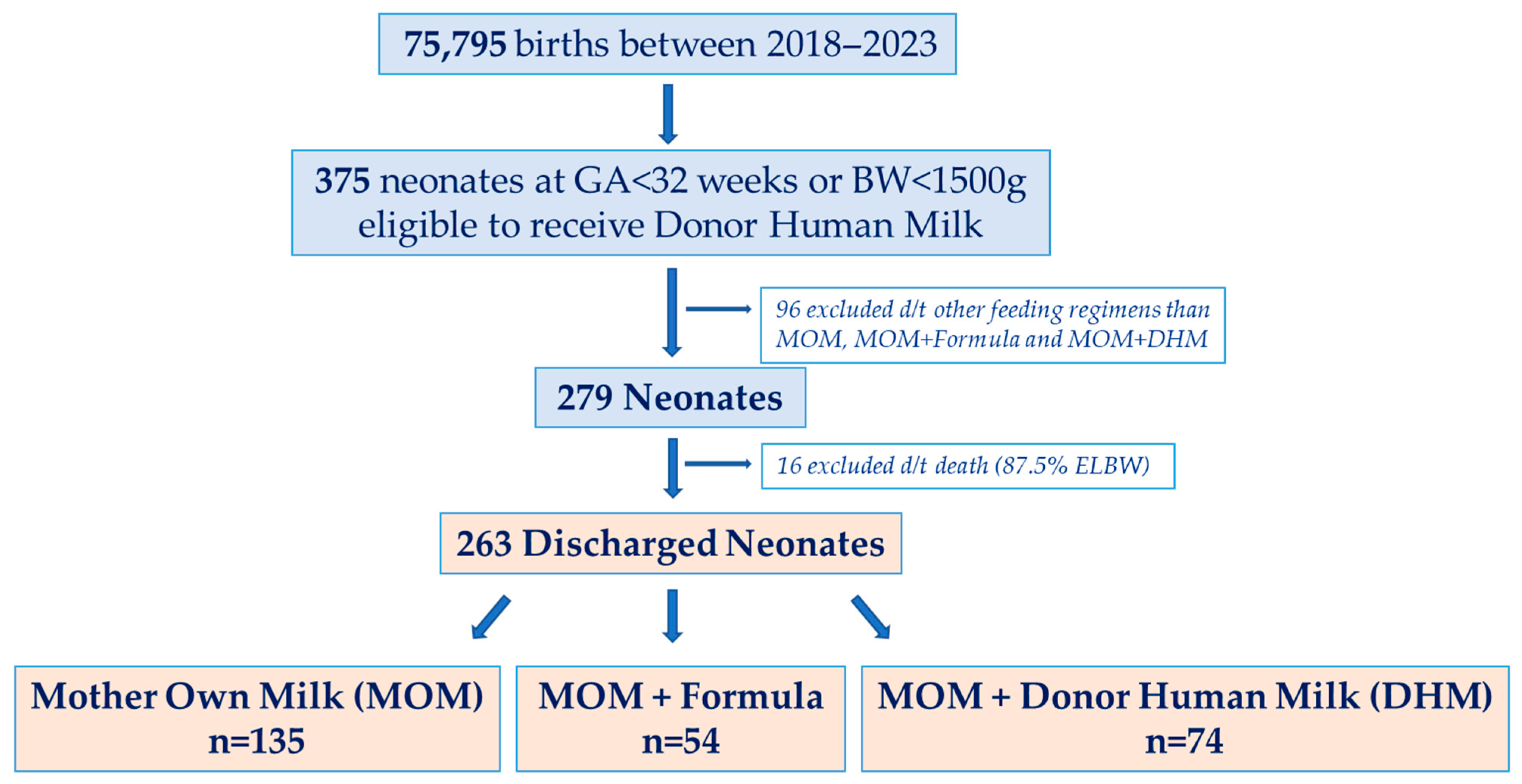
Figure 1. Flowchart of the cohort per feeding regimens.
Nutrients 17 03263 g001
Table 1. Comparison of demographic and clinical characteristics of the cohort across feeding groups.
CharacteristicsMOM
N = 135
MOM + Formula
N = 54
MOM + DHM
N = 74
p-Value
Mode of delivery 0.188
Spontaneous vaginal delivery40 (29.6)19 (35.2)30 (40.5)
Emergency Cesarean section94 (69.6)33 (61.1)42 (56.8)
Elective Cesarean section1 (0.7)2 (3.7)2 (2.7)
Infant of diabetic mothers10 (7.4)8 (14.8)11 (14.9)0.157
Celestone treatment99 (73.3)42 (77.8)61 (82.4)0.323
Intrauterine growth restriction (IUGR)24 (17.8)14 (25.9)8 (11)0.090
Gestational age (GA) (week)28 [27–30]30 [29–31]30 [28–31]<0.001
Birth weight (BW) (g)1070
[825–1320]
1450
[1157.5–1612.5]
1362.5
[1073.8–1486.3]
<0.001
Small for Gestational Age27 (20)15 (27.8)12 (16.2)0.272
Gender (Female)75 (55.6)37 (68.5)43 (58.1)0.258
Day of initiating enteral feeding (day)2 [1–2]2 [1–2]2 [1–2]0.058
Day at reaching BW10 [7–13.3]9 [6–11]9 [7–12]0.260
Duration of parenteral nutrition (day)15
[10–29]
9
[4.5–10.5]
9
[6–13]
<0.001
Day of reaching full enteral feeding (day)15
[11–28]
10
[6–12]
9
[6.5–13]
<0.001
Length of hospital stay (day)68
[45–95]
39
[31–46.3]
43
[37–74.5]
<0.001
Feeding Intolerance41 (30.4)9 (16.7)14 (18.9)0.062
Surfactant use74 (54.8)17 (31.5)26 (35.1)0.002
Necrotizing Enterocolitis25 (18.5)3 (5.6)9 (12.2)0.059
Respiratory Distress Syndrome126 (93.3)38 (70.4)55 (74.3)<0.001
Bronchopulmonary Dysplasia87 (64.4)8 (14.8)26 (35.1)<0.001
Blood Stream Infection27 (20)3 (5.6)6 (8.1)0.009
Retinopathy of Prematurity15 (11.1)2 (3.7)8 (10.8)0.264
Hypoglycemia27 (20)10 (18.5)16 (21.6)0.909
Percentage of MOM (%)10079.5 *
[64.4–90]
90 *
[79–93]
0.013 *
Data are expressed as mean ± standard deviation or median [Q1–Q3] or n (%). Abbreviations: MOM: Mother Own Milk; DHM: Donor Human Milk. In bold, significant p values for Kruskal–Wallis, ANOVA or Chi-square tests as appropriate. * Mann–Whitney test between mixed feeding group.
Table 2. Bone metabolism markers across the feeding groups.
CharacteristicsMOM
N = 135
MOM + Formula
N = 54
MOM + DHM
N = 74
p-Value
Alkaline Phosphatase (ALKP, IU/L) first measurement189
[156–235]
191
[162–254]
216.5
[170.8–276.8]
0.056
ALKP (IU/L) on day 7261 *
[216.5–337]
277
[205–351]
303 *
[256–378]
0.024 *
ALKP > 500 IU/L on day 711 (8.3)2 (3.8)3 (4.2)0.482
ALKP (IU/L) on day 14397 *
[300.8–506]
343 *
[269.5–413]
350.5
[262.3–457.0]
0.005 *
ALKP > 500 IU/L on day 1435 (26.9)3 (5.7)7 (9.7)<0.001
ALKP (IU/L) on day 28354.5
[285–473.8]
386.5
[279.8–457.0]
349
[289.5–420.0]
0.609
ALKP > 500 IU/L on day 2823 (18)8 (17.4)7 (10.1)0.331
Phosphorus (mg/dL) first measurement5.8 [4.9–6.6]5.8 [4.9–6.5]5.5 [4.8–6.4]0.796
Phosphorus (mg/dL) on day 75.7 [4.8–6.6]6 [5.4–6.9]5.8 [5.0–6.7]0.145
Phosphorus < 4.8 mg/dL on day 732 (24.2)5 (10)13 (18.8)0.096
Phosphorus (mg/dL) on day 146.1 ± 1.1
(3.0–8.4)
6.8 ± 1
(4.9–8.9)
6.6 ± 1.1
(3.3–8.8)
<0.001
Phosphorus < 4.8 mg/dL on day 1412 (9.7)0 (0)4 (6.1)0.046
Phosphorus (mg/dL) on day 286.4 ± 0.8
(4.2–8.0)
6.6 ± 0.6
(5.5–8.1)
6.6 ± 0.8
(4.4–8.1)
0.218
Phosphorus < 4.8 mg/dL on day 285 (4.0)01 (1.5)0.341
Calcium (mg/dL) first measurement8.7 ± 0.8
(6.9–10.4)
9.0 ± 0.8
(7.8–11.1)
9.1 ± 0.9
(6.7–11.6)
<0.001
Calcium (mg/dL) on day 79.9 [9.3–10.3]10.3 [9.8–10.7]10.3 [9.8–10.7]<0.001
Calcium < 7 mg/dL on day 7000NA
Calcium (mg/dL) on day 1410.0
[9.7–10.5]
10.4
[10.1–10.7]
10.4
[10.0–10.8]
<0.001
Calcium < 7 mg/dL on day 14001 (1.5)0.483
Calcium (mg/dL) on day 2810.2 *
[9.8–10.5]
10.2
[9.9–10.5]
10.4 *
[10.0–10.8]
0.033 *
Calcium < 7 mg/dL on day 28000NA
Data are expressed as mean ± standard deviation or median [Q1–Q3] or n (%). Abbreviations: MOM: Mother Own Milk; DHM: Donor Human Milk. In bold, significant p values for Kruskal–Wallis, ANOVA or Chi-square tests as appropriate, * Adjusted p value following Bonferroni correction for pairwise comparisons.
Disclaimer/Publisher’s Note: The statements, opinions and data contained in all publications are solely those of the individual author(s) and contributor(s) and not of MDPI and/or the editor(s). MDPI and/or the editor(s) disclaim responsibility for any injury to people or property resulting from any ideas, methods, instructions or products referred to in the content.