Abstract
Background/Objectives: With increasing knowledge of the role of the microbiota in health and disease, the need for the reliable simulation of its behavior in response to various factors, such as diet and probiotic administration in in vitro conditions, has emerged. Although many studies utilize developed systems, data on how accurately these systems represent individual microbiota responses are scarce. Methods: In the present study, the Simulator of Human Intestinal Microbial Ecosystem (SHIME®) was exposed to experimental conditions mimicking the application of probiotics and dietary changes in the study participant. Next-generation 16S rRNA sequencing was used to reveal the structure of the microbial communities in the analyzed samples. Results: Analysis of 17 samples revealed that predominantly diet and, to a lesser extent, probiotics had a divergent effect on the microbiota’s fluctuations dependent on the culture environment. Despite this, results from both in vitro and in vivo conditions aligned well with previously published data on the expected impact of dietary changes on the intestinal microbial community. Conclusions: The anecdotal evidence presented in this study suggested that current in vitro technology enables the reproduction of some of the microbiota responses that are well known from in vivo research. However, further work is required to enable simulations of an individual microbiota.
1. Introduction
The human gut microbiota plays a crucial role in maintaining the host’s health. It has been shown to influence processes such as immune modulation, metabolism, nutrient absorption, and protection against pathogens [1,2,3]. Disruptions in its composition, known as dysbiosis, have been associated with numerous diseases, including obesity, type II diabetes, inflammatory bowel disease, and neurological disorders [4,5]. The growing recognition of the microbiota’s role has sparked interest in how various interventions, such as dietary changes or probiotic supplementation, can be used to shape its composition and function.
Due to ethical, logistical, and biological constraints in human studies, in vitro gut models have become valuable tools for microbiota research, providing physiologically relevant insights into food–microbiota interactions and serving as important complements to in vivo studies, allowing for mechanistic validation under controlled conditions [6]. One of the most advanced systems is the Simulator of the Human Intestinal Microbial Ecosystem (SHIME®, referred to as SHIME through the text), which enables controlled, reproducible simulations of the human gut environment, including compartmentalized sections of the gastrointestinal tract (GIT) [7]. Recent research has increasingly focused on identifying in vitro models that closely replicate in vivo conditions, and SHIME has shown promise as a valuable predictive model to investigate microbial responses and interactions, particularly in the fields of food and nutrition, pharmacology, and gut health [8].
Although advanced in vitro models are widely used, data on how precisely they mirror individual in vivo conditions remain limited. Few works using the SHIME model have demonstrated qualitative similarities to microbiota shifts reported in human studies following similar interventions [9]. Nevertheless, several knowledge gaps can be pointed out. Most studies utilize SHIME and compare its output with findings from the literature [10,11,12]. Whereas fewer investigations are pointed towards directly comparing these changes to the microbiota of the donor, whose feces were used to inoculate the SHIME system under matched interventions [13]. Several studies have demonstrated that the composition of the original microbiota is not preserved after the transfer to the simulator [13,14,15]. This raises questions about whether such microbiota retain the ability to respond to a stimulus in a similar way as they would in their natural environment.
Access to in vitro tools that allow reliable simulations of an individual microbiota’s behavior could support advances in personalized health management, including precision nutrition. The term “precision nutrition” was introduced in the early 2010s and reflects a shift toward integrating omics data—genomic, metabolomic, and microbiomics—to design interventions that maximize individual health benefits [16,17]. In this context, the gut microbiota is both a target and a mediator of personalized dietary strategies. In vitro models offer a valuable platform to assess microbiota shifts and estimate diet-induced microbial metabolite production [18]. However, currently, data are insufficient to learn how a pool of macronutrient-level dietary changes, rather than isolated compounds like prebiotics or polyphenols, influences the microbiota composition in the SHIME relative to in vivo conditions. Bridging this gap would enhance the validity of in vitro systems for simulating complex host–microbiota–diet interactions.
Besides diet, a known approach to modulate the microbiota is the administration of probiotics. Lacticaseibacillus rhamnosus GG (LGG) is one of the most extensively studied probiotic strains, known for its beneficial effects on gut barrier function, immune response, and microbial balance. Studies have shown that LGG can temporarily colonize the GIT and promote shifts in microbial compositions, often characterized by increases in beneficial taxa and metabolic outputs such as short-chain fatty acids [19,20].
In this study, dietary and LGG interventions were applied to the SHIME system and the human donor in parallel to discern whether the behavior of individual microbiota may be modelled in an in vitro setting.
To our knowledge, this is the first study that attempted a comparison of the microbiota response to dietary and probiotic interventions in humans and simulators in parallel.
2. Materials and Methods
2.1. Study Design
The design of this study and methodological details were previously described by Rudzka et al. (2023) [13]. This previous publication reported a subset of data from the same experiment; hence, to avoid repetition, methodological details contained in the current work were limited to a minimum.
Briefly, a 39-year-old female volunteer with mild hypercholesterolemia but who was otherwise healthy donated a sample of feces that was used to inoculate the SHIME system (ProDigest, Gent, Belgium). The system operated in a MultiSHIME mode, focusing on the distal colon (two replicates of the following setup: one stomach, one duodenum, one proximal colon, and three distal colon bioreactors). Before the experiment, SHIME was stabilized for 14 days, using the standard SHIME media as per the manufacturer’s protocol. The volunteer provided 28-day dietary records before the start of the experiment. The experiment, during which the interventions were administered, lasted 28 days.
2.2. Interventions
Both the SHIME model and the volunteer were subjected to parallel interventions: a 28-day dietary modification and supplementation with a commercial formulation containing LGG (ATCC 53103) administered during the first 14 days of dietary intervention.
A commercially available LGG supplement in the form of capsules, claimed to contain 6 × 10⁹ LGG cells per capsule (other ingredients and additives: maltodextrin, hydroxypropyl methylcellulose, magnesium salts of fatty acids), was used in this study. Both the volunteer and the SHIME ingested the supplement twice a day. The volunteer consumed the capsules, while SHIME was inoculated with a small volume of sterile water in which the contents of 3 capsules were suspended (1.5 capsules in each stomach/ileum vessel). SHIME’s stomach was administered LGG right after filling with the fresh feed medium at pH 2, thereby exposing the probiotic to simulated GIT conditions from the stomach to the colon.
The volunteer was fed a nutritionally balanced diet with a calculated content of dietary macronutrients. To match macronutrient changes in the volunteer’s diet, the standard SHIME nutrient medium was modified by adjusting the proportions of its components that simulated dietary residues of animal protein (special peptone), non-animal protein (yeast extract), fiber (xylan, arabinogalactan, pectin, starch), and sugars (glucose). Fat was not included, as in this study, we chose to modify the standard SHIME feed by an exclusive adjustment of nutrient proportions. Nevertheless, the diet of the volunteer was not fat-free.
The mean intakes of dietary macronutrients and the nutritional compositions of SHIME feed media before and during the intervention are summarized in Table 1. Daily detailed nutrition data, including information about the meals given to the volunteer, were deposited in an open data resource [21,22].

Table 1.
Content of nutrients in standard and experimental diets for the volunteer and SHIME, mean ± standard deviation.
The modifications of the standard SHIME feed media were guided by an estimated proportion of food residues that were expected to reach the colon of the volunteer. This estimate was derived from a study on ileostomised humans, where the authors found that on average, 83 ± 15% of fiber, 1.1 ± 0.3% of carbohydrates, and 16 ± 5% of protein that were ingested reached the ileostomies [23]. These residue fractions were used to calculate nutrient concentrations that were included in the SHIME media while applying a normalization to soluble fiber content. To calculate SHIME medium composition, the following nutrient-to-ingredient conversion factors were used—1:1 for animal protein to special peptone, resistant starch to starch, arabinogalactan + arabinoxylan to sum of xylan and gum arabic, pectin to pectin, and 1 to 1.54 for non-animal protein to yeast extract. The glucose content of the media was corrected for the presence of glucose in yeast extract (2.9%).
2.3. Sampling
Twelve samples of feces (samples denoted with letter K and I-inoculum) and 11 samples of liquid from SHIME’s distal colon compartments (samples denoted with letter L) were collected. The sampling scheme is depicted in Figure 1. In total, three samples were collected before the experiment (I; and directly before the application of the intervention: SK and SL). Whereas during the intervention, sampling was performed 10 times, of which 5 samples were collected during the combined dietary and probiotic interventions (on experimental days 3, 6, 8, 10, 12) and the remaining during probiotic washout and continued dietary intervention (on experimental days 16, 18, 22, 25, 28). The sampling scheme relied on the volunteer’s availability to provide a fresh fecal sample on-site. The volunteer was instructed on the required number of samples for each study stage, with the condition that successive samples be collected at least one day apart. Samples from SHIME were collected in parallel with fecal samples provided by the volunteer.
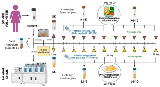
Figure 1.
Experimental design and sampling scheme used in this study. Created with Biorender.com.
2.4. Sample Preparation and Analysis
Methodological details of sample preparation and analysis were described previously [19]. Briefly, the DNA was extracted in three replicates from each fecal sample and from three of SHIME’s distal colon compartments. Then, the quality of the DNA was confirmed spectroscopically and via polymerase chain reaction. To reveal the structure of microbial community, a high-throughput sequencing of the V3–V4 regions of 16S rRNA was performed using DNA extracts pooled from three replicates taken for each sample. In total, 23 samples described in this study were sequenced.
2.5. Statistical Analysis
The statistical analysis presented in the current publication was performed with the use of three types of software. Statistica ver. 13 (StatSoft, Kraków, Poland) was used to obtain Spearman’s correlation coefficients for sensitivity analysis and run Wilcoxon signed-rank test. R built into R studio (Posit, Boston, MA, USA) was used to 1. calculate and visualize the microbial diversity indices and obtain the Jaccard distance matrix for the principal coordinate analysis (PCoA), 2. calculate data for heatmaps and draw them, and 3. perform linear modeling as the main analysis to compare the response of the microbiota to interventions in two different environments and draw a biplot. The linear modeling was preceded by data transformation. The abundance of microbial genera/phyla was centered log-ratio transformed (CLR) [21]. Dietary macronutrient residues expressed as g/day (volunteers) or g/L (SHIME) were standardized within each environment separately using z-transformation. Then, the principal component analysis (PCA) was performed on both CLR-transformed microbial counts and standardized macronutrient content, and the first two principal components (PCs) for each analysis were retained. These PCs were then used in the linear model described by relationship (1):
where
PC1_micro or PC2_micro ~ (PC1_diet + PC2_diet + Probiotic) × Environment
PC1_micro and PC2_micro—principal components of microbial community abundance.
PC1_diet and PC2_diet—principal components of the concentration of macronutrient residues in both in vitro and in vivo settings.
Probiotic—presence/absence of LGG supplementation.
Environment—in vitro (SHIME) or in vivo (volunteer) microbiota cultivation setting.
Linear models were evaluated by means of Type III Analysis of Variance (ANOVA) to test main and interaction effects.
Microsoft Excel (Microsoft, Redmond, WA, USA) was used to obtain the remaining figures.
All statistical tests assumed a significance level of 0.05. For multiple comparisons, either Benjamini–Hochberg false discovery rate (FDR) or Bonferroni corrections were used, as specified in the results. Compositional data of microbiota abundance were CLR transformed for correlation analyses.
3. Results
3.1. The Composition of the Microbiota
Substantial differences in the microbiota composition were observed between fecal and SHIME samples on all taxonomical levels, from phylum to genus. The dissimilarities in the microbial composition between the SHIME and stool microbiota were clearly visualized on the PCoA plot (Figure 2), where distinct groupings for each of the settings can be noticed. However, the plot also suggested the existence of some outliers. Three outlying samples were identified in each of the stool and SHIME datasets. These samples included L3, L4, and L10 for SHIME and K3, K4, and K10 for the stool. Consequently, they were excluded from the further statistical analyses presented in this manuscript.
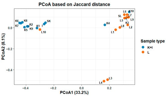
Figure 2.
Principal coordinate analysis of the composition of microbiota in SHIME (L) and stool (K), including inoculum (I) samples.
The bioinformatic analysis identified 112 bacterial genera in the whole dataset from the study; however, only 20 and 25 of these genera appeared in the majority of the analyzed fecal (at least 9 out of 12) and SHIME (at least 8 of 11) samples, respectively. The abundance of the most prevalent genera in all samples (including outliers) is shown in Figure 3. Eight of these genera were shared between the SHIME and stool, including Bacteroides, Bifidobacterium, Akkermansia, Alistipes, Sutterella, Parabacteroides, Phascolarctobacterium, and Victivallis. Their abundance varied between in vivo and in vitro settings and fluctuated over time. Among these genera, only Bacteroides was observed in appreciable levels in all the samples. Notably, the SHIME microbiota was richer in Bifidobacterium, Akkermansia, Lachnoclostridium, and Fusobacterium, while Alistipes, Subdoligogranulum, Oscillospiraceae (UCG-002), and members of the Christensenllaceae (R-7 group) were more abundant in fecal samples.
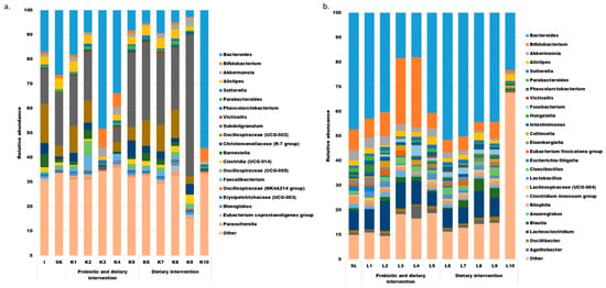
Figure 3.
The most prevalent bacterial genera in the feces of the volunteer ((a), includes 20 genera detected in at least 9 samples) and in the liquid from SHIME ((b), includes 25 genera detected in at least 8 samples).
Overall, the data shown in Figure 3 suggest that the microbial community was undergoing dynamic changes during the experiment in both in vitro and in vivo conditions, without any time-dependent trends.
Similarly to the taxonomic abundance, the Firmicutes to Bacteroidetes (F/B) ratio fluctuated during the experiment in both in vivo and in vitro conditions. This fluctuation was independent of the experimental phase, and its patterns were dissimilar between the SHIME and feces (supporting information, Figures S1 and S2). Overall, the F/B ratio was greater in fecal (mean: 4.9) than in SHIME samples (mean: 0.7). Except for outliers, neither of the fecal samples was characterized by an F/B ratio below one, but all of the SHIME samples fell below this threshold.
3.2. Microbiota Genetic Diversity
The α-diversity: The Chao1, Shannon, and Simpson indices of the microbial communities differed between fecal samples from the study participant and those from the SHIME model (Figure 4).
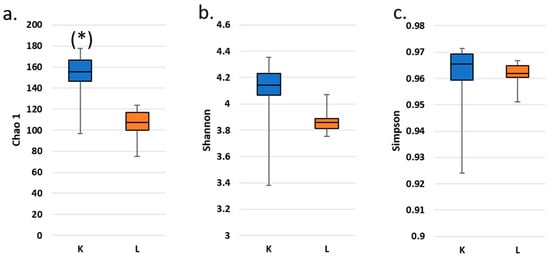
Figure 4.
Box plots of fecal (K) and SHIME (L) microbial α-diversity: Chao1 (a), Shannon (b) and Simpson (c) indices along the experimental trial. Reported data exclude outliers and the inoculum. (*) denotes a statistically significant difference between α-diversity in K and L according to a Wilcoxon’s signed-rank test (p < 0.05). No significant differences between pairs of data were detected after Bonferroni correction.
Fecal samples were characterized with consistently higher operational taxonomic unit (OTU) richness regardless of the α-diversity expression. This difference was statistically significant after Wilcoxon’s signed-rank test (p < 0.05) only for the Chao1 index; however, it did not survive the Bonferroni correction.
Twenty of the genera detected in the inoculum were absent from all SHIME samples. These genera were, however, each present at <4% abundance within the inoculum. On the other hand, seven genera were specific to SHIME samples, albeit also at a very low abundance (<2%). These genera included Enterococcus, Clostridium sensu stricto 1, Eubacterium hallii group, Megasphaera, Hafnia-Obesumbacterium, Bacillus, and Stenotrophomonas, which are all known members of intestinal microbiota.
3.3. The Comparison of the Microbiota Response to the Probiotic and Dietary Intervention
In previous sections, differences between the microbiota composition of a donor in the original in vivo conditions and after establishment in the in vitro conditions were presented. However, the main research question of this study was whether the microbiota behaved in a similar fashion regardless of the environment in which it was cultured.
During the probiotic supplementation, certain shifts in the abundance of the most prevalent genera were noticed (Figure 3). In fecal samples, the relative abundances of Phascolarcobacterium and Christensenellaceae (R-7 group) increased, while Bacteroides showed a consistent decline throughout the intervention, from the baseline (sample SK) through K1 and K2 to K5. On the other hand, SHIME samples exhibited a consistent increase only in the relative abundances of Sutterella and Victivallis.
Interestingly, the changes in the abundance of bacterial genera observed during the probiotic intervention did not reverse after its cessation. Furthermore, some shifts in the microbiota were noted during the post-supplementation probiotic washout period. These included a steady increase in Akkermansia and a decrease in Barnesiella in the volunteer’s microbiota. In the SHIME system, this period was marked by a decline in Bacteroides and increases in Oscillibacter and Bilophila.
Although an increase in the relative abundance of the former group of Lactobacillus (as classed under the older nomenclature and identified using the SILVA database for OTU assignment) was expected during the probiotic intervention as well as a decrease after its cessation, only in SHIME was such a phenomenon observed. The relative abundance of Lactobacillus was at 0.5% before the supplementation period (sample SL) and ranged from 2.23 to 2.63% while it lasted (samples L1, 2, and 5) and ranged from 1.21 to 1.89% after its cessation (samples L6, 7, 8, and 9). Lactobacillus was detected in all SHIME samples. On the other hand, in feces, this genus was only present in the sample used for the inoculum preparation at a relative abundance of 2.2%. After that, it was not detected in any of the samples collected during or after the probiotic intervention.
The dietary intervention did not seem to have an immediate effect on the microbiota structure in either of the two culturing environments. Correlation analysis between the relative abundance of microbial genera and macronutrient concentrations—measured either in food residues from the volunteers’ diet or in SHIME media from the day before sampling—revealed that only four of the most prevalent genera in each microbiota culturing environment increased in abundance in response to higher concentrations of at least one of eleven macronutrients (Spearman’s correlation coefficient > 0.7). To explore potential cumulative effects, a sensitivity analysis with a moving average for macronutrient residues was performed (results—Figure 5, full dataset available in data repository [22]). The analysis suggested that the optimal averaging window (the greatest number of genera with abundance correlating positively with at least one macronutrient) was 9 days for the volunteer and 12 days for SHIME.
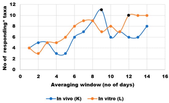
Figure 5.
Sensitivity analysis of nutrient content averaging window to correct for a lag in microbial response to nutritional changes; * responding taxa were defined as those whose abundance correlated positively (Spearman’s ρ > 0.7) with at least one macronutrient or group of macronutrients (protein, animal protein, non-animal protein, arabinogalactan, arabinoxylan, arabinogalactan + arabinoxylan, pectin, soluble fiber, starch, sugars, protein-to-soluble-ratio). Black markers indicate averaging windows that were selected for further statistical analyses.
Although the microbiota is known to respond quickly to dietary changes (shifts are noticeable within 24 h [24]), validation studies carried out in SHIME showed that these responses stabilize after a certain period (12 days for a standard, unchanged nutritional medium [25]). This could explain why a low number of correlations was found with the 24 h data in the present study and prompted the use of an optimal macronutrient content averaging window in all further analyses.
To quantitatively assess whether the probiotic and dietary intervention had different outcomes in two microbiota culturing environments, a linear model was fitted to bacterial abundance and intervention data (Section 2.5). The analysis focused on eight genera prevalent in the majority of SHIME and fecal samples (Section 3.1) and four selected macronutrients (soluble fiber, sugars, non-animal protein, and animal protein). A parallel analysis, at a phylum level, confirmed that the significance of the effects translated to the whole studied microbial population (open data [22]). The type III ANOVA analysis did not detect significant main effects for the intervention variables across the pooled dataset for eight selected genera (SHIME and volunteer considered together). In contrast, a strong main effect of the Environment on PC1_micro (explaining ~62% variance) was noted. Furthermore, a significant interaction between the Environment and PC1_diet and Environment and Probiotic on PC1_micro (~7% and 4% variance explained, respectively) was identified. Out of these results, at the phylum level, only the interaction effect between the Environment and Probiotic was not significant, whereas a significant main effect of PC1_diet and its interaction with the Environment on PC2_micro was found.
In summary, the structure of microbial communities was significantly dependent on the culturing environment (volunteer vs. SHIME), and the microbiota responded differently to the dietary intervention in each environment (regardless of whether the whole population or a small subset of eight genera common for both SHIME and the volunteer was considered). The biplot (Figure 6) illustrated this divergence well. Fecal and SHIME samples were clearly grouped in separate clusters, with an overlap of 95% confidence areas (overlap not observed at a phylum level [22]). In general, the SHIME microbiota was characterized with a greater abundance of the most prevalent genera shared between the two environments, and samples taken during the probiotic intervention presented an even greater enrichment. The only exception was Alistipes, which seemed to be more abundant in fecal microbiota.
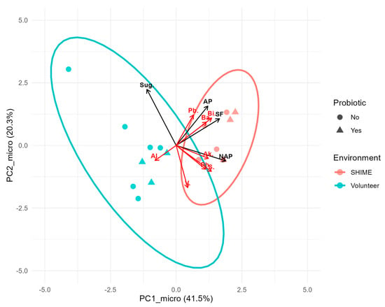
Figure 6.
Biplot of principal components based on the abundance of the eight most prevalent genera shared between SHIME and fecal samples. Points, red vectors, and black vectors represent samples, rescaled genera loadings, and overlaid principal components of dietary macronutrients, respectively. Ellipses mark 95% confidence regions for each microbiota culturing environment. Abbreviations: Pb.—Parabacteroides, Ph.—Phascolarcobacterium, Ba.—Bacteroides, Bi.—Bifidobacterium, V.—Victivallis, Al.—Alistipes, Ak.—Akkermansia, S.—Sutterella, Sug.—sugar, AP—animal protein, SF—Soluble fiber, and NAP- non-animal protein.
The biplot included overlaid vectors from a PCA on macronutrient concentrations. The abundance of most genera selected for analysis showed a positive correlation with at least one macronutrient, either animal protein, non-animal protein, or soluble fiber. Since the microbiota composition was significantly affected by the interaction between the Environment and PC1_diet, a separate analysis investigating responses to macronutrients in each environment was performed. For this purpose, a series of Spearman’s correlation analyses was conducted (Figure 7). Besides the eight genera highly prevalent in SHIME and fecal samples, broader descriptors of the microbiota’s condition, such as α-diversity indices and the F/B ratio, were also included.
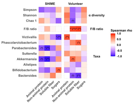
Figure 7.
Heatmap of Spearman’s correlation coefficients between the optimal averaging window content of macronutrients in SHIME feed and food residues and the α-diversity, F/B ratio, and relative abundance of the most prevalent genera common for the microbiota populating SHIME and human feces. Statistically significant correlations after FDR correction were marked with ** < 0.01, and * < 0.05, and raw p values are in parentheses. FDR corrections were applied for each level of data (alpha diversity, F/B ratio, and 8 genera) separately.
Sperman’s correlation analysis revealed that before FDR corrections, there were some statistically significant relationships between macronutrients and microbiota across all analyzed levels. After applying the FDR adjustment, a significant increase in the response to macronutrients (non-animal protein and soluble fiber) was found only for the F/B ratio in the stool. Other positive correlations that were initially significant but did not survive the correction displayed a strong relationship (Spearman’s ρ > 0.7). Such relationships were identified on the genus level but not for α-diversity indices. In stool, the abundance of Akkermansia correlated positively with non-animal protein; Phascolarcobacterium with soluble fiber; and Victivallis with the animal protein content in the dietary residues. In contrast, in SHIME the abundance of Akkermansia correlated negatively with the non-animal protein content, which remained significant even after the FDR correction. The other two genera showed a positive but weak association with their respective macronutrients.
4. Discussion
There are not many studies evaluating the fate of the microbiota of a particular donor in SHIME and its natural environment by means of modern high-throughput DNA sequencing techniques. Early validation studies (from the end of the XX and beginning of the XXI century) used such indicators as the metabolism of the microbiota, plate counts of selected taxa, and the stability of the microbiota after inoculation [13]. Outcomes were typically compared with data from existing human trials that did not include fecal sample donors; however, these comparisons allowed the authors of previous studies to demonstrate that the system was able to mimic the behavior of human microbiota. Multiple modern studies, which also relied on these non-parallel comparisons, demonstrated that high-throughput DNA sequencing techniques allowed for the validation of the outcomes from SHIME, e.g., [9,26]. Thus, it is not surprising that in the modern literature, SHIME has established itself as a state-of-the-art, validated method for studying reactions of the microbiota to various interventions in in vitro conditions.
In this study, it was demonstrated that the changes in the microbiota composition in response to applied interventions varied noticeably between in vitro and in vivo conditions. Hence, to validate the present findings, we followed an approach driven by previous records and evaluated how the responses of microbiota to probiotic and dietary interventions reported in this study aligned with the existing scientific evidence.
The former group of Lactobacillus was not detected in fecal samples during and after the LGG supplementation. This was unexpected, as LGG is well-characterized, and it has been known to appear in feces not only during but also up to 12 days post-supplementation [27]. On the other hand, in the SHIME model the former group of Lactobacillus persisted through the whole experiment, during and after the probiotic supplementation. This observation aligns well with both in vivo and in vitro studies. The latter showed that the system may allow LGG proliferation in the ascending colon for at least 10 days and promotes its persistence in the distal colon for at least two weeks [26,28].
In contrast to the human gastrointestinal tract, the SHIME model provides stable and controlled conditions that facilitate the persistence and proliferation of supplemented strains, whereas in vivo their colonization is often limited by microbial competition, rapid intestinal transit, and host immune responses. Moreover, colonization by Lactobacillus is frequently transient, meaning that their presence in feces may be sporadic and below the detection threshold of 16S rRNA sequencing. In addition, the 16S rRNA may suffer from a primer-dependent amplification bias towards low-abundance DNA, which ultimately can result in false negatives. Moreover, LGG may transiently adhere to the mucosal surface of the small intestine or proximal colon rather than being shed in feces, which limits its detectability in stool-based sequencing.
Besides the increase in the former Lactobacillus genus abundance, the literature reported some other minor effects of LGG supplementation on the microbiota. Taxa that were previously shown to respond to such supplementation in human and animal studies reported increases in Lachnospiraceae and Porphyromonadaceae families [29] and genera such as Bacteroides, Ruminococcus, Butyricicoccus, Erysipelatoclostridium, Flavonifractor, and Bacillus [30,31]). In our study these taxa were present at very low abundances, except for Bacteroides, which tended to decrease during the probiotic intervention in the volunteer’s feces. Furthermore, in vitro research with the use of the SHIME model inoculated with a western-diet consuming donor’s microbiota reported a minor decrease in the F/B ratio after 6 days from a single introduction of LGG into the system [28]. In contrast, in this study, the F/B ratio increased during the intervention in both in vitro and in vivo conditions and fluctuated after its cessation (supporting information, Figures S1 and S2). These slight changes and lack of clear trends in their reversal during the LGG washout period confirmed that dietary intervention was a major factor driving the fluctuations in the structure of the microbial community in this study.
The literature indicates a positive relationship between the diversity of the microbiota and dietary content of animal protein, plant protein (particularly soy), and fiber—especially branched arabinoxylan, fermented foods, and overall dietary variety [32,33,34,35]. On the other hand, diets high in fat and sugar, as well as the excessive consumption of ultra-processed foods, are known to reduce bacterial diversity [35,36]. This is consistent with the broader understanding that the diminished diversity of the microbiota is detrimental to host health and is associated with diseases, including diet-related conditions such as obesity or type 2 diabetes [37,38,39].
This study did not find any strong positive associations between macronutrients and microbiota’s α-diversity in either in vitro or in vivo conditions (Figure 7). On the other hand, a strong (but not statistically significant) negative relationship between the content of non-animal protein seemed to decrease the diversity of fecal microbiota. Such negative associations should be interpreted with care. Macronutrients do not directly contribute to the reduced ability of specific microorganisms to thrive. Rather, they may promote competing microorganisms, suppressing the ones that cannot readily utilize particular energy sources. Therefore, a decline of α-diversity resulting from the loss of the abundance of specific groups of microorganisms in response to a nutrient is likely a result of coadaptation issues. These challenges are particularly relevant to in vitro systems, such as SHIME, where the microbiota composition is affected during the transfer from in vivo conditions [14,15].
Apart from changes in diversity, research often refers to the F/B ratio to assess the condition of the microbiota and its reaction to a particular intervention. In this study, higher levels of non-animal protein and soluble fiber promoted Firmicutes over Bacteroidetes (Figure 7) but only in fecal samples. These findings align well with the literature reports, where an increase in the abundance of Firmicutes was reported in response to whole grain consumption [33] and in individuals adhering to a healthy diet pattern [40]. Furthermore, a decrease in Firmicutes after a few days (ca. 5) of an animal-based diet has been reported [41].
Although the elevated F/B ratio is an established indicator of gut microbiota dysbiosis and has been associated with diseases, such as obesity, in multiple studies [42,43], it is important to note that many beneficial microbes, including the former genus Lactobacillus, belong to the Firmicutes. In this study, SHIME and fecal samples contained an appreciable proportion of Firmicutes composed predominantly of three families: Lachnospiraceae, Ruminococcaceae, and Monoglobaceae. These families are known to feed on dietary fiber, with Lachnospiraceae and Ruminococcaceae being well-established short-chain fatty acid (SCFA) producers [44]. A high abundance of Lachnospiraceae and Ruminococcaceae has been associated with good health and favorable dietary habits, i.e., adherence to the Mediterranean diet [45,46]. In addition, such a microbiota is characteristic of vegetarian and vegan over omnivorous feeding patterns [47].
Considering a deeper taxonomic level, this study found strong positive associations between the relative abundance of Akkermansia, Phascolarcobacterium, Victicvallis, and Bifidobacterium and the concentration of some of the macronutrients in either in vitro or in vivo conditions (Figure 7).
A very well-known Akkermansia species is Akkermansia muciniphila. This species is related to healthy metabolism [48] and is promoted by various dietary fibers, including arabinogalactan, arabinoxylan, resistant starch, and pectin [49]. Interestingly, even fiber-restricted diets, such as the low-FODMAP [50] and ketogenic [51] diets, were shown to increase its abundance in vivo. Conversely, high-animal-protein diets could diminish the population of Akkermansia [52], while dietary sugars may promote their abundance [53]. Animal studies point to a positive effect of unsaturated fat consumption on the levels of A. muciniphila [32], a finding partly supported by a human study where people with mild cognitive decline followed a Mediterranean–ketogenic diet and also experienced an increase in Akkermansia [54]. Our results appear to support the role of plant-based diets in promoting Akkermansia. We have found a strong, positive correlation between its abundance and the concentration of non-animal protein in food residues in vivo. Notably, this association was not replicated in vitro; in fact, an inverse relationship was observed in SHIME. This discrepancy could result from non-animal protein being represented in SHIME feed by the yeast extract.
In our study, the abundance of Phascolarcobacterium, associated with good metabolic health [55], showed a strong positive correlation with the content of soluble fiber in the residues from the volunteers’ diet (Figure 7), confirming the literature findings [56,57,58,59]. However, this effect was not replicated in SHIME. Moreover, the abundance of Victivallis was positively correlated with the content of animal protein in the residues within the volunteer’s diet and pectin in SHIME (Figure 7), which was also in agreement with the existing literature [53,59,60,61]. The role of Victivallis in human health is not yet fully understood. Its abundance was found to be positively associated with the increased risk of hypertension and radiotherapy-induced oral mucositis in patients with head and neck cancer [62,63]. On the other hand, this genus belongs to SCFA producers and has been shown to increase in abundance in response to a reduced-sucrose and starch diet, alleviating symptoms in patients with irritable bowel syndrome [62,64].
The current study also found that increased levels of glucose promoted Bifidobacterium in SHIME (strong, but not significant correlation) but not in fecal samples (Figure 7). Bifidobacterium is a broadly recognized probiotic genus with anti-inflammatory and pathogen-protective effects [65,66]. It ferments a broad range of carbohydrates, including digestible and indigestible fractions, producing beneficial SCFAs [32,67]. Unsurprisingly, Bifidobacterium is promoted by carbohydrate-rich foods and supplements, such as corn flakes, apple pomace, inulin, β-glucan, or type 2 and 4 resistant starch [32,68,69,70]. Murine studies have shown that a diet high in unsaturated fats also supports this genus [32]. In contrast, low-carbohydrate and high-fat feeding patterns contribute to the decrease in Bifidobacterium [54,71].
Overall, observed associations between the macronutrient concentration and the abundance of specific taxa were all in good agreement with previously published findings from human and animal studies. This suggests that although we failed to reproduce the response of the studied individual microbiota to interventions in in vitro conditions, the SHIME system still provided outcomes that reflected well-known microbial responses to dietary inputs from human and animal studies.
One of the multiple reasons for the failure to reproduce the behavior of an individual microbiota in vitro could be a difference in its structure at the beginning of the experimental intervention period. Existing research underscores the variability of microbiota responses to identical interventions. For example, recently Devarakonda et al. [72] reported that the microbiota of different subjects exhibited diverse responses to the supplementation with type 2 and 4 resistant starch. The greatest interindividual variation was found in the abundance of Ruminococcus bromii and Parabacteroides distasonis, both known to react to this fiber fraction. Similarly, an earlier study by Salonen et al. [73] showed that the responsiveness of microbiota to dietary interventions in obese men was highly individualized. Furthermore, studies imply that the interactions of the intestinal microbial communities with medication are also specific to each person and can influence the efficacy of treatments [74]. These findings suggest that enabling in vitro research capable of evaluating the response of particular microbiota to nutritional or bioactive compounds could advance the development of targeted and effective interventions. In this context, the recreation of the microbiota of a particular donor in the SHIME and similar systems seems of great importance.
It is well understood that in vitro systems designed for human microbiota cultivation are imperfect in their reflection of realistic in vivo conditions. They simply lack the complexity of bodily functions that govern secretory activity, digestion, and absorption. The efficiency of these functions varies between individuals and is influenced by numerous factors such as diet, physical activity, sex, health status, mood, and many other personalized physiological responses. Such complexity of the individual GIT functions shapes the structure of the microbial community, and therefore, a shift in the microbiota composition upon transfer to in vitro conditions is expected.
However, the nature and extent of changes during the transfer of the microbiota to a simulator are not reproducible across different studies nor even among different donors within the same study [15]. For instance, here we did not detect a significant change in the α-diversity. This aligns with the findings of Liu et al. [14], who also studied fecal microbiota in SHIME. In contrast, another study reported a decrease in the Shannon index for the synthetic inoculum in the same system [75].
In the current study, the F/B ratio, crucial for distinguishing “healthy” and “dysbiotic” statuses, decreased approximately three-fold after the stabilization of the microbiota in vitro (supporting information, Figure S2). Other studies that used the SHIME system with its standard startup protocol showed smaller changes in the F/B ratio (less than two-fold) and indicated that establishing a “dysbiotic” microbiota (with F/B ratio > 1) in SHIME was possible [14,15,42].
At deeper taxonomic levels, changes in the microbiota structure after the transfer to in vitro conditions were also reported [14,15,75], which we similarly observed. However, specific changes varied between studies, most likely due to differences in the structure and quality of the microbiota comprising the inoculum.
Beyond the inability to accurately reflect the complexity of the human GIT, the literature identified several particular factors that hamper the preservation of the microbiota’s structure during its establishment in the simulator. These factors include stochasticity, coadaptation, stressors acting upon the inoculum, and the feed media’s composition.
A study utilizing a synthetic inoculum, based on 23 species isolated from human feces and cultivated in a system of Multifors bioreactors, highlighted that stochasticity was the key driver of the microbiota structure during its establishment in vitro [76]. The authors failed to reproduce the structure of the microbial community between replicates of the same cultivation conditions.
Coadaptation issues were also reported, particularly if the members of microbiota from different donors were combined into a single inoculum [76]. While the mentioned study provides valuable insights into the dynamics of synthetic microbiota establishment in the bioreactor, further research is needed to understand how these findings translate to more complex inocula, such as the full fecal microbiota of a single person used here.
Research with the use of a synthetic inoculum showed that microbiota establishment in SHIME is strongly influenced by the feed medium composition. Differences in feed media accounted for approximately 38% of the observed variability in the microbial community structure [75]. In contrast, the proportions of the microorganisms within the inoculum seemed to have little effect on its structure after the stabilization process.
When using complex fecal microbiota, the medium composition seems to play a critical role not only during the experiment but perhaps predominantly during the transition into an in vitro setting. Marzoratti et al. [77] demonstrated that feed formulations enriched in either dietary fiber or protein led to distinct microbial communities in SHIME, even though the microbiota originated from the same donor. While the F/B ratio was similar for both conditions, the high-protein feed increased the level of Proteobacteria in SHIME [77].
These findings suggest that any efforts focused on reproducing the microbiota of the donor in SHIME should begin with carefully tailoring the feed composition, primarily during the stabilization period. In the present study, the SHIME feed was only matched with feed residues that could originate from the volunteer’s diet arbitrarily and only after the standard stabilization period. This likely contributed to adaptive shifts in the microbial community that may have limited its fidelity to the original human microbiota. Furthermore, the lack of some food residues, such as fat and insoluble fiber, in the standard (and our experimental) SHIME feed may contribute to these shifts. Moreover, other factors differentiating what ultimately reaches the microbiota in the colonic environment in vitro and in vivo cannot be neglected. For example, pancreatic juice used in the SHIME model (containing potent gut microbiota modulators such as bile acids) may induce excessive changes in the microbiota composition due to the absence of reabsorption and bacterial metabolites of excessive bile acids.
Besides the coadaptation and medium composition, a range of stressors could impact the establishment of the particular microbiota in the in vitro system. These include all the differences between the human organism and bioreactors, as well as the procedural steps involved in the inoculum preparation. The literature suggests that some of these stressors, such as frozen storage before inoculation, help to reproduce the microbiota structure in vitro at least at the phylum level [15]. Low temperatures can alter the gene expression of microorganisms, consequently making them less sensitive to other environmental changes [78,79]. As a result, freezing the inoculum before its introduction into the bioreactor may partially stabilize the microbial community and mitigate compositional shifts during the transition to in vitro conditions. Nevertheless, here we have chosen to work with a freshly prepared fecal inoculum (a standard approach), which may partly explain a large shift in the microbial community’s structure after stabilization in SHIME.
In summary, findings of this and other studies suggest that standard protocols used for running in vitro experiments do not preserve the original structure of human microbiota. This likely contributes to shifts in the microbiota responses to interventions. Further studies should hence explore modifications to standard protocols aimed at the preservation of an individual microbiota structure. Such improvements could enable the use of in vitro technology to support developments in personalized health management strategies, including precision nutrition.
5. Conclusions
In this study, we found that the same initial microbiota exhibited different responses to probiotic supplementation and dietary changes in in vivo and in vitro settings. Thus, we failed to simulate the behavior of the individual microbiota in SHIME. Despite this, the system enabled microbiota responses that were known from multiple earlier human and animal studies. Particularly, we observed a positive correlation of the Bifidobacterium abundance with the increase in glucose in the feed medium and the enrichment of the former Lactobacillus genus during probiotic supplementation.
Although this study included no replication and only a single donor, to our knowledge, it presents the first attempt to compare the response of an individual microbiota to probiotic and dietary interventions in the donor and SHIME in parallel. We propose that the alteration of the microbial community during the establishment in the in vitro setting, shown here as well as in previous studies, is one of the several factors contributing to the observed divergence of the individual microbiota behavior between artificial and natural environments.
Nevertheless, the ability to replicate the reaction of an individual’s microbiota to a dietary, probiotic, drug, or any other intervention holds promise for the development of precision nutrition or personalized medical strategies. Further advances in the in vitro technology that would be aimed at the preservation of the structure of microbial communities contained in the inoculum, e.g., preparing frozen rather than fresh inocula and adjusting the feed media composition, are essential to enhance the predictive power of such systems.
Supplementary Materials
The following supporting information can be downloaded at https://www.mdpi.com/article/10.3390/nu17193093/s1: Figure S1: F/B ratio in fecal samples and Figure S2: F/B ratio in SHIME samples.
Author Contributions
A.R. conceptualization, investigation, methodology, data analysis, data curation, funding acquisition, project administration, writing—original draft preparation; O.P. investigation, writing—original draft preparation, writing—review and editing; M.P. data analysis, data curation, writing—original draft preparation, writing—review and editing; M.Z. investigation, writing—review and editing; T.K. investigation, writing—review and editing; M.O. investigation, writing—review and editing; D.K.-K. resources, writing—review and editing; M.K. (Marcin Kruk) data analysis, data curation, investigation, writing—review and editing; M.K. (Marcelina Karbowiak) investigation, writing—original draft preparation; writing—review and editing; W.M. investigation, writing—original draft preparation; writing—review and editing; D.Z. supervision, investigation, resources, writing—original draft preparation; writing—review and editing. All authors have read and agreed to the published version of the manuscript.
Funding
This research was funded by the National Science Centre, Poland 2021/05/X/NZ4/01613. The APC was covered by Jan Dlugosz University in Czestochowa.
Institutional Review Board Statement
This study was conducted in accordance with the Declaration of Helsinki and approved by the Research Ethics Committee at Jan Dlugosz University in Czestochowa (decision KE-U/12/2022, date of approval: 27 June 2022).
Informed Consent Statement
Informed consent was obtained from the volunteer involved in this study.
Data Availability Statement
The full dataset that allowed for the generation of results reported in this study was archived in an open repository under: https://doi.org/10.5281/zenodo.14974398 (also shared in a previous publication) and under: https://doi.org/10.5281/zenodo.16751627 (exclusively for the current report).
Acknowledgments
The authors of this study would like to thank all employees of the Institute of Human Nutrition Sciences, Warsaw University of Life Sciences, for their patience and technical advice during the experimental work on SHIME. Chat GPT 5 (OpenAI) was used in this study for support in the R script-writing.
Conflicts of Interest
The authors declare no conflicts of interest.
Abbreviations
The following abbreviations are used in this manuscript:
| CFU | Colony-Forming Unit |
| DNA | Deoxyribonucleic Acid |
| F/B ratio | Firmicutes to Bacteroidetes Ratio |
| FODMAP | Fermentable Oligosaccharides, Disaccharides, Monosaccharides, and Polyols |
| GIT | Gastro-Intestinal Tract |
| LGG | Lacticaseibacillus Rhamnosus Strain GG |
| OTU | Operational Taxonomic Unit |
| PCoA | Principal Coordinate Analysis |
| rRNA | Ribosomal Ribonucleic Acid |
| SCFAs | Short-Chain Fatty Acids |
| SHIME | Simulator of the Human Intestinal Microbial Ecosystem |
References
- Afzaal, M.; Saeed, F.; Islam, F.; Ateeq, H.; Asghar, A.; Shah, Y.A.; Ofoedu, C.E.; Chacha, J.S. Nutritional Health Perspective of Natto: A Critical Review. Biochem. Res. Int. 2022, 2022, 5863887. [Google Scholar] [CrossRef] [PubMed]
- Fan, Y.; Pedersen, O. Gut Microbiota in Human Metabolic Health and Disease. Nat. Rev. Microbiol. 2021, 19, 55–71. [Google Scholar] [CrossRef]
- Rudzka, A.; Kapusniak, K.; Zielińska, D.; Kołożyn-Krajewska, D.; Kapusniak, J.; Barczyńska-Felusiak, R. The Importance of Micronutrient Adequacy in Obesity and the Potential of Microbiota Interventions to Support It. Appl. Sci. 2024, 14, 4489. [Google Scholar] [CrossRef]
- Napolitano, M.; Fasulo, E.; Ungaro, F.; Massimino, L.; Sinagra, E.; Danese, S.; Mandarino, F.V. Gut Dysbiosis in Irritable Bowel Syndrome: A Narrative Review on Correlation with Disease Subtypes and Novel Therapeutic Implications. Microorganisms 2023, 11, 2369. [Google Scholar] [CrossRef]
- Tan, J.; Taitz, J.; Nanan, R.; Grau, G.; Macia, L. Dysbiotic Gut Microbiota-Derived Metabolites and Their Role in Non-Communicable Diseases. Int. J. Mol. Sci. 2023, 24, 15256. [Google Scholar] [CrossRef] [PubMed]
- Li, C.; Zhang, X. Current in Vitro and Animal Models for Understanding Foods: Human Gut–Microbiota Interactions. J. Agric. Food Chem. 2022, 70, 12733–12745. [Google Scholar] [CrossRef]
- Van de Wiele, T.; Van den Abbeele, P.; Ossieur, W.; Possemiers, S.; Marzorati, M. The Simulator of the Human Intestinal Microbial Ecosystem (SHIME®). In The Impact of Food Bioactives on Health: In Vitro and Ex Vivo Models; Verhoeckx, K., Cotter, P., López-Expósito, I., Kleiveland, C., Lea, T., Mackie, A., Requena, T., Swiatecka, D., Wichers, H., Eds.; Springer International Publishing: Cham, Switzerland, 2015; pp. 305–317. ISBN 978-3-319-16104-4. [Google Scholar]
- Zhu, W.; Zhang, X.; Wang, D.; Yao, Q.; Ma, G.-L.; Fan, X. Simulator of the Human Intestinal Microbial Ecosystem (SHIME®): Current Developments, Applications, and Future Prospects. Pharmaceuticals 2024, 17, 1639. [Google Scholar] [CrossRef]
- Duysburgh, C.; Van den Abbeele, P.; Kamil, A.; Fleige, L.; De Chavez, P.J.; Chu, Y.; Barton, W.; O’Sullivan, O.; Cotter, P.D.; Quilter, K.; et al. In Vitro–in Vivo Validation of Stimulatory Effect of Oat Ingredients on Lactobacilli. Pathogens 2021, 10, 235. [Google Scholar] [CrossRef]
- Chen, P.; Chen, X.; Hao, L.; Du, P.; Li, C.; Han, H.; Xu, H.; Liu, L. The Bioavailability of Soybean Polysaccharides and Their Metabolites on Gut Microbiota in the Simulator of the Human Intestinal Microbial Ecosystem (SHIME). Food Chem. 2021, 362, 130233. [Google Scholar] [CrossRef]
- Marzorati, M.; Van den Abbeele, P.; Bubeck, S.; Bayne, T.; Krishnan, K.; Young, A. Treatment with a Spore-Based Probiotic Containing Five Strains of Bacillus Induced Changes in the Metabolic Activity and Community Composition of the Gut Microbiota in a SHIME® Model of the Human Gastrointestinal System. Food Res. Int. 2021, 149, 110676. [Google Scholar] [CrossRef] [PubMed]
- De Oliveira, F.L.; Salgaço, M.K.; de Oliveira, M.T.; Mesa, V.; Sartoratto, A.; Peregrino, A.M.; Ramos, W.S.; Sivieri, K. Exploring the Potential of Lactobacillus Helveticus R0052 and Bifidobacterium Longum R0175 as Promising Psychobiotics Using SHIME. Nutrients 2023, 15, 1521. [Google Scholar] [CrossRef] [PubMed]
- Rudzka, A.; Patloka, O.; Płecha, M.; Królikowski, T.; Oczkowski, M.; Zborowski, M.; Kołożyn-Krajewska, D.; Zielińska, D. Changes in the Microbiome of a Human and in the Simulator of Human Intestinal Microbial Ecosystem (SHIME®) in Response to a Diet and Probiotic Supplementation. Food Sci. Technol. Qual. 2023, 134, 53–72. [Google Scholar] [CrossRef]
- Liu, L.; Firrman, J.; Tanes, C.; Bittinger, K.; Thomas-Gahring, A.; Wu, G.D.; Van den Abbeele, P.; Tomasula, P.M. Establishing a Mucosal Gut Microbial Community In Vitro Using an Artificial Simulator. PLoS ONE 2018, 13, e0197692. [Google Scholar] [CrossRef]
- Da Ros, A.; Polo, A.; Rizzello, C.G.; Acin-Albiac, M.; Montemurro, M.; Di Cagno, R.; Gobbetti, M. Feeding with Sustainably Sourdough Bread Has the Potential to Promote the Healthy Microbiota Metabolism at the Colon Level. Microbiol. Spectr. 2021, 9, e00494-21. [Google Scholar] [CrossRef]
- Abeltino, A.; Hatem, D.; Serantoni, C.; Riente, A.; De Giulio, M.M.; De Spirito, M.; De Maio, F.; Maulucci, G. Unraveling the Gut Microbiota: Implications for Precision Nutrition and Personalized Medicine. Nutrients 2024, 16, 3806. [Google Scholar] [CrossRef]
- Kirk, D.; Catal, C.; Tekinerdogan, B. Precision Nutrition: A Systematic Literature Review. Comput. Biol. Med. 2021, 133, 104365. [Google Scholar] [CrossRef]
- Singh, V.; Son, H.; Lee, G.; Lee, S.; Unno, T.; Shin, J.-H. Role, Relevance, and Possibilities of in Vitro Fermentation Models in Human Dietary, and Gut-Microbial Studies. Biotechnol. Bioeng. 2022, 119, 3044–3061. [Google Scholar] [CrossRef]
- Liu, Y.; Chen, Y.; Liao, H.; Sun, S.; Zhang, X.; Xie, L.; Liu, H. Research Progress on the Application of Lacticaseibacillus rhamnosus GG in Pediatric Respiratory Diseases. Front. Nutr. 2025, 12, 1553674. [Google Scholar] [CrossRef] [PubMed]
- Xavier-Santos, D.; Scharlack, N.K.; Pena, F.d.L.; Antunes, A.E.C. Effects of Lacticaseibacillus rhamnosus GG Supplementation, via Food and Non-Food Matrices, on Children’s Health Promotion: A Scoping Review. Food Res. Int. 2022, 158, 111518. [Google Scholar] [CrossRef] [PubMed]
- Rudzka, A.; Patloka, O.; Płecha, M.; Królikowski, T.; Oczkowski, M.; Zborowski, M.; Kołożyn-Krajewska, D.; Zielińska, D. Dataset Used for the Needs of Publication Entitled “Changes in the microbiome of a human and in the Simulator of Human Intestinal Microbial Ecosystem (SHIME®) in response to a diet and probiotic supplementation”. ZENODO 2025. [Google Scholar] [CrossRef]
- Rudzka, A.; Patloka, O.; Płecha, M.; Zborowski, M.; Królikowski, T.; Oczkowski, M.; Kolozyn-Krajewska, D.; Kruk, M.; Karbowiak, M.; Mosiej, W.; et al. Dataset Used for the Needs of Publication Entitled “Comparison of the Response of the Human Intestinal Microbiota to Probiotic and Nutritional Intervention In Vitro and In Vivo—A Case Study”. ZENODO 2025. [Google Scholar] [CrossRef]
- Isaksson, H.; Landberg, R.; Sundberg, B.; Lundin, E.; Hallmans, G.; Zhang, J.-X.; Tidehag, P.; Erik Bach Knudsen, K.; Moazzami, A.A.; Aman, P. High-Fiber Rye Diet Increases Ileal Excretion of Energy and Macronutrients Compared with Low-Fiber Wheat Diet Independent of Meal Frequency in Ileostomy Subjects. Food Nutr. Res. 2013, 57, 18519. [Google Scholar] [CrossRef][Green Version]
- David, L.A.; Maurice, C.F.; Carmody, R.N.; Gootenberg, D.B.; Button, J.E.; Wolfe, B.E.; Ling, A.V.; Devlin, A.S.; Varma, Y.; Fischbach, M.A.; et al. Diet Rapidly and Reproducibly Alters the Human Gut Microbiome. Nature 2014, 505, 559–563. [Google Scholar] [CrossRef]
- Possemiers, S.; Verthé, K.; Uyttendaele, S.; Verstraete, W. PCR-DGGE-Based Quantification of Stability of the Microbial Community in a Simulator of the Human Intestinal Microbial Ecosystem. FEMS Microbiol. Ecol. 2004, 49, 495–507. [Google Scholar] [CrossRef] [PubMed]
- Duysburgh, C.; Abbeele, P.V.d.; Morera, M.; Marzorati, M. Lacticaseibacillus rhamnosus GG and Saccharomyces Cerevisiae Boulardii Supplementation Exert Protective Effects on Human Gut Microbiome Following Antibiotic Administration In Vitro. Benef. Benefic. Microbes 2021, 12, 59–73. [Google Scholar] [CrossRef] [PubMed]
- Alander, M.; Satokari, R.; Korpela, R.; Saxelin, M.; Vilpponen-Salmela, T.; Mattila-Sandholm, T.; von Wright, A. Persistence of Colonization of Human Colonic Mucosa by a Probiotic Strain, Lactobacillus Rhamnosus GG, after Oral Consumption. Appl. Environ. Microbiol. 1999, 65, 351–354. [Google Scholar] [CrossRef]
- Mahalak, K.K.; Firrman, J.; Bobokalonov, J.; Narrowe, A.B.; Bittinger, K.; Daniel, S.; Tanes, C.; Mattei, L.M.; Zeng, W.-B.; Soares, J.W.; et al. Persistence of the Probiotic Lacticaseibacillus rhamnosus Strain GG (LGG) in an In Vitro Model of the Gut Microbiome. Int. J. Mol. Sci. 2022, 23, 12973. [Google Scholar] [CrossRef]
- Segers, C.; Mysara, M.; Coolkens, A.; Baatout, S.; Leys, N.; Lebeer, S.; Verslegers, M.; Mastroleo, F. Limnospira Indica PCC 8005 or Lacticaseibacillus Rhamnosus GG Dietary Supplementation Modulate the Gut Microbiome in Mice. Appl. Microbiol. 2022, 2, 636–650. [Google Scholar] [CrossRef]
- Closs, G.; Bhandari, M.; Helmy, Y.A.; Kathayat, D.; Lokesh, D.; Jung, K.; Suazo, I.D.; Srivastava, V.; Deblais, L.; Rajashekara, G. The Probiotic Lacticaseibacillus rhamnosus GG Supplementation Reduces Salmonella Load and Modulates Growth, Intestinal Morphology, Gut Microbiota, and Immune Responses in Chickens. Infect. Immun. 2025, 93, e00420-24. [Google Scholar] [CrossRef]
- Lee, S.U.; Jang, B.-S.; Na, Y.R.; Lee, S.H.; Han, S.; Chang, J.H.; Kim, H.J. Effect of Lactobacillus Rhamnosus GG for Regulation of Inflammatory Response in Radiation-Induced Enteritis. Probiotics Antimicrob. Proteins 2024, 16, 636–648. [Google Scholar] [CrossRef]
- Singh, R.K.; Chang, H.-W.; Yan, D.; Lee, K.M.; Ucmak, D.; Wong, K.; Abrouk, M.; Farahnik, B.; Nakamura, M.; Zhu, T.H.; et al. Influence of Diet on the Gut Microbiome and Implications for Human Health. J. Transl. Med. 2017, 15, 73. [Google Scholar] [CrossRef] [PubMed]
- Vacca, M.; Celano, G.; Calabrese, F.M.; Portincasa, P.; Gobbetti, M.; De Angelis, M. The Controversial Role of Human Gut Lachnospiraceae. Microorganisms 2020, 8, 573. [Google Scholar] [CrossRef]
- Huang, X.; Gao, Y.; Chen, W.; Hu, Q.; He, Z.; Wang, X.; Li, D.; Lin, R. Dietary Variety Relates to Gut Microbiota Diversity and Abundance in Humans. Eur. J. Nutr. 2022, 61, 3915–3928. [Google Scholar] [CrossRef]
- Zhang, P. Influence of Foods and Nutrition on the Gut Microbiome and Implications for Intestinal Health. Int. J. Mol. Sci. 2022, 23, 9588. [Google Scholar] [CrossRef] [PubMed]
- Valicente, V.M.; Peng, C.-H.; Pacheco, K.N.; Lin, L.; Kielb, E.I.; Dawoodani, E.; Abdollahi, A.; Mattes, R.D. Ultraprocessed Foods and Obesity Risk: A Critical Review of Reported Mechanisms. Adv. Nutr. 2023, 14, 718–738. [Google Scholar] [CrossRef] [PubMed]
- Chen, Z.; Radjabzadeh, D.; Chen, L.; Kurilshikov, A.; Kavousi, M.; Ahmadizar, F.; Ikram, M.A.; Uitterlinden, A.G.; Zhernakova, A.; Fu, J.; et al. Association of Insulin Resistance and Type 2 Diabetes with Gut Microbial Diversity: A Microbiome-Wide Analysis from Population Studies. JAMA Netw. Open 2021, 4, e2118811. [Google Scholar] [CrossRef]
- Chanda, D.; De, D. Meta-Analysis Reveals Obesity Associated Gut Microbial Alteration Patterns and Reproducible Contributors of Functional Shift. Gut Microbes 2024, 16, 2304900. [Google Scholar] [CrossRef]
- Patloka, O.; Komprda, T.; Franke, G. Review of the Relationships Between Human Gut Microbiome, Diet, and Obesity. Nutrients 2024, 16, 3996. [Google Scholar] [CrossRef]
- Malinowska, A.M.; Kok, D.E.; Steegenga, W.T.; Hooiveld, G.J.E.J.; Chmurzynska, A. Human Gut Microbiota Composition and Its Predicted Functional Properties in People with Western and Healthy Dietary Patterns. Eur. J. Nutr. 2022, 61, 3887–3903. [Google Scholar] [CrossRef]
- Bandopadhyay, P.; Ganguly, D. Chapter Six—Gut Dysbiosis and Metabolic Diseases. In Progress in Molecular Biology and Translational Science; Das, B., Singh, V., Eds.; Human Microbiome in Health and Disease—Part A; Academic Press: Cambridge, MA, USA, 2022; Volume 191, pp. 153–174. [Google Scholar]
- Pinart, M.; Dötsch, A.; Schlicht, K.; Laudes, M.; Bouwman, J.; Forslund, S.K.; Pischon, T.; Nimptsch, K. Gut Microbiome Composition in Obese and Non-Obese Persons: A Systematic Review and Meta-Analysis. Nutrients 2021, 14, 12. [Google Scholar] [CrossRef]
- Lim, Y.Y.; Lee, Y.S.; Ooi, D.S.Q. Engineering the Gut Microbiome for Treatment of Obesity: A Review of Current Understanding and Progress. Biotechnol. J. 2020, 15, e2000013. [Google Scholar] [CrossRef]
- Machado, D.T.; Dias, B.d.C.; Cayô, R.; Gales, A.C.; Marques de Carvalho, F.; Vasconcelos, A.T.R. Uncovering New Firmicutes Species in Vertebrate Hosts through Metagenome-Assembled Genomes with Potential for Sporulation. Microbiol. Spectr. 2024, 12, e02113-24. [Google Scholar] [CrossRef]
- Ishiguro, E.; Haskey, N.; Campbell, K. Chapter 3—Gut Microbiota Throughout the Lifespan. In Gut Microbiota; Ishiguro, E., Haskey, N., Campbell, K., Eds.; Academic Press: Cambridge, MA, USA, 2018; pp. 41–55. ISBN 978-0-12-810541-2. [Google Scholar]
- Galié, S.; García-Gavilán, J.; Camacho-Barcía, L.; Atzeni, A.; Muralidharan, J.; Papandreou, C.; Arcelin, P.; Palau-Galindo, A.; Garcia, D.; Basora, J.; et al. Effects of the Mediterranean Diet or Nut Consumption on Gut Microbiota Composition and Fecal Metabolites and Their Relationship with Cardiometabolic Risk Factors. Mol. Nutr. Food Res. 2021, 65, 2000982. [Google Scholar] [CrossRef]
- Sidhu, S.R.K.; Kok, C.W.; Kunasegaran, T.; Ramadas, A. Effect of Plant-Based Diets on Gut Microbiota: A Systematic Review of Interventional Studies. Nutrients 2023, 15, 1510. [Google Scholar] [CrossRef] [PubMed]
- Kurina, I.; Popenko, A.; Klimenko, N.; Koshechkin, S.; Chuprikova, L.; Filipenko, M.; Tyakht, A.; Alexeev, D. Development of qPCR Platform with Probes for Quantifying Prevalent and Biomedically Relevant Human Gut Microbial Taxa. Mol. Cell Probes 2020, 52, 101570. [Google Scholar] [CrossRef]
- Zhang, Y.; Hu, J.; Tan, H.; Zhong, Y.; Nie, S. Akkermansia muciniphila, an Important Link between Dietary Fiber and Host Health. Curr. Opin. Food Sci. 2022, 47, 100905. [Google Scholar] [CrossRef]
- Zhou, K. Strategies to Promote Abundance of Akkermansia muciniphila, an Emerging Probiotics in the Gut, Evidence from Dietary Intervention Studies. J. Funct. Foods 2017, 33, 194–201. [Google Scholar] [CrossRef]
- Olson, C.A.; Vuong, H.E.; Yano, J.M.; Liang, Q.Y.; Nusbaum, D.J.; Hsiao, E.Y. The Gut Microbiota Mediates the Anti-Seizure Effects of the Ketogenic Diet. Cell 2018, 173, 1728–1741.e13. [Google Scholar] [CrossRef] [PubMed]
- Wu, S.; Bhat, Z.F.; Gounder, R.S.; Mohamed Ahmed, I.A.; Al-Juhaimi, F.Y.; Ding, Y.; Bekhit, A.E.-D.A. Effect of Dietary Protein and Processing on Gut Microbiota-A Systematic Review. Nutrients 2022, 14, 453. [Google Scholar] [CrossRef] [PubMed]
- Ali, I.; Liu, K.; Long, D.; Faisal, S.; Hilal, M.G.; Ali, I.; Huang, X.; Long, R. Ramadan Fasting Leads to Shifts in Human Gut Microbiota Structured by Dietary Composition. Front. Microbiol. 2021, 12, 642999. [Google Scholar] [CrossRef]
- Nagpal, R.; Neth, B.J.; Wang, S.; Craft, S.; Yadav, H. Modified Mediterranean-Ketogenic Diet Modulates Gut Microbiome and Short-Chain Fatty Acids in Association with Alzheimer’s Disease Markers in Subjects with Mild Cognitive Impairment. eBioMedicine 2019, 47, 529–542. [Google Scholar] [CrossRef]
- Xiao, R.; Chen, Y.; Zhu, X.; Wang, L.; Tian, P.; Jin, X.; Liang, M.; Chen, Z.; Zhang, T.; Qian, L.; et al. A Randomised Double-Blind Placebo-Controlled Trial of a Probiotic Combination for Manipulating the Gut Microbiota and Managing Metabolic Syndrome. Food Biosci. 2024, 59, 104076. [Google Scholar] [CrossRef]
- Fang, F.; He, Y.-X.; Wang, H.-Q.; Zhang, Y.-L.; Zhong, Y.; Hu, X.-T.; Nie, S.-P.; Xie, M.-Y.; Hu, J.-L. Impact of Eight Extruded Starchy Whole Grains on Glycemic Regulation and Fecal Microbiota Modulation. Food Hydrocoll. 2025, 160, 110756. [Google Scholar] [CrossRef]
- Tran, T.T.T.; Cousin, F.J.; Lynch, D.B.; Menon, R.; Brulc, J.; Brown, J.R.-M.; O’Herlihy, E.; Butto, L.F.; Power, K.; Jeffery, I.B.; et al. Prebiotic Supplementation in Frail Older People Affects Specific Gut Microbiota Taxa but Not Global Diversity. Microbiome 2019, 7, 39. [Google Scholar] [CrossRef] [PubMed]
- Zhou, X.; Zhang, Z.; Huang, F.; Yang, C.; Huang, Q. In Vitro Digestion and Fermentation by Human Fecal Microbiota of Polysaccharides from Flaxseed. Molecules 2020, 25, 4354. [Google Scholar] [CrossRef]
- Vázquez-Cuesta, S.; Lozano García, N.; Rodríguez-Fernández, S.; Fernández-Avila, A.I.; Bermejo, J.; Fernández-Avilés, F.; Muñoz, P.; Bouza, E.; Reigadas, E. Impact of the Mediterranean Diet on the Gut Microbiome of a Well-Defined Cohort of Healthy Individuals. Nutrients 2024, 16, 793. [Google Scholar] [CrossRef]
- Zhang, L.; Ouyang, Y.; Li, H.; Shen, L.; Ni, Y.; Fang, Q.; Wu, G.; Qian, L.; Xiao, Y.; Zhang, J.; et al. Metabolic Phenotypes and the Gut Microbiota in Response to Dietary Resistant Starch Type 2 in Normal-Weight Subjects: A Randomized Crossover Trial. Sci. Rep. 2019, 9, 4736. [Google Scholar] [CrossRef] [PubMed]
- Granado-Serrano, A.B.; Martín-Garí, M.; Sánchez, V.; Riart Solans, M.; Lafarga Giribets, A.; Berdún, R.; Vilaprinyó, E.; Portero-Otín, M.; Serrano, J.C.E. Colonic Microbiota Profile Characterization of the Responsiveness to Dietary Fibre Treatment in Hypercholesterolemia. Nutrients 2022, 14, 525. [Google Scholar] [CrossRef]
- Miao, C.; Xu, X.; Huang, S.; Kong, L.; He, Z.; Wang, Y.; Chen, K.; Xiao, L. The Causality between Gut Microbiota and Hypertension and Hypertension-Related Complications: A Bidirectional Two-Sample Mendelian Randomization Analysis. Hell. J. Cardiol. 2024, 83, 38–50. [Google Scholar] [CrossRef]
- Al-Qadami, G.; Bowen, J.; Van Sebille, Y.; Secombe, K.; Dorraki, M.; Verjans, J.; Wardill, H.; Le, H. Baseline Gut Microbiota Composition Is Associated with Oral Mucositis and Tumour Recurrence in Patients with Head and Neck Cancer: A Pilot Study. Support. Care Cancer 2023, 31, 98. [Google Scholar] [CrossRef] [PubMed]
- Nilholm, C.; Manoharan, L.; Roth, B.; D’Amato, M.; Ohlsson, B. A Starch- and Sucrose-Reduced Dietary Intervention in Irritable Bowel Syndrome Patients Produced a Shift in Gut Microbiota Composition along with Changes in Phylum, Genus, and Amplicon Sequence Variant Abundances, without Affecting the Micro-RNA Levels. United Eur. Gastroenterol. J. 2022, 10, 363–375. [Google Scholar] [CrossRef]
- Hill, C.; Guarner, F.; Reid, G.; Gibson, G.R.; Merenstein, D.J.; Pot, B.; Morelli, L.; Canani, R.B.; Flint, H.J.; Salminen, S.; et al. Expert Consensus Document: The International Scientific Association for Probiotics and Prebiotics Consensus Statement on the Scope and Appropriate Use of the Term Probiotic. Nat. Rev. Gastroenterol. Hepatol. 2014, 11, 506–514. [Google Scholar] [CrossRef]
- Tomova, A.; Bukovsky, I.; Rembert, E.; Yonas, W.; Alwarith, J.; Barnard, N.D.; Kahleova, H. The Effects of Vegetarian and Vegan Diets on Gut Microbiota. Front. Nutr. 2019, 6, 47. [Google Scholar] [CrossRef] [PubMed]
- Mukherjee, A.; Lordan, C.; Ross, R.P.; Cotter, P.D. Gut Microbes from the Phylogenetically Diverse Genus Eubacterium and Their Various Contributions to Gut Health. Gut Microbes 2020, 12, 1802866. [Google Scholar] [CrossRef] [PubMed]
- Yamauchi, Y.; Masutomi, H.; Ishihara, K.; Hartanto, T.; Lee, C.G.; Fukuda, S. The Differential Effect of Two Cereal Foods on Gut Environment: A Randomized, Controlled, Double-Blind, Parallel-Group Study. Front. Nutr. 2024, 10, 1254712. [Google Scholar] [CrossRef]
- Martínez, I.; Kim, J.; Duffy, P.R.; Schlegel, V.L.; Walter, J. Resistant Starches Types 2 and 4 Have Differential Effects on the Composition of the Fecal Microbiota in Human Subjects. PLoS ONE 2010, 5, e15046. [Google Scholar] [CrossRef]
- Cronin, P.; Joyce, S.A.; O’Toole, P.W.; O’Connor, E.M. Dietary Fibre Modulates the Gut Microbiota. Nutrients 2021, 13, 1655. [Google Scholar] [CrossRef]
- Brinkworth, G.D.; Noakes, M.; Clifton, P.M.; Bird, A.R. Comparative Effects of Very Low-Carbohydrate, High-Fat and High-Carbohydrate, Low-Fat Weight-Loss Diets on Bowel Habit and Faecal Short-Chain Fatty Acids and Bacterial Populations. Br. J. Nutr. 2009, 101, 1493–1502. [Google Scholar] [CrossRef]
- Devarakonda, S.L.S.; Superdock, D.K.; Ren, J.; Johnson, L.M.; Loinard-González, A.P.; Poole, A.C. Gut Microbial Features and Dietary Fiber Intake Predict Gut Microbiota Response to Resistant Starch Supplementation. Gut Microbes 2024, 16, 2367301. [Google Scholar] [CrossRef]
- Salonen, A.; Lahti, L.; Salojärvi, J.; Holtrop, G.; Korpela, K.; Duncan, S.H.; Date, P.; Farquharson, F.; Johnstone, A.M.; Lobley, G.E.; et al. Impact of Diet and Individual Variation on Intestinal Microbiota Composition and Fermentation Products in Obese Men. ISME J. 2014, 8, 2218–2230. [Google Scholar] [CrossRef] [PubMed]
- Rudzka, A.; Zielińska, D.; Neffe-Skocińska, K.; Sionek, B.; Szydłowska, A.; Górnik-Horn, K.; Kołożyn-Krajewska, D. The Role of Intestinal Microbiota and Dietary Fibre in the Regulation of Blood Pressure Through the Interaction with Sodium: A Narrative Review. Microorganisms 2025, 13, 1269. [Google Scholar] [CrossRef] [PubMed]
- Gnanasekaran, T.; Sarathi, A.; Fang, Q.; Azarm, A.; Assis Geraldo, J.; Nigro, E.; Arumugam, M. Quantitative Differences in Synthetic Gut Microbial Inoculums Do Not Affect the Final Stabilized in Vitro Community Compositions. mSystems 2023, 8, e01249-22. [Google Scholar] [CrossRef] [PubMed]
- Oliphant, K.; Parreira, V.R.; Cochrane, K.; Allen-Vercoe, E. Drivers of Human Gut Microbial Community Assembly: Coadaptation, Determinism and Stochasticity. ISME J. 2019, 13, 3080–3092. [Google Scholar] [CrossRef]
- Marzorati, M.; Vilchez-Vargas, R.; Bussche, J.V.; Truchado, P.; Jauregui, R.; El Hage, R.A.; Pieper, D.H.; Vanhaecke, L.; Van de Wiele, T. High-Fiber and High-Protein Diets Shape Different Gut Microbial Communities, Which Ecologically Behave Similarly under Stress Conditions, as Shown in a Gastrointestinal Simulator. Mol. Nutr. Food Res. 2017, 61, 1600150. [Google Scholar] [CrossRef]
- Wing, K.M.; Phillips, M.A.; Baker, A.R.; Burke, M.K. Consequences of Cryopreservation in Diverse Natural Isolates of Saccharomyces Cerevisiae. Genome Biol. Evol. 2020, 12, 1302–1312. [Google Scholar] [CrossRef]
- Dudkiewicz, A.; Masmejean, L.; Arnaud, C.; Onarinde, B.A.; Sundara, R.; Anvarian, A.H.P.-T.; Tucker, N. Approaches for Improvement in Digestive Survival of Probiotics, a Comparative Study. Pol. J. Food Nutr. Sci. 2020, 70, 265–273. [Google Scholar] [CrossRef]
Disclaimer/Publisher’s Note: The statements, opinions and data contained in all publications are solely those of the individual author(s) and contributor(s) and not of MDPI and/or the editor(s). MDPI and/or the editor(s) disclaim responsibility for any injury to people or property resulting from any ideas, methods, instructions or products referred to in the content. |
© 2025 by the authors. Licensee MDPI, Basel, Switzerland. This article is an open access article distributed under the terms and conditions of the Creative Commons Attribution (CC BY) license (https://creativecommons.org/licenses/by/4.0/).