Article

Effects of Two Compound Probiotic Formulations on Gastrointestinal Symptoms and Gut Microbiota: A 4-Week Randomized, Double-Blind Intervention Trial

1
College of Food and Bioengineering, Henan University of Science and Technology, Luoyang 471000, China
2
Henan Engineering Research Center of Food Material, Henan University of Science and Technology, Luoyang 471023, China
3
Wecare Probiotics R&D Centers (WPC), Wecare Probiotics Co., Ltd., Suzhou 215200, China
4
Henan Engineering Research Center of Food Microbiology, Luoyang 471000, China
*
Authors to whom correspondence should be addressed.
These authors contributed equally to this work.
Nutrients 2025, 17(17), 2886; https://doi.org/10.3390/nu17172886
Submission received: 2 August 2025 / Revised: 26 August 2025 / Accepted: 3 September 2025 / Published: 6 September 2025
(This article belongs to the Section Prebiotics and Probiotics)

Abstract

Background/Objectives: Probiotic interventions can alleviate gastrointestinal (GI) discomfort, but evidence comparing multi-strain combinations at different doses remains limited. We evaluated whether formulation potency influences clinical and microbiome outcomes. Methods: In a 4-week, randomized, double-blind trial, 100 eligible adults received one of two higher-dose multi-strain probiotic formulations at different dosages (Wec120B vs Wec300B). Weekly Gastrointestinal Symptom Rating Scale (GSRS) scores tracked symptom trajectories. Gut microbiota composition and diversity were profiled by 16S rRNA gene sequencing. Biomarkers included lipopolysaccharide (LPS), fecal calprotectin (FC), and immunoglobulin A (IgA). Results: Results indicated that the Wec120B group showed more significant improvement in abdominal pain during the early phase of intervention, while the Wec300B group was more effective in relieving reflux symptoms. In terms of biomarkers, Wec120B was more effective in reducing lipopolysaccharide (LPS) levels, whereas Wec300B showed a greater increase in immunoglobulin A (IgA) and a more pronounced reduction in fecal calprotectin (FC) levels. Both formulations significantly increased the abundance of beneficial genera such as Bifidobacterium, Blautia, [Eubacterium]_hallii_group, and Anaerostipes, while suppressing the growth of potential pathogens including Prevotella and Escherichia-Shigella. Conclusions: These findings suggest that both compound probiotic products can significantly improve GI symptoms and modulate gut microbiota structure, with Wec300B showing a superior performance in microbial regulation, likely due to its higher dosage of probiotics. This study provides reference evidence for the rational application of probiotic products in gut health management.

1. Introduction

With the continued advancement of the “Healthy China 2030” initiative, gut health has emerged as a critical foundation for population-wide well-being. According to a 2021 global survey conducted by the Rome Foundation, approximately 40.3% of individuals met the diagnostic criteria for functional gastrointestinal disorders (FGIDs), and the prevalence of functional dyspepsia in China has reached 23.5%. Regional studies have reported similar trends, such as a 32.0% overall prevalence of FGIDs in Hainan Province, with 10.7% specifically attributed to functional dyspepsia [1]. Gastrointestinal dysfunction not only significantly reduces quality of life but is also closely associated with increased risks of depression [2] and metabolic syndrome [3]. In 2018, the United States spent an estimated USD 119.6 billion on GI-related healthcare, underlining the substantial economic burden associated with these conditions [4].
In the post-COVID-19 era, antibiotic overuse has further disrupted gut microbiota composition. Data from the U.S. Centers for Disease Control and Prevention (CDC) in 2022 revealed that over 236 million outpatient antibiotic prescriptions were issued, with at least 28% considered unnecessary [5]. In response, the global probiotics market has seen an explosive growth. The global probiotics market is poised for substantial growth. Forecasts indicate that the market will surge, expected to reach a staggering $220.14 billion by 2030. This exponential growth represents a compound annual growth rate (CAGR) of 14.0% from 2023 to 2030 [6].
However, the 2023 CCTV “3·15” Consumer Rights Day program exposed several cases of false advertising related to probiotic products, highlighting the reality that the fundamental research in this field lags behind rapid industrial development. In particular, regarding the synergistic effects of multi-strain probiotic formulations and their dose–response relationships, no scientific consensus has yet been established [7]. The influence of different strain combinations and dosages on clinical outcomes remains complex and variable, warranting further in-depth studies to elucidate the underlying mechanisms and to define standardized guidelines.
The gut microbiota has been vividly referred to as the host’s “second genome” [8], with its extensive genomic repertoire and diverse metabolites exerting profound effects on the host’s physiological functions [9]. Among these metabolites, short-chain fatty acids (SCFAs) not only serve as an energy source for intestinal epithelial cells but also maintain mechanical, chemical, microbial, and immune barriers of the gut, thereby regulating immune homeostasis and the balance of digestion and absorption [10]. In addition, gut microbes utilize bile acid metabolic pathways to generate secondary bile acids, which act through bile acid receptors to participate in immune modulation and metabolic regulation [11]. More importantly, microbially derived SCFAs, secondary bile acids, and indole derivatives can activate enteroendocrine cells, promoting the secretion of glucagon-like peptide-1 (GLP-1) and thereby influencing host neuroendocrine regulation and energy metabolism [12]. Notably, an imbalanced Firmicutes-to-Bacteroidetes ratio has been linked to an increased severity of irritable bowel syndrome (IBS) symptoms [13]. Several studies have demonstrated the beneficial effects of probiotics in alleviating IBS and modulating immune responses [14]. For instance, Lactobacillus rhamnosus GG has been shown to enhance mucin (MUC2) expression and improve gut barrier integrity [15], while multi-strain probiotics may provide broader metabolic benefits, such as increasing SCFA production through complementary fermentation pathways [16].
Nevertheless, key challenges remain. Firstly, the dose–response relationship remains poorly defined, with most trials using fixed doses and lacking gradient comparisons. Secondly, the mechanisms underlying inter-strain synergy are largely unexplored, as many clinical studies prioritize symptomatic outcomes over mechanistic insights into microbial–host metabolic interactions. Finally, objective biomarkers for probiotic efficacy remain underdeveloped.
To address these gaps, we evaluated two multi-strain probiotic formulations delivered as 2-g sachets that differed in dosage, 120 billion (Wec120B) vs 300 billion (Wec300B) colony-forming units (CFU) per dose. In a 4-week randomized, double-blind, controlled trial in adults with GI discomfort, we assessed effects on gastrointestinal symptoms and characterized changes in gut microbiota composition, immune biomarkers, and gut-barrier indicators.

2. Materials and Methods

2.1. Study Design

This was a randomized, double-blind, placebo-controlled clinical trial conducted between December 2024 and January 2025 at the School of Food and Biological Engineering, Henan University of Science and Technology. This study followed the principles of the Declaration of Helsinki [17] and was approved by the Medical Ethics Review Committee of Henan University of Science and Technology (Approval No. 2024-002, 13 December 2024). The trial was registered at ClinicalTrials.gov (NCT07025798).
Participants were recruited voluntarily through community advertisements and outpatient clinics. All participants were screened for eligibility based on predefined inclusion and exclusion criteria. After screening, eligible participants were randomly assigned (1:1) to one of two intervention groups. The intervention lasted for 4 weeks, during which participants consumed the assigned probiotic product daily. They completed weekly follow-up questionnaires (weeks 0, 1, 2, 3, and 4) and provided fecal samples at weeks 0, 2, and 4.
A CONSORT flow diagram (Figure 1) illustrates the participant recruitment, randomization, follow-up, and analysis. To ensure safety and compliance, participants were instructed to record daily intake and any adverse events in a diary card. Study personnel monitored adherence through sachet counts and the recovery of unused products at the end of the study. Written informed consent was obtained from all participants prior to enrollment.

2.2. Inclusion and Exclusion Criteria

Inclusion criteria: (1) voluntary participation with written informed consent; (2) ability to comply with the study protocol; (3) aged between 18 and 65 years; (4) diagnosed with gastrointestinal dysfunction according to the “Chinese Consensus on Precision Health Communication—Public Gut Health Guidelines”; (5) symptoms including irregular bowel movements, lose or hard stools, borborygmi, bloating, belching, excessive flatulence, abdominal pain, acid reflux, heartburn, hunger-induced stomach or abdominal pain, and nausea; (6) clinical signs such as abdominal pain or bloating, diarrhea or constipation, dyspepsia, acid regurgitation, halitosis or foul-smelling gas, skin issues, and abnormal stool color or shape.
Exclusion criteria: (1) use of medications affecting gut microbiota (e.g., antibiotics, probiotics, intestinal mucosal protectants, traditional Chinese medicines) for more than one week within one month prior to screening; (2) recent intake of substances with similar functions that may interfere with the outcomes; (3) use of antibiotics during illness; (4) presence of severe systemic disease or malignancy; (5) pregnancy, lactation, or plans for conception during the study; (6) inability to participate due to personal reasons; (7) deemed unsuitable for participation by the investigators.

2.3. Sample Size and Randomization

The sample size was estimated based on previous studies investigating the effects of probiotics on gastrointestinal symptoms, assuming an effect size of 0.6 for GSRS score reduction, α = 0.05, and 80% power. The calculation indicated that at least 45 participants were required per group. Considering a potential dropout rate of 10–20%, a total of 106 participants were initially recruited. Eligibility was assessed according to predefined inclusion and exclusion criteria, and all participants were provided with a blank informed consent form and a detailed explanation of the study. After excluding 6 individuals, 100 participants (50 in each group) were finally enrolled.
Randomization was performed using a computer-generated random number sequence (SPSS randomization module). Allocation concealment was ensured by using sequentially numbered, opaque, sealed envelopes prepared by an independent statistician not involved in study procedures. Both participants and investigators were blinded to group assignments until data analysis was completed.

2.4. Participants and Intervention

A total of 100 participants diagnosed with functional gastrointestinal disorders were screened and randomly assigned to either the Wec120B group or the Wec300B group (n = 50 per group). Participants in the Wec120B group consumed two sachets of Wec120B daily (2 g/sachet, release-tested viable count ≥ 1.2 × 10¹¹ CFU/sachet). This formulation consisted of nine probiotic strains: Bifidobacterium animalis subsp. lactis BLa80, Weizmannia coagulans BC99, Lacticaseibacillus rhamnosus LRa05, Lactiplantibacillus plantarum Lp05, Pediococcus acidilactici PA53, Bifidobacterium longum subsp. longum BL21, Bifidobacterium breve BBr60, Lacticaseibacillus paracasei LC86, and Lactobacillus acidophilus LA85. Participants in the Wec300B group consumed two sachets of Wec300B daily (2 g/sachet, release-tested viable count ≥ 3 × 10¹¹ CFU/sachet). This formulation consisted of nine probiotic strains: Bifidobacterium animalis subsp. lactis BLa80, Weizmannia coagulans BC99, Lacticaseibacillus rhamnosus LRa05, Lactiplantibacillus plantarum Lp05, Pediococcus acidilactici PA53, Bifidobacterium longum subsp. longum BL21, Bifidobacterium breve BBr60, Lacticaseibacillus paracasei LC86, and Lactobacillus acidophilus LA85. The intervention lasted for 4 weeks. Questionnaires were collected at weeks 0, 1, 2, 3, and 4, and fecal samples were obtained at weeks 0, 2, and 4. All remaining products and packaging were recovered after the study.
In the present study, two probiotic formulations with similar strain compositions but different viable counts were intentionally selected. This design was based on the study objective of examining whether the level of probiotic activity influences clinical outcomes and gut microbiota modulation in individuals with gastrointestinal discomfort. By maintaining comparable strain diversity while varying CFU counts, we aimed to isolate the dose–response relationship of probiotics, thereby reducing confounding factors related to differences in strain combinations. Such a strategy not only reflects real-world conditions in which probiotic products are frequently marketed at different doses, but also provides valuable insights into whether higher probiotic activity confers incremental benefits beyond those achievable with a standard-dose formulation.

2.5. Primary and Secondary Outcomes

The primary outcome was the improvement of gastrointestinal function assessed weekly using the Gastrointestinal Symptom Rating Scale (GSRS). Secondary outcomes included changes in gut microbiota composition and diversity, and fecal levels of immune markers (IgA, FL), inflammatory markers (FC, NGAL), and gut barrier indicators (LPS, D-LA, DAO).

2.6. Gastrointestinal Symptom Assessment

The GSRS questionnaire was administered weekly via the Wenjuanxing platform (a validated online survey system in China). The GSRS evaluates the severity of common GI symptoms on a Likert scale (1 = no discomfort, 7 = very severe discomfort), covering five domains: reflux, abdominal pain, indigestion, diarrhea, and constipation.

2.7. Fecal Sample Collection and Biomarker Analysis

Fresh fecal samples were collected after an overnight fast of at least 10 h and stored at −80 °C under sterile conditions. Fecal immune (IgA, FL), inflammatory (FC, NGAL), and gut barrier (LPS, D-LA, DAO) biomarkers were quantified using commercial enzyme-linked immunosorbent assay (ELISA) kits (Hepeng (Shanghai) Biotechnology Company, China; Catalog number: IgA HP-E11520, FL HP-E11678, FC HP-E10812, NGAL HP-E10253, LPS HP-E10787, D-LA HP-E10918, DAO HP-E10940), following the manufacturers’ protocols.

2.8. Gut Microbiota Analysis

Fecal samples from the Wec120B and Wec300B groups were analyzed using 16S rDNA sequencing to profile gut microbial communities. The sequencing workflow included genomic DNA extraction and quality assessment, PCR amplification of the target region, the purification of amplification products, library construction, and quality control, followed by high-throughput sequencing. Raw paired-end reads were merged based on overlapping regions, and low-quality sequences as well as chimeric reads were filtered to generate high-quality clean data. Subsequently, denoising procedures were applied to remove PCR and sequencing errors, yielding representative amplicon sequence variants (ASVs) and corresponding abundance tables. The resulting ASV data were subjected to analyses of α-diversity, β-diversity, taxonomic composition, and differential taxa, as well as functional composition and differential functional profiles.

2.9. Statistical Analysis

All statistical analyses were performed using GraphPad Prism 10.0 and SPSS 22.0 software [18]. Quantitative data were expressed as mean ± standard deviation (SD). Between-group and within-group comparisons were conducted using t-tests. A p-value < 0.05 was considered statistically significant.

3. Results

3.1. Baseline Characteristics

All 100 participants completed the 4-week intervention. In the Wec120B group, there were 29 males and 21 females with an average age of 21.1 ± 5.18 years. The Wec300B group included 24 males and 26 females, with an average age of 20.42 ± 2.7 years. The participant demographic details are summarized in Table 1.

3.2. Effects on Immune, Inflammatory, and Gut Barrier Markers

The post-intervention changes in fecal immune markers are shown (Figure 2A,B). Both groups demonstrated a significant increase in fecal IgA and a decrease in fecal lactoferrin (FL) levels. The Wec300B group exhibited a more pronounced IgA elevation (from 1249.63 μg/mL to 1552.87 μg/mL). IgA is a key mucosal antibody involved in the immune exclusion of pathogens and microbiota homeostasis [19], while elevated FL is indicative of neutrophil-driven intestinal inflammation [20].
As shown (Figure 2C,D), the levels of fecal calprotectin (FC) and neutrophil gelatinase-associated lipocalin (NGAL) decreased significantly in both groups, with a stronger FC reduction in the Wec300B group (from 696.94 ng/mL to 477.56 ng/mL). FC is a stable neutrophil protein and a reliable marker of intestinal inflammation [21]. NGAL is involved in barrier integrity and microbial regulation, making it a valuable indicator of gut mucosal health [22].
The figures illustrate the modulation of gut barrier markers including LPS, D-lactate (D-LA), and diamine oxidase (DAO) (Figure 2E,F). All three markers decreased significantly after 4 weeks, with the Wec120B group showing a greater reduction in LPS (from 375.72 EU/L to 277.72 EU/L). Elevated LPS reflects increased intestinal permeability and endotoxemia risk [23], while D-LA and DAO are indicators of compromised mucosal integrity [24].

3.3. Comparison of Gastrointestinal Symptoms Before and After Intervention

Gastrointestinal symptom changes were assessed weekly using the GSRS questionnaire over 4 weeks. The GSRS was subdivided into five domains: dyspepsia (bloating, increased flatulence, belching, borborygmi), abdominal pain (including hunger-induced pain and nausea), reflux (heartburn, acid reflux), constipation, and diarrhea [25]. Each item was scored on a standardized Likert scale: 1–6 for dyspepsia, 1–7 for most symptoms, and 0–3 for stool consistency categories [26].
As shown (Figure 3, Table 2 and Table 3), both groups exhibited symptom improvement after intervention. The Wec120B group showed earlier relief, particularly for dyspepsia and reflux in week 1. The Wec300B group demonstrated more pronounced improvement in weeks 3–4, especially in reflux and constipation. Wec120B was more effective in reducing abdominal pain and diarrhea scores, while Wec300B showed greater improvement in reflux and constipation symptoms. These findings suggest that both formulations are beneficial for GI health, with symptom improvement becoming more pronounced over time.

3.4. Modulation of Gut Microbiota Composition

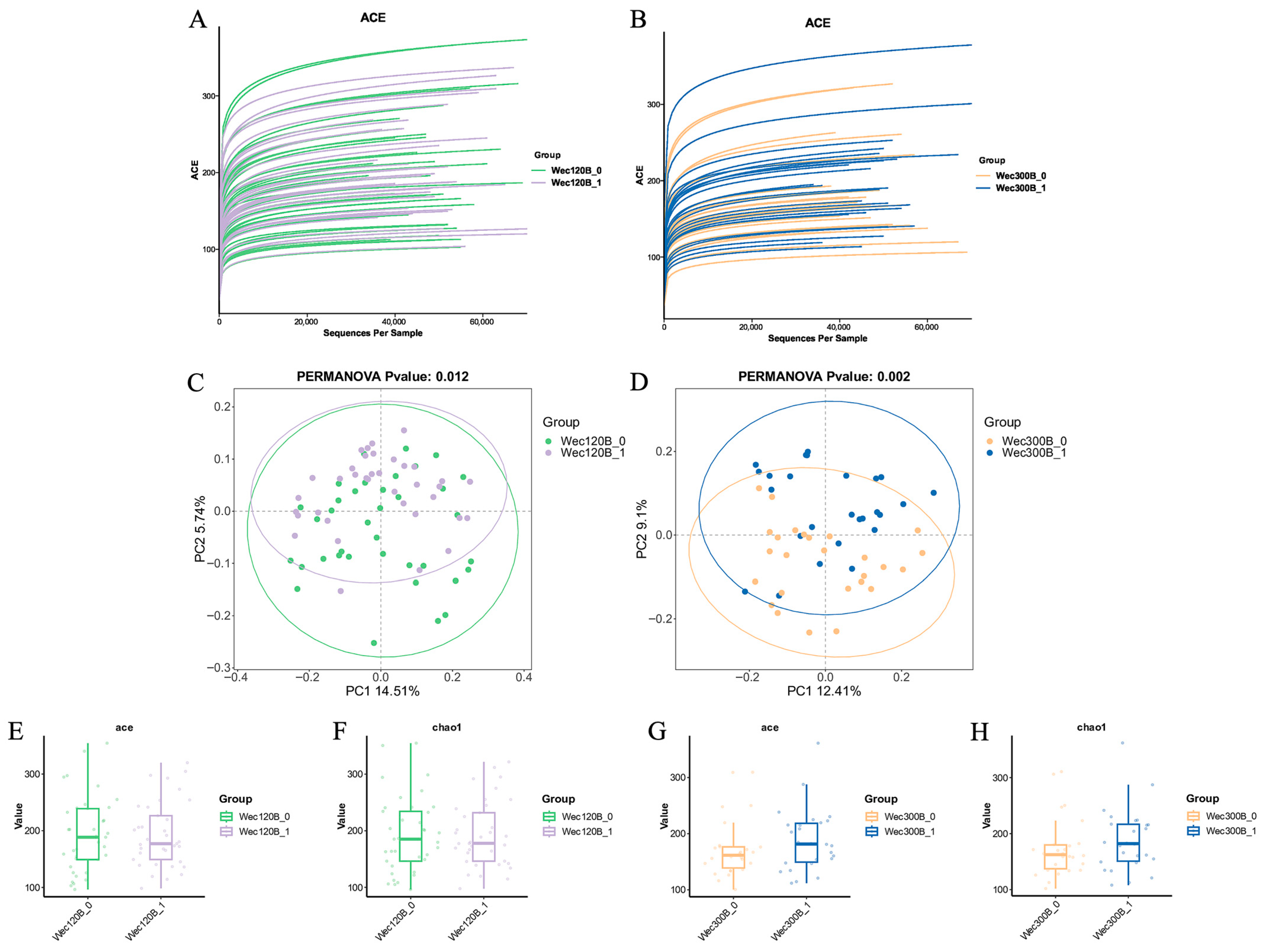
We analyzed the participants’ fecal microbiota using 16S rRNA gene sequencing. Rarefaction curves were used to assess sequencing depth and species richness across samples. The curve for the Wec120B group (Figure 4A) plateaued, indicating adequate sequencing coverage. The Wec300B group (Figure 4B) showed a similar trend with increasing ASV counts stabilizing at higher sequencing depths.
Beta-diversity, which reflects the differences in microbial community composition among groups, was assessed using non-metric multidimensional scaling (NMDS). While the pre- and post-intervention ellipses in both groups showed some overlap, distinct shifts in sample points post-intervention indicated changes in microbial community structure (Figure 4C,D).
Alpha-diversity was evaluated using the Chao1 and ACE indices. A slight decrease in these indices was observed in the Wec120B group, suggesting reduced proportions of low-abundance species and a more optimized microbiota. In contrast, the Wec300B group exhibited increased alpha-diversity post-intervention, reflecting greater microbial richness and diversity (Figure 4E,H).
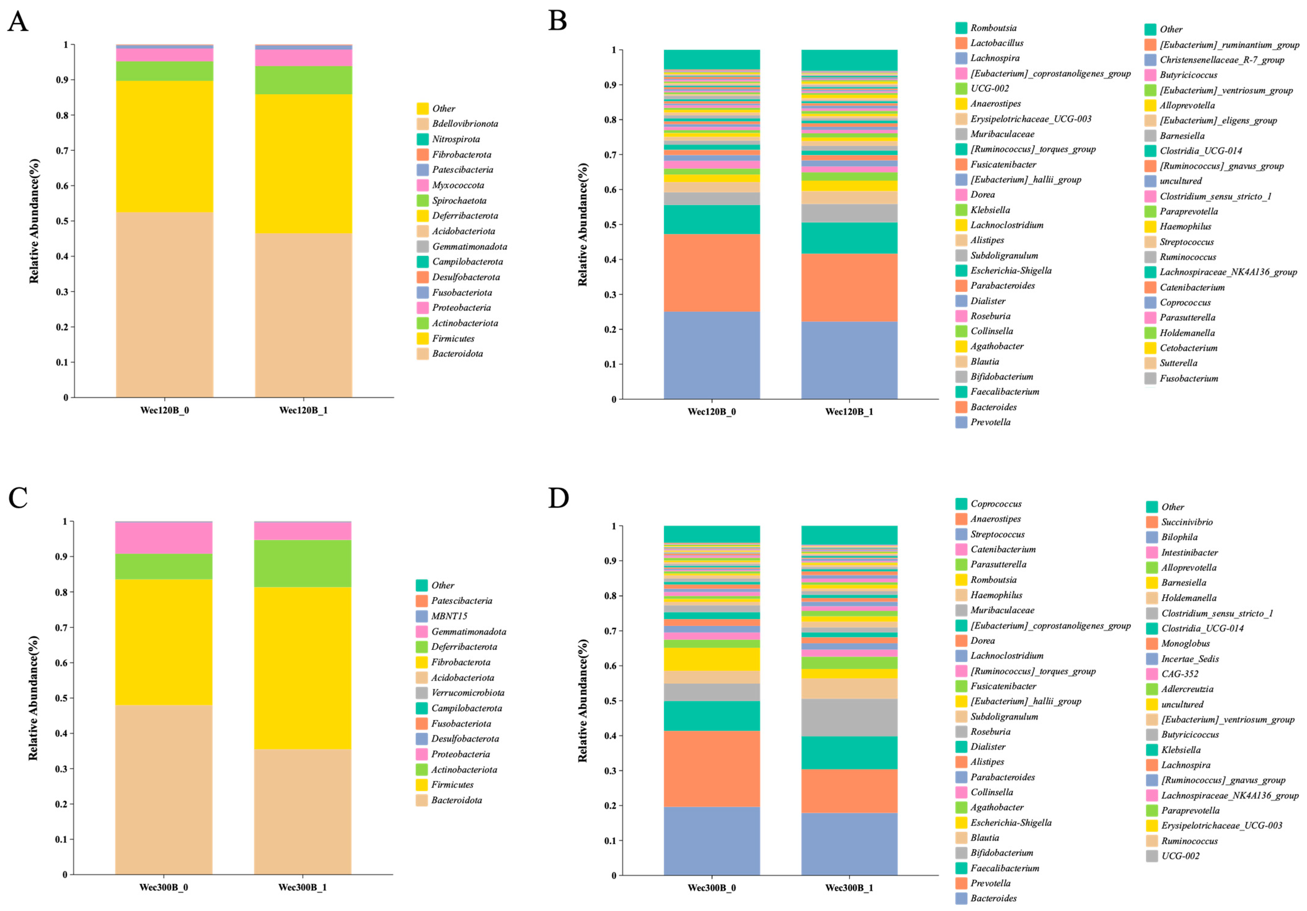
According to the species annotation results, the top 15 bacterial phyla with the highest abundance at the phylum level in each group were selected to generate a bar graph of species relative abundance. As shown in Figure 5A,C, the dominant taxa included Firmicutes, Bacteroidota, Actinobacteriota, and Proteobacteria. Post-intervention, Wec120B increased Firmicutes, Proteobacteria, and Actinobacteriota and reduced Bacteroidota. Wec300B increased Firmicutes and Actinobacteriota, while decreasing both Bacteroidota and Proteobacteria.
At the genus level (Figure 5B,D), both groups showed post-intervention increases in beneficial genera including Faecalibacterium, Bifidobacterium, Blautia, Agathobacter, Subdoligranulum, and Lactobacillus, and reductions in potentially pathogenic genera such as Prevotella, Escherichia-Shigella, and [Ruminococcus]_gnavus_group. A decrease in Bacteroides was observed in both groups, which may result from competition with Bifidobacterium for dietary fiber and oligosaccharides [27]. In the Wec300B group, minor genera including Dialister, Parabacteroides, and Alistipes also decreased, possibly due to environmental changes such as pH shifts driven by increased SCFA production [28]. The greater genus-level improvements in the Wec300B group reflect its higher dosage of probiotics and the synergistic action of its multi-strain composition.
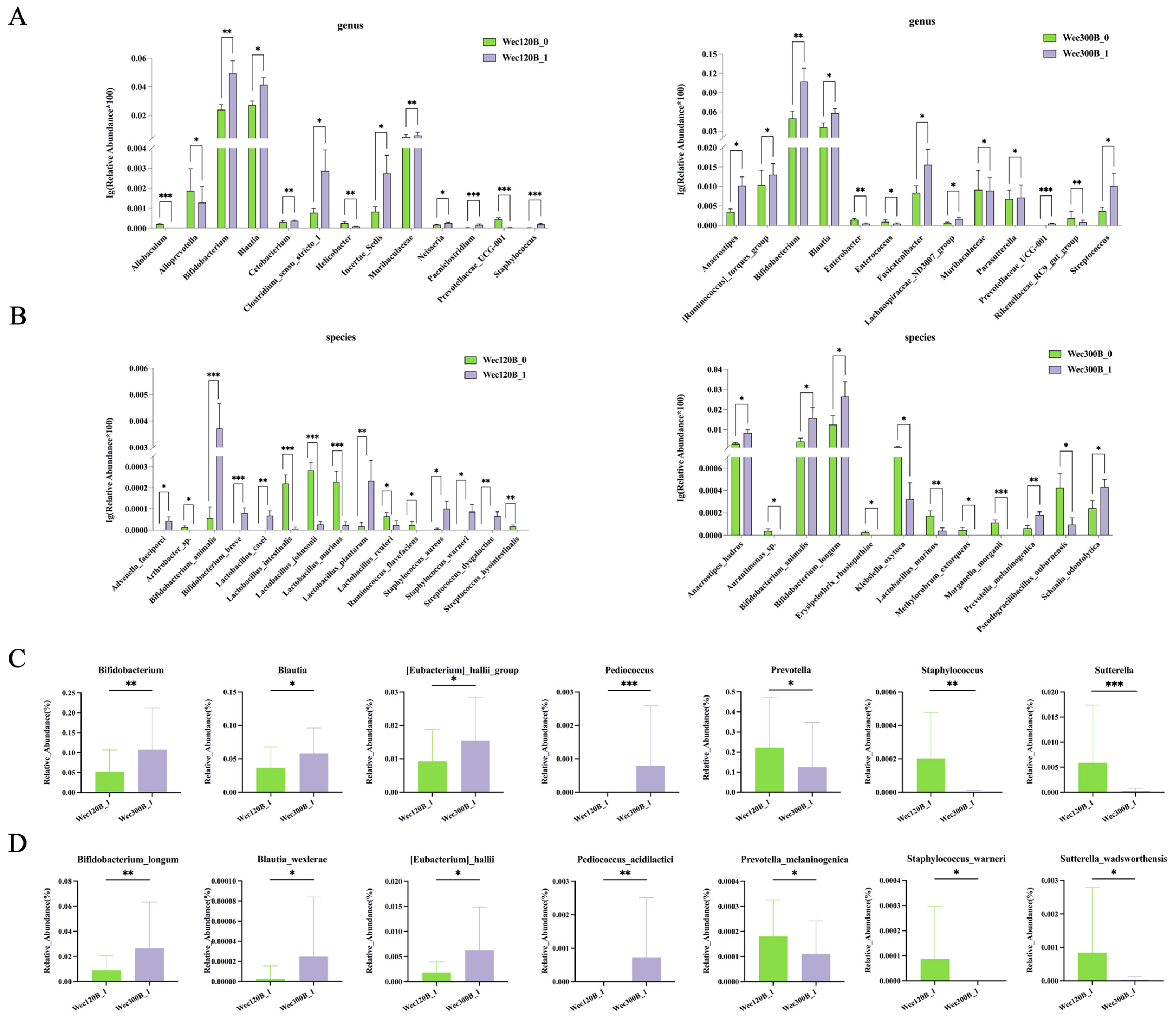
Subsequently, statistical analysis was performed using the Wilcoxon algorithm at both the genus and species levels. The post-intervention Wec120B group was compared with the Wec300B group. As shown (Figure 6A,C), significant differences in multiple bacterial genera were observed between the two groups. Specifically, the Wec300B group exhibited significantly higher relative abundances of beneficial bacteria such as Bifidobacterium, Blautia, [Eubacterium]_hallii_group, and Pediococcus compared with the Wec120B group, while showing significantly lower relative abundances of pathogenic bacteria including Prevotella, Staphylococcus, and Sutterella. At the species level (Figure 6B,D), significant differences were observed in Bifidobacterium_longum, Blautia_wexlerae, [Eubacterium]_hallii, Pediococcus_acidilactici, Prevotella_melaninogenica, Staphylococcus_warneri, and Sutterella_wadsworthensis. These bacterial species may represent key strains associated with the alleviation of gastrointestinal symptoms
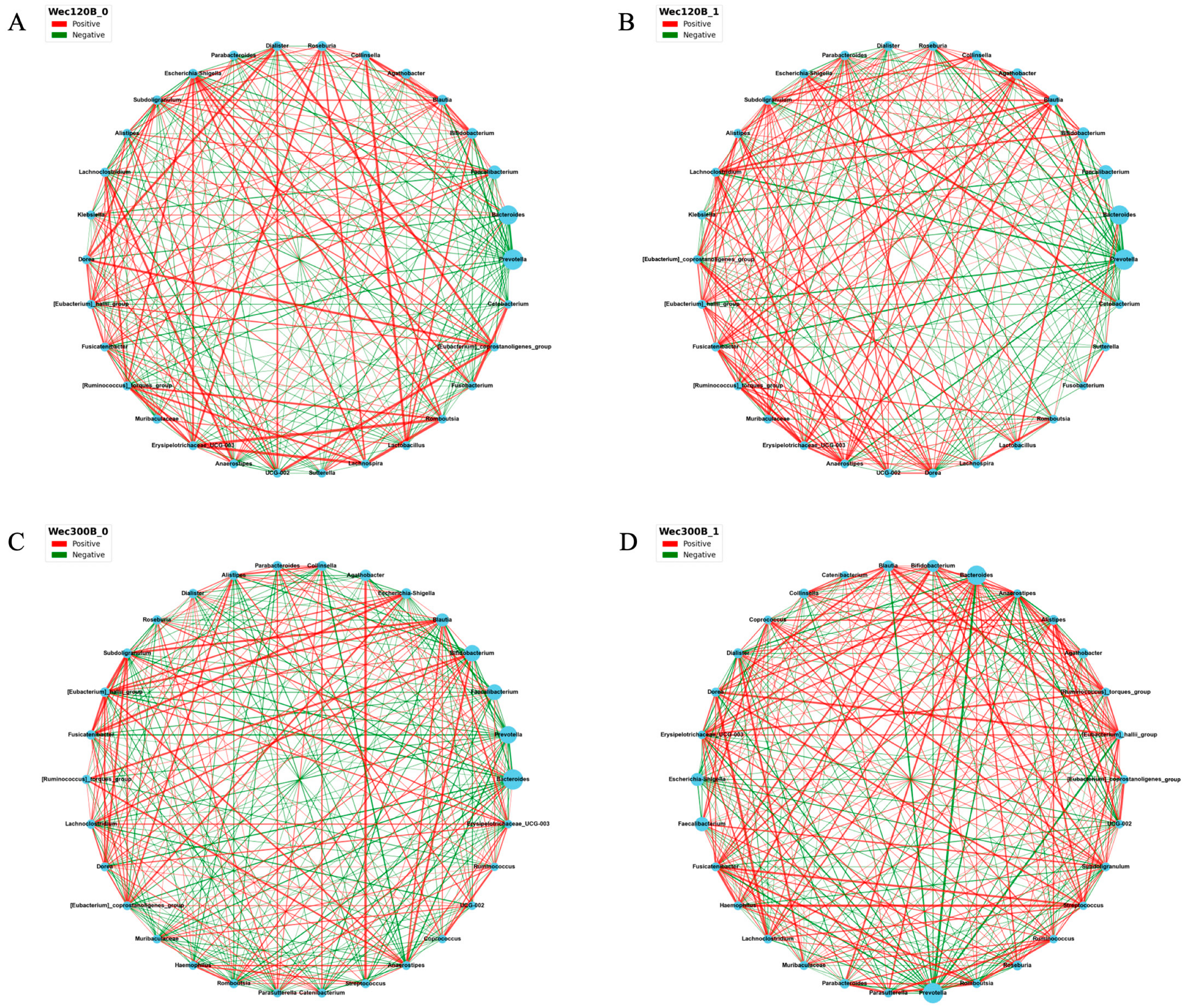
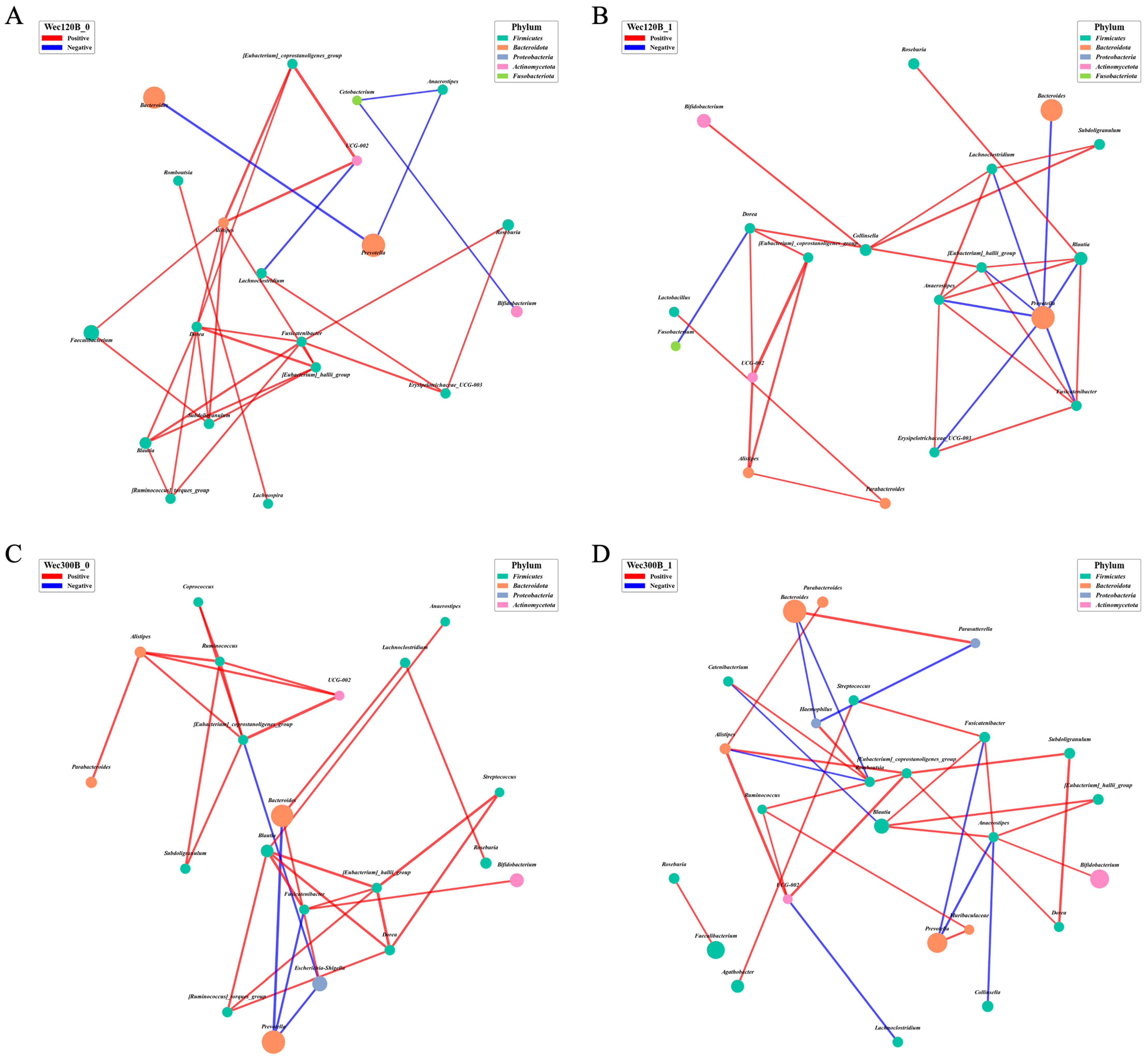
To further investigate the probiotic-induced changes in microbial ecosystem stability and function, we performed Spearman’s correlation analysis to construct genus-level co-occurrence networks before and after the intervention (p < 0.05). In these networks, nodes represent genera and edges represent significant correlations (positive or negative). Network density and connectivity reflect ecosystem complexity and stability. Post-intervention (Figure 7B,D), Bifidobacterium, Blautia, and Pediococcus increased in abundance in both groups and showed enhanced correlations with beneficial taxa such as Lactobacillus, Subdoligranulum, Fusicatenibacter, and Anaerostipes. These genera also exhibited strong negative correlations with pathogens such as Prevotella and Escherichia-Shigella. Bifidobacterium and Blautia became central hubs in the networks, suggesting key roles in microbial interaction and ecosystem functionality. In the Wec300B group, network topology improved significantly, with denser connections and more synergistic relationships involving probiotics, indicating their role as potential “network stabilizers.” The concurrent decrease in pathogen abundance and network marginalization further supports enhanced microbial stability.
To better explore the correlations among gut microbial genera, Spearman’s correlation analysis was performed based on relative abundance data across samples. This analysis identified intra-group relationships between taxa and was used to construct microbial co-occurrence networks. The top 30 genera in total abundance were selected, and pairwise Spearman’s correlation coefficients were calculated to visualize associations among dominant taxa. Only correlations with p < 0.05 were considered significant, and the top 30 most significant edges were displayed in the resulting networks.
Following intervention, the Wec120B group showed increased relative abundances of Bifidobacterium, Blautia, and [Eubacterium]_hallii_group, along with stronger negative correlations with Prevotella. Additionally, positive correlations were enhanced among beneficial genera including Bifidobacterium, Blautia, [Eubacterium]_hallii_group, Anaerostipes, and Collinsella (Figure 8A,B). Notably, the correlation between Bifidobacterium and Collinsella/Dorea was strengthened, suggesting that Bifidobacterium may have integrated into the symbiotic network and exerted regulatory effects through metabolic cross-feeding, such as lactate production [29]. Furthermore, a negative correlation between Blautia and Escherichia was observed, indicating that Blautia may competitively inhibit pro-inflammatory taxa. In the Wec300B group (Figure 8C,D), Bifidobacterium, Blautia, and [Eubacterium]_hallii_group exhibited stronger negative correlations with Prevotella, particularly through the central node Anaerostipes. Both Bifidobacterium and Blautia emerged as key network hubs, forming more cooperative associations with other genera and moving closer to the network core. This highlights their pivotal role in microbial interaction and ecological function, and suggests that they may act as “network stabilizers.” Meanwhile, the decreased abundance and peripheral positioning of certain potential pathogens further support enhanced microbial ecosystem stability following probiotic intervention.
At the species level, the top 30 most abundant taxa were selected for network construction. Only edges with p < 0.05 and within the top 30 strongest correlations were retained, and unconnected nodes were excluded. Post-intervention analysis revealed that in the Wec120B group (Figure 9A,B), several core probiotic species—including Bifidobacterium_animalis, Bifidobacterium_longum, Collinsella_aerofaciens, Bifidobacterium_adolescentis, and Bifidobacterium_bifidum—exhibited enhanced network integration. Specifically, B. animalis [30] showed strengthened positive correlations with SCFA-producing species such as Dialister_sp. [31], C. aerofacien [32], and Bacteroides_vulgatus [33], and increased connections with anti-inflammatory species such as Bacteroides_ovatus and Parabacteroides_distasonis [34]. These findings suggest that B. animalis may facilitate cooperative interactions among beneficial bacteria while suppressing potential pathogens.
In the Wec300B group (Figure 9C,D), the probiotic intervention induced more pronounced network reconfiguration. Bacteroides_ovatus emerged as a central hub in the post-intervention network, forming highly synergistic relationships with multiple functional species including B. animalis, Adlercreutzia_equolifaciens, Bacteroides_uniformis, [Eubacterium]_hallii, P. distasonis, C. aerofaciens, and Dialister_sp. These taxa are known to influence host metabolism, immune regulation, and the coordination of inflammatory responses. Additionally, B. longum exhibited enhanced positive correlations with Anaerostipes_hadrus [35] and Bifidobacterium_catenulatum [36], suggesting strengthened cross-feeding relationships and the increased production of butyrate. Butyrate alleviates intestinal inflammation by inhibiting histone deacetylase (HDAC) activity and modulating G protein-coupled signaling pathways [37]. In the Wec300B group, post-intervention analysis revealed the formation of a tighter and more functionally diverse co-occurrence network, with significantly enhanced integration and cooperative connectivity among core probiotic species. Notably, Wec300B promoted stronger interactions among SCFA-producing bacteria, anti-inflammatory taxa, and mucin-metabolizing microbes. This restructuring of microbial interactions may provide a key microecological basis for the superior efficacy of Wec300B in alleviating gastrointestinal discomfort symptoms.

4. Discussion

GI discomfort is a common clinical functional disorder characterized by symptoms such as bloating, abdominal pain, belching, acid reflux, constipation, or diarrhea, which severely affect quality of life. In recent years, the prevalence of GI discomfort has increased due to lifestyle changes [38] and Westernized dietary patterns [39], particularly among young and middle-aged adults. Conventional therapies, including proton pump inhibitors (PPIs), prokinetic agents, and laxatives, primarily target symptom relief but have limited effects on restoring gastrointestinal function. Long-term use may also cause drug dependence and increase safety concerns. Although PPIs are effective, prolonged administration has been linked to higher risks of infections, bone fractures, impaired mineral and vitamin absorption, kidney injury, as well as the disruption of the gastrointestinal barrier and microbiota, warranting a careful evaluation of the risk–benefit ratio [40]. Prokinetic agents may improve gastrointestinal motility, but their clinical benefits are restricted and potential adverse effects, such as movement disorders, arrhythmias, and neurological complications, necessitate the strict control of dosage and treatment duration during long-term use [41]. Increasing evidence has shown that GI dysfunction is closely related to gut microbiota dysbiosis, which may contribute to symptoms via multiple mechanisms, including mucosal barrier disruption and elevated inflammatory mediators [42], impaired gut–brain axis regulation [43], and the reduced production of beneficial metabolites such as short-chain fatty acids (SCFAs), which affect gut motility and immune balance [44]. Therefore, restoring microbial balance and functionality has emerged as a promising strategy for managing GI symptoms, and probiotics have gained attention as a non-pharmacological intervention.
This 4-week randomized, double-blind intervention trial compared two high-potency multi-strain probiotic formulations in alleviating GI discomfort and modulating gut microecology. Symptomatically, the Wec120B group showed faster relief of abdominal pain and dyspepsia, while the Wec300B group demonstrated more sustained improvements in reflux and constipation. Regarding fecal biomarkers, both groups exhibited significant increases in IgA and reductions in FL, FC, NGAL, DAO, D-LA, and LPS, indicating enhanced mucosal immunity, reduced inflammation, and improved gut barrier integrity. Notably, Wec300B achieved greater increases in IgA and reductions in FC, suggesting stronger immunomodulatory effects.
Gut microbial diversity analyses revealed varying degrees of post-intervention changes, indicating the restructuring of the microbial community following probiotic colonization. Community heatmaps and differential taxa bar plots showed increased relative abundances of Bifidobacterium, Blautia, [Eubacterium]_hallii_group, and Pediococcus, alongside decreases in Prevotella, Staphylococcus, and Sutterella. Bifidobacterium may inhibit pathogen colonization [45], produce SCFAs (e.g., acetate and butyrate) to enhance barrier function and immune regulation [46], and promote anti-inflammatory cytokine secretion [47]. Blautia is also strongly associated with host health, particularly in metabolic regulation and immune modulation [48]. Furthermore, GI dysfunction can itself reshape the gut ecosystem via changes in motility [49], gastric acid secretion, and bile acid metabolism [50], altering pH, redox state, and substrate availability, thereby affecting microbial colonization and function. In chronic GI disorders, microbial features typically include reduced diversity, structural imbalance, and an enrichment of pro-inflammatory taxa. For example, beneficial bacteria such as Bifidobacterium, Faecalibacterium, and Lactobacillus are often depleted, while Escherichia-Shigella, Fusobacterium, and Clostridium sensu stricto 1 are elevated. Genera like Ruminococcus and Prevotella also show abnormal trends, suggesting roles in barrier function, immune modulation, and neuroregulation. These features not only underlie GI dysfunction pathophysiology but also represent potential targets for probiotic therapy and microbial diagnostics.
In addition to taxonomic composition, the ecological stability of the gut microbiota determines its resilience and functional potential. Microbial co-occurrence network analysis allows deeper insights into synergistic and antagonistic relationships between genera and structural changes in the microbial ecosystem. Our findings revealed that both Wec120B and Wec300B interventions enhanced microbial network complexity and stability, as reflected by increased node counts, strengthened cooperation among symbiotic genera, and the emergence of key probiotics (Bifidobacterium, Blautia, [Eubacterium]_hallii_group, Anaerostipes) as core network members, replacing previously dominant potential pathogens. This structural remodeling may enhance pathogen resistance and improve ecosystem resilience and metabolic capacity.
Spearman’s correlation-based co-occurrence networks further illustrated the differential effects of probiotic activity levels on microbial structure and cooperative function. Both formulations promoted the increased integration and connectivity of core beneficial genera/species, with Wec300B producing stronger effects. This supports the hypothesis that network-level remodeling may underlie the microecological mechanism by which probiotics improve GI symptoms.
At the genus level, the Wec120B group showed increased relative abundances of Bifidobacterium, Blautia, and [Eubacterium]_hallii_group after intervention, with more positive edges observed—particularly with SCFA producers like Anaerostipes and Collinsella—indicating that low-dose probiotics can support beneficial microbial interactions. The enhanced positive correlations between Bifidobacterium and Dorea/Collinsella may reflect lactate-driven cross-feeding and energy network integration, while the increased negative correlation between Blautia and Escherichia suggests antagonism via competitive metabolism or inhibitory metabolites. These effects were amplified in the Wec300B group, where Bifidobacterium and Blautia acted as network hubs forming cooperative structures with Anaerostipes, [Eubacterium]_hallii_group, and others, organized around the suppression of Prevotella. This anti-inflammatory network reflects enhanced microbial homeostasis and suggests that multi-strain probiotics may act as “network stabilizers.”
At the species level, Wec120B intervention enhanced the network integration of several Bifidobacterium species (B. animalis, B. longum, B. adolescentis) and Collinsella aerofaciens. B. animalis displayed stronger positive correlations with SCFA producers such as Dialister and B. vulgatus, as well as increased links to anti-inflammatory species like B. ovatus and P. distasonis, indicating its involvement in barrier protection and inflammation control via metabolic coupling. In the Wec300B group, network restructuring was more pronounced. Not only did B. animalis maintain its hub status, but B. ovatus emerged as a highly connected node, forming a synergistic cluster with Adlercreutzia equolifaciens, B. uniformis, [Eubacterium]_hallii, P. distasonis, and others—covering SCFA production, immune regulation, and inflammation control.
Notably, in Wec300B, B. longum exhibited stronger positive associations with Anaerostipes hadrus and B. catenulatum, potentially enhancing butyrate production, which may suppress histone deacetylase (HDAC) activity and modulate G-protein signaling pathways to alleviate inflammation. Additionally, although low-abundance probiotic species such as Weissella cibaria did not form stable networks in Wec120B, they showed positive associations with Akkermansia muciniphila in Wec300B, suggesting potential cooperation in mucosal barrier metabolism.
In summary, compound probiotic supplementation restructured microbial functionality by reinforcing the synergistic interactions among beneficial taxa, marginalizing potential pathogens, and promoting the formation of anti-inflammatory and energy-related networks. The high-activity formulation Wec300B demonstrated superior effects in enhancing microbial structure and ecological coordination, supporting its mechanistic advantage in the clinical management of GI dysfunction.
Although this study provides valuable evidence regarding the effects of high-activity multi-strain probiotics on gastrointestinal symptoms and gut microbiota, several limitations should be noted.
The absence of a placebo control group represents an important methodological limitation. The randomized, double-blind design ensured a certain degree of rigor, yet without a placebo arm it is difficult to fully exclude potential placebo effects, especially in the context of subjective symptom outcomes such as abdominal pain, bloating, or stool frequency. Placebo responses are well recognized in functional gastrointestinal disorders and may account for a considerable proportion of symptom improvement. Therefore, while our results strongly suggest beneficial effects of probiotic intervention, the causal relationship between treatment and clinical benefit cannot be established with the highest level of certainty. Future trials should incorporate placebo control groups with adequate sample sizes to provide more robust evidence and enhance the reliability of conclusions.
The evaluation of fecal metabolites relied on ELISA-based assays targeting a limited panel of biomarkers. While these markers are clinically relevant and provide practical insights into intestinal immune and barrier function, they do not fully capture the complexity of host–microbiota metabolic interactions. More advanced technologies such as high-resolution mass spectrometry (HRMS) or nuclear magnetic resonance (NMR) could enable untargeted metabolomic profiling, allowing the discovery of novel metabolic signatures linked to probiotic intervention. Moreover, HRMS would facilitate pathway-level analysis and integration with microbiome data, thereby advancing our understanding of the functional consequences of microbial modulation. Given resource and cost constraints, we were unable to employ such techniques in the current trial, but future studies should adopt comprehensive metabolomics to provide mechanistic depth.
In particular, the use of ELISA for measuring gut barrier dysfunction and inflammation-related biomarkers has been well supported in clinical populations. For example, serum D-lactate and diamine oxidase (DAO) levels have been measured by ELISA and shown to be reliable indicators of intestinal permeability in Crohn’s disease patients [51]. Similarly, fecal calprotectin (FC) and lactoferrin (FL) have been extensively assessed using ELISA in clinical cohorts, where FC-ELISA demonstrated a high accuracy in distinguishing inflammatory bowel disease (IBD) from functional disorders [52]. Furthermore, neutrophil gelatinase-associated lipocalin (NGAL) has also been evaluated by ELISA in clinical studies as a biomarker of gut inflammation and barrier integrity [53].
These findings underscore the clinical relevance, robustness, and broad acceptance of ELISA-based biomarker assessment in both gastrointestinal disease and probiotic intervention trials. While HRMS could indeed provide broader metabolomic insights, it was beyond the scope and budget of the present clinical trial.

5. Conclusions

In summary, both compound probiotic formulations effectively alleviated GI symptoms in the short term, improved inflammatory (FC, NGAL) and immune (IgA, FL) markers, enhanced intestinal barrier function (LPS, D-LA, DAO), and remodeled the structure and function of the gut microbiota. Compared with Wec120B, the superior performance of Wec300B may be attributed to its higher dosage of probiotics. Key genera such as Bifidobacterium, Blautia, Faecalibacterium, Agathobacter, and Subdoligranulum may underlie its enhanced effects. These findings offer experimental evidence for the use of compound probiotics in managing GI dysfunction and highlight the importance of strain synergy, dosage, and host-specific factors in shaping probiotic efficacy. Future research should focus on mechanistic studies and personalized intervention strategies to advance the precision application of high-activity probiotics in functional GI disorders.

Author Contributions

Conceptualization, Z.Q. and Y.W.; Data curation, Z.Q., Y.J. and J.F.; Formal analysis, Y.J. and J.F.; Investigation, Z.Q. and Y.D.; Methodology, Z.Q., L.C., Y.D. and Y.W.; Project administration, S.G. and S.F.; Resources, Y.W. and S.G.; Software, Z.Q., Y.J., and J.F.; Supervision, S.G.; Validation, Y.J.; Visualization, J.F., Y.D. and L.C.; Writing—original draft, Z.Q.; Writing—review and editing, Y.W., L.C., Y.D. and S.G. All authors have read and agreed to the published version of the manuscript.

Funding

This work was supported by Major Science and Technology Special Projects in Henan Province (Grant Number 231100310200) and Key R&D Projects in Henan Province (Grant Number 241111314200) from the Project of Henan Provincial Department of Science and Technology.

Institutional Review Board Statement

This study was conducted in accordance with the Declaration of Helsinki, and approved by the Ethics Committee of the First Affiliated Hospital of Henan University of Science and Technology (2024-002, 13 December 2024).

Informed Consent Statement

Informed consent was obtained from all subjects involved in the study.

Data Availability Statement

All original contributions presented in the study are included in the article; further inquiries can be directed to the corresponding author.

Conflicts of Interest

Authors Yao Dong and Shuguang Fang were employed by the company Wecare Probiotics Co., Ltd. Y.D. participated in investigation, methodology, visualization, review and editing in the study. S.F. participated in project administration in the study. The role of the company was research participating units. The remaining authors declare that the research was conducted in the absence of any commercial or financial relationships that could be construed as a potential conflict of interest.

References

  1. Chen, C.; Zhang, D.-Y.; Chen, S.; Huang, S.; Zeng, F.; Li, D.; Lv, Y.-T.; Xiang, X.; Chen, R.-X.; Zhang, X.; et al. Prevalence, Types, and Risk Factors of Functional Gastrointestinal Diseases in Hainan Province, China. Sci. Rep. 2024, 14, 4553. [Google Scholar] [CrossRef] [PubMed]
  2. Sasso, J.M.; Ammar, R.M.; Tenchov, R.; Lemmel, S.; Kelber, O.; Grieswelle, M.; Zhou, Q.A. Gut Microbiome-Brain Alliance: A Landscape View into Mental and Gastrointestinal Health and Disorders. ACS Chem. Neurosci. 2023, 14, 1717–1763. [Google Scholar] [CrossRef] [PubMed]
  3. Volarić, M.; Šojat, D.; Majnarić, L.T.; Vučić, D. The Association between Functional Dyspepsia and Metabolic Syndrome-The State of the Art. Int. J. Environ. Res. Public Health 2024, 21, 237. [Google Scholar] [CrossRef]
  4. Peery, A.F.; Crockett, S.D.; Murphy, C.C.; Jensen, E.T.; Kim, H.P.; Egberg, M.D.; Lund, J.L.; Moon, A.M.; Pate, V.; Barnes, E.L.; et al. Burden and Cost of Gastrointestinal, Liver, and Pancreatic Diseases in the United States: Update 2021. Gastroenterology 2022, 162, 621–644. [Google Scholar] [CrossRef]
  5. Hamilton, A.; Poleon, S.; Cherian, J.; Cosgrove, S.; Laxminarayan, R.; Klein, E. COVID-19 and Outpatient Antibiotic Prescriptions in the United States: A County-Level Analysis. Open Forum Infect. Dis. 2023, 10, ofad096. [Google Scholar] [CrossRef]
  6. Abdul Manan, M. Progress in Probiotic Science: Prospects of Functional Probiotic-Based Foods and Beverages. Int. J. Food Sci. 2025, 2025, 5567567. [Google Scholar] [CrossRef]
  7. Tremblay, A.; Fatani, A.; Ford, A.L.; Piano, A.; Nagulesapillai, V.; Auger, J.; MacPherson, C.W.; Christman, M.C.; Tompkins, T.A.; Dahl, W.J. Safety and Effect of a Low- and High-Dose Multi-Strain Probiotic Supplement on Microbiota in a General Adult Population: A Randomized, Double-Blind, Placebo-Controlled Study. J. Diet. Suppl. 2021, 18, 227–247. [Google Scholar] [CrossRef]
  8. Qin, J.; Li, R.; Raes, J.; Arumugam, M.; Burgdorf, K.S.; Manichanh, C.; Nielsen, T.; Pons, N.; Levenez, F.; Yamada, T.; et al. A Human Gut Microbial Gene Catalogue Established by Metagenomic Sequencing. Nature 2010, 464, 59–65. [Google Scholar] [CrossRef]
  9. de Vos, W.M.; Tilg, H.; Hul, M.V.; Cani, P.D. Gut Microbiome and Health: Mechanistic Insights. Gut 2022, 71, 1020–1032. [Google Scholar] [CrossRef]
  10. Ma, J.; Piao, X.; Mahfuz, S.; Long, S.; Wang, J. The Interaction among Gut Microbes, the Intestinal Barrier and Short Chain Fatty Acids. Anim. Nutr. 2022, 9, 159–174. [Google Scholar] [CrossRef]
  11. Tyagi, A.; Kumar, V. The Gut Microbiota-Bile Acid Axis: A Crucial Regulator of Immune Function and Metabolic Health. World J. Microbiol. Biotechnol. 2025, 41, 215. [Google Scholar] [CrossRef]
  12. Masse, K.E.; Lu, V.B. Short-Chain Fatty Acids, Secondary Bile Acids and Indoles: Gut Microbial Metabolites with Effects on Enteroendocrine Cell Function and Their Potential as Therapies for Metabolic Disease. Front. Endocrinol. 2023, 14, 1169624. [Google Scholar] [CrossRef]
  13. Carco, C.; Young, W.; Gearry, R.B.; Talley, N.J.; McNabb, W.C.; Roy, N.C. Increasing Evidence That Irritable Bowel Syndrome and Functional Gastrointestinal Disorders Have a Microbial Pathogenesis. Front. Cell Infect. Microbiol. 2020, 10, 468. [Google Scholar] [CrossRef] [PubMed]
  14. Zhang, T.; Zhang, C.; Zhang, J.; Sun, F.; Duan, L. Efficacy of Probiotics for Irritable Bowel Syndrome: A Systematic Review and Network Meta-Analysis. Front. Cell Infect. Microbiol. 2022, 12, 859967. [Google Scholar] [CrossRef] [PubMed]
  15. Gao, J.; Li, Y.; Wan, Y.; Hu, T.; Liu, L.; Yang, S.; Gong, Z.; Zeng, Q.; Wei, Y.; Yang, W.; et al. A Novel Postbiotic from Lactobacillus Rhamnosus GG with a Beneficial Effect on Intestinal Barrier Function. Front. Microbiol. 2019, 10, 477. [Google Scholar] [CrossRef]
  16. Segui-Perez, C.; Huang, L.Z.X.; Paganelli, F.L.; Lievens, E.; Strijbis, K. Probiotic Bifidobacterium bifidum Strains Desialylate MUC13 and Increase Intestinal Epithelial Barrier Function. Sci. Rep. 2025, 15, 8778. [Google Scholar] [CrossRef]
  17. World Medical Association (WMA). WMA Declaration of Helsinki—Ethical Principles for Medical Research Involving Human Participants. World Medical Association. 2022. Available online: https://www.wma.net/policies-post/wma-declaration-of-helsinki/ (accessed on 31 October 2024).
  18. IBM Corp. IBM SPSS Statistics for Windows, Version 25.0; Released 2017; IBM Corp.: Armonk, NY, USA, 2017. [Google Scholar]
  19. Bunker, J.J.; Bendelac, A. IgA Responses to Microbiota. Immunity 2018, 49, 211–224. [Google Scholar] [CrossRef]
  20. Abraham, B.P.; Kane, S. Fecal Markers: Calprotectin and Lactoferrin. Gastroenterol. Clin. 2012, 41, 483–495. [Google Scholar] [CrossRef]
  21. Ikhtaire, S.; Shajib, M.S.; Reinisch, W.; Khan, W.I. Fecal calprotectin: Its scope and utility in the management of inflammatory bowel disease. J. Gastroenterol. 2016, 51, 434–446. [Google Scholar] [CrossRef] [PubMed]
  22. Lu, F.; Inoue, K.; Kato, J.; Minamishima, S.; Morisaki, H. Functions and Regulation of Lipocalin-2 in Gut-Origin Sepsis: A Narrative Review. Crit. Care 2019, 23, 269. [Google Scholar] [CrossRef]
  23. Ghosh, S.S.; Wang, J.; Yannie, P.J.; Ghosh, S. Intestinal Barrier Dysfunction, LPS Translocation, and Disease Development. J. Endocr. Soc. 2020, 4, bvz039. [Google Scholar] [CrossRef]
  24. Zhang, Q.; Gao, X.; Wu, J.; Chen, M. The Correlation between Endotoxin, D-Lactate, and Diamine Oxidase with Endoscopic Activity in Inflammatory Bowel Disease. Dis. Markers 2022, 2022, 9171436. [Google Scholar] [CrossRef]
  25. Svedlund, J.; Sjödin, I.; Dotevall, G. GSRS—A Clinical Rating Scale for Gastrointestinal Symptoms in Patients with Irritable Bowel Syndrome and Peptic Ulcer Disease. Dig. Dis. Sci. 1988, 33, 129–134. [Google Scholar] [CrossRef] [PubMed]
  26. Kulich, K.R.; Madisch, A.; Pacini, F.; Piqué, J.M.; Regula, J.; Van Rensburg, C.J.; Újszászy, L.; Carlsson, J.; Halling, K.; Wiklund, I.K. Reliability and Validity of the Gastrointestinal Symptom Rating Scale (GSRS) and Quality of Life in Reflux and Dyspepsia (QOLRAD) Questionnaire in Dyspepsia: A Six-Country Study. Health Qual. Life Outcomes 2008, 6, 12. [Google Scholar] [CrossRef] [PubMed]
  27. Yin, P.; Du, T.; Yi, S.; Zhang, C.; Yu, L.; Tian, F.; Chen, W.; Zhai, Q. Response Differences of Gut Microbiota in Oligofructose and Inulin Are Determined by the Initial Gut Bacteroides/Bifidobact. Ratios. Food Res. Int. 2023, 174, 113598. [Google Scholar] [CrossRef] [PubMed]
  28. Lordan, C.; Roche, A.K.; Delsing, D.; Nauta, A.; Groeneveld, A.; MacSharry, J.; Cotter, P.D.; van Sinderen, D. Linking Human Milk Oligosaccharide Metabolism and Early Life Gut Microbiota: Bifidobacteria and Beyond. Microbiol. Mol. Biol. Rev. 2024, 88, e00094-23. [Google Scholar] [CrossRef]
  29. Hiraku, A.; Nakata, S.; Murata, M.; Xu, C.; Mutoh, N.; Arai, S.; Odamaki, T.; Iwabuchi, N.; Tanaka, M.; Tsuno, T.; et al. Early Probiotic Supplementation of Healthy Term Infants with Bifidobacterium longum Subsp. infantis M-63 Is Safe and Leads to the Development of Bifidobacterium-Predominant Gut Microbiota: A Double-Blind, Placebo-Controlled Trial. Nutrients 2023, 15, 1402. [Google Scholar] [CrossRef]
  30. Alatawi, H.; Mosli, M.; Saadah, O.I.; Annese, V.; Al-Hindi, R.; Alatawy, M.; Al-Amrah, H.; Alshehri, D.; Bahieldin, A.; Edris, S. Attributes of Intestinal Microbiota Composition and Their Correlation with Clinical Primary Non-Response to Anti-TNF-α Agents in Inflammatory Bowel Disease Patients. Biomol. Biomed. 2022, 22, 412–426. [Google Scholar] [CrossRef]
  31. Lin, J.; Li, E.; Li, C. Increasing Degree of Substitution Inhibits Acetate While Promotes Butyrate Production during in Vitro Fermentation of Citric Acid-Modified Rice Starch. Int. J. Biol. Macromol. 2024, 281, 136385. [Google Scholar] [CrossRef]
  32. Wang, Z.; Yuan, X.; Zhu, Z.; Pang, L.; Ding, S.; Li, X.; Kang, Y.; Hei, G.; Zhang, L.; Zhang, X.; et al. Multiomics Analyses Reveal Microbiome–Gut–Brain Crosstalk Centered on Aberrant Gamma-Aminobutyric Acid and Tryptophan Metabolism in Drug-Naïve Patients with First-Episode Schizophrenia. Schizophr. Bull. 2024, 50, 187–198. [Google Scholar] [CrossRef]
  33. Xu, W.; Wang, Q.; Yang, Y.; Sun, S.; Qi, H.; He, J.; Jin, T.; Yao, P.; Wang, J.; Lin, F. Integrated Analysis of Gut Microbiota, Fecal and Serum Metabolites in Type 2 Diabetes Mellitus with Peripheral Neuropathy. J. Endocrinol. Investig. 2025. [Google Scholar] [CrossRef]
  34. Gaifem, J.; Mendes-Frias, A.; Wolter, M.; Steimle, A.; Garzón, M.J.; Ubeda, C.; Nobre, C.; González, A.; Pinho, S.S.; Cunha, C.; et al. Akkermansia muciniphila and Parabacteroides distasonis synergistically protect from colitis by promoting ILC3 in the gut. mBio 2024, 15, e00078-24. [Google Scholar] [CrossRef]
  35. Tian, C.; Yang, Q.; Lv, H.; Yue, F. Integrative Analysis of Gut Microbiota and Fecal Metabolites in Cynomolgus Monkeys with Spontaneous Type 2 Diabetes Mellitus. Microb. Pathog. 2025, 199, 107228. [Google Scholar] [CrossRef]
  36. Li, M.; Wang, L.; Peng, Z.; Jiang, L.; Yan, Y.; Xia, Y.; Wang, Y.; Guo, L.; Miao, J.; Bian, Y. Causal Associations of Gut Microbiota Species with Lymphoma: A Two-Sample Mendelian Randomization Study. Hematol. Oncol. 2025, 43, e70046. [Google Scholar] [CrossRef]
  37. Luo, C.-H.; Lai, A.C.-Y.; Chang, Y.-J. Butyrate Inhibits Staphylococcus Aureus-Aggravated Dermal IL-33 Expression and Skin Inflammation through Histone Deacetylase Inhibition. Front. Immunol. 2023, 14, 1114699. [Google Scholar] [CrossRef]
  38. Sebastián Domingo, J.J. Irritable Bowel Syndrome. Med. Clin. (Barc.) 2022, 158, 76–81. [Google Scholar] [CrossRef] [PubMed]
  39. Hosseini, Z.; Rostami, M.; Whiting, S.J.; Vatanparast, H. Fast-Food Dietary Pattern Is Linked to Higher Prevalence of Metabolic Syndrome in Older Canadian Adults. J. Nutr. Metab. 2021, 2021, 5712844. [Google Scholar] [CrossRef] [PubMed]
  40. Castellana, C.; Pecere, S.; Furnari, M.; Telese, A.; Matteo, M.V.; Haidry, R.; Eusebi, L.H. Side effects of long-term use of proton pump inhibitors: Practical considerations. Pol. Arch. Intern. Med. 2021, 131, 541–549. [Google Scholar] [CrossRef] [PubMed]
  41. Quigley, E.M.M. Prokinetics in the Management of Functional Gastrointestinal Disorders. Curr. Gastroenterol. Rep. 2017, 19, 53. [Google Scholar] [CrossRef]
  42. Maideen, N.M.P. Adverse Effects Associated with Long-Term Use of Proton Pump Inhibitors. Chonnam Med. J. 2023, 59, 115–127. [Google Scholar] [CrossRef]
  43. San Mauro Martín, I.; López Oliva, S.; Garicano Vilar, E.; Sánchez Niño, G.M.; Penadés, B.F.; Terrén Lora, A.; Sanz Rojo, S.; Collado Yurrita, L. Effects of Gluten on Gut Microbiota in Patients with Gastrointestinal Disorders, Migraine, and Dermatitis. Nutrients 2024, 16, 1228. [Google Scholar] [CrossRef]
  44. Zhao, L.-P.; Wu, J.; Quan, W.; Zhou, Y.; Hong, H.; Niu, G.-Y.; Li, T.; Huang, S.-B.; Qiao, C.-M.; Zhao, W.-J.; et al. DSS-Induced Acute Colitis Causes Dysregulated Tryptophan Metabolism in Brain: An Involvement of Gut Microbiota. J. Nutr. Biochem. 2023, 115, 109282. [Google Scholar] [CrossRef]
  45. Zhang, D.; Zhou, Y.; Ma, Y.; Chen, P.; Tang, J.; Yang, B.; Li, H.; Liang, M.; Xue, Y.; Liu, Y.; et al. Gut Microbiota Dysbiosis Correlates with Long COVID-19 at One-Year After Discharge. J. Korean Med. Sci. 2023, 38, e120. [Google Scholar] [CrossRef]
  46. Gavzy, S.J.; Kensiski, A.; Lee, Z.L.; Mongodin, E.F.; Ma, B.; Bromberg, J.S. Bifidobacterium Mechanisms of Immune Modulation and Tolerance. Gut Microbes 2023, 15, 2291164. [Google Scholar] [CrossRef] [PubMed]
  47. Jin, H.; Wang, H.; Feng, X.; Ma, X.; Zhai, Z.; Hao, Y. Bifidobacterium longum BL300 Alleviates DSS-Induced Colitis by Promoting IgA-Mediated Gut Microbiota Homeostasis. Food Funct. 2025, 16, 4303–4314. [Google Scholar] [CrossRef]
  48. Benítez-Páez, A.; Gómez del Pugar, E.M.; López-Almela, I.; Moya-Pérez, Á.; Codoñer-Franch, P.; Sanz, Y. Depletion of Blautia Species in the Microbiota of Obese Children Relates to Intestinal Inflammation and Metabolic Phenotype Worsening. mSystems 2020, 5, e00857-19. [Google Scholar] [CrossRef] [PubMed]
  49. Waclawiková, B.; Codutti, A.; Alim, K.; El Aidy, S. Gut Microbiota-Motility Interregulation: Insights from in Vivo, Ex Vivo and in Silico Studies. Gut Microbes 2022, 14, 1997296. [Google Scholar] [CrossRef]
  50. Staley, C.; Weingarden, A.R.; Khoruts, A.; Sadowsky, M.J. Interaction of Gut Microbiota with Bile Acid Metabolism and Its Influence on Disease States. Appl. Microbiol. Biotechnol. 2017, 101, 47–64. [Google Scholar] [CrossRef]
  51. Cai, J.; Chen, H.; Weng, M.; Jiang, S.; Gao, J. Diagnostic and Clinical Significance of Serum Levels of D-Lactate and Diamine Oxidase in Patients with Crohn’s Disease. Gastroenterol. Res. Pract. 2019, 2019, 8536952. [Google Scholar] [CrossRef]
  52. Wang, Y.; Pei, F.; Wang, X.; Sun, Z.; Hu, C.; Dou, H. Diagnostic Accuracy of Fecal Lactoferrin for Inflammatory Bowel Disease: A Meta-Analysis. Int. J. Clin. Exp. Pathol. 2015, 8, 12319–12332. [Google Scholar] [PubMed]
  53. Sutton, K.A.; He, M.; Ma, C.; Liu, T.-C.; Faubion, W.A.; Hoffmann, J.; Linneman, L.; Rodriguez, C.; Holtz, L.R. Noninvasive Markers of Inflammation and Protein Loss Augment Diagnosis of Pediatric Celiac Disease. Clin. Transl. Gastroenterol. 2024, 15, e00695. [Google Scholar] [CrossRef] [PubMed]
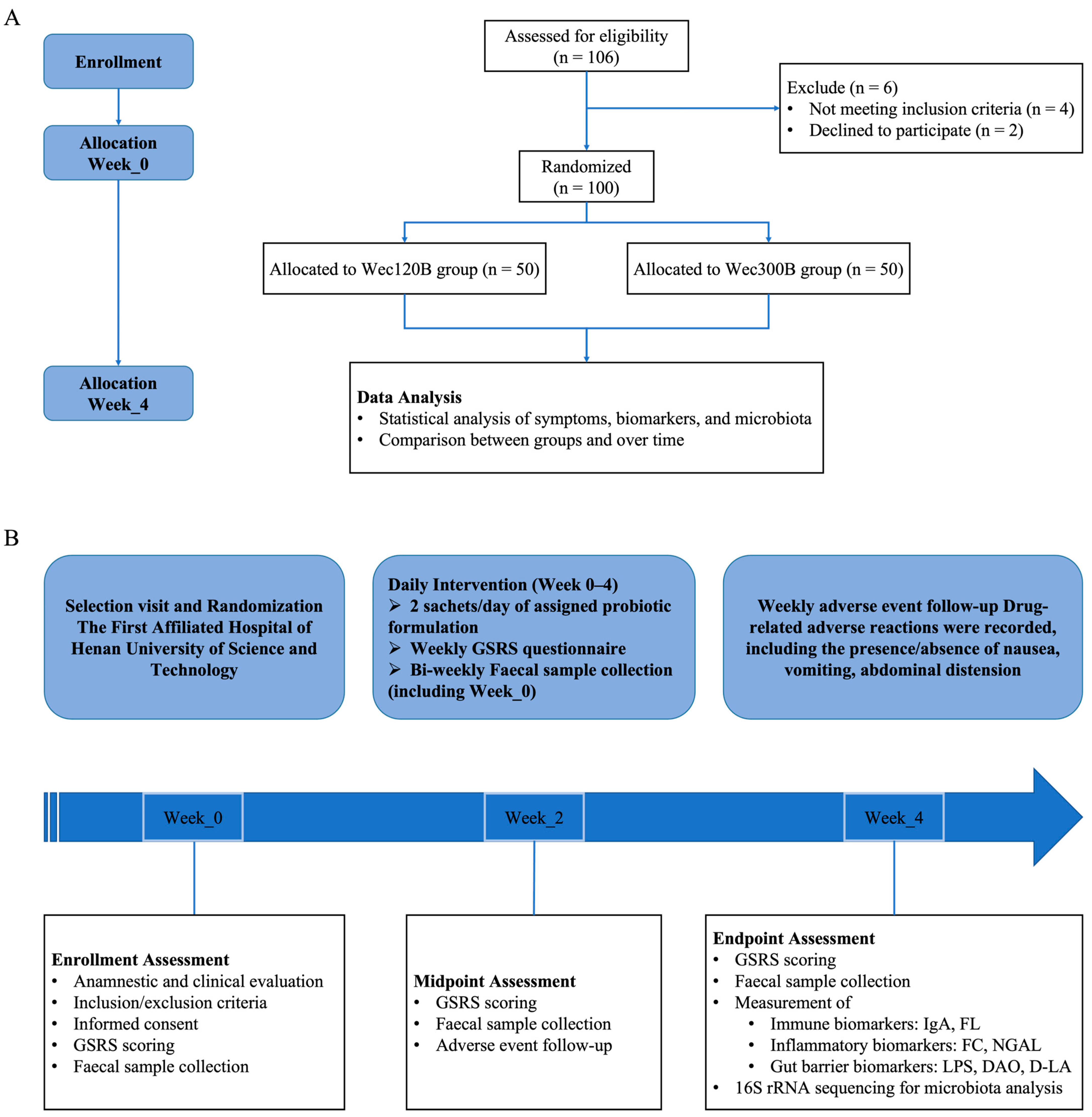
Figure 1. Flowchart for this study. Process of the study implementation. (A) The design of the study. (B) Flow diagram of this study selection.
Nutrients 17 02886 g001
Figure 2. Effects of probiotic intervention on immune factors, inflammatory factors, and intestinal barrier markers. (A) Wec120B group immune factors; (B) Wec300B group immune factors; (C) Wec120B group inflammatory factors; (D) Wec300B group inflammatory factors; (E) Wec120B group intestinal barrier markers; (F) Wec300B group intestinal barrier markers. * p < 0.05, ** p < 0.01.
Nutrients 17 02886 g002
Figure 3. Changes in GSRS domain scores over time. (A) Dyspepsia; (B) reflux; (C) abdominal pain; (D) constipation; (E) diarrhea. ns p ≥ 0.05, * p < 0.05, ** p < 0.01, *** p < 0.001, **** p < 0.0001.
Nutrients 17 02886 g003
Figure 4. Microbial diversity analysis. (A,B) Rarefaction curves; (C,D) NMDS plots; (EH) ACE and Chao1 indices.
Nutrients 17 02886 g004
Figure 5. Microbial composition at phylum and genus levels for Wec120B and Wec300B groups. (A,C) Relative abundance of bacterial communities at the phylum level; (B,D) relative abundance of bacterial communities at the genus level.
Nutrients 17 02886 g005
Figure 6. Significant differences in intestinal microbiota at the genus and species levels. (A) Species with genus-level differences in the Wec120B group and Wec300B group before and after the intervention; (B) species-level differences between Wec120B group and Wec300B group before and after intervention; (C) species with genus-level differences between the Wec120B group and the Wec300B group after the intervention; (D) species-level differences between the Wec120B group and the Wec300B group after intervention. * p < 0.05, ** p < 0.01, *** p < 0.001.
Nutrients 17 02886 g006
Figure 7. Microbial community interaction network analysis. (A) Wec120B group before intervention; (B) Wec120B group after intervention; (C) Wec300B group before intervention; (D) Wec300B group after intervention.
Nutrients 17 02886 g007
Figure 8. Genus-level microbiome correlation network. (A) Wec120B group before intervention; (B) Wec120B group after intervention; (C) Wec300B group before intervention; (D) Wec300B group after intervention.
Nutrients 17 02886 g008
Figure 9. Species-level microbiome correlation network. (A) Wec120B group before intervention; (B) Wec120B group after intervention; (C) Wec300B group before intervention; (D) Wec300B group after intervention.
Nutrients 17 02886 g009
Table 1. Baseline characteristics of study participants.
GroupAge ( x ¯ ± s )Gender
MaleFemale
Wec120B group (n = 50)21.1 ± 5.1829 (58%)21 (42%)
Wec300B group (n = 50)20.42 ± 2.724 (48%)26 (52%)
Table 2. Analysis of GSRS in Wec120B group.
DomainEvaluationScorep-Value
(vs. Baseline)
95% CI
(vs. Baseline)
IndigestionBaseline7.54 ± 2.56--
W16.64 ± 2.100.057−0.02815, 1.82815
W26.44 ± 1.790.0140.22461, 1.97539
W36.52 ± 1.580.0180.17625, 1.86375
W46.16 ± 1.540.00080.57955, 2.18025
RefluxBaseline2.74 ± 1.38--
W12.44 ± 0.880.199−0.16052, 0.76052
W22.62 ± 1.050.626−0.36675, 0.60675
W32.26 ± 0.600.0270.05715, 0.90285
W42.22 ± 0.550.0150.10296, 0.93704
BellyacheBaseline5.10 ± 2.38--
W14.24 ± 1.520.0330.06863, 1.65137
W24.10 ± 1.400.0120.22569, 1.77431
W33.58 ± 1.000.0000.79758, 2.24242
W43.48 ± 0.76<0.00010.91984, 2.32016
ConstipationBaseline0.52 ± 0.58--
W10.38 ± 0.600.239−0.09467, 0.37467
W20.36 ± 0.480.138−0.05215, 0.37215
W30.32 ± 0.590.090−0.03157, 0.43157
W40.26 ± 0.440.0130.05518, 0.46482
DiarrheaBaseline0.54 ± 0.73--
W10.38 ± 0.640.247−0.11251, 0.43251
W20.34 ± 0.660.155−0.07672, 0.47672
W30.30 ± 0.540.066−0.01647, 0.49647
W40.18 ± 0.390.0030.12691, 0.59309
Table 3. Analysis of GSRS in Wec120B group.
DomainEvaluationScorep-Value
(vs. Baseline)
95% CI
(vs. Baseline)
IndigestionBaseline7.63 ± 2.18--
W16.73 ± 2.050.0340.07031, 1.73361
W26.73 ± 2.150.0380.05014, 1.75379
W36.53 ± 2.000.0090.27520, 1.92088
W46.10 ± 1.680.00020.73682, 2.32270
RefluxBaseline2.94 ± 1.33--
W12.57 ± 0.920.104−0.07770, 0.82280
W22.61 ± 1.080.168−0.14298, 0.80965
W32.27 ± 0.570.0010.26412, 1.06922
W42.29 ± 0.670.0030.23236, 1.06176
BellyacheBaseline4.92 ± 2.35--
W14.20 ± 2.000.096−0.13144, 1.58242
W24.27 ± 1.670.112−0.15412, 1.44824
W33.98 ± 1.610.0200.15084, 1.73152
W43.75 ± 1.400.0030.41730, 1.93564
DiarrheaBaseline0.51 ± 0.61--
W10.49 ± 0.670.878−0.23346, 0.27268
W20.38 ± 0.600.255−0.10065, 0.37516
W30.31 ± 0.580.101−0.03875, 0.43091
W40.35 ± 0.630.0390.01334, 0.49647
ConstipationBaseline0.51 ± 0.58--
W10.55 ± 0.730.764−0.29796, 0.21953
W20.32 ± 0.650.110−0.04524, 0.43739
W30.22 ± 0.540.0090.07406, 0.51418
W40.14 ± 0.350.0000.18501, 0.56008
Disclaimer/Publisher’s Note: The statements, opinions and data contained in all publications are solely those of the individual author(s) and contributor(s) and not of MDPI and/or the editor(s). MDPI and/or the editor(s) disclaim responsibility for any injury to people or property resulting from any ideas, methods, instructions or products referred to in the content.