Article

Clinical Presentations of Celiac Disease: Experience of a Single Italian Center

1
Nutritional Rehabilitation Unit, Bambino Gesù Children Hospital IRCCS, 00165 Rome, Italy
2
Clinical Pediatrics, Department of Molecular Medicine and Development, University of Siena, 53100 Siena, Italy
3
Gastroenterology and Nutrition Unit, Bambino Gesù Children Hospital IRCCS, 00165 Rome, Italy
*
Author to whom correspondence should be addressed.
Nutrients 2025, 17(1), 129; https://doi.org/10.3390/nu17010129
Submission received: 10 November 2024 / Revised: 13 December 2024 / Accepted: 20 December 2024 / Published: 31 December 2024
(This article belongs to the Special Issue Nutrition and Immunobiology of Celiac Disease)

Abstract

:
Background/Objectives: In Italy, the prevalence of celiac disease (CeD) among children exceeds 1.5% and has steadily increased with a linear trend over the past 25 years. The clinical presentation is heterogenous and a change in onset symptoms has been described in recent years. The aim of the study is to describe the pattern of clinical presentation of CeD during the last 12 years in a single Italian center. Methods: We retrospectively enrolled all children diagnosed with CeD at Bambino Gesù Children Hospital, Rome between 1 March 2011 and 22 June 2023. To investigate the changes in pattern of clinical presentation, we divided the patient population into three groups of approximately 4 years each (respectively: 49, 48 and 48 months). Patients who previously received a CeD diagnosis in other centers were excluded. Results: Overall, 4478 patients were diagnosed with CeD at our center. 1082 were excluded, leaving 3396 available for analysis. We divided the study cohort into three groups: group 1 (n = 909), group 2 (n = 1103), and group 3 (n = 1384). Diagnoses of CeD increased by 17.5%. The trend of the non-classic form shows a significative increase (p = 0.000064), showing a high prevalence of bloating and abdominal pain and a significant reduction in celiac crisis (p < 0.0001). Conclusions: Annual diagnoses of CeD increased during the study period, and the clinical presentation has changed in recent years, showing an increase in the non-classic form and a reduction in more severe forms of celiac crisis.

1. Introduction

Celiac disease (CeD) is an autoimmune enteropathy triggered by gluten intake, arising in genetically predisposed individuals. It is currently estimated to have a worldwide prevalence of 0.7–1.4% based, respectively, on serology and biopsy, with a female predominance and an almost double incidence in the pediatric population [1]. In Italy, the prevalence of celiac disease among children exceeds 1.5% and has presented a steady increase with a linear trend over the past 25 years [2]. A recent multi-center cross-sectional study conducted in Italy [3] reported a 1.65% overall prevalence of celiac disease (CeD) in the country, with the highest prevalence recorded in the southern regions of the island at 1.93%. Despite this, celiac disease remains a largely underdiagnosed condition with a 70% rate of misdiagnosed cases [4]. One of the prominent causes of such alarming rates is the heterogeneous clinical manifestation; in fact, in addition to typical gastrointestinal symptoms, many cases begin with atypical manifestations due to extra-intestinal signs, or even without any symptoms. The classification currently used is based on the Oslo criteria [5]. Based on these criteria, “typical forms” are usually present in younger children as failure to thrive, chronic diarrhea, abdominal distention, muscle wasting and hypotonia, poor appetite, and unhappy behavior; “atypical forms” are characterized by unusual intestinal complaints (vomiting) or by extra-intestinal manifestations (e.g., pubertal delay, iron deficiency, aphthosis, dental enamel defects, and abnormalities in liver function). In conclusion, asymptomatic patients are usually screened for associated conditions such as diabetes mellitus type 1, autoimmune thyroiditis, epilepsy, Down Syndrome or a family history of CeD. Recent evidence [6,7,8,9] has shown a change in clinical presentation with a decreasing presence of malabsorption symptoms in the onset clinical picture.
The aim of this study is to describe the pattern of clinical presentation of CeD in Italian children during the last 12 years, diagnosed in a single tertiary clinical center, highlighting any difference from the past.

2. Materials and Methods

We retrospectively enrolled all children diagnosed with CeD at tertiary care academic hospital Bambino Gesù Children Hospital, Rome between 1 March 2011 and 22 June 2023. All patients were diagnosed according to current ESPGHAN guidelines [10,11,12]. To investigate the changes in pattern of clinical presentation, we divided the patient population into three groups of approximately 4 years each (respectively: 49, 48 and 48 months). Patients who previously received a CeD diagnosis in other centers were excluded. For all patients, demographic and anthropometric characteristics, symptoms, laboratory tests and antibodies values, and familiarity for CeD or other autoimmune diseases were collected. Symptoms were collected according to Oslo Classification.
We excluded patients who had previously received a CeD diagnosis in other centers and those with missing data.

Statistical Analysis
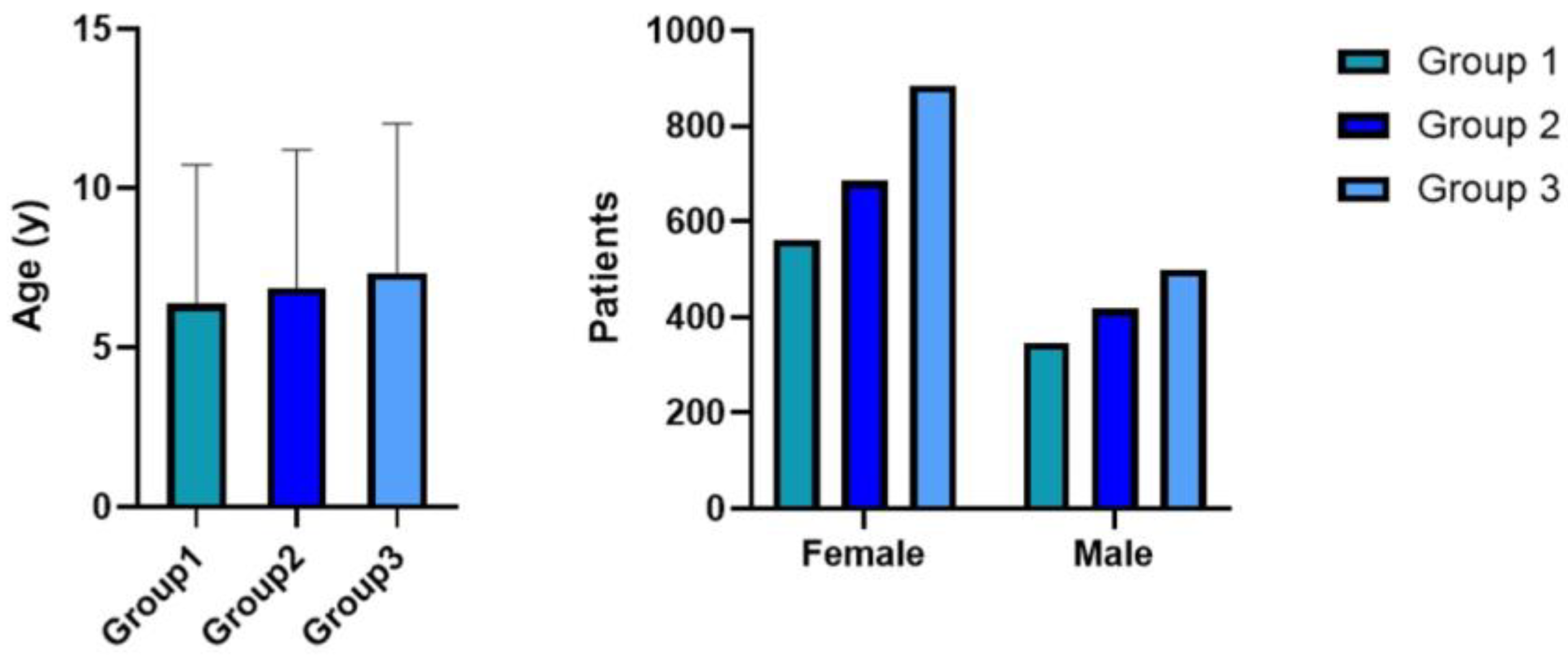
Statistical analysis was performed using the statistical software R (version 4.2.2, R Core Team, Vienna). Furthermore, Pearson’s Chi-square was used to measure whether prevalence of the different symptoms changed according to time of diagnosis. Significance was set at p < 0.05.

3. Results

The study initially included 4478 patients who visited Bambino Gesù Pediatric Hospital for their first consultation regarding celiac disease. Of these, 1070 were excluded due to a prior diagnosis at another center. Additionally, 2 patients were excluded because of missing data, and 10 others were excluded because their diagnosis fell outside the study period (Figure 1).
Ultimately, 3396 patients (female = 2131, median age = 6.9 years old, age range = 8 months−18.8 years) were enrolled in the study cohort. The study population was divided in three groups, according to the diagnosis date:
  • Group 1 (n = 909): patients who received a diagnosis from 1 March 2011 to 30 April 2015;
  • Group 2 (n = 1103): patients who received a diagnosis from 1 May 2015 to 30 May 2019;
  • Group 3 (n = 1384): patients who received a diagnosis from 1 June 2019 to 22 June 2023.
Out of 3396 patients, 2314 (68.1%) patients received a diagnosis with biopsy-sparing protocol, according to ESPGHAN 1990, ESPGHAN 2012 or ESPGHAN 2020 criteria [10,11,12]. Of the 1082 biopsied children, Marsh–Oberhuber grade 3 lesions were present in 989 patients, Marsh–Oberhuber grade 2 lesions in 42 patients, and Marsh–Oberhuber grade 1 lesions in 51 patients, respectively.
The rate of diagnosis of celiac disease increased by 52.2% from the period 2011–2015 to the period 2019–2023.
Group 1 comprised 562 females (61.8%) and 347 males (38.2%), with a median age of 6.4 years. Group 2 included 685 females (62.1%) and 418 males (37.9%), with a median age of 6.9 years. Group 3 consisted of 885 females (63.9%) and 499 males (36.1%), with a median age of 7.3 years (Figure 2). A significant increase in age at diagnosis among the three groups was observed (F (2, 3391) = 13.18, p < 0.0001).
Conversely, no significant differences were found in the distribution of males and females among the three groups (χ²(2) = 1374, p = 0.508).
Clinical features of the study cohort were classified according to Oslo criteria. Overall, 2305 patients (67.8%) debuted with a classic form, with signs and symptoms of malabsorption, while 1006 patients (29.6%) debuted with a non-classic form. 85 patients out of 3396 (2.5%) showed an asymptomatic presentation.
The percentage of the classic form in the three groups was 69% (n = 629), 70% (n = 770) and 65% (n = 906), respectively; the percentage of the non-classic form was 27% (n = 249) in group 1, 29% (n = 320) in group 2 and 31.6% (n = 437) in group 3. The chi-square statistic was 17.36 with a p-value of 0.0016.
In order to observe the trend of non-classical form across the three-time periods, the Cochran Armitage trend test was also applied. The results (statistic-z = 3.99; p-value = 0.000064) confirm a significant difference (Figure 3).
The distribution of single symptoms is reported in Table 1. Bloating and abdominal pain showed a notable variation, indicating that the prevalence of these symptoms differ significantly among the three groups. Similarly, weight loss, failure to thrive, iron deficiency anemia, asymptomatic presentation, and celiac crisis were also significant for p < 0.05. Conversely, several symptoms did not exhibit statistically significant differences among the groups, presenting a substantial stability over time. Group 1 and group 3 were also compared individually, yielding consistent results in most cases. However, for symptoms such as constipation and oral stomatitis, a significant increase was observed from Group 1 and group 3. A reduction in celiac crisis was also observed. In group 1 a total of 33 children (3.6%) presented a celiac crisis at the onset of disease, while in group 3 the percentage decreased to 0.8% (11 children with celiac crisis out of 1384 diagnoses of celiac disease), overtaken by group 2 with a percentage of 1.2%. The statistical analysis using the chi-square test revealed a χ2 value of approximately 28.15 (p-value < 0.00001).
Table 2 summarizes diagnoses of CeD in other associated conditions. Regarding other autoimmune diseases, 2% of new celiac disease diagnoses were made in patients with type 1 diabetes and 1% in patients with rheumatoid arthritis. The incidence rate has remained stable over the analyzed years, as well as the associated syndromes.

4. Discussion

Diagnosis of celiac disease has continued to increase in recent years. Recent data show that the prevalence of CeD in Italy, estimated between 2017 and 2020, was 1.65% [3]. Data in the literature show an increase in the number of annual diagnoses [7]; we likewise observed a percentage growth of celiac diagnosis of 52% from 2011–2015 to 2019–2023. The reasons behind this partly lie in improved screening methods and partly in the increase of true pathology due to changes in environmental factors and dietary habits [13]. It can also be argued that awareness of CeD is also increasing, thanks to media attention directed to this topic [14]. Additionally, the average age at diagnosis has shifted from under 2 years to 6–9 years in many developed countries [15]. Our findings corroborate this trend, with an average diagnosis age of 7 years in our cohort from 2019 to 2023.
In recent decades, a change in clinical presentation is occurring. New symptoms previously unrelated to CeD are now recognized as part of a broader spectrum of manifestations. As an example, neurological disorders such as migraines and ataxia are now considered among “non-classical” symptoms in children [16], and there is a growing interest in other manifestations in adult CeD, such as infertility and sexual dysfunction [17,18,19]. A retrospective study on the Dutch population, conducted from 2007 to 2016, showed a shift in the clinical spectrum of presenting symptoms in pediatric CeD towards an atypical presentation [6]. A North American retrospective study, including patients from 1994 to 2014, showed a light prevalence of the non-classic form of CeD (43%) compared to the classic form (34%) or asymptomatic form diagnosed by screening (23%) [7]. A recent Spanish study [20] examined the clinical presentation between 2011 and 2017 and found that, while the classical form remained the most common, its prevalence significantly declined over the study period. These findings align with our results, which indicate an increase in non-classical presentations at the expense of classical forms, although the latter remains the most frequent.
Considering each symptom individually, gastrointestinal issues are the most prevalent. However, their prevalence is shifting over time. A study conducted in western New York showed that abdominal pain and constipation were the most common symptoms in newly diagnosed celiac patients and that these symptoms occur more frequently in children older than CeD children with classic presentation [21]. In a multicentric study conducted in the US, abdominal pain was reported in 57% of the study cohort [22]. In Greece, patients presenting with malabsorption signs were less than 45%; among the most frequent onset symptoms were constipation, recurrent abdominal pain, and vomiting [8]. An Italian study [9] recently analyzed CeD clinical presentation in children for 30 years, reporting a reduction in diarrhea and an increase in constipation and bloating. Our results confirm the prevalence of gastrointestinal symptoms (i.e., intestinal meteorism, 77.6%, diarrhea, 34%, abdominal pain, 54.5%). In agreement with Pedretti et al. [9], in this study the prevalence of intestinal meteorism and constipation increased, while diarrhea did not show a significant difference during the study period analyzed. Other symptoms demonstrate a decrease over time, such as iron deficiency anemia, weight loss, and failure to thrive. Moreover, a reduction in the incidence of celiac crisis was observed, underlining a greater attention by both clinicians and families to the appearance of warning symptoms of celiac disease. It is important to remember that celiac crisis is a rare presentation of CeD, although it can occur at any age, including in elderly patients [23]. Moreover, regarding clinical presentation, it is important to not forget the “strange” and rare form of presentation as a dermatologic sign (e.g., acrodermatitis [24] and pemphigus vulgaris [25]).
Recent data show an increase in the incidence of autoimmune diseases; our results show a stable percentage of co-occurrence of type 1 diabetes and CeD. A strict GFD is beneficial for the long-term management of these autoimmune conditions, as shown by a decrease in the serum concentration of different autoantibodies [26]. In light of the upcoming national screening program of T1DM and CeD, this figure is likely to change significantly [27,28]. On top of T1DM, several other autoimmune diseases should be assessed and included in CeD evaluation and screening, ranging from autoimmune thyroid disease to autoimmune hepatitis [29,30].
Our study has several strengths, including the large sample size and the extended duration of data collection and observation. At the same time, the main limitations of this study are its retrospective design and the involvement of only one center.

5. Conclusions

The increasing diversity in the clinical presentation of CeD highlights the critical importance of recognizing and diagnosing this condition. The established shift towards atypical symptoms needs increased awareness among healthcare providers to ensure timely identification and management. With new screening campaigns on the horizon, we can anticipate a further rise in celiac disease diagnoses in the coming years. This underscores the need for continuous education and vigilance, not only among clinicians but also within the general public, to recognize the wide spectrum of symptoms associated with CeD. Addressing this growing awareness is crucial for improving patient outcomes and enhancing the quality of life for those affected by this condition.

Author Contributions

Conceptualization, C.M.T. and F.F.; Data curation, C.M.T.; Formal analysis, C.M.T.; Investigation, M.A., G.B., T.C., S.C., A.A. and M.M.; Methodology, C.M.T.; Supervision, S.G., P.D.A. and A.D.; Visualization, G.E. and F.B.; Writing—original draft, C.M.T., A.P.D.B., G.B., S.C. and A.A.; Writing—review and editing, T.C. and D.K. All authors have read and agreed to the published version of the manuscript.

Funding

This research received no external funding.

Institutional Review Board Statement

The study was conducted in accordance with the Declaration of Helsinki, and approved by Ethics Committee of Ospedale Pediatrico Bambino Gesù (PNRR-POC-2022-12375779).

Informed Consent Statement

Informed consent was obtained from all subjects involved in the study.

Data Availability Statement

Data for the present study are available from the corresponding author upon reasonable request.

Conflicts of Interest

The authors declare no conflicts of interest for the present study.

References

  1. Singh, P.; Arora, A.; Strand, T.A.; Leffler, D.A.; Catassi, C.; Green, P.H.; Kelly, C.P.; Ahuja, V.; Makharia, G.K. Global Prevalence of Celiac Disease: Systematic Review and Meta-analysis. Clin. Gastroenterol. Hepatol. 2018, 16, 823–836.e2. [Google Scholar] [CrossRef] [PubMed]
  2. Gatti, S.; Lionetti, E.; Balanzoni, L.; Verma, A.K.; Galeazzi, T.; Gesuita, R.; Scattolo, N.; Cinquetti, M.; Fasano, A.; Catassi, C.; et al. Increased Prevalence of Celiac Disease in School-age Children in Italy. Clin. Gastroenterol. Hepatol. 2020, 18, 596–603. [Google Scholar] [CrossRef] [PubMed]
  3. Lionetti, E.; Pjetraj, D.; Gatti, S.; Catassi, G.; Bellantoni, A.; Boffardi, M.; Cananzi, M.; Cinquetti, M.; Francavilla, R.; Malamisura, B.; et al. Prevalence and detection rate of celiac disease in Italy: Results of a SIGENP multicenter screening in school-age children. Dig. Liver Dis. 2023, 55, 608–613. [Google Scholar] [CrossRef] [PubMed]
  4. Catassi, C.; Verdu, E.F.; Bai, J.C.; Lionetti, E. Coeliac disease. Lancet 2022, 399, 2413–2426. [Google Scholar] [CrossRef] [PubMed]
  5. Ludvigsson, J.F.; Leffler, D.A.; Bai, J.C.; Biagi, F.; Fasano, A.; Green, P.H.; Hadjivassiliou, M.; Kaukinen, K.; Kelly, C.P.; Leonard, J.N.; et al. The Oslo definitions for coeliac disease and related terms. Gut 2013, 62, 43–52. [Google Scholar] [CrossRef]
  6. Van Kalleveen, M.W.; de Meij, T.; Plotz, F.B. Clinical spectrum of paediatric coeliac disease: A 10-year single-centre experience. Eur. J. Pediatr. 2018, 177, 593–602. [Google Scholar] [CrossRef]
  7. Almallouhi, E.; King, K.S.; Patel, B.; Wi, C.; Juhn, Y.J.; Murray, J.A.; Absah, I. Increasing Incidence and Altered Presentation in a Population-based Study of Pediatric Celiac Disease in North America. J. Pediatr. Gastroenterol. Nutr. 2017, 65, 432–437. [Google Scholar] [CrossRef]
  8. Roma, E.; Panayiotou, J.; Karantana, H.; Constantinidou, C.; Siakavellas, S.I.; Krini, M.; Syriopoulou, V.P.; Bamias, G. Changing pattern in the clinical presentation of pediatric celiac disease: A 30-year study. Digestion 2009, 80, 185–191. [Google Scholar] [CrossRef]
  9. Pedretti, M.; Sbravati, F.; Allegri, D.; Labriola, F.; Lombardo, V.; Spisni, E.; Zarbo, C.; Alvisi, P. Is the clinical pattern of pediatric celiac disease changing? A thirty-years real-life experience of an Italian center. Ital. J. Pediatr. 2021, 47, 235. [Google Scholar] [CrossRef]
  10. Revised criteria for diagnosis of coeliac disease. Report of Working Group of European Society of Paediatric Gastroenterology and Nutrition. Arch. Dis. Child. 1990, 65, 909–911. [CrossRef]
  11. Husby, S.; Koletzko, S.; Korponay-Szabo, I.R.; Mearin, M.L.; Phillips, A.; Shamir, R.; Troncone, R.; Giersiepen, K.; Branski, D.; Catassi, C.; et al. European Society for Pediatric Gastroenterology, Hepatology, and Nutrition guidelines for the diagnosis of coeliac disease. J. Pediatr. Gastroenterol. Nutr. 2012, 54, 136–160. [Google Scholar] [CrossRef] [PubMed]
  12. Husby, S.; Koletzko, S.; Korponay-Szabo, I.; Kurppa, K.; Mearin, M.L.; Ribes-Koninckx, C.; Shamir, R.; Troncone, R.; Auricchio, R.; Castillejo, G.; et al. European Society Paediatric Gastroenterology, Hepatology and Nutrition Guidelines for Diagnosing Coeliac Disease 2020. J. Pediatr. Gastroenterol. Nutr. 2020, 70, 141–156. [Google Scholar] [CrossRef] [PubMed]
  13. Bolia, R.; Thapar, N. Celiac Disease in Children: A 2023 Update. Indian J. Pediatr. 2024, 91, 481–489. [Google Scholar] [CrossRef] [PubMed]
  14. Trovato, C.M.; Montuori, M.; Oliva, S.; Cucchiara, S.; Cignarelli, A.; Sansone, A. Assessment of public perceptions and concerns of celiac disease: A Twitter-based sentiment analysis study. Dig. Liver Dis. 2020, 52, 464–466. [Google Scholar] [CrossRef]
  15. Di Biase, A.R.; Marasco, G.; Ravaioli, F.; Colecchia, L.; Dajti, E.; Lecis, M.; Passini, E.; Alemanni, L.V.; Festi, D.; Iughetti, L.; et al. Clinical Presentation of Celiac Disease and Diagnosis Accuracy in a Single-Center European Pediatric Cohort over 10 Years. Nutrients 2021, 13, 4131. [Google Scholar] [CrossRef]
  16. Trovato, C.M.; Raucci, U.; Valitutti, F.; Montuori, M.; Villa, M.P.; Cucchiara, S.; Parisi, P. Neuropsychiatric manifestations in celiac disease. Epilepsy Behav. 2019, 99, 106393. [Google Scholar] [CrossRef]
  17. Glimberg, I.; Haggard, L.; Lebwohl, B.; Green, P.H.R.; Ludvigsson, J.F. The prevalence of celiac disease in women with infertility—A systematic review with meta-analysis. Reprod. Med. Biol. 2021, 20, 224–233. [Google Scholar] [CrossRef]
  18. Castano, M.; Gomez-Gordo, R.; Cuevas, D.; Nunez, C. Systematic Review and Meta-Analysis of Prevalence of Coeliac Disease in Women with Infertility. Nutrients 2019, 11, 1950. [Google Scholar] [CrossRef]
  19. Romano, L.; Pellegrino, R.; Sciorio, C.; Barone, B.; Gravina, A.G.; Santonastaso, A.; Mucherino, C.; Astretto, S.; Napolitano, L.; Aveta, A.; et al. Erectile and sexual dysfunction in male and female patients with celiac disease: A cross-sectional observational study. Andrology 2022, 10, 910–918. [Google Scholar] [CrossRef]
  20. Perez Solis, D.; Cilleruelo Pascual, M.L.; Ochoa Sangrador, C.; Garcia Burriel, J.I.; Sanchez-Valverde Visus, F.; Eizaguirre Arocena, F.J.; Garcia Calatayud, S.; Martinez-Ojinaga Nodal, E.; Donat Aliaga, E.; Barrio Torres, J.; et al. Spanish National Registry of Paediatric Coeliac Disease: Changes in the Clinical Presentation in the 21st Century. J. Pediatr. Gastroenterol. Nutr. 2022, 74, 805–811. [Google Scholar] [CrossRef]
  21. Khatib, M.; Baker, R.D.; Ly, E.K.; Kozielski, R.; Baker, S.S. Presenting Pattern of Pediatric Celiac Disease. J. Pediatr. Gastroenterol. Nutr. 2016, 62, 60–63. [Google Scholar] [CrossRef] [PubMed]
  22. Krueger, A.; Fahey, L.; Sun, Q.; Regis, S.; Khavari, N.; Jericho, H.; Badalyan, V.; Absah, I.; Verma, R.; Leonard, M.M.; et al. Clinical presentation and factors associated with gluten exposure in children with celiac disease. J. Pediatr. Gastroenterol. Nutr. 2024, 79, 895–904. [Google Scholar] [CrossRef] [PubMed]
  23. Soldera, J.; Coelho, G.P.; Heinrich, C.F. Life-Threatening Diarrhea in an Elderly Patient. Gastroenterology 2021, 160, 26–28. [Google Scholar] [CrossRef] [PubMed]
  24. Ancona, S.; Bianchin, S.; Zampatti, N.; Nosratian, V.; Bigatti, C.; Ferro, J.; Trambaiolo Antonelli, C.; Viglizzo, G.; Gandullia, P.; Malerba, F.; et al. Cutaneous Disorders Masking Celiac Disease: Case Report and Mini Review with Proposal for a Practical Clinical Approach. Nutrients 2023, 16, 83. [Google Scholar] [CrossRef]
  25. Goldberg, I.M.; Brooks, J.K.; Ghita, I.; Sultan, A.S.; MacKelfresh, J.B. Oral and cutaneous pemphigus vulgaris: An atypical clinical presentation. Gen. Dent. 2022, 70, 22–26. [Google Scholar]
  26. Tiberti, C.; Montuori, M.; Panimolle, F.; Trovato, C.M.; Anania, C.; Valitutti, F.; Vestri, A.R.; Lenzi, A.; Cucchiara, S.; Morano, S. Screening for Type 1 Diabetes-, Thyroid-, Gastric-, and Adrenal-Specific Humoral Autoimmunity in 529 Children and Adolescents With Celiac Disease at Diagnosis Identifies as Positive One in Every Nine Patients. Diabetes Care 2017, 40, e10–e11. [Google Scholar] [CrossRef]
  27. Catassi, C.; Vincentini, O.; Pricci, F.; Agrimi, U.; Silano, M.; Bosi, E. Pediatric screening for type 1 diabetes and celiac disease: The future is today in Italy. Minerva Pediatr. 2024, 76, 461–463. [Google Scholar] [CrossRef]
  28. Bosi, E.; Catassi, C. Screening type 1 diabetes and celiac disease by law. Lancet Diabetes Endocrinol. 2024, 12, 12–14. [Google Scholar] [CrossRef]
  29. Panetta, F.; Nobili, V.; Sartorelli, M.R.; Papa, R.E.; Ferretti, F.; Alterio, A.; Diamanti, A. Celiac disease in pediatric patients with autoimmune hepatitis: Etiology, diagnosis, and management. Paediatr. Drugs 2012, 14, 35–41. [Google Scholar] [CrossRef]
  30. Tiberti, C.; Panimolle, F.; Borghini, R.; Montuori, M.; Trovato, C.M.; Filardi, T.; Lenzi, A.; Picarelli, A. Type 1 diabetes, thyroid, gastric and adrenal humoral autoantibodies are present altogether in almost one third of adult celiac patients at diagnosis, with a higher frequency than children and adolescent celiac patients. Scand. J. Gastroenterol. 2020, 55, 549–554. [Google Scholar] [CrossRef]
Figure 1. Flowchart of study population.
Nutrients 17 00129 g001
Figure 2. Distribution of age and sex at diagnosis in the three groups.
Nutrients 17 00129 g002
Figure 3. Classic, non-classic and asymptomatic forms variation during the time. Group 1, n= 909, diagnosed between 1 March 2011 and 30 April 2015; group 2, n= 1103, diagnosed between 1 May 2015 and 30 May 2019; group 3, n= 1384, diagnosed between 1 June 2019 and 22 June 2023.
Nutrients 17 00129 g003
Table 1. Clinical presentation of celiac disease. Gr1 = group 1; Gr2 = group 2; Gr3 = group 3; χ2 (G1-G2-G3) = chi square derived from the comparison among the three groups; χ2 (G1-G3) = chi square derived from the comparison between group 1 and group 3.
SymptomsnGroup 1
(n = 909)
Group 2
(n = 1103)
Group 3
(n = 1384)
χ2 (G1-G2-G3)χ2 (G1-G3)
Diarrhea1159332 (36.5%)386 (35%)441 (31.8%)χ2 = 5.84
p = 0.053
χ2 = 5.33
p = 0.021
Bloating2636629 (69.1%)909 (82.4%)1098 (79.3%)χ2 = 54.05
p < 0.00001
χ2 = 30.32
p < 0.00001
Abdominal pain1853526 (57.8%)650 (58.9%)677 (48.9%)χ2 = 30.28
p < 0.00001
χ2 = 46.20
p < 0.00001
Vomiting931236 (25.9%)321 (29.1%)356 (35.7%)χ2 = 4.10
p = 0.128
χ2 = 0.016
p = 0.8978
Constipation1017248 (27.2%)328 (29.7%)441 (31.8%)χ2 = 5.52
p = 0.063
χ2 = 7.55
p = 0.006
Weight loss703219 (24%)207 (18.7%)277 (20%)χ2 = 9.27
p = 0.009
χ2 = 5.38
p = 0.0204
Failure to thrive1650464 (51%)576 (52.2%)610 (44%)χ2 = 19.31
p = 0.00006
χ2 = 10.69
p = 0.0011
Change in mood852250 (27,5%)270 (24.4%)332 (24%)χ2 = 3.92
p = 0.140
χ2 = 3.58
p = 0.0586
Iron deficiency anemia1166341 (37.5%)418 (37.8%)407 (29.4%)χ2 = 25.18
p < 0.00001
χ2 = 16.39
p = 0.00001
Hypertransaminasemia15337 (4%)55 (4.9%)61 (4.4%)χ2 = 1.023
p = 0.599
χ2 = 0.15
p = 0.6963
Recurrent aphthous stomatitis21269 (7.6%)70 (6.3%)73 (5.3%)χ2 = 5.058
p = 0.079
χ2 = 5.064
p = 0.0244
Dental enamel defects21045 (4.9%)102 (9.2%)63 (4.5%)χ2 = 26.58
p < 0.00001
χ2 = 0.194
p = 0.6596
Celiac crisis5833 (3.6%)14 (1.2%)11 (0.8%)χ2 = 28.15
p < 0.00001
χ2 = 23.43
p < 0.00001
Asymptomatic8531 (3.4%)13 (1.2%)41 (2.9%)χ2 = 12.19
p = 0.0022
χ2 = 0.362
p = 0.5475
Table 2. Associated conditions.
Associated ConditionnGroup 1
(n = 909)
Group 2
(n= 1103)
Group 3
(n = 1384)
Type 1 Diabetes21345 (4.9%)102 (9.2%)66 (4.7%)
Chronic autoimmune thyroiditis8645 (4.9%)17 (1.5%)24 (1.73%)
Rheumatoid arthritis3212 (1.32%)5 (0.45%)15 (1%)
Down Syndrome407 (0.77%)18 (1.63%)15 (1%)
Turner Syndrome51 (0.11%)4 (0.36%)0
Other Syndromes71 (0.11%)3 (0.27%)3 (0.2%)
Disclaimer/Publisher’s Note: The statements, opinions and data contained in all publications are solely those of the individual author(s) and contributor(s) and not of MDPI and/or the editor(s). MDPI and/or the editor(s) disclaim responsibility for any injury to people or property resulting from any ideas, methods, instructions or products referred to in the content.

