Article

Early Probiotic Supplementation of Healthy Term Infants with Bifidobacterium longum subsp. infantis M-63 Is Safe and Leads to the Development of Bifidobacterium-Predominant Gut Microbiota: A Double-Blind, Placebo-Controlled Trial

1
Food Ingredients and Technology Institute, R & D Division, Morinaga Milk Industry Co., Ltd., 5-1-83, Higashihara, Zama 252-8583, Japan
2
Department of Pediatrics, Matsumoto City Hospital, 4417-180, Hata, Matsumoto 390-1401, Japan
3
Next Generation Science Institute, R & D Division, Morinaga Milk Industry Co., Ltd., 5-1-83, Higashihara, Zama 252-8583, Japan
4
Department of neurosurgery, Matsumoto City Hospital, 4417-180, Hata, Matsumoto 390-1401, Japan
*
Author to whom correspondence should be addressed.
Nutrients 2023, 15(6), 1402; https://doi.org/10.3390/nu15061402
Submission received: 14 February 2023 / Revised: 25 February 2023 / Accepted: 2 March 2023 / Published: 14 March 2023
(This article belongs to the Special Issue Nutrition and the Gut Microbiome in Infants)

Abstract

:
Bifidobacteria are important intestinal bacteria that provide a variety of health benefits in infants. We investigated the efficacy and safety of Bifidobacterium longum subsp. infantis (B. infantis) M-63 in healthy infants in a double-blind, randomized, placebo-controlled trial. Healthy term infants were given B. infantis M-63 (n = 56; 1 × 10⁹ CFU/day) or placebo (n = 54) from postnatal age ≤ 7 days to 3 months. Fecal samples were collected, and fecal microbiota, stool pH, short-chain fatty acids, and immune substances were analyzed. Supplementation with B. infantis M-63 significantly increased the relative abundance of Bifidobacterium compared with the placebo group, with a positive correlation with the frequency of breastfeeding. Supplementation with B. infantis M-63 led to decreased stool pH and increased levels of acetic acid and IgA in the stool at 1 month of age compared with the placebo group. There was a decreased frequency of defecation and watery stools in the probiotic group. No adverse events related to test foods were observed. These results indicate that early supplementation with B. infantis M-63 is well tolerated and contributes to the development of Bifidobacterium-predominant gut microbiota during a critical developmental phase in term infants.

1. Introduction

The establishment of a healthy gut microbiota in the early developmental stages of human life plays an important role in later health [1]. Bifidobacteria are important intestinal bacteria that provide a variety of health benefits for infants, including the production of vitamins and organic acids [2], maintenance of gut homeostasis [3], improved vaccination response [4,5], prevention from infection [6], suppression of gut inflammation [7], and allergy prevention [8]. Reduction of bifidobacteria in infants could cause a variety of disorders and has been associated with an increased prevalence of obesity, diabetes, metabolic disorders, and all-cause mortality in later life [9,10,11]. The formation of Bifidobacterium-dominant microbiota is delayed in infants born by cesarean section [12], in newborns who used antibiotics at birth [13], and in low-birth-weight infants. Low-birth-weight infants with delayed colonization of Bifidobacterium have been reported to be at high risk for sepsis and necrotizing enterocolitis [14].
Human milk oligosaccharides (HMOs), a complex mixture of over 200 molecular species including fucose and sialic acid, are the third most abundant solids in breast milk after lactose and lipids and are the selective growth factors of bifidobacteria. HMOs are not digested in the small intestine because they cannot be broken down by the infant itself; instead, they reach the large intestine, where they are selectively capitalized upon by intestinal bacteria such as bifidobacteria and Bacteroides. The Bifidobacterium longum subsp. infantis (B. infantis) microorganisms have metabolic pathways that allow them to preferentially uptake and utilize HMOs, such as fucosyl-lactose (FL) transporters, fucosidases, and sialidases [15], and they utilize HMOs for their own bifidobacterial growth. B. infantis, which becomes the predominant bacterial species in the gut due to HMOs in breast milk, benefits infant health by promoting the maturation of immune function, inhibiting excessive inflammation, enhancing gut barrier function, and increasing acetate production [16].
Administration of probiotics to infants could maintain mucosal barrier integrity, regulate appropriate bacterial colonization, and modulate immune function by promoting IgA secretion in the mucosa and suppressing intestinal inflammation [17]. Early postnatal use of probiotics such as Bifidobacterium spp. in low-birth-weight infants has been reported to be effective in preventing sepsis and necrotizing enterocolitis and establishing enteral nutrition early by the formation of a Bifidobacterium-dominant microbiota [18,19,20,21]. In clinical studies in which B. infantis was administered to preterm or undernourished infants, anti-inflammatory effects and growth-promoting effects on infant development were reported [10,22]. However, current guidelines from the European Society of Pediatric Gastroenterology, Hepatology and Nutrition and the American Academy of Pediatrics state that there is insufficient evidence to recommend the use of probiotics for all neonates [17].
B. infantis M-63 was isolated from the feces of infants [22] and has been shown to grow well in human breast milk due to its high capability to capitalize on HMOs [23,24] and its tolerance to lysozyme [25]. From clinical studies in which probiotics containing B. infantis M-63 and a mixture of several other species of Bifidobacterium and Lactobacillus were administered to infants and children, the following effects have been reported: early establishment of Bifidobacterium-predominant gut microbiota and suppression of intestinal staphylococcal colonization in low-birth-weight infants [19], suppression of dysbiosis and growth promotion in neonates with congenital gastrointestinal surgical conditions (CGISC) [26], improvement of symptoms of gastrointestinal discomfort such as overflowing milk and bloating in colicky infants [27], reduction in the duration of crying or agitation [28], prevention and alleviation of the onset of allergies such as seasonal rhinitis and atopic dermatitis [29], alleviation of functional constipation [3], and improvement of QOL in children with functional gastrointestinal disorders [30]. However, the effect of administering the M-63 strain alone to full-term infants during the early postnatal period has not been examined to date. In this study, the effects of B. infantis M-63 as a single probiotic on gut microbiota formation, intestinal environment, gastrointestinal function, and immune parameters in healthy full-term infants up to 3 months of age were investigated in a double-blind, randomized, placebo-controlled trial.

2. Materials and Methods

2.1. Study Population

Between October 2019 and August 2021, healthy women who were scheduled to give birth or deliver a healthy full-term baby at Matsumoto City Hospital were recruited and subsequently provided written informed consent for participation in the study. Eligible participants were apparently healthy children born within 7 days whose gestational age at birth was ≥37 weeks and ˂42 weeks. Exclusion criteria for mothers were as follows: mothers who were diagnosed with any severe liver, renal, cardiovascular, respiratory, endocrine, metabolic, or mental disease or planned to administer any other probiotic supplements to their infants during the study period. Mothers with gestational diabetes or gestational hypertension were not excluded. Exclusion criteria for infants were as follows: infants born with multiple births and infants born with medical complications such as small for gestational age (SGA), large for gestational age (LGA), blood, liver, heart, kidneys, digestive disease, or suspected immunodeficiency or who had exposure to any oral or intravenous antibiotics or who were judged to be inappropriate to participate in the trial by the principal investigator.

2.2. Study Design

This study was a single-center, placebo-controlled, double-blinded randomized trial of probiotics in healthy full-term infants conducted in Matsumoto City Hospital. This study protocol was performed in compliance with the Helsinki Declaration of 1975 as revised in 2013 and the Ethical Guidelines for Medical and Health Research Involving Human Subjects proposed by the Ministry of Education, Culture, Sports, Science and Technology and the Ministry of Health, Labor and Welfare; it was reviewed and approved by the Research Ethics Committee of Matsumoto City Hospital (27 September 2019). This study was registered on the UMIN Clinical Trials Registry (UMIN000038351).
Prior to the initiation of the study, the investigator recruited participants from mothers giving birth at Matsumoto City Hospital and explained the details of the study according to the informed consent form. After giving birth, mothers completed a written consent form as a surrogate, and the infants who met the eligibility criteria were stratified by randomization schemes based on delivery mode (vaginal delivery/cesarean section) or sex (male/female) and assigned to two groups. Stratified randomization was utilized because delivery mode and gender have been shown to be influencing factors on early intestinal gut microbiota [31]. The infants in each group were fed B. infantis M-63 (1 billion CFU/1.0 g of sachet) or placebo (sterilized dextrin only/1.0 g of sachet) daily from within 7 days after birth to 3 months after birth. The test foods were suspended in a small amount of sterile water in a sterilized feeding bottle at room temperature, and mothers fed the suspension to their infants using a feeding bottle or a sterilized medicine dropper. After giving birth, the mother received support at the hospital to promote breastfeeding as much as possible, and the infants who were judged to require supplementation with infant formula were begun on mixed nutrition. None of the infants used HMOs-fortified infant formula.

2.3. Infant Gastrointestinal (GI) Tolerability and Health Examination

Infant GI tolerability and health was assessed by mothers on a daily basis from Day 1 until 3 months of age during test food supplementation period. Mothers were instructed to record the following information about their infants in daily logs: consumption of the amount of test foods, intake of any oral antibiotics or medicines, any symptoms including episodes of fever (≥38 °C), and hospital visits. For the 7 days before 1 week, 1 month, and 3 months of age, mothers recorded in daily logs the number of breast milk-fed and infant formula-fed infants, number of stools, stool consistency, duration and episodes of crying, number of regurgitations and vomiting episodes after feeding. Consistency of stool was assayed by mothers using a modified Amsterdam infant scale [32] (4-point scale with 1: watery; 2: soft; 3: formed; 4: hard). Episodes of crying were defined as more than 30 min per day. Episodes of colic were defined as 3 h per day for at least 3 days per week [33]. The investigator examined infant health and physical growth (length, body weight, and head circumference) immediately after birth and during hospitalization after delivery, before starting the study, and at 1 month and 3 months of age and checked the daily logs recorded by mothers.
A safety evaluation was conducted on all participants who consumed the study food once or more. Throughout the study period, all adverse events related to subjective and objective symptoms were recorded in the daily logs. The degree of symptoms and the causal relationship were evaluated according to the revised “National Cancer Institute, Common Terminology Standards for Adverse Events Version 4.0, Japanese Translation JCOG Version”.

2.4. Fecal Sample Collection

Two types of fecal samples were prepared: fresh fecal samples were collected before intake and at 1 month of age, and fecal samples with the preservative solution, guanidine thiocyanate solution, were collected before intake, 1 week after intervention, and at 1 month and 3 months of age. Both fecal samples were collected from infants’ diapers in each stool collection tube (Techno Suruga Laboratory Co., Ltd., Shizuoka, Japan). Fresh fecal samples were collected within 24 h of the hospital visit and were stored in a foamed styrene box with cooling agent at home, whereas fecal samples with the preservative solution were stored at room temperature at home. Both samples taken at home were transferred to the hospital and stored in −20 °C freezers. After being transported to the laboratory with dry ice, they were stored at −80 °C before DNA extraction. Fresh fecal samples were used for the analysis of real-time PCR, pH, SCFAs, and biomarkers, whereas fecal samples with the preservative solution were used for 16S rRNA gene sequencing. All participant samples were blinded to the process and analysis.

2.5. Fecal DNA Extraction and 16S rRNA Gene Sequencing

Fecal DNA was extracted according to previously described methods [34]. In brief, 200 μL of fecal sample in GuSCN solution was lysed with glass beads (300 mg, 0.1 mm diameter) and 300 μL of lysis buffer (No. 10 buffer, Kurabo Industries Ltd., Osaka, Japan) using a FastPrep-245G homogenizer (MP Biomedicals LLC, Santa Ana, CA, USA) at a 5-power level for 45 s with 5 min cooling intervals on ice. After centrifugation at 12,000 rpm for 5 min, DNA was extracted from 200 μL of the supernatant by utilizing the GENE PREP STAR PI-480 instrument (Kurabo Industries Ltd., Osaka, Japan) according to the manufacturer’s protocol. Amplification of the V3-V4 region of the bacterial 16S rRNA gene through PCR and subsequent DNA sequencing were carried out as previously described [35], using by the Illumina MiSeq instrument (Illumina, San Diego, CA, USA).
Following the removal of sequences that aligned with data from the Genome Reference Consortium human build 38 (GRCh38) and phiX reads from the raw Illumina paired-end reads, the remaining sequences were analyzed using the QIIME2 software package version 2017.10 (https://qiime2.org/). DADA2 [36] was employed to remove potential chimeric sequences, followed by trimming 30 and 90 bases of the 3′ region of the forward and reverse reads, respectively. Taxonomical classification was conducted by utilizing the naive Bayes classifier trained on Greengenes13.8 with a 99% threshold of full-length sequence operational taxonomic units. Alpha diversities were calculated using QIIME2 software. We used R software (ver. 3.6.0) for principal coordinate analysis (PCoA) based on Jensen–Shannon distance (JSD) and partitioning around medoid (PAM) clustering [37]. The optimal number of clusters was estimated by utilizing the Calinski–Harabasz (CH) index.

2.6. Quantification of Bifidobacterium Species by Real-Time PCR

Fecal DNA for real-time PCR was extracted as previously described [38]. In brief, 200 μL of fecal sample in GTC buffer was lysed using a Precellys Evolution (Bertin Instruments, FRA) system, and DNA was extracted using the GENE PREP STAR PI-480 instrument (Kurabo Industries Ltd., Osaka, Japan) in accordance with the manufacturer’s protocol. Real-time PCR was conducted using an ABI PRISM 7500 fast real-time PCR system (Thermo Fisher Scientific, Waltham, MA, USA) with TB Green Premix Ex Taq™ Tli RNaseH Plus (TaKaRa Bio Inc., Shiga, Japan) in accordance with the manufacturer’s protocol. Quantification of each Bifidobacteria species was performed using the following primers (Table S1). Primers and amplification methods used were determined based on prior studies [39,40,41]. Bacterial copy numbers were determined by utilizing bacterial solutions with established counts as a standard. Duplicate assays were performed for all samples.

2.7. Fecal pH, Short-Chain Fatty Acids (SCFAs) and Biomarker Analysis

Fecal pH was measured with an electrode-fitted pH meter LAQUAtwinB-712 (HORIBA, Kyoto, Japan) after suspending fecal samples in Milli-Q water and sterilizing at 85 °C for 15 min for sterilization of fecal samples.
Fecal SCFAs (acetate, propionate, n-butyrate, iso-butyrate, n-valerate, iso-valerate, n-caproic acid) in ethyl acetate extract were determined by gas chromatography system GC-FID (7890B, Agilent Technologies, Santa Clara, CA, USA) and a DB-WAXetr column (30 m, 0.25 mm id, 0.25 μm film thickness, 1.2 mL/min) as described previously [42].
Commercially available ELISA kits were used to determine fecal secretory immunoglobulin A (sIgA: Human IgA ELISA Core Kit, LABISKOMA, Seoul, Republic of Korea) and calprotectin (IDK® Calprotectin MRP 8/14 ELISA kit, Immundiagnostik AG, Bensheim, Germany).

2.8. Sample Size

In a previous study on Japanese infants [15], the relative abundance of bifidobacteria in feces at 1 month of age was 55.40 ± 40%. Assuming the relative abundance of bifidobacteria would increase to 75% by intake of test food, to detect an intergroup difference with 80% power and α = 0.05 and a 15% attrition rate, 75 infants were needed in each group.

2.9. Primary and Secondary Outcomes

Evaluation of the primary outcome involved determining whether B. infantis M-63 supplementation could increase the relative abundance of Bifidobacteriaceae, the proportion of infants with Bifidobacteriaceae-predominant microbiota, and the fecal copy number of Bifidobacteriaceae in healthy term infants. Secondary outcomes included the effect of B. infantis M-63 on the abundance of intestinal bacteria and diversity, GI tolerability (stool frequency and consistency, number of regurgitation and vomiting after feeding), duration and episodes of crying, episodes of fever (≥38 °C), hospital visit, fecal pH, SCFAs, sIgA, and calprotectin.

2.10. Statistics

Intergroup differences in the microbiota at the amplicon sequence variant (ASV) level were analyzed by ALDEx2 [43]. A Q-value < 0.05 was considered significant. To assess the variation in microbiota composition explained by each factor, a permutational multivariate analysis of variance (PERMANOVA) test for JSD was used for multivariate analysis. For the relative abundance of Bifidobacteria, a normal log transformation was performed for each time point and for each test food group, and the mean ± standard error of the mean (SEM) was calculated. When the assumption of a normal distribution was not ruled out, an analysis of covariance (ANCOVA) was performed with the values at each time point as the objective variable, the test food group as the explanatory variable, and the baseline values as covariates. The changes from the baseline value were calculated for each time point, and a paired t test was performed. When the assumption of a normal distribution was not verified, the change from the baseline values was calculated for each time point and compared between the groups using the Wilcoxon rank sum test. To identify changes from baseline values, a Wilcoxon signed ranks test was performed. The same analysis was performed for the absolute number of bifidobacteria per gram of feces (copy number), occupancy of each bacterial species and diversity index in gut microbiota analysis, and physical and chemical analysis of stool (pH, amount of short-chain fatty acids, IgA, and calprotectin, pH). For frequency of defecation and fecal characteristics, the average number of defecations (times/day) and average fecal characteristics (score/times) were calculated for each time point and analyzed in the same way. The percentage of infants whose most prevalent bacterial species was Bifidobacterium was calculated for each time point, and Fisher’s exact test was performed. The breastfeeding rate was defined as the number of times breastfeeding occurred relative to the number of feedings. Correlations between the breastfeeding rate and the relative abundance of Bifidobacterium were analyzed by calculating Spearman’s rank correlation coefficients. For the health condition of the infants, duration and frequency of crying, frequency of regurgitation and vomiting, frequency of fever, and hospital visits, the mean values per week were calculated for each time point, and a Wilcoxon rank sum test was performed.
For the primary endpoint, subgroups were analyzed according to the following factors affecting gut microbiota: (1) mode of delivery (vaginal delivery, cesarean section); (2) whether antibiotics were administered to the mother during delivery; and (3) mode of nutrition (exclusively breastfeeding, mixed feeding, exclusively formula feeding). For height, weight, and head circumference of children, the mean ± SE was calculated for each time point, and Student’s t test was performed. Adverse events and side effects were tested for incidence (person/person) using Fisher’s exact test in the ITT population of patients that consumed the study foods. Statistical analysis software was IBM SPSS Statistics (version 28.0), and a p value of < 0.05 was adopted to indicate a significant difference.

3. Results

3.1. Infant and Maternal Characteristics

Figure 1 shows the process from subject enrollment to analysis: 111 subjects were enrolled, and after randomization (54 in the placebo group and 57 in the M-63 group), one subject in the M-63 group withdrew consent before consuming the test food, and one subject in the placebo group was excluded from the analysis due to noncompliance involving intake of less than 50% of the test food. The analysis was performed in the per protocol set (PPS) population (53 patients in the placebo group and 56 patients in the M-63 group). No infant received antibiotics or oral probiotics other than the test food, although one infant used antibacterial eye drops due to discharge from the eyes during the study period.
The subject background of the newborns and mothers is shown in Table 1. There were no significant differences between groups in the number of weeks of gestation, mode of delivery (vaginal delivery, cesarean section), sex, height at birth, weight, head circumference, APGAR score (5 min after birth), and intake rate of test foods among the newborns. There were also no significant differences between the two groups in maternal age, number of deliveries (first-time mothers and term mothers), prepregnancy body mass index (BMI, kg/m2), weight gain during pregnancy, number of women taking antibiotics at delivery, history of allergies, and smoking habits.

3.2. Infant Feeding

The intake rate of the test foods was 92.4 ± 1.4% for the placebo group and 94.4 ± 0.8% for the M-63 group with no significant difference between groups (p = 0.5082). Table S2 shows the number of feedings and the percentage of breastfeeding in each group at 1 week, 1 month, and 3 months of age (calculated from the number of breastfeeds/number of feedings per day). The percentage of breastfeeding increased in both groups at 1 month and 3 months of age with no significant differences between the groups. At 1 month and 3 months of age, 27.5% and 52.3% of the infants were exclusively breastfed, 70.6% and 41.3% were mixed fed, and very few were exclusively formula fed: one case at 1 week, two cases at 1 month, and seven cases at 3 months of age. Therefore, when subgroup analysis was performed, depending on the infant’s feeding status, the analysis was performed in the groups of either exclusive breastfeeding or mixed feeding with exclusive formula feeding.
There were 14 cases of mothers taking antibiotics during the lactation period (6 in the placebo group and 8 in the M-63 group), and there was no significant difference between the two groups. The reasons for taking the drugs were mastitis (three cases), cold (two cases), treatment of cesarean section wounds (one case), uterine restoration failure (one case), treatment of placental abruption (one case), asthma (one case), sinusitis (one case), cystitis (one case), dental treatment (one case), stye (one case), and unknown (one case).

3.3. Microbiota Analysis

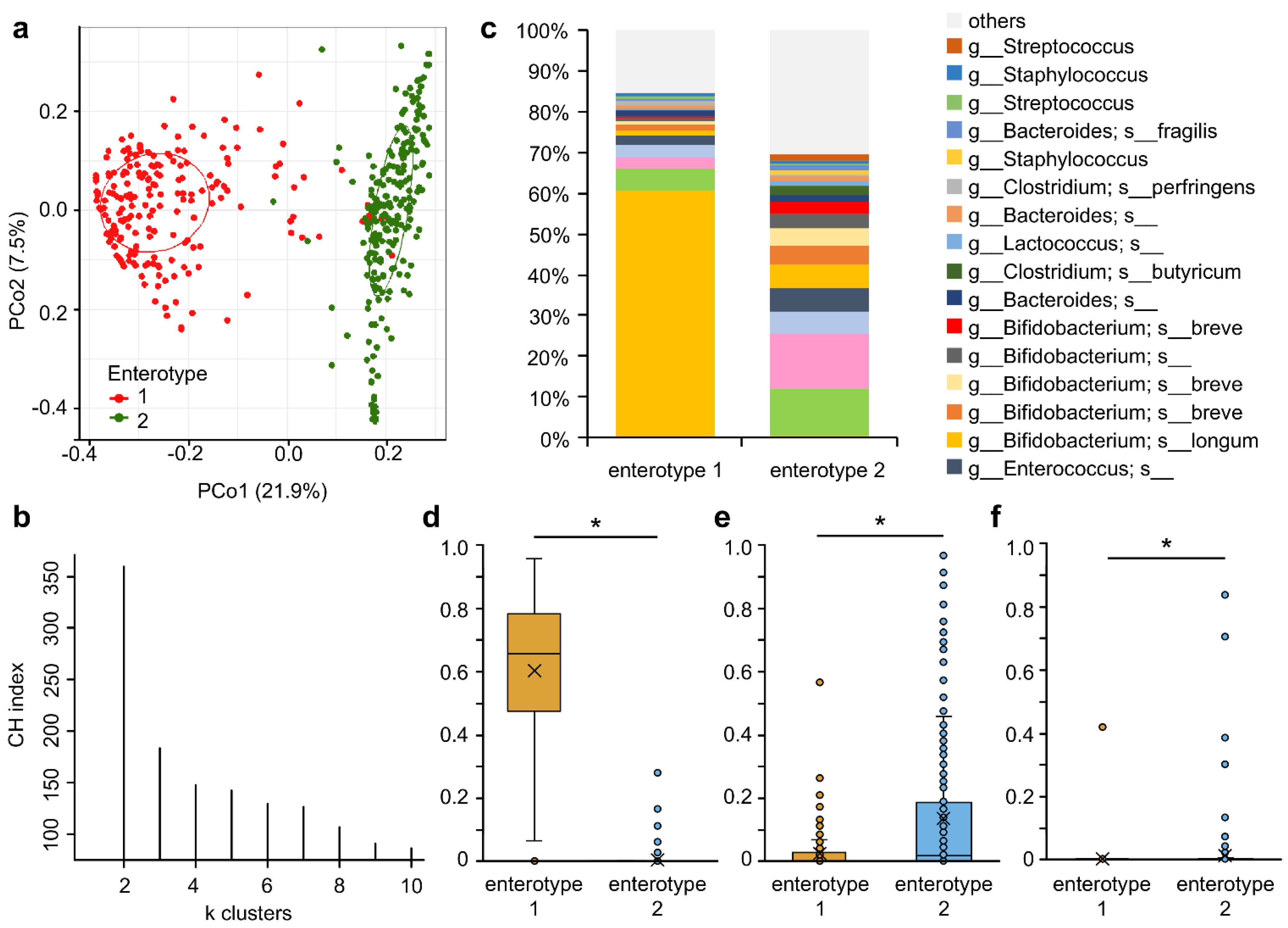
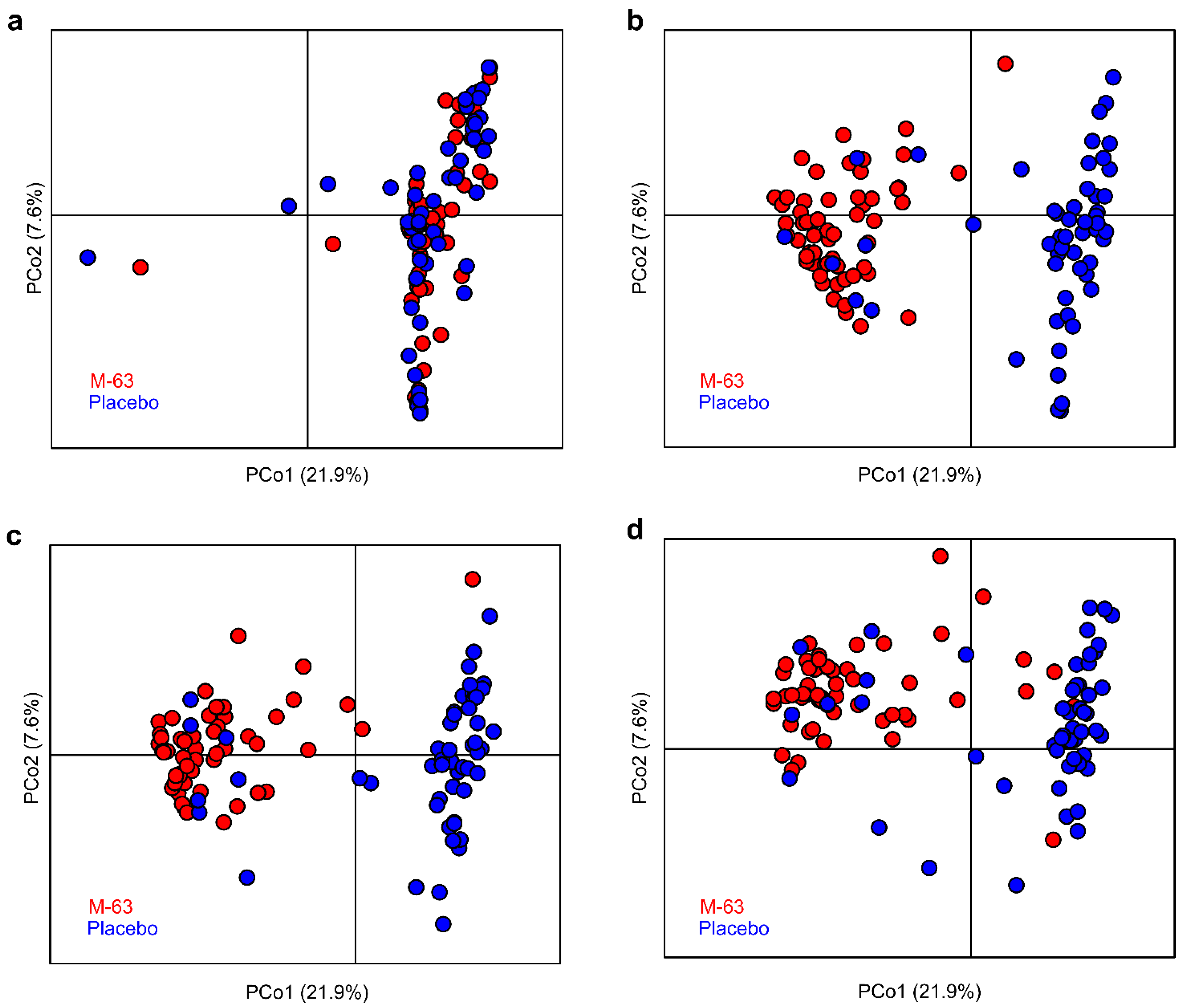
We then investigated the fecal microbiota of all samples during the intervention. PCoA data clearly showed two enterotypes (Figure 2a,b) enriched in Bifidobacterium (enterotype 1) and several taxa, such as Enterobacteriaceae (enterotype 2, Figure 2c–f). Within a week of birth, 101 of 106 subjects had enterotype 2 microbiota (Figure 3a). However, the enterotype of all subjects in the M-63 group except one transitioned to the Bifidobacterium-dominated enterotype (enterotype 1) after 1 week of administration of B. infantis M-63, while the enterotype of most subjects in the placebo group was stable in enterotype 2 (Figure 3). This polarization was maintained until the end of the intervention.

3.4. Bifidobacterial Colonization

The relative abundance of Bifidobacterium and the percentage of infants for whom Bifidobacterium spp. was the most dominant bacterial genus are shown in Table 2. There was no significant difference between the two groups in the relative abundance of Bifidobacterium before intake of the test food, and the relative abundance of Bifidobacterium increased with the age of the infants (1 week, 1 month, and 3 months of age), and a significant increase was observed in the M-63 group over the placebo group. The percentage of infants in the M-63 group with Bifidobacterium as the most dominant genus was also significantly higher than that of the placebo group at the first week of intake and at one month of age. This trend was also observed in all subgroups, regardless of the delivery method (vaginal or cesarean section), regardless of the mode of nutrition in the infants or whether antibiotics were used by the mothers at delivery.
The relative abundance of bifidobacteria in the placebo group was significantly lower in infants born to mothers who received antibiotics at delivery than in those whose mothers did not receive antibiotics before intake and was also lower in the placebo group at 1 week (Table S3). There was no significant difference in the relative abundance of bifidobacteria between cesarean section and vaginal delivery infants within the population whose mothers received antibiotics in the placebo group, and no association was found between mode of delivery and the relative abundance of bifidobacteria. Even in the case of vaginal delivery, bifidobacteria were found to be significantly lower in the antibiotic-treated group than in the antibiotic-untreated group.

3.5. Bacterial Species of Colonized Bifidobacteria

The abundance and detection rate of the genus Bifidobacterium and each Bifidobacterium species at 1 month after birth, measured by quantitative PCR, are shown in Table 3. There were no significant differences in the abundance and detection rate of the genus Bifidobacterium and each Bifidobacterium species before the ingestion point. However, the abundance and detection rate of the genera Bifidobacterium and B. infantis in the M-63 group were significantly higher than those in the placebo group. In short, the results of quantitative PCR at 1 month of age showed that most of the Bifidobacterium species in the M-63 group were B. infantis, whereas B. infantis was barely detected in the placebo group and B. breve and B. longum subsp. longum (B. longum) were detected in the placebo group. These results indicate that B. infantis was the main Bifidobacterium species that colonized the intestines of the infants in the M-63 group, while B. breve and B. longum were the main species in the placebo group.

3.6. Correlation between Breastfeeding and Bifidobacterial Occupancy

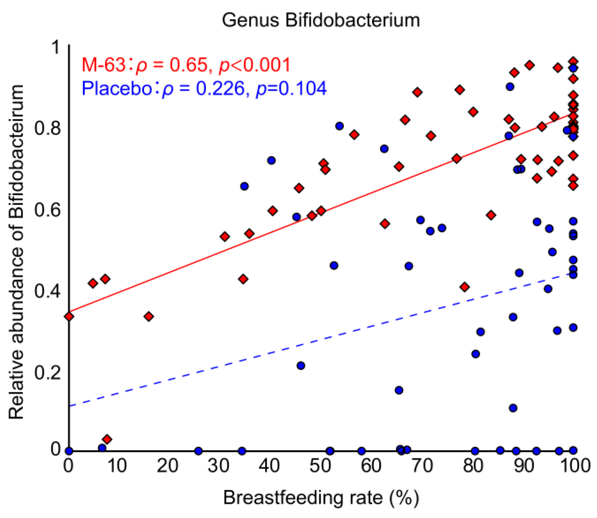
We analyzed the correlation at 1 month of age between the breastfeeding rate and the relative abundance of bifidobacteria (Figure 4). A significant positive correlation was seen between the breastfeeding rate and the relative abundance of Bifidobacterium in the M-63 group. However, there was no significant positive correlation between them in the placebo group. Although the breastfeeding rate was more than 80%, some infants did not have increased bifidobacteria in the gut in the placebo group.

3.7. Gut Fermentation Patterns and Immunologic Parameters in Stools

The pH, amount of short-chain fatty acid, IgA, and calprotectin in the stools before intake and one month after birth are shown in Table 4. In the M-63 group, the pH in the stools was significantly lower and the amount of acetic acid in the stools increased at 1 month of age compared to the placebo group. There was no difference between the two groups in terms of calprotectin in the feces, but IgA levels in the feces were significantly higher in the M-63 group than in the placebo group at one month of age.

3.8. GI Tolerability and Health Condition of Infants

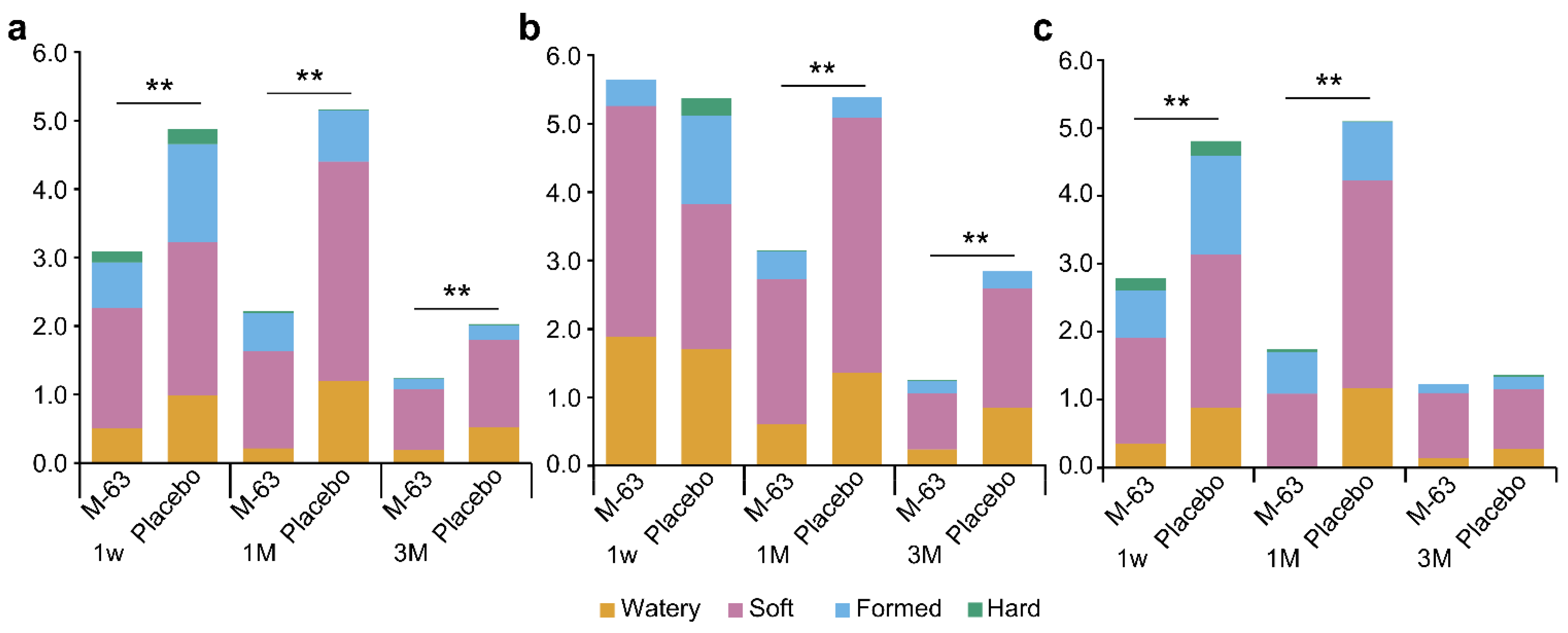
The number of defecations per day and fecal characteristics of the infants are shown in Figure 5. The placebo group showed a decrease in defecation frequency from 1 to 3 months of age, whereas the M-63 group showed a gradual decrease in defecation frequency from 1 week to 3 months of age, with a significant decrease in the M-63 group compared to the placebo group”. “Watery” stools and “soft” stools were significantly decreased in the M-63 group as compared to the placebo group at 1 month of age (Figure 5a). Stratified analysis by type of nutrition is shown in Figure 5b,c. In the exclusively breastfed infants, defecation frequency was lower in the M-63 group than in the placebo group at 1 and 3 months of age (Figure 5b), and in the mixed-fed infants and exclusively formula-fed infants, defecation frequency was lower in the M-63 group than in the placebo group at 1 week and 1 month of age (Figure 5c). These results indicate that M-63 modulates the defecation function of infants regardless of the type of nutrition.
The number of times (times/day) and average duration (minutes/time) that infants cried for more than 30 min are shown in Table S4. Neither group included infants who cried for an average of more than 3 h per day or who were suspected of having so-called “colic”, and there were no significant differences between the two groups.
The number of times and number of infants with regurgitation and vomiting of milk are shown in Table S5. There were significantly fewer infants in the M-63 group than in the placebo group who had regurgitation of milk at 1 week after intake. Both regurgitation and vomiting of milk were mild and not pathological.
The growth of the infant’s height, weight, and head circumference at 1 and 3 months after birth was favorable and comparable in both groups (Table S6).

3.9. Adverse Events

Adverse events during the study period are shown in Table S7. No adverse events attributable to bifidobacteria administration were identified, and the incidence of respiratory, gastrointestinal, and skin symptoms did not differ between the two groups.

4. Discussion

In this study, B. infantis M-63 was administered as a single species probiotic at a dose of 1 billion/day to healthy full-term infants up to 3 months of age to investigate its effects on gut microbiota formation, intestinal environment, gastrointestinal function and fecal immune parameters. Administration of B. infantis M-63 was associated with a decreased frequency of defecation and watery stools (Figure 5), suggesting that B. infantis M-63 may modulate gastrointestinal function in infants. Ingestion of B. infantis M-63 promoted the formation of Bifidobacterium-dominant gut microbiota within one week (Figure 2 and Figure 3), and this effect was confirmed even in infants with low postnatal bifidobacterial occupancy, such as cesarean section infants and newborn infants born to mothers who had taken antibiotics during delivery (Table 2). In the M-63 group, the relative abundance of Bifidobacterium was higher as the frequency of breast milk intake increased (Figure 4), and the main Bifidobacterium species that colonized the infants was B. infantis (Table 3), indicating that B. infantis is compatible with breast milk. Administration of B. infantis M-63 was shown to decrease stool pH by increasing the amount of acetic acid in the stool and to increase IgA in the stool (Table 4), which plays an important role in mucosal immunity. No serious adverse events were observed in neonates that could be attributed to B. infantis M-63 administration (Table S7). These results indicate that administration of B. infantis M-63 at 1 billion/day to healthy full-term neonates has a beneficial effect on the health maintenance of infants by promoting the formation of Bifidobacterium-dominant gut microbiota.
The frequency of defecation during the intervention decreased over the first 3 months of life in both groups (Figure 5). This is consistent with reports that defecation frequency decreases physiologically during the neonatal period. This may be due to the maturation of the digestive and absorptive capacity of nutrients and water as the intestinal tract grows [44,45,46,47]. The frequency of defecation was significantly decreased in the M-63 group compared to the placebo group, especially the decrease in watery stools, which was remarkable. A clinical trial using B. infantis EVC001 in neonates showed a similar effect [48]. The authors speculate that B. infantis consumption promoted the maturation of the intestinal mucosa, based on the fact that Bifidobacterium enhances the barrier function of the intestinal tract [6] and that B. infantis increases the expression of mRNA for tight junction proteins in intestinal epithelial cells [49]. On the other hand, in the present study, a modulatory effect of B. infantis M-63 administration on defecation frequency and fecal characteristics was observed from 1 week after ingestion. Since this effect was observed immediately after ingestion, it is possible that administration of M-63 directly regulates intestinal functions via a decrease in intestinal pH, etc., rather than promoting the development of intestinal functions. Considering these results and reports, it is possible that administration of M-63 to neonates regulates intestinal functions through direct modulatory effects via a decrease in intestinal pH, etc., in addition to promoting the development of intestinal functions.
The gut microbiota of breastfed infants was reported to be dominated by Bifidobacterium until the cessation of breastfeeding [50,51]. Even though almost all infants were breastfed or mixed-fed at 1 week after intake (Table S2), in the placebo group, the percentage of infants where Bifidobacterium was the most dominant genus was 41.5%, and this proportion was still only 51% at 1 month of age (Table 2). Furthermore, B. infantis was not detected in any except four of the placebo group infants by 1 month of age (Table 3). On the other hand, B. infantis was not detected before intake in the M-63 group but was detected in 54 of 56 infants at 1 month of age (Table 3), and the proportion of infants with Bifidobacterium as the most prevalent species increased sharply to 94.6% (Table 2). The majority of bifidobacteria that caused the increased with intake of M-63 were B. infantis, while the amounts of B. longum, B. bifidum, and B. breve, which were present before the intervention, did not change significantly (Table 3). It has been reported that B. infantis is undetectable in many adults and is detected in infants after 2 months of age, indicating the possibility of horizontal transmission from the skin or environment rather than vertical transmission from mother to infant during delivery [52]. In the present study, B. infantis was detected in most of the infants in the M-63 group at 1 week after intake (Table 3), i.e., less than 1 month after birth, and we speculate that the B. infantis M-63 administered soon after birth colonized the intestine and formed a bifidobacteria-dominant gut microbiota earlier than in the nonintervention population.
In the M-63 group, there was a significant positive correlation between the breastfeeding rate and the relative abundance of Bifidobacterium (Figure 4), and the main Bifidobacterium species that colonized the infants in the M-63 group was B. infantis (Table 3). These results suggest that breast milk intake increases bifidobacteria in the infant gut, especially B. infantis among the bifidobacteria species. On the other hand, no correlation was found between the proportion of breastfeeding and bifidobacteria occupancy in the placebo group (Figure 4). A certain number of infants in the placebo group showed no bifidobacterial colonization even though they were breastfed a higher proportion of the time. These results suggest that there are cases in which Bifidobacteria do not colonize even with breastfeeding and that supplementation with B. infantis in the early postnatal period and either breastfeeding or supplementation of alternative breast milk components may be necessary to establish a high percentage of Bifidobacteria.
Maternal antimicrobial use at delivery has been reported to have a stronger effect than the mode of delivery on the gut microbiota, in particularly colonization of bifidobacteria [13,53]. In the present study, neonates of mothers who used antimicrobials at delivery had lower bifidobacterial occupancy within the first 7 days of life, whereas mode of delivery had no clear effect on neonatal bifidobacterial abundance (Table S3), suggesting that the establishment of bifidobacteria in the infant intestinal tract is more influenced by the mother’s use of antimicrobials during delivery than by the mode of delivery. Administration of B. infantis M-63 was found to be effective in increasing bifidobacterial abundance, which was low due to the use of antimicrobials at delivery, by 1 week after administration (Table 2).
Evaluation of intestinal metabolites at 1 month of intervention showed a significant increase in acetic acid in stools and a decrease in n-butanoic acid in the M-63 group compared to the placebo group (Table 4). This is consistent with reports of other bifidobacteria administered neonatally and changes in short-chain fatty acids in the gut [54]. Acetic acid and butyric acid are beneficial short-chain fatty acids and have been reported to change composition gradually in the infant’s intestine [55]. Butyrate is an important short-chain fatty acid that is a major source of energy for intestinal epithelial cells, but it is not a major source during lactation as it has been reported to increase with intestinal Clostridiales occupancy after lactation cessation [55]. Patients in the M-63 group showed a significant decrease in fecal pH after 1 month of intervention compared to the placebo group (Table 4), and there was a significant negative correlation between the number of bifidobacteria in the feces and pH (ρ = −0.716, p < 0.01. A review of studies of infant fecal pH and gut microbiota over the past 100 years indicates an increase in infant fecal pH and a concomitant decrease in bifidobacteria, a major component of infant gut commensal bacteria [56]. More interestingly, in relation to B. infantis, infants lacking B. infantis have been reported to have significantly higher fecal pH, higher levels of potential pathogens and mucus-eating bacteria in their intestinal flora, and signs of chronic enteritis, suggesting that acidification in the gut by B. infantis may contribute to the suppression of harmful bacteria associated with the induction of inflammation [56]. The study confirmed that consumption of B. infantis M-63 has the effect of increasing acetic acid concentration and lowering pH in the gut, which may have beneficial effects on the health of newborns.
Ingestion of B. infantis M-63 resulted in increased IgA in stool one month after intervention (Table 4). IgA is a mucosal immunoglobulin that predominates in mucosal tissues such as the intestinal tract and plays a crucial role in protection against antigens, toxins, and potential pathogens. It has been reported that IgA is increased in breast-fed infants due to the supply of IgA from breast milk. Although the IgA concentration in breast milk was not measured in this study, the breast milk intake rate was similar to that of the placebo group (Table S2), and no significant correlation was observed between the breast milk intake rate and IgA concentration in placebo group (ρ = −0.005, p = 0.973). A trend toward increased IgA was observed in the M-63 group even in the mixed-fed infants and exclusively formula-fed infants (M-63: 1832 ± 249 µg/g, placebo: 1352 ± 187 µg/g; p = 0.299), suggesting that the increase in B. infantis may have promoted IgA secretion from the intestinal immune tissues. Similar to our findings, bifidobacteria in the intestine, especially B. infantis and B. breve, have been demonstrated to increase fecal sIgA and anti-poliovirus-specific IgA in healthy full-term infants. Acetic acid has been reported to promote IgA secretion in the intestinal tract by regulating the IgA class switch of B cells by intestinal dendritic cells via GPR43, thereby maintaining host-intestinal bacterial symbiosis and exhibiting anti-inflammatory effects [57]. The effect of B. infantis M-63 on the host immune response requires detailed analysis, but ingestion of B. infantis M-63 may promote IgA secretion into the intestinal tract by increasing acetic acid in the intestinal tract.
Frese et al. [58] reported that administration of B. infantis EVC001 at a dose of 18 billion CFU/day to full-term infants significantly increased bifidobacteria in the intestine. In the present study, administration of a lower dose of B. infantis M-63 at 1 billion CFU/day to full-term infants significantly increased B. infantis in the gut (Table 2), and the relative abundance of bifidobacteria, most of which were presumed to be B. infantis species in the M-63 group, correlated with the proportion of breastfeeding. B. infantis is known to have a generally high capacity to utilize HMOs [59], and B. infantis M-63 has also been reported to have a high capacity to utilize the major HMOs in human breast milk [23,24]. It is likely that B. infantis M-63 preferentially utilizes HMOs in the gut of breast-fed infants, allowing it to grow efficiently and form a Bifidobacterium-dominated microbiota even at low doses.
Mothers recorded the number of episodes and duration of crying for more than 30 min and the number of instances of regurgitation and vomiting milk in their diaries for the seven days prior to 1 week, 1 month, and 3 months of age. There was no difference between the groups in the number of episodes and duration of crying for more than 30 min (Table S4). Regarding regurgitation and vomiting milk, the frequency of regurgitation tended to be higher in the M-63 group at the age of 3 months, but they were not pathological or serious events (Table S5). There were no differences between both groups in other adverse events recorded in the logs during the study period (Table S7), and no adverse events were identified that could be attributed to the consumption of the test food. There was also no difference in the growth of the infants up to 3 months of age (Table S6). These results indicate that ingestion of B. infantis M-63 is safe and well tolerated in neonates.
Potential limitations of this study are that it was conducted at a single center in Japan and that we were unable to evaluate the effects of the composition of breast milk on the infant’s gut microbiota and immune indices. However, the effects of breast milk should have been limited to some extent, as confirmed by the fact that breast milk frequency was studied and was comparable in both feeding groups. To obtain a comprehensive understanding of early bifidobacterial intervention and its beneficial effects on healthy full-term infants, a detailed analysis of the association between gut microbiota and clinical benefit is needed, and the results of this analysis will be reported as a separate study. Further studies, including long-term follow-ups, are also needed to determine the impact of early bifidobacterial intervention on the health of growing infants.

5. Conclusions

In conclusion, supplementation with B. infantis M-63 in healthy term infants was well tolerated and beneficially modulated the infant gut microbiota toward higher Bifidobacterium levels, accompanied by softer stool consistency. B. infantis M-63 enhances the secretion of intestinal acetic acid and sIgA, providing beneficial effects on digestive function.

Supplementary Materials

The following supporting information is available to download at https://www.mdpi.com/article/10.3390/nu15061402/s1: Table S1: PCR primers for the detection of infant intestinal bifidobacteria; Table S2: The number of feedings and percentage of breastfed infants in each group; Table S3: The relative abundance of Bifidobacterium in the placebo group in relation to antibiotics and mode of delivery; Table S4: The number of times and average duration that infants cried for more than 30 min; Table S5: The number of times and number of infants with regurgitation and vomiting of milk; Table S6: Growth of the infant’s height, weight, and head circumference at 1 and 3 months after birth; Table S7: Summary of adverse events during the study period.

Author Contributions

Conceptualization, N.I. and M.T.; methodology, A.H. and N.I.; investigation, S.N. and T.T.; data curation, A.H. and N.M.; formal analysis, A.H., M.M., C.X., T.O. and N.I.; writing—original draft preparation, M.M., C.X., S.A. and N.I.; writing—review and editing, S.N., M.T., T.T. and M.N.; supervision, M.N. All authors have read and agreed to the published version of the manuscript.

Funding

This research was financially supported by Morinaga Milk Industry, Co., Ltd.

Institutional Review Board Statement

The study was conducted in accordance with the Declaration of Helsinki and approved by the Research Ethics Committee of Matsumoto City Hospital (27 September 2019).

Informed Consent Statement

Prior to their participation, informed consent was obtained from all participants (mothers provided consent on behalf of their newborns).

Data Availability Statement

The data presented in this study can be found in this published article and its Supplementary Information files.

Acknowledgments

The authors sincerely thank the following: the clinical research coordinators of Matsumoto City Hospital and the staff of Matsumoto City and Matsumoto Health Lab for their courteous and helpful work; parents of study infants for providing informed consent for participation in this trial; and study infants for participating in this trial. We would like to express our gratitude to Ryuji Takeda of Kansai University of Welfare Sciences for providing valuable comments on the statistical analysis and Jin-zhong Xiao of Morinaga Milk Industry, Co., Ltd. For proofreading.

Conflicts of Interest

A.H., M.M., C.X., N.M., S.A., T.O., N.I. and M.T. are employed by Morinaga Milk Industry, Co., Ltd. S.N., T.T. and M.N. have no conflict of interest.

References

  1. Korpela, K.; de Vos, W.M. Infant gut microbiota restoration: State of the art. Gut Microbes 2022, 14, 2118811. [Google Scholar] [CrossRef] [PubMed]
  2. Wong, C.B.; Sugahara, H.; Odamaki, T.; Xiao, J.Z. Different physiological properties of human-residential and non-human-residential bifidobacteria in human health. Benef. Microbes 2018, 9, 111–122. [Google Scholar] [CrossRef] [PubMed]
  3. Russo, M.; Giugliano, F.P.; Quitadamo, P.; Mancusi, V.; Miele, E.; Staiano, A. Efficacy of a mixture of probiotic agents as complementary therapy for chronic functional constipation in childhood. Ital. J. Pediatr. 2017, 43, 24. [Google Scholar] [CrossRef] [Green Version]
  4. Huda, M.N.; Lewis, Z.; Kalanetra, K.M.; Rashid, M.; Ahmad, S.M.; Raqib, R.; Qadri, F.; Underwood, M.A.; Mills, D.A.; Stephensen, C.B. Stool microbiota and vaccine responses of infants. Pediatrics 2014, 134, e362–e372. [Google Scholar] [CrossRef] [Green Version]
  5. Huda, M.N.; Ahmad, S.M.; Alam, M.J.; Khanam, A.; Kalanetra, K.M.; Taft, D.H.; Raqib, R.; Underwood, M.A.; Mills, D.A.; Stephensen, C.B. Bifidobacterium abundance in early infancy and vaccine response at 2 years of age. Pediatrics 2019, 143, e20181489. [Google Scholar] [CrossRef] [Green Version]
  6. Fukuda, S.; Toh, H.; Hase, K.; Oshima, K.; Nakanishi, Y.; Yoshimura, K.; Tobe, T.; Clarke, J.M.; Topping, D.L.; Suzuki, T.; et al. Bifidobacteria can protect from enteropathogenic infection through production of acetate. Nature 2011, 469, 543–547. [Google Scholar] [CrossRef] [PubMed]
  7. Henrick, B.M.; Chew, S.; Casaburi, G.; Brown, H.K.; Frese, S.A.; Zhou, Y.; Underwood, M.A.; Ra, J.T. Colonization by B. infantis EVC001 modulates enteric inflammation in exclusively breastfed infants. Pediatr. Res. 2019, 86, 749–757. [Google Scholar] [CrossRef] [Green Version]
  8. Fiocchi, A.; Pawankar, R.; Cuello-Garcia, C.; Ahn, K.; Al-Hammadi, S.; Agarwal, A.; Beyer, K.; Burks, W.; Canonica, G.W.; Ebisawa, M.; et al. World allergy organization-mcmaster university guidelines for allergic disease prevention (GLAD-P): Probiotics. World Allergy Organ. J. 2015, 8, 4. [Google Scholar] [CrossRef] [Green Version]
  9. Milani, C.; Duranti, S.; Bottacini, F.; Casey, E.; Turroni, F.; Mahony, J.; Belzer, C.; Palacio, S.D.; Montes, S.A.; Mancabelli, L.; et al. The first microbial colonizers of the human gut: Composition, activities, and health implications of the infant gut microbiota. Microbiol. Mol. Biol. Rev. 2017, 81, e00036-17. [Google Scholar] [CrossRef] [Green Version]
  10. Sutharsan, R.; Mannan, M.; Doi, S.A.; Mamun, A.A. Caesarean delivery and the risk of offspring overweight and obesity over the life course: A systematic review and bias-adjusted meta-analysis. Clin. Obes. 2015, 5, 293–301. [Google Scholar] [CrossRef]
  11. Korpela, K.; Zijlmans, M.A.C.; Kuitunen, M.; Kukkonen, K.; Savilahti, E.; Salonen, A.; de Weerth, C.; de Vos, W.M. Childhood BMI in relation to microbiota in infancy and lifetime antibiotic use. Microbiome 2017, 5, 26. [Google Scholar] [CrossRef] [Green Version]
  12. Mitselou, N.; Hallberg, J.; Stephansson, O.; Almqvist, C.; Melén, E.; Ludvigsson, J.F. Cesarean delivery, preterm birth, and risk of food allergy: Nationwide Swedish cohort study of more than 1 million children. J. Allergy Clin. Immunol. 2018, 142, 1510–1514.e2. [Google Scholar] [CrossRef] [PubMed] [Green Version]
  13. Imoto, N.; Morita, H.; Amanuma, F.; Maruyama, H.; Watanabe, S.; Hashiguchi, N. Maternal antimicrobial use at delivery has a stronger impact than mode of delivery on bifidobacterial colonization in infants: A pilot study. J. Perinatol. 2018, 38, 1174–1181. [Google Scholar] [CrossRef] [PubMed] [Green Version]
  14. Henderickx, J.G.E.; Zwittink, R.D.; van Lingen, R.A.; Knol, J.; Belzer, C. The preterm gut microbiota: An inconspicuous challenge in nutritional neonatal care. Front. Cell. Infect. Microbiol. 2019, 9, 85. [Google Scholar] [CrossRef]
  15. Matsuki, T.; Yahagi, K.; Mori, H.; Matsumoto, H.; Hara, T.; Tajima, S.; Ogawa, E.; Kodama, H.; Yamamoto, K.; Yamada, T.; et al. A key genetic factor for fucosyllactose utilization affects infant gut microbiota development. Nat. Commun. 2016, 7, 11939. [Google Scholar] [CrossRef] [PubMed]
  16. Underwood, M.A.; German, J.B.; Lebrilla, C.B.; Mills, D.A. Bifidobacterium longum subspecies infantis: Champion colonizer of the infant gut. Pediatr. Res. 2014, 77, 229–235. [Google Scholar] [CrossRef] [Green Version]
  17. Hickey, L.; Jacobs, S.E.; Garland, S.M.; ProPrems Study Group. Probiotics in neonatology. J. Paediatr. Child Health 2012, 48, 777–783. [Google Scholar] [CrossRef]
  18. Bajorek, S.; Duar, R.M.; Corrigan, M.; Matrone, C.; Winn, K.A.; Norman, S.; Mitchell, R.D.; Cagney, O.; Aksenov, A.A.; Melnik, A.V.; et al. B. infantis EVC001 is well-tolerated and improves human milk oligosaccharide utilization in preterm infants in the neonatal intensive care unit. Front. Pediatr. 2021, 9, 795970. [Google Scholar] [CrossRef]
  19. Ishizeki, S.; Sugita, M.; Takata, M.; Yaeshima, T. Effect of administration of bifidobacteria on intestinal microbiota in low-birth-weight infants and transition of administered bifidobacteria: A comparison between one-species and three-species administration. Anaerobe 2013, 23, 38–44. [Google Scholar] [CrossRef]
  20. Patole, S.K.; Rao, S.C.; Keil, A.D.; Nathan, E.A.; Doherty, D.A.; Simmer, K.N. Benefits of Bifidobacterium breve M-16V supplementation in preterm neonates—A retrospective cohort study. PLoS ONE 2016, 11, e0150775. [Google Scholar] [CrossRef] [Green Version]
  21. Tobias, J.; Olyaei, A.; Laraway, B.; Jordan, B.K.; Dickinson, S.L.; Golzarri-Arroyo, L.; Fialkowski, E.; Owora, A.; Scottoline, B. Bifidobacterium longum subsp. infantis EVC001 administration is associated with a significant reduction in the incidence of necrotizing enterocolitis in very low birth weight infants. J. Pediatr. 2022, 244, 64–71.e2. [Google Scholar] [CrossRef] [PubMed]
  22. Abe, F.; Yaeshima, T.; Iwatsuki, K. Safety evaluation of two probiotic bifidobacterial strains, Bifidobacterium breve M-16V and Bifidobacterium infantis M-63, by oral toxicity tests using rats. Biosci. Microflora 2009, 28, 7–15. [Google Scholar] [CrossRef] [Green Version]
  23. Walsh, C.; Lane, J.A.; van Sinderen, D.; Hickey, R.M. Human milk oligosaccharide-sharing by a consortium of infant derived Bifidobacterium species. Sci. Rep. 2022, 12, 4143. [Google Scholar] [CrossRef] [PubMed]
  24. Thongaram, T.; Hoeflinger, J.L.; Chow, J.; Miller, M.J. Human milk oligosaccharide consumption by probiotic and human-associated bifidobacteria and lactobacilli. J. Dairy Sci. 2017, 100, 7825–7833. [Google Scholar] [CrossRef] [PubMed] [Green Version]
  25. Minami, J.; Odamaki, T.; Hashikura, N.; Abe, F.; Xiao, J.Z. Lysozyme in breast milk is a selection factor for bifidobacterial colonisation in the infant intestine. Benef. Microbes 2016, 7, 53–60. [Google Scholar] [CrossRef] [PubMed]
  26. Rao, S.; Esvaran, M.; Chen, L.; Keil, A.D.; Gollow, I.; Simmer, K.; Wemheuer, B.; Conway, P.; Patole, S. Probiotic supplementation in neonates with congenital gastrointestinal surgical conditions: A pilot randomised controlled trial. Pediatr. Res. 2022, 92, 1122–1131. [Google Scholar] [CrossRef]
  27. Dupont, C.; Rivero, M.; Grillon, C.; Belaroussi, N.; Kalindjian, A.; Marin, V. α-Lactalbumin-enriched and probiotic-supplemented infant formula in infants with colic: Growth and gastrointestinal tolerance. Eur. J. Clin. Nutr. 2010, 64, 765–767. [Google Scholar] [CrossRef] [Green Version]
  28. Rozé, J.C.; Barbarot, S.; Butel, M.J.; Kapel, N.; Waligora-Dupriet, A.J.; De Montgolfier, I.; Leblanc, M.; Godon, N.; Soulaines, P.; Darmaun, D.; et al. An α-lactalbumin-enriched and symbiotic-supplemented v. a standard infant formula: A multicentre, double-blind, randomised trial. Br. J. Nutr. 2012, 107, 1616–1622. [Google Scholar] [CrossRef] [Green Version]
  29. Del Giudice, M.M.; Indolfi, C.; Capasso, M.; Maiello, N.; Decimo, F.; Ciprandi, G. Bifidobacterium mixture (B longum BB536, B infantis M-63, B breve M-16V) treatment in children with seasonal allergic rhinitis and intermittent asthma. Ital. J. Pediatr. 2017, 43, 25. [Google Scholar] [CrossRef] [Green Version]
  30. Giannetti, E.; Maglione, M.; Alessandrella, A.; Strisciuglio, C.; De Giovanni, D.; Campanozzi, A.; Miele, E.; Staiano, A. A Mixture of 3 bifidobacteria decreases abdominal pain and improves the quality of life in children with irritable bowel syndrome. J. Clin. Gastroenterol. 2017, 51, e5–e10. [Google Scholar] [CrossRef]
  31. Martin, R.; Makino, H.; Yavuz, A.C.; Ben-Amor, K.; Roelofs, M.; Ishikawa, E.; Kubota, H.; Swinkels, S.; Sakai, T.; Oishi, K.; et al. Early-life events, including mode of delivery and type of feeding, siblings and gender, shape the developing gut microbiota. PLoS ONE 2016, 11, e0158498. [Google Scholar] [CrossRef] [Green Version]
  32. Bekkali, N.; Hamers, S.L.; Reitsma, J.B.; Van Toledo, L.; Benninga, M.A. Infant stool form scale: Development and results. J. Pediatr. 2009, 154, 521–526.e1. [Google Scholar] [CrossRef]
  33. Wolke, D.; Bilgin, A.; Samara, M. Systematic review and meta-analysis: Fussing and crying durations and prevalence of colic in infants. J. Pediatr. 2017, 185, 55–61.e4. [Google Scholar] [CrossRef] [Green Version]
  34. Komatsu, Y.; Kumakura, D.; Seto, N.; Izumi, H.; Takeda, Y.; Ohnishi, Y.; Nakaoka, S.; Aizawa, T. Dynamic associations of milk components with the infant gut microbiome and fecal metabolites in a mother–infant model by microbiome, NMR metabolomic, and time-series clustering analyses. Front. Nutr. 2022, 8, 813690. [Google Scholar] [CrossRef] [PubMed]
  35. Odamaki, T.; Bottacini, F.; Kato, K.; Mitsuyama, E.; Yoshida, K.; Horigome, A.; Xiao, J.Z.; van Sinderen, D. Genomic diversity and distribution of Bifidobacterium longum subsp. longum across the human lifespan. Sci. Rep. 2018, 8, 85. [Google Scholar] [CrossRef] [PubMed] [Green Version]
  36. Callahan, B.J.; McMurdie, P.J.; Rosen, M.J.; Han, A.W.; Johnson, A.J.A.; Holmes, S.P. DADA2: High-resolution sample inference from illumina amplicon data. Nat. Methods 2016, 13, 581–583. [Google Scholar] [CrossRef] [Green Version]
  37. Arumugam, M.; Raes, J.; Pelletier, E.; Le Paslier, D.; Yamada, T.; Mende, D.R.; Fernandes, G.R.; Tap, J.; Bruls, T.; Batto, J.M.; et al. Enterotypes of the human gut microbiome. Nature 2011, 473, 174–180. [Google Scholar] [CrossRef] [PubMed] [Green Version]
  38. Takahashi, S.; Tomita, J.; Nishioka, K.; Hisada, T.; Nishijima, M. Development of a prokaryotic universal primer for simultaneous analysis of bacteria and archaea using next-generation sequencing. PLoS ONE 2014, 9, e105592. [Google Scholar] [CrossRef] [Green Version]
  39. Tso, L.; Bonham, K.S.; Fishbein, A.; Rowland, S.; Klepac-Ceraj, V. Targeted high-resolution taxonomic identification of Bifidobacterium longum subsp. infantis using human milk oligosaccharide metabolizing genes. Nutrients 2021, 13, 2833. [Google Scholar] [CrossRef]
  40. Murakami, R.; Hashikura, N.; Yoshida, K.; Xiao, J.Z.; Odamaki, T. Growth-promoting effect of alginate on Faecalibacterium prausnitzii through cross-feeding with Bacteroides. Food Res. Int. 2021, 144, 110326. [Google Scholar] [CrossRef]
  41. Kato, K.; Odamaki, T.; Mitsuyama, E.; Sugahara, H.; Xiao, J.Z.; Osawa, R. Age-related changes in the composition of gut Bifidobacterium species. Curr. Microbiol. 2017, 74, 987–995. [Google Scholar] [CrossRef] [PubMed]
  42. García-Villalba, R.; Giménez-Bastida, J.A.; García-Conesa, M.T.; Tomás-Barberán, F.A.; Espín, J.C.; Larrosa, M. Alternative method for gas chromatography-mass spectrometry analysis of short-chain fatty acids in faecal samples. J. Sep. Sci. 2012, 35, 1906–1913. [Google Scholar] [CrossRef]
  43. Fernandes, A.D.; Reid, J.N.S.; Macklaim, J.M.; McMurrough, T.A.; Edgell, D.R.; Gloor, G.B. Unifying the analysis of high-throughput sequencing datasets: Characterizing RNA-seq, 16S rRNA gene sequencing and selective growth experiments by compositional data analysis. Microbiome 2014, 2, 15. [Google Scholar] [CrossRef] [PubMed] [Green Version]
  44. Weaver, L.T.; Steiner, H. The bowel habit of young children. Arch. Dis. Child. 1984, 59, 649–652. [Google Scholar] [CrossRef]
  45. Weaver, L.T.; Ewing, G.; Taylor, L.C. The bowel habit of milk-fed infants. J. Pediatr. Gastroenterol. Nutr. 1988, 7, 568–571. [Google Scholar] [CrossRef]
  46. Tham, E.B.A.; Nathan, R.; Davidson, G.P.; Moore, D.J. Bowel habits of healthy Australian children aged 0–2 years. J. Paediatr. Child Health 1996, 32, 504–507. [Google Scholar] [CrossRef] [PubMed]
  47. Nyhan, W.L. Stool frequency of normal infants in the first week of life. Pediatrics 1952, 10, 414–425. [Google Scholar] [CrossRef]
  48. Smilowitz, J.T.; Moya, J.; Breck, M.A.; Cook, C.; Fineberg, A.; Angkustsiri, K.; Underwood, M.A. Safety and tolerability of Bifidobacterium longum subspecies infantis EVC001 supplementation in healthy term breastfed infants: A phase I clinical trial. BMC Pediatr. 2017, 17, 180. [Google Scholar] [CrossRef] [Green Version]
  49. Chichlowski, M.; De Lartigue, G.; German, J.B.; Raybould, H.E.; Mills, D.A. Bifidobacteria isolated from infants and cultured on human milk oligosaccharides affect intestinal epithelial function. J. Pediatr. Gastroenterol. Nutr. 2012, 55, 321–327. [Google Scholar] [CrossRef] [Green Version]
  50. Subramanian, S.; Blanton, L.V.; Frese, S.A.; Charbonneau, M.; Mills, D.A.; Gordon, J.I. Cultivating healthy growth and nutrition through the gut microbiota. Cell 2015, 161, 36–48. [Google Scholar] [CrossRef] [Green Version]
  51. Bäckhed, F.; Roswall, J.; Peng, Y.; Feng, Q.; Jia, H.; Kovatcheva-Datchary, P.; Li, Y.; Xia, Y.; Xie, H.; Zhong, H.; et al. Dynamics and stabilization of the human gut microbiome during the first year of life. Cell Host Microbe 2015, 17, 852. [Google Scholar] [CrossRef] [Green Version]
  52. Taft, D.H.; Lewis, Z.T.; Nguyen, N.; Ho, S.; Masarweh, C.; Dunne-Castagna, V.; Tancredi, D.J.; Huda, M.N.; Stephensen, C.B.; Hinde, K.; et al. Bifidobacterium species colonization in infancy: A global cross-sectional comparison by population history of breastfeeding. Nutrients 2022, 14, 1423. [Google Scholar] [CrossRef] [PubMed]
  53. Imoto, N.; Kano, C.; Aoyagi, Y.; Morita, H.; Amanuma, F.; Maruyama, H.; Nojiri, S.; Hashiguchi, N.; Watanabe, S. Administration of β-lactam antibiotics and delivery method correlate with intestinal abundances of Bifidobacteria and Bacteroides in early infancy, in Japan. Sci. Rep. 2021, 11, 6231. [Google Scholar] [CrossRef] [PubMed]
  54. Wang, C.; Shoji, H.; Sato, H.; Nagata, S.; Ohtsuka, Y.; Shimizu, T.; Yamashiro, Y. Effects of oral administration of Bifidobacterium breve on fecal lactic acid and short-chain fatty acids in low birth weight infants. J. Pediatr. Gastroenterol. Nutr. 2007, 44, 252–257. [Google Scholar] [CrossRef] [PubMed]
  55. Tsukuda, N.; Yahagi, K.; Hara, T.; Watanabe, Y.; Matsumoto, H.; Mori, H.; Higashi, K.; Tsuji, H.; Matsumoto, S.; Kurokawa, K.; et al. Key bacterial taxa and metabolic pathways affecting gut short-chain fatty acid profiles in early life. ISME J. 2021, 15, 2574–2590. [Google Scholar] [CrossRef]
  56. Duar, R.M.; Kyle, D.; Casaburi, G. Colonization resistance in the infant gut: The role of B. infantis in reducing pH and preventing pathogen growth. High Throughput 2020, 9, 7. [Google Scholar] [CrossRef] [Green Version]
  57. Wu, W.; Sun, M.; Chen, F.; Cao, A.T.; Liu, H.; Zhao, Y.; Huang, X.; Xiao, Y.; Yao, S.; Zhao, Q.; et al. Microbiota metabolite short-chain fatty acid acetate promotes intestinal IgA response to microbiota which is mediated by GPR43. Mucosal Immunol. 2017, 10, 946–956. [Google Scholar] [CrossRef] [Green Version]
  58. Frese, S.A.; Hutton, A.A.; Contreras, L.N.; Shaw, C.A.; Palumbo, M.C.; Casaburi, G.; Xu, G.; Davis, J.C.C.; Lebrilla, C.B.; Henrick, B.M.; et al. Persistence of supplemented Bifidobacterium longum subsp. infantis EVC001 in breastfed infants. mSphere 2017, 2, e00501-17. [Google Scholar] [CrossRef] [Green Version]
  59. Ioannou, A.; Knol, J.; Belzer, C. Microbial glycoside hydrolases in the first year of life: An analysis review on their presence and importance in infant gut. Front. Microbiol. 2021, 12, 631282. [Google Scholar] [CrossRef]
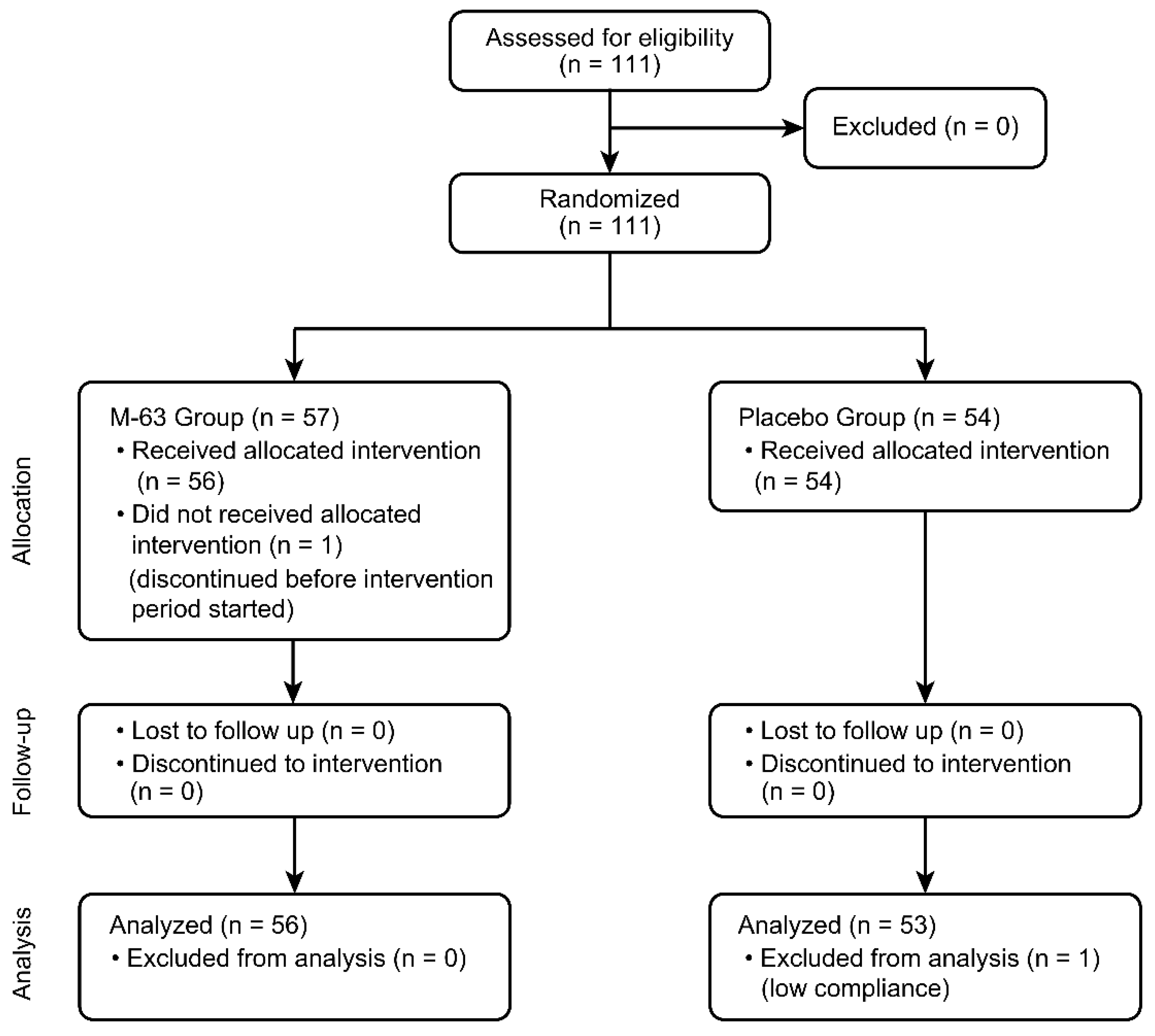
Figure 1. CONSORT flow diagram showing participant flow through the trial.
Nutrients 15 01402 g001
Figure 2. Enterotype of subjects during the intervention. (a) Principal coordinate analysis (PCoA) as seen through the first two principal coordinates (PCo1 and PCo2) based on Jensen–Shannon distance (JSD) calculated by the fecal microbiota composition at the ASV level; (b) PAMs clustering with JSD. The estimated suitability of the cluster number Calinski–Harabasz index; (c) top 20 ASVs in the fecal microbiota composition in each enterotype; (d) relative abundance of ASVs annotated as the B. longum group, including B. longum subsp. infantis and B. longum subsp. longum; (e) the relative abundance of ASVs annotated as Enterobacteriaceae; (f) the relative abundance of ASV annotated as Lactococcus. Asterisks (*) indicate q < 0.05 calculated by ALDEx2.
Nutrients 15 01402 g002
Figure 3. Dynamics of enterotype in each group during the intervention. PCoA of fecal microbiota in each group based on JSD calculated by the fecal microbiota composition at the ASV level. (a) Before ingestion; (b) 1 week after ingestion; (c) 1 month; (d) 3 months of age. Blue and red symbols indicate subjects in the placebo and M-63 groups, respectively.
Nutrients 15 01402 g003
Figure 4. Correlation between the breastfeeding rate and the relative abundance of Bifidobacterium at 1 month of age. Red squares () indicate the M-63 group, and blue circles () indicate the placebo group. Spearman’s correlation coefficients and the corresponding p values are shown.
Nutrients 15 01402 g004
Figure 5. Number of infant stools and consistency. (a) PPS population; (b) exclusively breastfed infants; (c) mixed-fed infants and exclusively formula-fed infants. ** p < 0.01 vs. placebo group (Wilcoxon rank sum test).
Nutrients 15 01402 g005
Table 1. Subject background of infants and mothers.
Placebo (n = 53)M-63 (n = 56)p Value
Infant
Gestational age, weeks39.0 ± 0.239.1 ± 0.20.704 a
Singleton, n (%)53 (100)56 (100)-
Sex, n male/female (%)24/29 (45.3/54.7)28/28 (50.0/50.0)0.702 b
Birth weight, g3037.5 ± 43.63058.7 ± 35.40.706 a
Birth height, cm49.2 ± 0.249.5 ± 0.20.319 a
Birth head circumference, cm33.4 ± 0.233.5 ± 0.20.496 a
Cesarean births, n (%)8 (15.1)11 (19.6)0.618 b
APGAR score (5 min after birth)8.96 ± 0.059.07 ± 0.040.087 a
Maternal
Maternal age, years31.8 ± 0.731.3 ± 0.60.534 a
Multiparous woman, n (%)36 (68.0)32 (57.1)0.323 b
Prepregnancy body mass index (BMI)21.0 ± 0.421.8 ± 0.40.119 a
Pregnancy weight gain, kg9.9 ± 0.49.7 ± 0.50.759 a
Antibiotics during labor, n (%)30 (56.6)30 (53.6)0.848 b
Pregnancy smoking habits, n (%)3 (5.7)3 (5.4)1.00 b
Values are represented as the mean ± SEM, a Student’s t test, b Fisher’s exact test.
Table 2. The relative abundance of Bifidobacterium and percentage of infants in which Bifidobacterium was the most dominant genus.
Relative Abundance of Bifidobacterium (%)Infants Where Bifidobacterium Is The Most Dominant Genus, n (%)
PlaceboM-63p Value aPlaceboM-63p Value b
PPS population
Before ingestion18.7 ± 4.017.1 ± 3.40.53116 (30.2)15 (26.8)0.832
1 week after ingestion28.3 ± 3.863.8 ± 2.2<0.00122 (41.5)53 (94.6)<0.001
1 month of age35.8 ± 4.171.0 ± 2.5<0.00127 (51.0)53 (94.6)<0.001
3 months of age44.3 ± 3.364.5 ± 3.0<0.00136 (68.0)47 (83.9)0.072
Vaginal delivery
Before ingestion20.2 ± 4.218.4 ± 3.80.66915 (33.3)13 (29.0)0.82
1 week after ingestion29.8 ± 4.262.1 ± 2.5<0.00120 (44.4)42 (93.3)<0.001
1 month of age37.6 ± 4.469.4 ± 2.9<0.00124 (53.3)42 (93.3)<0.001
3 months of age43.5 ± 3.763.4 ± 3.4<0.00129 (64.4)38 (84.4)0.052
Cesarean section
Before ingestion10.3 ± 10.211.8 ± 8.00.5601 (12.5)2 (18.2)1.00
1 week after ingestion20.4 ± 8.370.7 ± 4.2<0.0012 (25.0)11 (100.0)0.001
1 month of age26.0 ± 12.177.7 ± 3.50.0043 (37.5)11 (100.0)0.005
3 months of age48.5 ± 9.869.0 ± 6.20.0997 (87.5)9 (81.8)1.00
Not using antibiotics during labor
Before ingestion27.5 ± 5.527.3 ± 5.40.99211 (47.8)11 (42.3)0.778
1 week after ingestion37.8 ± 5.964.4 ± 3.80.00114 (60.9)25 (96.2)0.003
1 month of age40.5 ± 5.767.6 ± 4.3<0.00112 (52.2)23 (88.5)0.010
3 months of age42.7 ± 5.162.4 ± 4.80.00614 (60.9)21 (80.8)0.205
Using antibiotics during labor
Before ingestion12.0 ± 5.28.3 ± 3.60.3245 (16.7)4 (13.3)1.00
1 week after ingestion21.1 ± 4.663.3 ± 2.5<0.0018 (26.7)28 (93.3)<0.001
1 month of age32.2 ± 5.274.0 ± 2.6<0.00115 (50.0)30 (100.0)<0.001
3 months of age44.1 ± 4.566.4 ± 3.80.00122 (73.3)26 (86.7)0.333
Breast-fed infants
1 week after ingestion36.1 ± 10.368.0 ± 4.40.0224 (57.1)6 (100.0)0.192
1 month of age45.5 ± 8.581.3 ± 1.8<0.0017 (63.6)19 (100.0)0.012
3 months of age49.5 ± 5.672.9 ± 2.7<0.00118 (75.0)32 (97.0)0.034
Mixed-fed infants and formula-fed infants
1 week after ingestion27.2 ± 4.163.3 ± 2.4<0.00118 (39.1)47 (94.0)<0.001
1 month of age33.3 ± 4.765.7 ± 3.3<0.00120 (47.6)34 (91.9)<0.001
3 months of age38.5 ± 3.952.5 ± 5.30.05218 (62.1)15 (65.2)1.00
Values are represented as the mean ± SEM, p < 0.05 indicates statistical significance, a Wilcoxon rank sum test, b Fisher’s exact test.
Table 3. Abundance and detection rate of Bifidobacterium species.
Abundance (Log10 CFU/g Feces)Number of Infants Where Each Bifidobacterium Was Detected (Detection Rate, %)
PlaceboM-63p Value aPlaceboM-63p Value b
Genus Bifidobacterium
Before ingestion7.57 ± 0.277.85 ± 0.270.48724 (45.3)29 (51.2)0.567
1 month of age9.10 ± 0.2910.73 ± 0.11<0.00138 (71.7)55 (98.2)<0.001
Bifidobacterium bifidum
Before ingestion6.20 ± 0.126.25 ± 0.110.3743 (5.7)6 (10.7)0.490
1 month of age6.42 ± 0.176.52 ± 0.170.4256 (11.3)10 (17.9)0.421
Bifidobacterium breve
Before ingestion6.50 ± 0.166.68 ± 0.180.54310 (18.9)13 (23.2)0.643
1 month of age7.36 ± 0.257.07 ± 0.190.52321 (39.6)24 (42.9)0.846
Bifidobacterium longum
Before ingestion6.85 ± 0.197.19 ± 0.240.19215 (28.3)21 (37.5)0.318
1 month of age7.39 ± 0.257.22 ± 0.200.73524 (45.3)27 (48.2)0.848
Bifidobacterium infantis
Before ingestion6.15 ± 0.116.00 ± 0.000.1442 (3.8)0 (0)0.234
1 month of age6.35 ± 0.1710.36 ± 0.13<0.0014 (7.6)54 (96.4)<0.001
Values are represented as the mean ± SEM, p < 0.05 indicates statistical significance, a Wilcoxon rank sum test, b Fisher’s exact test.
Table 4. pH, amount of short-chain fatty acid, IgA, and calprotectin in the stools.
PlaceboM-63p Value 1
pH
Before ingestion5.92 ± 0.086.05 ± 0.080.209
1 month of age6.05 ± 0.115.53 ± 0.07<0.001
Short-chain fatty acids (µmol/g feces)
acetic acid
Before ingestion16.09 ± 1.4915.45 ± 1.160.884
1 month of age22.01 ± 1.6230.0 ± 1.44<0.001
propionic acid
Before ingestion0.54 ± 0.260.64 ± 0.310.644
1 month of age2.32 ± 0.662.54 ± 0.690.404
n-butanoic acid
Before ingestion0.43 ± 0.360.75 ± 0.340.105
1 month of age0.49 ± 0.170.15 ± 0.110.027
iso-butanoic acid
Before ingestion00.01 ± 0.010.326
1 month of age0.06 ± 0.020.08 ± 0.040.734
n-valeric acid
Before ingestion001.00
1 month of age0.02 ± 0.020.01 ± 0.010.931
iso-valeric acid
Before ingestion0.01 ± 0.010.01 ± 0.010.989
1 month of age0.15 ± 0.090.06 ± 0.030.795
n-caproic acid
Before ingestion0.01 ± 0.010.01 ± 0.010.989
1 month of age0.01 ± 0.0100.317
Calprotectin (µg/g feces)
Before ingestion277.06 ± 35.16346.07 ± 54.220.535
1 month of age212.54 ± 30.0257.52 ± 37.460.537
IgA (µg/g feces)
Before ingestion1528.85 ± 311.531700.63 ± 373.620.661
1 month of age1393.19 ± 152.911971.25 ± 200.900.033
All values are represented as the mean ± SEM, p < 0.05 indicates statistical significance. 1 Wilcoxon rank sum test.
Disclaimer/Publisher’s Note: The statements, opinions and data contained in all publications are solely those of the individual author(s) and contributor(s) and not of MDPI and/or the editor(s). MDPI and/or the editor(s) disclaim responsibility for any injury to people or property resulting from any ideas, methods, instructions or products referred to in the content.