Article

Kaempferitrin: A Flavonoid Marker to Distinguish Camellia oleifera Honey

1
Honeybee Research Institute, Jiangxi Agricultural University, Nanchang 330045, China
2
Jiangxi Province Key Laboratory of Honeybee Biology and Beekeeping, Jiangxi Agricultural University, Nanchang 330045, China
*
Author to whom correspondence should be addressed.
Nutrients 2023, 15(2), 435; https://doi.org/10.3390/nu15020435
Submission received: 3 December 2022 / Revised: 5 January 2023 / Accepted: 12 January 2023 / Published: 14 January 2023
(This article belongs to the Section Nutrition and Public Health)

Abstract

:
C. oleifera is an economically important oilseed crop and medical plant. However, as a characteristic honey resource, the standard protocol used to identify the composition of C. oleifera honey has not been established yet. Previously, distinctive flavonoid has been shown as an effective marker to trace the botanical origin of honey. In this study, we examined the flavonoid types in C. oleifera honey and nine other monofloral honeys by using liquid chromatography tandem-mass spectrometry (LC-MS/MS) and compared the differences and identified eight distinct flavonoids in C. oleifera honey. Then, comparing the 8 flavonoids with the 14 flavonoids common to C. oleifera honey and nectar, two distinct flavonoids were identified in C. oleifera honey and nectar. Finally, we identified kaempferitrin as the distinct flavonoid marker in C. oleifera honey using the degree of influence of the partial least-squares discriminant analysis (PLS-DA) model on C. oleifera honey and ployfloral honey.

1. Introduction

Honey is a sweet substance that is produced from the nectar of flowers, which is collected by foraging honeybees and mixed with the secreted enzyme and then stored in the hive comb until thoroughly mature [1]. Honeybees can collect nectar from one or more plants to make honey; thus, honey can be classified as monofloral or polyfloral (multi-floral) honey [2]. Honey composition comprises more than 200 different components such as sugar, water, organic acids, minerals, enzymes, proteins, vitamins, ash, polyphenolic compounds, and plant derivatives, etc., [3,4]. The chemical composition, color, and flavor of honey varies depending on the environment, where the plants were grown, and their geographical location, as well as being affected by weather conditions, processing, handling, packaging, and storage time [5].
Honey manifests a variety of medicinal and health benefits as a natural food supplement with a long history of utilization [6]. Honey was first registered as a topical pharmaceutical preparation in Australia in 1999; since then, a range of honey-based products have become available, including sterile Manuka honey ointments and dressings containing honey [6]. Currently, the study of the medical value of medicinal honey extracted from special medicinal plants is a popular research topic [7].
C. oleifera is one of the most valuable economic woody crops and medical plant grown in Asia and has been cultivated for more than 2300 years [8]. The main profit driver of growing C. oleifera is to obtain the C. oleifera seeds and the camellia oil with a highly economical value. C. oleifera seeds are abundant in a multitude of bioactive compounds, which have the effect of preventing cardiovascular diseases such as hypertension, coronary heart disease, and atherosclerosis [9]. Camellia oil is acquired from the seeds of C. oleifera with a superior color, aroma, and taste, and is commonly regarded as an excellent quality oil because it is easily absorbed and digested by the human body, with a variety of biological activities such as lowering blood pressure, blood lipids, and the softening blood vessels; moreover, the long-term consumption can enhance human immunity, etc., [10]. In addition, camellia oil has powerful antioxidant activity and can serve as a traditional medicine to prevent liver damage and gastrointestinal ulcers caused by oxidative stress [11].
C. oleifera honey is derived from nectar collected by honeybees foraging on C. oleifera Abel. plants. Previous studies have found that C. oleifera honey contains oligosaccharides (manninotriose, raffinose, and stachyose), and it can lead to the death of honeybee larvae and adult worker bees [12,13]. Raffinose and stachyose are classified as raffinose family oligosaccharides (RFOs), one type of prebiotic that has biological functions such as regulating gut flora, preventing inflammatory bowel disease, protecting the liver, and lowering blood sugar and blood lipids, etc., [14,15,16,17]. This implies the promising use of C. oleifera honey for the development of potential health promoters and dietary supplements, which is a prioritized direction for subsequent research in our laboratory.
Once the special pharmacological effects of C. oleifera honey that are beneficial for health-related functions are proven, then the commercial value of C. oleifera honey will increase dramatically. In addition, the wealth of C. oleifera growers and beekeepers will also increase owing to the side industry of C. oleifera honey. After the commercial value of C. oleifera honey has increased, its authenticity is well worth studying [18]. This is because the danger of adulterated honey is not only the use of cheap honey as high-priced honey to achieve a higher value, but, even more so, it will damage the health of consumers. First and foremost, we can initially identify the authenticity of C. oleifera honey in terms of its oligosaccharide components and concentration; however, there are limitations to this discriminatory approach. As the RFOs are highly water soluble, it would be feasible to isolate them from Glycine max, Stachys floridana, and Stachys sieboldii and then blend them into common honey [19], posing as C. oleifera honey. However, accumulating evidence reveals that the abundant but trace amounts of flavonoids (natural secondary metabolites derived from plants) in honey provide their own distinctive chemical markers [7,20,21]. Hence, the distinct flavonoid markers to identify the authenticity of C. oleifera honey is a reliable and novel strategy.
In order to identify the distinctive flavonoid markers in C. oleifera honey to facilitate the discrimination of C. oleifera honey from other commercial honeys, in the present study, we employed the liquid chromatography tandem-mass spectrometry (LC-MS/MS) technique to identify the types and absolute contents of flavonoid compounds in C. oleifera honey, as well as in nine other kinds of monofloral honey and one polyfloral honey. Additionally, the flavonoid species of C. oleifera honey with that of C. oleifera nectar from the parent plant were identified and applied using partial least-squares discriminant analysis (PLS-DA).

2. Materials and Methods

2.1. Chemicals and Reagents

Liquid-chromatography mass spectrometry (LC-MS) grade methanol and acetonitrile were purchased from Merck (Darmstadt, Germany). Formic acid (LC-MS grade) was obtained from Sigma–Aldrich (St. Louis, MO, USA). Fructose, glucose, sucrose, melibiose, manninotriose, raffinose, stachyose, and 5-hydroxymethylfurfural (5-HMF) standards (purity > 98%) were obtained from Sigma (St. Louis, MO, USA). Ultra-pure water from MilliQ-system (Millipore Corporation, Billerica, MA, USA) was used throughout the study. Next, 204 flavonoid standards (purity > 98%) were acquired from MedChemExpress Company (Shanghai, China), see details in Supporting Table S1.

2.2. Honey and Nectar Samples’ Collection

The mature C. oleifera honey (COH) was collected by honeybees (Apis mellifera) placed at the C. oleifera plantation in Shengqiao Town, Changning City, Hunan Province, and the sample time was from October to November 2021. The C. oleifera nectar (CON) was collected using a micro aspirator (Beijing Dalong Xingchuang Experimental Instrument Co., Beijing, China) in Shengqiao Town from three mother plants of C. oleifera. Moreover, the sampling schedule was October 2021. Citrus reticulata honey (CRH), Vitex negundo honey (VNH), Eriobotrya japonica honey (EJH), Litchi chinensis Sonn honey (LCSH), Lycium chinense Miller honey (LCMH), Ziziphus jujuba honey (ZJH), Tilia tuan honey (TTH), Brassica napus honey (BNH), Robinia pseudoacacia honey (RPH), nine types of monofloral honey, and one polyfloral honey were provided by Wuhan Baochun Bee Products Company (Wuhan, China). All honey or nectar samples were set up with three biological replicates and stored at −18 °C for subsequent analysis.

2.3. Honey and Nectar Preparation

Next, 0.2 g (±0.01 g) of the honey or nectar sample was weighed accurately in a 10 mL capacity centrifuge tube with screw-on caps, and 100 μL of the internal standard working solution at a concentration of 4000 nmol/L and 5000 μL of the 70% methanol solution were added. After 30 min of ultrasound, the samples were centrifuged (12,000 r/min for 5 min at room temperature). Finally, the supernatant was aspirated and filtered through a 0.22 μm filter membrane and 800 μL was transferred to a 1.5 mL injection vial for LC-MS/MS analysis.

2.4. Analysis the Chemical Parameters of C. oleifera Honey

2.4.1. Analysis Methods for the Sugar Composition in C. oleifera Honey

Thermo ICS 5000 liquid chromatography with an electrochemical detector (Thermo Fisher Technology Inc., Waltham, MA, USA) and a CarboPac PA20 liquid chromatographic column (150 × 3.0 mm, 4 μm) was employed for the analysis of sugar composition in honey samples. The mobile phases were A: H2O, B: 100 mM NaOH; the injection volume was 5 μL, the flow rate was 0.5 mL/min, and the column temperature was 30 °C. Elution gradient: 0.0~9 min, 5% B; 9~20 min, 5~100% B; 20~30 min, 100% B; 30~30.1 min, 100~5% B; 30.1~60 min, 5% B.

2.4.2. Analysis Methods for the Water, Acidity, and 5-HMF Composition in C. oleifera Honey

The water content of the honey samples was determined by reading the refractive index of each sample using an Abbe refractometer and brought into the formula: moisture (%) = 100 − [78 + 390.7 × (n − 1.4768)] to calculate the water content. Where n is the actual refractive index of the specimen honey measured at 40 °C.
Next, 4 g of sodium hydroxide was dissolved in 1 L of boiled and cooled water and its concentration was calibrated with potassium hydrogen phthalate (reference reagent) according to the following method: weigh the potassium hydrogen phthalate (reference reagent 0.8~0.9 g (accurate to 0.0002 g) that has been dried in advance at 125 °C, place it in a 250 mL conical flask, dissolve it in 50 mL of boiled and cooled water, and add 2~3 drops of 1% phenolphthalein. Add 2~3 drops of 1% phenolphthalein indicator and titrate with sodium hydroxide solution until the solution is pink, and the end point is that the color does not fade within 10 s.
The concentration of sodium hydroxide standard solution (mol/L) = 0.2042 m/v.
The meaning of the letters in the formula.
c: a concentration of sodium hydroxide standard solution (mol/L).
m: the mass of potassium hydrogen phthalate (g).
v: the volume of sodium hydroxide standard solution consumed at dropwise intervals (mL).
0.2042: a mass of potassium hydrogen phthalate per mL of standard solution of sodium hydroxide [c (NaOH) = 1.000 mol/L] (g).
Weigh 10 g of the honey sample (accurate to 0.001 g). Dissolve in 75 mL of boiled and cooled water, add 2–3 drops of phenolphthalein indicator; titrate with sodium hydroxide standard solution until the solution is pink and does not fade within 10 s as the end point.
The sample acidity (mL/kg) = CV100/m.
v: titration of the volume of sodium hydroxide standard solution was consumed (mL).
c: the molar concentration of sodium hydroxide standard solution (mol/L).
m: the mass of the sample (g).
Note: if the color of honey is too dark, weigh the sample 5 g, or use thymol blue indicator instead of phenolphthalein indicator.
The determination of 5-HMF in C. oleifera honey samples was performed on an Agilent 1260 Infinity II liquid chromatography workstation equipped with a Proshell SB C18 column (4.6 × 150 mm, 3.5 μm) (Agilent Technology Inc., Santa Clara, CA, USA). The flow rate was 0.2 m L/min, the column temperature was 30 °C, and the injection volume was 10 μL. Using methanol:water = 8:92 (v:v) as the mobile phase, the detection limit (LOD, S/N = 3) of 5-HMF was obtained as 12 mg/kg at 284 nm UV wavelength.

2.5. LC-MS/MS Method for the Determination of Flavonoids in Honey and Nectar

Regarding, the flavonoid standards’ preparation and construction of the standard curve, the 204 flavonoid standards were weighed and prepared into a master batch of 10 mmol/L by methanol–water (70:30) (the concentration of all 204 standards was 10 mmol/L). After that, the master batch was diluted with methanol–water (70:30) and formulated into standard curve working solutions of 0.5 nmol/L, 1 nmol/L, 5 nmol/L, 10 nmol/L, 20 nmol/L, 50 nmol/L, 100 nmol/L, 200 nmol/L, 500 nmol/L, 1000 nmol/L, 2000 nmol/L. Moreover, 100 μL of the internal standard working solution (daidzein) with a concentration of 4000 nmol/L was required and added in each working solution, and the ultimate volume of each working solution was 5 mL. The mass spectral peak intensity data of the corresponding quantitative signals of each concentration standard working solution were acquired. With the concentration ratio of the external standard to the internal standard as the horizontal coordinate and the area ratio of external standard to internal standard ratio as the vertical coordinate, the standard curves of different substances were plotted. The resulting 204 flavonoid standards curve demonstrated a good linearity from 0.5 nmol/L to 200 nmol/L (R2 ≥ 0.9900). Results are shown in Supporting Table S2.

2.6. LC-MS/MS Analysis

Quantification of flavonoids in honey or nectar was performed on an ultra-performance liquid chromatography system (ExionLC™ AD) coupled with tandem mass spectrometry (QTRAP® 6500+).
Separations were carried out using a Waters ACQUITY UPLC HSS T3 C18 column (1.8 µm, 100 mm × 2.1 mm, Waters, Milford, MA, USA). Analytes were separated using gradient elution with water (containing 0.05%, v/v formic acid) (A) and acetonitrile (containing 0.05% formic acid, v/v) (B) at a flow-rate of 0.35 mL/min. The linear gradient elution program was: 0.0~1.0 min, 10~20% B; 1.0~9.0 min, 20~70% B; 9.0~12.5 min, 70~95% B; 12.5~13.5 min, 95% B; 13.5~13.6 min, 95~10% B, 13.6~15 min, 10% B. The column was thermostated at 40 °C and injection volume was 2 μL. The electrospray ionization (ESI) source temperature was 550 °C, while the mass spectrometry voltage was 5500 V in positive ion mode, −4500 V in negative ion mode, and 35 psi of curtain gas (CUR). In the Q-Trap 6500+, each ion pair was scanned for detection based on the optimized declustering potential (DP) and collision energy (CE).

2.7. Conversion of Flavonoid Amounts in Honey and Nectar Samples

After substituting the integrated peak area ratio of all identified samples into the linear equation of the standard curve for calculation, and further substituting the calculation formula to calculate, the final data of the content of the substance in the actual sample was attained.
The amounts of flavonoids in the sample (nmol/g) = cV/1,000,000/m.
The meaning of the letters in the formula.
c: the sample concentration value (nmol/L) obtained by substituting the integral peak area ratio of the sample into the standard curve.
V: the volume of the solution used in the extraction (μL).
m: the mass of the sample weighed (g).

2.8. Data Processing

MultiQuant 3.0.3 software (AB SCIEX) was used to process the mass spectrometry data, and the retention time and peak shape information of the standards were referenced to guarantee the accuracy of the qualitative quantification by integrating and correcting the mass spectrometry peaks detected in different samples for the analytes. PLS-DA was fulfilled viva Wekemo Bioincloud (Shenzheng, China). Data are expressed as mean ± standard deviation (SD).

3. Results and Discussion

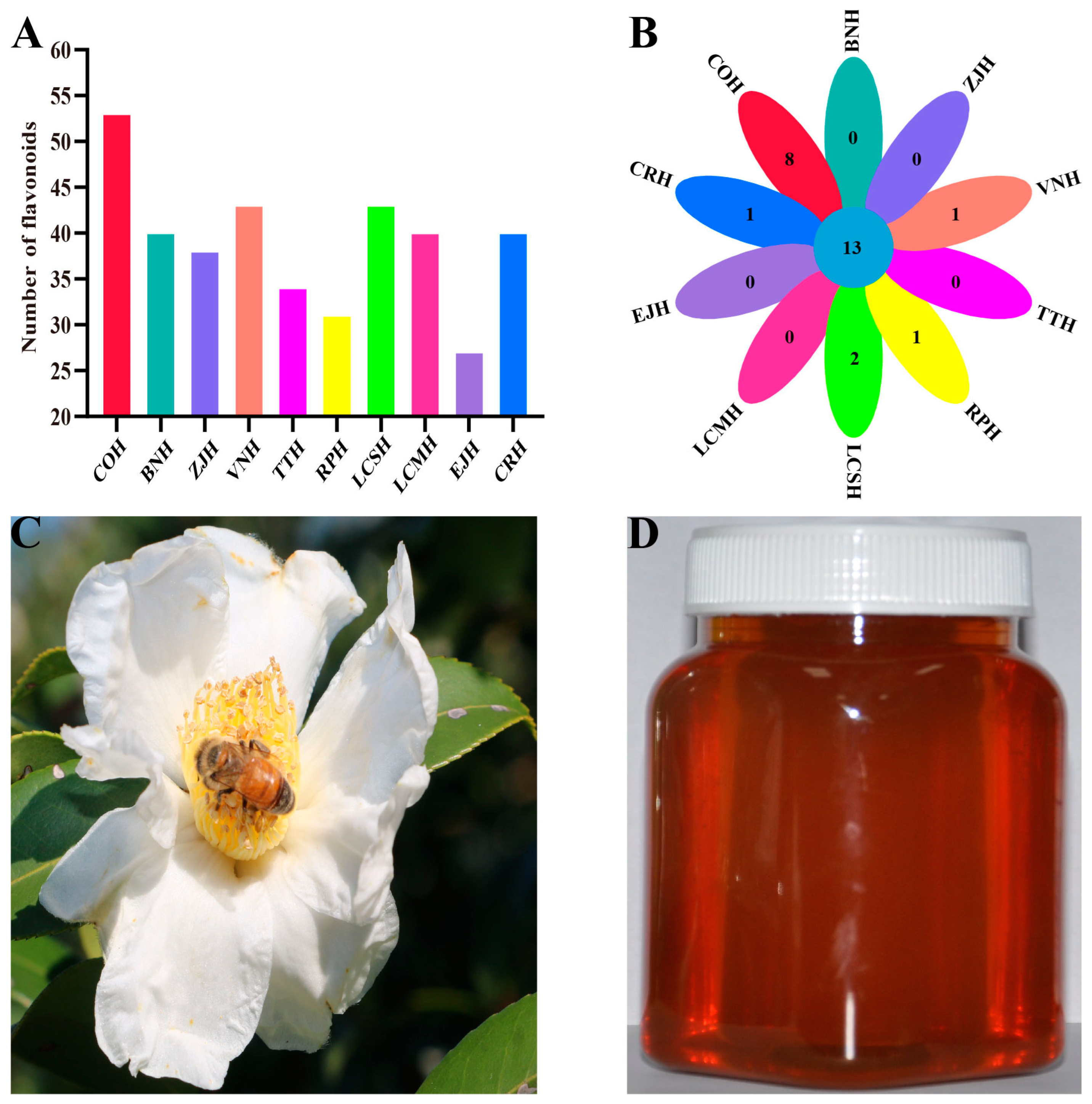
3.1. Differences in Flavonoid Species among C. oleifera Honey and Nine Kinds of Monofloral Honey

Table 1 shows the basic parameters of C. oleifera honey, in which the content of total reducing sugar was 65.71% (fructose content 38.27%, glucose content 27.44%), sucrose content 1.56%, and moisture content 17.62%, all of which were in accordance with European Union honey standards [7]. The harmful hydroxymethyl furfural was not detected in C. oleifera honey. Moreover, apart from the common fructose, glucose, and sucrose, C. oleifera honey also contains a minor amount of melibiose and manninotriose and a higher content of raffinose and stachyose. These parameters indicate that C. oleifera honey is a high-quality honey and has great potential to regulate the gut [19].
There were 54 flavonoids detected in C. oleifera honey using LC-MS/MS, which was higher than the remaining nine monofloral honeys (Figure 1A). Likewise, eight distinct flavonoids in C. oleifera honey were found, including kaempferitrin, phloretin, acacetin, scutellarein tetramethyl ether, 5,7-dihydroxy-3,4,5-trimethoxyflavone, scutellarin, sinensetin, and tectorigenin (Figure 1B). These differences in the composition and content of flavonoid compounds in different monofloral nectars were predominantly attributed to the pollen of nectar plants [22]. The blossom size of C. oleifera is about over six times larger than that of the nectar flowers of common plants and has a large amount of nectar and pollen (Figure 1C). Thus, the honeybees collect more pollen when collecting C. oleifera nectar, resulting in a greater variety of flavonoid compounds in C. oleifera honey.

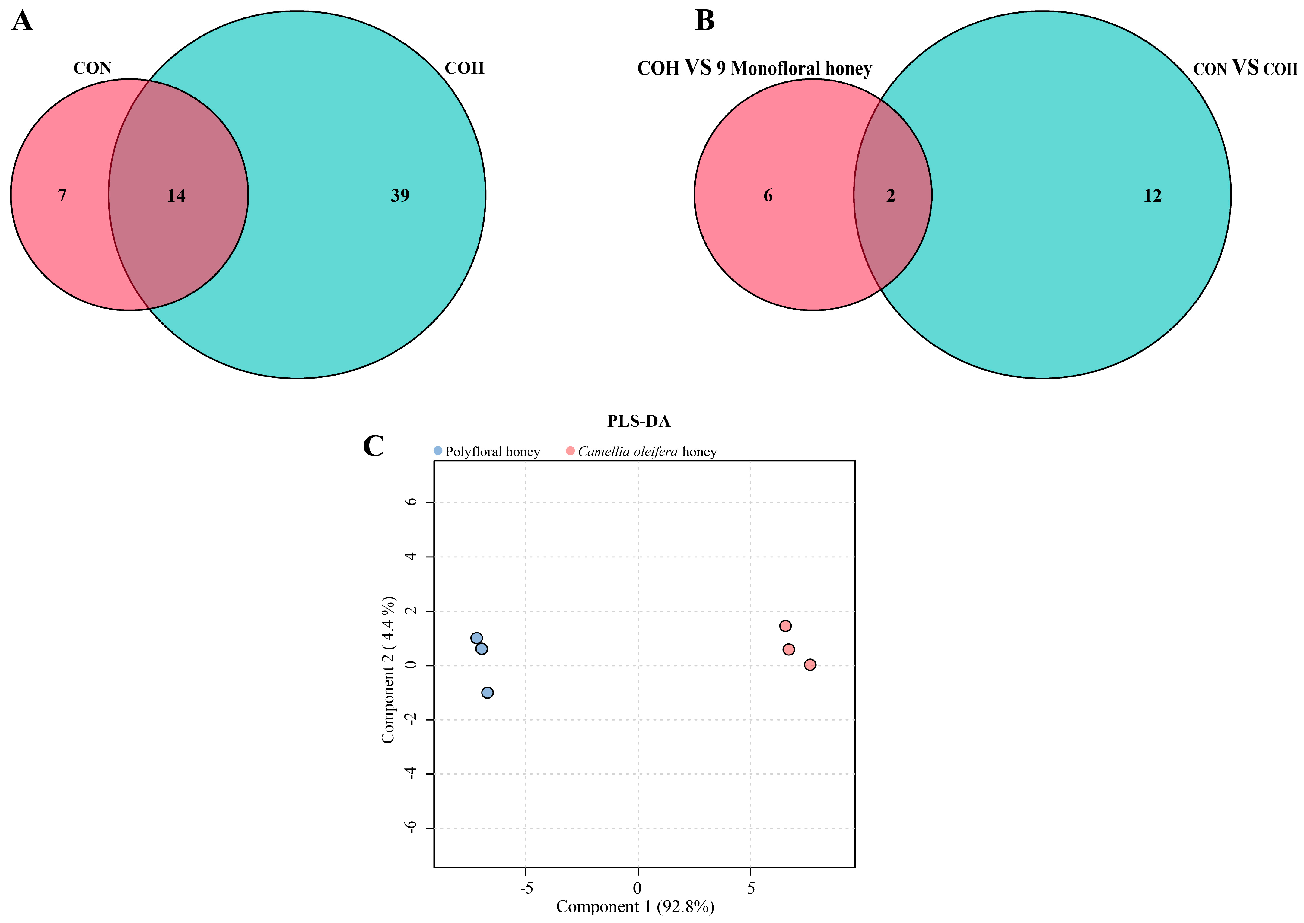
3.2. Identification of the Distinctive Flavonoid Marker in C. oleifera Honey

Flavonoids are a large family of phenolic pigments that are natural secondary metabolites derived from plants [23]. Flavonoids in C. oleifera honey originate from the nectar of C. oleifera and the pollen blended with the nectar. We further identified 21 flavonoid species in C. oleifera nectar utilizing LC-MS/MS (Figure 2A). There were 14 flavonoids shared between C. oleifera honey and C. oleifera nectar (Figure 2A). After further comparison of the 14 flavonoids in common with the 8 flavonoids formerly unique to C. oleifera honey relative to the 9 kinds of monofloral honey, two flavonoids (kaempferitrin and phloretin) unique to C. oleifera honey and nectar were identified (Figure 2B).
Moreover, we constructed one PLS-DA model based on the types and contents of flavonoids included in C. oleifera honey and polyfloral honey (Figure 2C). C. oleifera honey and polyfloral honey were well separated in the model, again illustrating that the use of flavonoid components to distinguish honey of different plant origin is a very feasible approach. The variable importance in the projection (VIP) value of kaempferitrin was 1.052, indicating that it had a greater effect on the PLS-DA model between polyfloral and C. oleifera honey samples (VIP > 1 is typically regarded as having the great impact on the model) (Supporting Table S3). In contrast, the VIP value of phloretin was 0.968, which had less effect on the PLS-DA model (Supporting Table S3). More importantly, phloretin was present in polyfloral honey as well. Finally, we identified the distinct flavonoid marker in C. oleifera honey as kaempferitrin. The chromatogram and mass spectra of kaempferitrin in C. oleifera honey and nectar are shown in Figure 3.

3.3. Kaempferitrin Quantification and Method Validation

C. oleifera, known as the source of camellia oil, is also a versatile plant. For instance, besides being a nutritious edible oil, camellia oil can also be used as anti-rust oil and lubricant for industrial purposes; the pressed C. oleifera cake is both a natural fungicide and fertilizer, and the seed peel of C. oleifera is a raw material for extracting tannin extract [24]. Kaempferitrin is mainly distributed in the new leaf buds of Camellia sinensis, and it exhibits analgesic, anti-inflammatory, antidiabetic, antitumor, and chemotherapeutic effects, as well as activating insulin signaling [25,26]. Therefore, the examination of kaempferitrin in C. oleifera honey not only enables the authentication of C. oleifera honey, it also has realistic value for the evaluation of biological activity of C. oleifera honey.
Here, a LC-MS/MS method was developed to detect of kaempferitrin: first, a standard curve was established using kaempferitrin standards, and then the intensity of kaempferitrin parent ions in C. oleifera honey and nectar samples could be quantified upon this curve and LC-MS/MS. We constructed a standard curve of kaempferitrin with good linearity (regression coefficient = 0.9989) in the range of 0.5–2000 nmol/L as y = 10,383.8424 x + 1676.5839 (Table 2). The limits of detection (LOD, signal to noise ratio = 3) and quantification (LOQ, signal to noise ratio = 10) of kaempferol were 0.07 nmol/kg and 0.25 nmol/kg, respectively (Table 2), and our determination results indicated that the honey and nectar of C. oleifera contain 5.98 ± 0.84 and 2.36 ± 0.82 nmol/kg of kaempferitrin, respectively (Table 2). To further validate the method, the RSD of C. oleifera honey and nectar were calculated as 1.23% and 1.38%, respectively (Table 2). Overall, the method is sensitive and reliable for the detection of kaempferitrin.

4. Conclusions

C. oleifera honey is one of the new byproducts of the C. oleifera industry, which boosts the income of beekeepers and C. oleifera growers and shows huge potential to serve as a medicinal honey. In this experiment, by comparing the flavonoid differences between C. oleifera honey and nine monofloral commercial honeys, kaempferitrin was identified as the distinct flavonoid marker of C. oleifera honey. An LC-MS/MS method was also developed to detect the content of kaempferitrin in C. oleifera honey and nectar samples. The identification of the distinctive flavonoid markers has a practical application for the authentication of C. oleifera honey.

Supplementary Materials

The following supporting information can be downloaded at: https://www.mdpi.com/article/10.3390/nu15020435/s1, Table S1: Standard information for 204 flavonoids; Table S2: Standard working curve information for 204 flavonoids; Table S3: Partial least-squares discriminant analysis (PLS-DA) results.

Author Contributions

Collecting nectar and honey samples, detection of samples, writing original manuscripts, Z.L.; improving manuscripts, Q.H.; collecting nectar and honey samples, detection of samples, Y.Z. (Yu Zheng); help with data analysis, Y.Z. (Yong Zhang); help with doing the experiment in the lab, B.L. and W.S.; resources, funding acquisition, improving manuscripts, Z.Z. All authors have read and agreed to the published version of the manuscript.

Funding

This research was funded by the Earmarked Fund for the China Agricultural Research System (Grant Number: CARS-44-KXJ15) and the National Natural Science Foundation of China, grant number (Grant Number: 32172790).

Institutional Review Board Statement

Not applicable.

Informed Consent Statement

Not applicable.

Data Availability Statement

The data in this study are available upon reasonable request.

Conflicts of Interest

The authors declare that they have no known competing financial interests or personal relationships that could have appeared to influence the work reported in this paper.

References

  1. Downey, G.; Hussey, K.; Kelly, J.D.; Walshe, T.F.; Martin, P. Preliminary contribution to the characterisation of artisanal honey produced on the island of Ireland by palynological and physico-chemical data. Food Chem. 2005, 91, 347–354. [Google Scholar] [CrossRef]
  2. Ramsay, E.I.; Rao, S.; Madathil, L.; Hegde, S.K.; Baliga-Rao, M.P.; George, T.; Baliga, M.S. Honey in oral health and care: A mini review. J. Oral Sci. 2019, 61, 32–36. [Google Scholar] [CrossRef] [PubMed]
  3. El Sohaimy, S.A.; Masry, S.; Shehata, M. Physicochemical characteristics of honey from different origins. Ann. Agric. Sci. 2015, 60, 279–287. [Google Scholar] [CrossRef] [Green Version]
  4. Valenzuela, R.; Das, U.N.; Videla, L.A.; Llorente, C.G. Nutrients and diet: A relationship between oxidative stress, aging, obesity, and related noncommunicable diseases. Oxid. Med. Cell. Longev. 2018, 2018, 7460453. [Google Scholar] [CrossRef] [PubMed]
  5. da Silva, P.M.; Gauche, C.; Gonzaga, L.V.; Costa, A.C.O.; Fett, R. Honey: Chemical composition, stability and authenticity. Food Chem. 2016, 196, 309–323. [Google Scholar] [CrossRef] [PubMed]
  6. Ahmed, S.; Sulaiman, S.A.; Baig, A.A.; Ibrahim, M.; Liaqat, S.; Fatima, S.; Jabeen, S.; Shamim, N.; Othman, N.H. Honey as a potential natural antioxidant medicine: An insight into its molecular mechanisms of action. Oxid. Med. Cell. Longev. 2018, 2018, 8367846. [Google Scholar] [CrossRef] [Green Version]
  7. Zhao, L.; Ren, C.; Xue, X.; Lu, H.; Wang, K.; Wu, L. Safflomin A: A novel chemical marker for Carthamus tinctorius L. (Safflower) monofloral honey. Food Chem. 2022, 366, 130584. [Google Scholar] [CrossRef] [PubMed]
  8. Song, Q.; Ji, K.; Yu, X.; Chen, L.; Wang, L.; Gong, W.; Yuan, D. Dynamic metabolic and transcriptomic profiling reveal synthetic characters and regulators of flavonoid biosynthesis in Camellia oleifera seeds. Ind. Crops Prod. 2022, 186, 115295. [Google Scholar] [CrossRef]
  9. Lee, C.P.; Yen, G.C. Antioxidant activity and bioactive compounds of tea seed (Camellia oleifera Abel.) oil. J. Agric. Food Chem. 2006, 54, 779–784. [Google Scholar] [CrossRef]
  10. Li, H.; Zhou, G.Y.; Zhang, H.Y.; Liu, J.A. Research progress on the health function of tea oil. J. Med. Plant 2011, 5, 485–489. [Google Scholar]
  11. Cheng, Y.T.; Lu, C.C.; Yen, G.C. Beneficial effects of camellia oil (Camellia oleifera Abel.) on hepatoprotective and gastroprotective activities. J. Nutr. Sci. Vitaminol. 2015, 61, S100–S102. [Google Scholar] [CrossRef] [PubMed]
  12. Su, R.; Dong, Y.; Dong, K.; He, S. The toxic honey plant Camellia oleifera. J. Apic. Res. 2012, 51, 277–279. [Google Scholar] [CrossRef]
  13. Li, Z.; Huang, Q.; Zheng, Y.; Zhang, Y.; Li, X.; Zhong, S.; Zeng, Z. Identification of the toxic compounds in Camellia oleifera honey and pollen to honey bees (Apis mellifera). J. Agric. Food Chem. 2022, 70, 13176–13185. [Google Scholar] [CrossRef]
  14. He, L.; Zhang, F.; Jian, Z.; Sun, J.; Chen, J.; Liapao, V.; He, Q. Stachyose modulates gut microbiota and alleviates dextran sulfate sodium-induced acute colitis in mice. Saudi J. Gastroenterol. 2020, 26, 153. [Google Scholar] [CrossRef] [PubMed]
  15. Liu, G.; Bei, J.; Liang, L.; Yu, G.; Li, L.; Li, Q. Stachyose improves inflammation through modulating gut microbiota of high-fat diet/streptozotocin-induced type 2 diabetes in rats. Mol. Nutr. Food Res. 2018, 62, 1700954. [Google Scholar] [CrossRef] [PubMed]
  16. Xi, M.; Zhao, S.; Ge, W.; Chen, Y.; Cui, X.; Sun, Q. Effects of stachyose on the intestinal microbiota and barrier in antibiotic-treated mice. J. Funct. Foods 2021, 83, 104493. [Google Scholar] [CrossRef]
  17. Zhang, R.; Zhao, Y.; Sun, Y.; Lu, X.; Yang, X. Isolation, characterization, and hepatoprotective effects of the raffinose family oligosaccharides from Rehmannia glutinosa Libosch. J. Agric. Food Chem. 2013, 61, 7786–7793. [Google Scholar] [CrossRef]
  18. Siddiqui, A.J.; Musharraf, S.G.; Choudhary, M.I. Application of analytical methods in authentication and adulteration of honey. Food Chem. 2017, 217, 687–698. [Google Scholar] [CrossRef] [PubMed]
  19. Elango, D.; Rajendran, K.; Van der Laan, L.; Sebastiar, S.; Raigne, J.; Thaiparambil, N.A.; El Haddad, N.; Raja, B.; Wang, W.; Ferela, A. Raffinose family oligosaccharides: Friend or foe for human and plant health? Front. Plant Sci. 2022, 13, 829118. [Google Scholar] [CrossRef]
  20. Martos, I.; Ferreres, F.; Tomás-Barberán, F.A. Identification of flavonoid markers for the botanical origin of Eucalyptus honey. J. Agric. Food Chem. 2000, 48, 1498–1502. [Google Scholar] [CrossRef]
  21. Truchado, P.; Ferreres, F.; Bortolotti, L.; Sabatini, A.G.; Tomás-Barberán, F.A. Nectar flavonol rhamnosides are floral markers of acacia (Robinia pseudacacia) honey. J. Agric. Food Chem. 2008, 56, 8815–8824. [Google Scholar] [CrossRef] [PubMed]
  22. Wang, J.; Li, Q.X. Chemical composition, characterization, and differentiation of honey botanical and geographical origins. Adv. Food Nutr. Res. 2011, 62, 89–137. [Google Scholar] [PubMed]
  23. Du Fall, L.A.; Solomon, P.S. Role of cereal secondary metabolites involved in mediating the outcome of plant-pathogen interactions. Metabolites 2011, 1, 64–78. [Google Scholar] [CrossRef] [PubMed] [Green Version]
  24. Luan, F.; Zeng, J.; Yang, Y.; He, X.; Wang, B.; Gao, Y.; Zeng, N. Recent advances in Camellia oleifera Abel: A review of nutritional constituents, biofunctional properties, and potential industrial applications. J. Funct. Foods 2020, 75, 104242. [Google Scholar] [CrossRef]
  25. Varshney, M.; Kumar, B.; Rana, V.S.; Sethiya, N.K. An overview on therapeutic and medicinal potential of poly-hydroxy flavone viz. Heptamethoxyflavone, Kaempferitrin, Vitexin and Amentoflavone for management of Alzheimer’s and Parkinson’s diseases: A critical analysis on mechanistic insight. Crit. Rev. Food Sci. Nutr. 2021, 1–24. [Google Scholar] [CrossRef]
  26. Yao, L.; Jiang, Y.; Datta, N.; Singanusong, R.; Liu, X.; Duan, J.; Raymont, K.; Lisle, A.; Xu, Y. HPLC analyses of flavanols and phenolic acids in the fresh young shoots of tea (Camellia sinensis) grown in Australia. Food Chem. 2004, 84, 253–263. [Google Scholar] [CrossRef]
Figure 1. (A) Flavonoid species identified in C. oleifera honey and 9 monofloral honeys. C. oleifera honey (COH), Citrus reticulata honey (CRH), Vitex negundo honey (VNH), Eriobotrya japonica honey (EJH), Litchi chinensis Sonn honey (LCSH), Lycium chinense Miller honey (LCMH), Ziziphus jujuba honey (ZJH), Tilia tuan honey (TTH), Brassica napus honey (BNH), Robinia pseudoacacia honey (RPH). (B) Flavonoid species distinctive to C. oleifera honey relative to 9 monofloral honeys. (C) Honeybee-visited C. oleifera. (D) C. oleifera honey.
Nutrients 15 00435 g001
Figure 2. (A) The flavonoid species that are shared in C. oleifera honey (COH) and C. oleifera nectar (CON). (B) Flavonoids distinctive to C. oleifera honey (COH) and nectar relative to 9 types of monofloral honey. (C) PLS-DA of the flavonoid compounds determined by LC-MS/MS of samples of polyfloral and C. oleifera honey. The first PLS component explains 92.8% (Component 1) and the second PLS component 4.4% (Component 2) of the variation of the data.
Nutrients 15 00435 g002
Figure 3. The typical chromatograms of the extracts from the positive/negative mode analyzed by LC-MS/MS: (A) Total ion current chromatogram (TIC) of C. oleifera nectar. (B) Extracted ion chromatogram (EIC) and mass spectrum of kaempferitrin in C. oleifera nectar. (C) TIC of C. oleifera honey. (D) EIC and mass spectrum of kaempferitrin in C. oleifera honey. (E) TIC of kaempferitrin standard. (F) EIC and mass spectrum of kaempferitrin standard.
Nutrients 15 00435 g003
Table 1. Chemical parameters of C. oleifera honey (n = 3).
ParameterMean ± SD
Fructose, %38.27 ± 1.06
Glucose, %27.44 ± 0.71
Sucrose, %1.56 ± 0.03
Melibiose, %0.11 ± 0.002
Manninotriose, %1.44 ± 0.03
Raffinose, %6.92 ± 0.21
Stachyose, %7.85 ± 0.21
Water, %17.62 ± 0.16
Acidity, mL/kg34.83 ± 0.82
5-HMF, mg/kgND
Note: “ND” means not detected.
Table 2. Kaempferitrin of standard curve, LOD (nmol/kg), LOQ (nmol/kg) and the content of kaempferitrin in C. oleifera honey (nmol/kg) and nectar (nmol/kg).
CompoundStandard CurveLODLOQRegression (R2)COH (n = 3)CON (n = 3)
Content RSD (%)Content RSD (%)
Kaempferitriny = 10,383.8424 x + 1676.58390.070.250.99895.98 ± 0.841.232.36 ± 0.821.38
Disclaimer/Publisher’s Note: The statements, opinions and data contained in all publications are solely those of the individual author(s) and contributor(s) and not of MDPI and/or the editor(s). MDPI and/or the editor(s) disclaim responsibility for any injury to people or property resulting from any ideas, methods, instructions or products referred to in the content.

