Communication

Codonopsis lanceolata Extract Restores Smooth Muscle Vasorelaxation in Rat Carotid Arteries Even under High Extracellular K+ Concentrations

1
Department of Basic Nursing Science, College of Nursing, Korea University, Seoul 02841, Republic of Korea
2
FOUR Program of Transdisciplinary Major in Learning Health Systems, Graduate School, Korea University, Seoul 02841, Republic of Korea
*
Author to whom correspondence should be addressed.
Nutrients 2023, 15(17), 3791; https://doi.org/10.3390/nu15173791
Submission received: 31 July 2023 / Revised: 23 August 2023 / Accepted: 29 August 2023 / Published: 30 August 2023
(This article belongs to the Section Nutrition and Public Health)

Abstract

:
Recent studies showed that Codonopsis lanceolata (CL) has antihypertensive effects. However, to date, no study has examined the effects of CL on vascular tone under a high extracellular K+ concentration ([K+]o). Thus, the present study examined the effect of an extract of Codonopsis lanceolata (ECL) on the vascular tension of rat carotid arteries exposed to high [K+]o. We used myography to investigate the effect of an ECL on the vascular tension of rat carotid arteries exposed to high [K+]o and the underlying mechanism of action. In arteries with intact endothelia, the ECL (250 μg/mL) had no effect on vascular tension in arteries exposed to normal or high [K+]o. In contrast, the ECL significantly increased vasorelaxation in endothelium-impaired arteries exposed to a physiologically normal or high [K+]o compared with control arteries exposed to the same [K+]o conditions in the absence of ECL. This vasorelaxing action was unaffected by a broad-spectrum K+ channel blocker and an ATP-sensitive K+ channel blocker. The ECL significantly inhibited the vasoconstriction induced by Ca2+ influx through voltage-dependent Ca2+ channels (VDCCs) but not Ca2+ influx induced via receptor-operated Ca2+ channels or the release of Ca2+ from the sarcoplasmic reticulum in the vascular smooth muscle. In summary, our study reveals that the ECL acts through VDCCs in vascular smooth muscle to promote the recovery of vasorelaxation even in arteries exposed to high [K+]o in the context of endothelial dysfunction and provides further evidence of the vascular-protective effects of ECL.

1. Introduction

Blood vessels play an essential role in the homeostatic regulation of peripheral vascular resistance through changes in lumen diameter which, in turn, is controlled by factors that promote vascular constriction or relaxation [1]. Vascular constriction or relaxation (i.e., vascular tone) is mainly influenced by alterations in the cytosolic free calcium (Ca2+) concentration [2] and also by changes in membrane potential [3].
Potassium (K+), one of the most abundant cations in the human body, plays a vital role in maintaining the resting membrane potential of cells [4]. Because only ~2% of the total K+ in the human body is located extracellularly [5], the concentration of K+ ([K+]o) in the extracellular space can easily become elevated in varied pathological settings, such as chronic kidney disease (CKD) [6], rhabdomyolysis [7], and burns [8].
Vascular smooth muscle cells express various types of K+ channels [9]; notably, changes in [K+]o lead to changes in the cell membrane potential that induce vasoconstriction (elevated [K+]o) or vasodilation (decreased [K+]o) [10]. Inappropriate changes in vascular function caused by uncontrolled elevations in [K+]o can lead to secondary diseases such as systemic and pulmonary hypertension, hypercholesterolemia, and arteriosclerosis [11]. Therefore, it would be very beneficial to prevent pathological vascular function changes induced by elevated [K+]o.
Codonopsis lanceolata (CL), generally known as a traditional therapeutic plant, has been widely used in East Asian countries owing to its various medicinal effects, which include anti-inflammatory, antioxidant, and antimicrobial properties [12]. Various components of CL contribute to its medicinal effects, including saponins, polyphenols, tannins, and flavonoids [13]. For instance, lancemaside A and echinocystic acid, saponin components of an extract of Codnopsis lanceolata (ECL) and its metabolite, respectively, relieved 2, 4, 6-trinitrobenzenesulfonic acid-induced colitis mice [14,15]. Also, total polysaccharides, which are crucial bioactive compounds in ECL, ameliorated insulin resistance in mice fed a high-fat/high-fructose diet via anti-oxidative actions [16].
Furthermore, recent studies have suggested that an ECL and its components can also improve vascular function [17,18,19,20]. Specifically, an ECL was shown to significantly decrease systolic blood pressure in both a rat model of hypertension and in prehypertensive adults [19,20]. Some studies demonstrated that a saponin component of an ECL improves endothelial cell function by activating endothelial nitric oxide synthase [17] and modulating nitric oxide bioavailability in hypertensive rat aorta tissue [18]. Based on the previous studies, it is likely that that Ca2+ channels are involved in the working mechanism of CL, similar to the case of nifedipine, a well-known Ca2+ channel blocker that functions by inhibiting intracellular Ca2+ influx into the blood vessels and heart. For example, an ECL contributed to Ca2+ homeostasis in vascular endothelial and smooth muscle cells [21]. Also, an ECL showed a significant hypotensive effect in a hypertensive rat model, although not to the same degree as nifedipine [19].
In short, no previous study has examined how an ECL affects vascular tone among rat carotid arteries exposed to high [K+]o. Accordingly, we sought to investigate how an ECL affects changes in the vascular tension of rat carotid arteries that are exposed to high [K+]o and to determine the possible underlying mechanism of this action.

2. Materials and Methods

2.1. Preparation of Plant Extracts and Chemicals

An extract of Codonopsis lanceolata (ECL) was prepared as previously described [19]. Briefly, sliced CL roots obtained from Panax Korea were lyophilized and extracted at 60 °C for 270 min, using 55% ethanol using a condenser. The extract was filtered (Whatman Inc., Clifton, NJ, USA) to remove undissolved residue, concentrated using a rotary evaporator (N-1000S; EYELA, Tokyo, Japan), and lyophilized into a powder. Phenylephrine (PE), acetylcholine (ACh), Nω-nitro-L-arginine methyl ester (L-NAME), potassium chloride (KCl), calcium chloride (CaCl2), ethylene glycol-bis (β-aminoethyl ether)-N,N,N′,N′-tetraacetic acid (EGTA), tetraethylammonium (TEA), and glibenclamide (GLB) were purchased from Sigma Aldrich (St. Louis, MO, USA). All chemicals were dissolved in distilled water with the exception of GLB, which was dissolved in dimethyl sulfoxide.

2.2. Experimental Animals and Myography

Male 3-week-old Sprague Dawley rats (72.79 ± 0.65 g) from YoungBio (Seongnam, Republic of Korea) were used in these experiments. The rats were acclimatized to their environment for 1 week prior to the initiation of the experiment. The rats were 4 weeks old after the acclimatization period. All the rats were raised in standard environmental conditions (a 12 h dark/light cycle at 22 ± 1 °C with lights on/off at 08:00/20:00 and 50 ± 5% humidity), with standard chow and tap water provided ad libitum. The rats were anesthetized with isoflurane and sacrificed, after which their carotid arteries were carefully dissected and cut into rings measuring 2–3 mm in length. The vessel rings were then mounted on thin tungsten wires in an organ chamber (DMT620M; Aarhus, Denmark) filled with Krebs solution (118.3 mM of NaCl, 4.78 mM of KCl, 25 mM of NaHCO3, 1.22 mM of KH2PO4, 11.1 mM of glucose, 2.5 mM of CaCl2·2H2O, and 1.2 mM if MgCl2·6H2O) and maintained at 37 °C with continuous aeration (95% O2 and 5% CO2). The resting tension was set to 1 g for 60 min. After the resting tension of the carotid arteries became stable, PE (10 μM) was added to evoke maximum vasoconstriction under normal [K+]o conditions. Vasoconstriction and vasorelaxation were confirmed via the sequential administration of 12 mM or 18 mM of KCl (high [K+]o, [22]) and ACh. Vasorelaxation was calculated as a percentage relative to that which caused maximum vasoconstriction. The vessel rings were pretreated with 50 μg/mL or 250 μg/mL of an ECL (ECL50 and ECL250, respectively) or 100 of μM L-NAME 30 min before the addition of PE. All procedures were carried out in accordance with the institutional guidelines for the use and care of laboratory animals and were approved by the Ethics Committee of Korea University (Protocol Number: KUIACUC-2021-0088).

2.3. Involvements of Extracellular Ca2+ Influx and Intracellular Ca2+ Release in the Effects of ECL on Vascular Tension

To investigate whether the ECL mediates changes in intracellular Ca2+ via Ca2+ channels in vascular smooth muscle cells, we evaluated two Ca2+ channels—voltage-dependent Ca2+ channels (VDCCs) and receptor-operated Ca2+ channels (ROCCs)—using different protocols [23]. Changes in vasoconstriction mediated by VDCCs or ROCCs were monitored after the addition of CaCl2 (10−6 to 10−2 M) to endothelium-denuded rat carotid artery rings which were pre-constricted with 60 mM of KCl or 10 μM of PE to promote Ca2+ influx via VDCCs or ROCCs in a Ca2+-free Krebs solution containing 10−3 M of EGTA. For experiments assessing the role of the endothelium, endothelial function was crippled by inhibiting the production of the major endothelium-derived vasodilator, nitric oxide (NO), using 100 μM of L-NAME, which was included in the bath solutions. After washing the vessel rings three times, they were pretreated with the ECL (250 μg/mL), followed by addition of KCl or PE. The vasoconstrictions induced by CaCl2 were calculated relative to those induced by 10−2 M CaCl2 in the absence of ECL, defined as 100% [24].
To assess whether the ECL was involved in intracellular Ca2+ release, we indirectly evaluated the release of Ca2+ from the sarcoplasmic reticulum (SR) by comparing the two PE-induced constrictions. Carotid artery rings were incubated in a Ca2+-free Krebs solution containing 100 μM of L-NAME and 10−3 M of EGTA. After first monitoring the PE (10 μM)-induced vasoconstriction for 20 min, the vessels were washed three times with a normal Krebs solution and then pretreated with the ECL (250 μg/mL) for 15 min. Thereafter, PE was again added, and the second PE-induced vasoconstriction was monitored [24].

2.4. Involvement of K+ Channels in the Effects of ECL on Vascular Tension

To identify K+ channels that might be involved in mediating the effect of the ECL, we used two K+ channel blockers [24], the broad-spectrum K+ channel blocker TEA (1 mM) and the ATP-sensitive K+ (KATP) channel blocker GLB (1 μM). In myography experiments (described above), each of these K+ channel-blocking agents was added 10 min before the addition of PE.

2.5. Statistical Analysis

Statistical analyses were performed using SPSS statistics version 23.0 software (IBM, Chicago, IL, USA). Data are presented as means ± the standard error of the mean (SEM). Differences between two groups were analyzed using paired t-tests, and differences among multiple groups were analyzed via a one-way analysis of variance (ANOVA) followed by Tukey’s post hoc test. Statistical significance was accepted for p-values < 0.05.

3. Results

3.1. Effects of ECL on Vascular Tension According to [K+]o

Changes in vasoconstriction and vasorelaxation in the rat carotid arteries with intact endothelia as [K+]o was increased from normal to high KCl are displayed in Figure 1A–D. When [K+]o increased from 6 mM to 12 mM KCl, the K+ alone (control group) demonstrated significantly increased vasoconstriction, measured as vascular tension (0.55 ± 0.02 g to 0.60 ± 0.03 g; n = 11, p = 0.002), as well as when added to the ECL50 group (0.51 ± 0.03 g to 0.59 ± 0.03 g; n = 15, p < 0.001) or ECL250 group (0.49 ± 0.03 g to 0.53 ± 0.03 g; n = 15, p < 0.001). Similarly, K+-dependent vasoconstriction was significantly elevated in the control group (0.59 ± 0.03 g to 0.69 ± 0.04 g; n = 10, p < 0.001), ECL50 group (0.50 ± 0.03 g to 0.63 ± 0.04 g; n = 22, p < 0.001), and ECL250 group (0.50 ± 0.03 g to 0.56 ± 0.03 g; n = 17, p < 0.001) as [K+]o increased from 6 mM to 18 mM KCl (Figure 1A,B). Although increasing [K+]o from 6 mM to 18 mM had no significant effect on vasoconstriction, there was a trend toward a reduction in K+-induced vasoconstriction in the ECL250 group compared with the control group. Figure 1C,D display the different degrees of vasorelaxation observed with 6 mM of KCl, 12 mM of KCl, and 18 mM of KCl. Vasorelaxation was not significantly different among the vessels of the control group with intact endothelia, ECL50 group, and ECL250 group in the presence of 6 mM of KCl, 12 mM of KCl, or 18 mM of KCl.
Figure 1E,H show changes in vasoconstriction and vasorelaxation in endothelium-impaired arteries as [K+]o was increased from normal to high levels of KCl. In contrast to the arteries with intact endothelia, in the presence of L-NAME (the L-NAME group, L-NAME + ECL50 group, and L-NAME + ECL250 group), K+ failed to increase vasoconstriction. There was no difference in vasoconstriction between 6 mM and 12 mM or 6 mM and 18 mM of KCl in the L-NAME group, L-NAME + ECL50 group, or L-NAME + ECL250 group (Figure 1E,F). In endothelium-impaired vascular beds exposed to 6 mM of KCl plus the L-NAME-induced disruption of NO production, vasorelaxation was significantly increased in the L-NAME + ECL250 group (54.96 ± 5.56%; n = 6, p < 0.001) compared to the L-NAME group (20.20 ± 2.06%; n = 6). This phenomenon in the L-NAME + ECL250 group was also observed even when [K+]o was elevated to 12 mM (55.40 ± 5.02%; n = 6, p < 0.001) or 18 mM of KCl (31.85 ± 4.10%; n = 6, p = 0.027) compared to the L-NAME group (18.15 ± 1.39%; n = 5, 16.50 ± 3.84%; n = 6) (Figure 1G,H).

3.2. Involvement of Ca2+ Influx or Release in the Effect of ECL

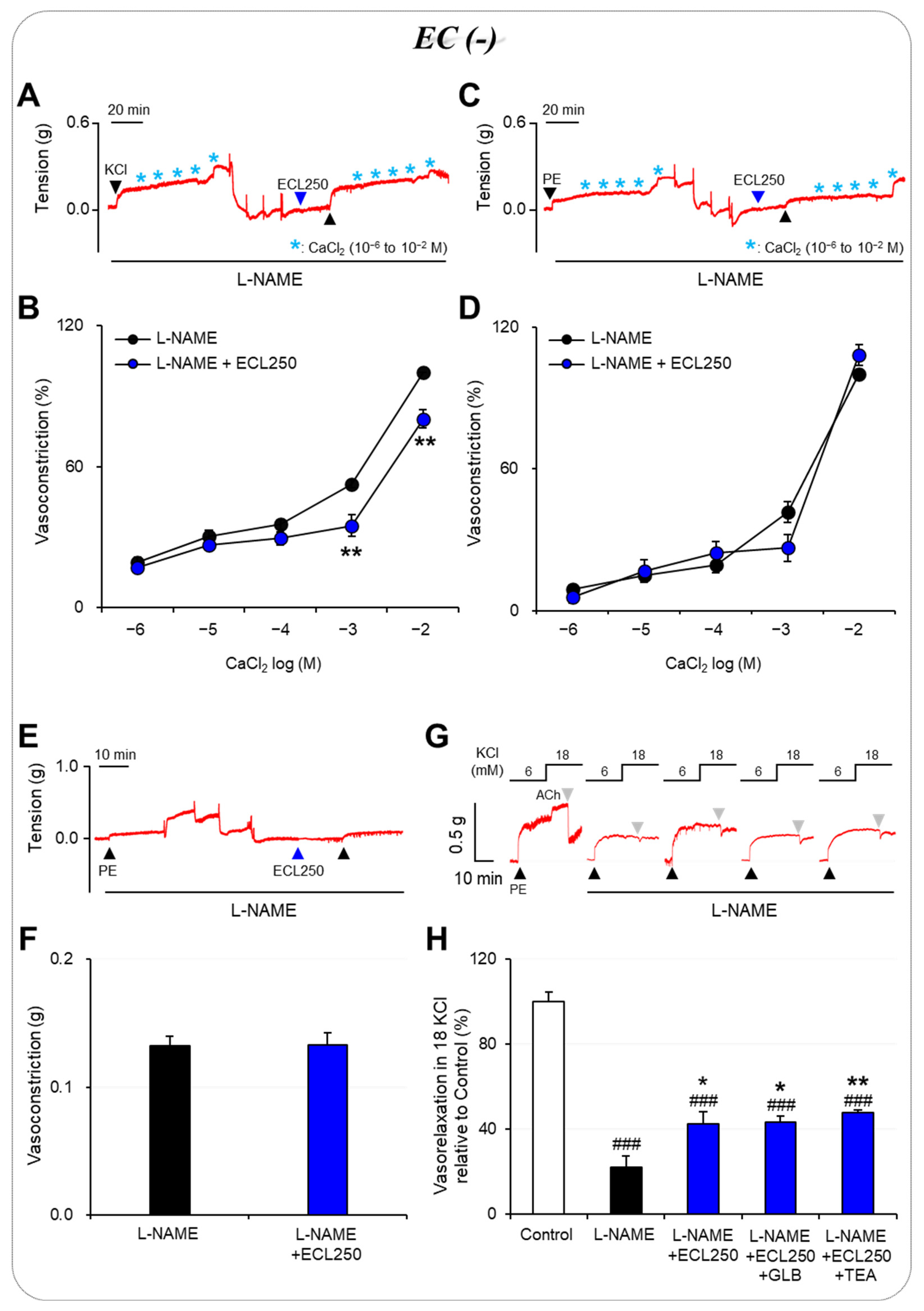
To determine whether the effect of the ECL is related to Ca2+ influx, we investigated VDCCs by using 60 mM of KCl and ROCCs by using 10 μM of PE. These experiments showed that pretreatment with ECL significantly reduced CaCl2-induced vasoconstriction in Ca2+-free solutions compared with the control group only under conditions in which the vessel was pre-constricted with KCl (p < 0.01; Figure 2A–D). This suggests that the ECL does not block Ca2+ influx via ROCCs but instead blocks VDCCs. To test whether the ECL affects vascular tone through the interruption of the release of Ca2+ from the SR, we measured PE-induced vasoconstriction under Ca2+-free conditions. A comparison of vasoconstriction in the presence and absence of the ECL showed no significant difference (Figure 2E,F), indicating that the mechanism through which the ECL regulates vascular tone is not affected by the release of Ca2+ from the SR.

3.3. Involvement of K+ Channels in the Effect of ECL

Moreover, pretreatment with TEA (1 mM) or GLB (1 μM), which are broad-spectrum K+ channel and KATP channel blockers, respectively, did not affect the recovery of ECL-induced vasorelaxation in the L-NAME–pretreated carotid arteries exposed to 18 mM of KCl (Figure 2G,H). These results imply that neither the broad range of K+ channels inhibited by TEA nor the KATP channels in vascular smooth muscle cells are involved in mediating the effects of ECL under high [K+]o conditions.

4. Discussion

The purpose of this study was to investigate the effect of the ECL on changes in the vascular tone of rat carotid arteries following exposure to high [K+]o and to determine the possible underlying mechanism of action. In the context of endothelial dysfunction, vessel rings pretreated with 250 μg/mL of ECL showed significantly more vasorelaxation than control rings, even under high [K+]o. Our results suggest that ECL contributes to improving the vasorelaxant responses of carotid arteries in an endothelium-independent manner, even in the presence of high [K+]o. VDCC, not ROCC, the release of Ca2+ from intracellular Ca2+ stores, KATP channels, and a broad range of K+ channels were involved in mediating the effects of ECL under high [K+]o conditions (Figure 3).
The endothelium contributes to vasomotor homeostasis by releasing substances that are important in vasorelaxation, primarily NO and endothelium-derived relaxing factors. Therefore, endothelial dysfunction disables important vasorelaxation mechanisms, leading to impaired vasomotor homeostasis. Alterations in the vasomotor properties of peripheral blood vessels can influence the regulatory functions controlling peripheral blood vessel resistance, thereby affecting proper blood pressure control. Numerous studies have highlighted the pivotal role of the endothelium in the effect of ECL on blood vessels. For instance, lancemaside A enhances endothelial function through the activation of endothelial NO synthase [17] and prevents hypertension by improving NO bioavailability in rats [18]. In a separate study by Han et al. (2018), an ECL was shown to have a significant blood pressure-reducing effect and tended to increase ACh- and SNP-induced vasorelaxation in a hypertensive rat model [19]. However, our current findings demonstrate that, despite endothelial dysfunction, the ECL exerted a prominent vasorelaxing effect even in carotid arteries exposed to high [K+]o, countering the vasoconstricting effect of this latter condition.
The Ca2+ channel-mediated alteration of intracellular Ca2+ concentrations in both endothelial and vascular smooth muscle cells is important for vasomotion. Vascular smooth muscle cells express various types of Ca2+ channels that play important roles in controlling vascular tone [25]. Here, we evaluated two such channels expressed in smooth muscle, namely, VDCCs and ROCCs, for their involvement in the ability of the ECL to modulate vascular tone. High [K+]o promotes Ca2+ influx through VDCCs, whereas PE-mediated α-receptor activation promotes Ca2+ influx through ROCCs [24]. Therefore, we estimated the CaCl2-induced vasoconstrictive responses of the arteries that had been pre-constricted via KCl or PE in a Ca2+-free solution containing EGTA. Our results showed that the ECL did not significantly change vasoconstriction induced by Ca2+ influx through ROCCs but significantly reduced vasoconstriction induced by Ca2+ influx through VDCCs. We also evaluated the involvement of the release of Ca2+ from intracellular Ca2+ stores and found no significant difference in vasoconstriction induced by Ca2+ release from the SR following pretreatment with the ECL. Collectively, these findings suggest that one of the mechanisms through which ECL improves vasorelaxation is through the inhibition of Ca2+ influx via VDCCs in vascular smooth muscle. In a previous study, the oral administration of an ECL (200 or 400 mg/kg) in a rat model of hypertension rats had less of an effect compared with the administration of the L-type Ca2+ channel blocker nifedipine, but it nonetheless significantly reduced systolic blood pressure compared with rats in a vehicle control group [19]. Moreover, lancemaside A, a major saponin component of an ECL, contributed to Ca2+ homeostasis by inhibiting Ca2+ influx through store-operated Ca2+ entry in vascular smooth muscle cells [21], which suggests that effects of the ECL in this study could be induced by lancemaside A. Increased [K+]o is a crucial factor that can cause changes in vascular tone [26]. Specifically, high [K+]o causes the depolarization of the cell membrane, which activates VDCCs, increasing Ca2+ influx and promoting the Ca2+-dependent vasoconstriction of vascular smooth muscle [27,28]. Our results collectively suggest that the ECL contributes to improving impaired vasorelaxant responses by inhibiting the ability of VDCCs in vascular smooth muscle to respond to the vasoconstrictive effects of high [K+]o.
Various pathophysiological situations can cause endothelial dysfunction; some of them also carry a risk of elevated [K+]o. For example, rhabdomyolysis was reported to induce damage to endothelial cells in mice [29], and simultaneous cell death due to rhabdomyolysis can acutely increase [K+]o because most K+ ions are contained within the intracellular compartment [30,31]. Similarly, CKD patients are vulnerable to [K+]o elevation due to deterioration of the kidney’s K+ excretion function [6]. Numerous factors in CKD can impair the bioavailability of nitric oxide [32], and CKD-related cardiovascular disease is primary based on endothelial dysfunction [33]. In this context, our results demonstrate that the ECL significantly improves vascular vasorelaxant responses caused by endothelial dysfunction under elevated [K+]o.
Here, we found that the ECL effectively restores impaired vasorelaxation caused by endothelial dysfunction even when exposed to high [K+]o. Previous studies have suggested that most of the beneficial effects of an ECL or a major saponin of CL on blood vessels apply only in pathological situations, such as in a hypertensive rat model [18,19]. Interestingly, however, our present results showed that the ECL improved the vasorelaxation induced by endothelial dysfunction under elevated [K+]o and it did not negatively affect vascular tone under normal physiological [K+]o or when the endothelium remained intact.

5. Conclusions

In conclusion, this study showed for the first time that an ECL significantly restored of smooth muscle vasorelaxation in endothelium-impaired rat carotid arteries even under high [K+]o. The present results add to the existing knowledge of the beneficial properties of ECLs on vascular function and indicate that an ECL could be developed as an alternative medicinal agent in patients with elevated [K+]o who are at risk for endothelial dysfunction. Because the present experiments were only conducted on the carotid arteries of rats, further clinical research on the safety and efficacy of using an ECL in humans is needed.

Author Contributions

Conceptualization, G.H.S.; methodology, G.H.S.; formal analysis, U.K., Y.K.S., J.P. and G.H.S.; investigation, U.K., Y.K.S. and J.P.; writing—original draft, U.K., Y.K.S. and G.H.S.; writing—review and editing, G.H.S.; visualization, U.K. and G.H.S.; supervision, G.H.S.; project administration, G.H.S.; funding acquisition, G.H.S. All authors have read and agreed to the published version of the manuscript.

Funding

This work was supported by the National Research Foundation of Korea (NRF) grant funded by the Korean government (MSIT) (No. 2021R1A2C2004118) and the Institute of Nursing Research, Korea University Grant. This manuscript is a revision of UK’s master’s thesis from Korea University.

Institutional Review Board Statement

All procedures were carried out in accordance with the institutional guidelines for the use and care of laboratory animals and were approved by the Ethical Committee of the Korea University (Protocol Number: KUIACUC-2021-0088).

Informed Consent Statement

Not applicable.

Data Availability Statement

Data are available upon reasonable request from the corresponding author.

Conflicts of Interest

The authors declare no conflict of interest.

References

  1. Garland, C.J.; Dora, K.A. Endothelium-Dependent Hyperpolarization: The Evolution of Myoendothelial Microdomains. J. Cardiovasc. Pharmacol. 2021, 78, S3–S12. [Google Scholar] [CrossRef]
  2. Akata, T. General anesthetics and vascular smooth muscle: Direct actions of general anesthetics on cellular mechanisms regulating vascular tone. Anesthesiology 2007, 106, 365–391. [Google Scholar] [CrossRef]
  3. Daghbouche-Rubio, N.; López-López, J.R.; Pérez-García, M.T.; Cidad, P. Vascular smooth muscle ion channels in essential hypertension. Front. Physiol. 2022, 13, 1016175. [Google Scholar] [CrossRef]
  4. Ekmekcioglu, C.; Elmadfa, I.; Meyer, A.L.; Moeslinger, T. The role of dietary potassium in hypertension and diabetes. J. Physiol. Biochem. 2016, 72, 93–106. [Google Scholar] [CrossRef]
  5. Palmer, B.F.; Clegg, D.J. Physiology and pathophysiology of potassium homeostasis. Adv. Physiol. Educ. 2016, 40, 480–490. [Google Scholar] [CrossRef]
  6. Sarnowski, A.; Gama, R.M.; Dawson, A.; Mason, H.; Banerjee, D. Hyperkalemia in Chronic Kidney Disease: Links, Risks and Management. Int. J. Nephrol. Renovasc. Dis. 2022, 15, 215–228. [Google Scholar] [CrossRef]
  7. Emmett, M. Review of Clinical Disorders Causing Metabolic Acidosis. Adv. Chronic Kidney Dis. 2022, 29, 355–363. [Google Scholar] [CrossRef]
  8. Navarrete, N. Hyperkalemia in electrical burns: A retrospective study in Colombia. Burns 2018, 44, 941–946. [Google Scholar] [CrossRef]
  9. Nwokocha, C.; Palacios, J.; Ojukwu, V.E.; Nna, V.U.; Owu, D.U.; Nwokocha, M.; McGrowder, D.; Orie, N.N. Oxidant-induced disruption of vascular K+ channel function: Implications for diabetic vasculopathy. Arch. Physiol. Biochem. 2022, 1–12. [Google Scholar] [CrossRef]
  10. Knox, M.; Vinet, R.; Fuentes, L.; Morales, B.; Martínez, J.L. A Review of Endothelium-Dependent and -Independent Vasodilation Induced by Phytochemicals in Isolated Rat Aorta. Animals 2019, 9, 623. [Google Scholar] [CrossRef]
  11. Dogan, M.F.; Yildiz, O.; Arslan, S.O.; Ulusoy, K.G. Potassium channels in vascular smooth muscle: A pathophysiological and pharmacological perspective. Fundam. Clin. Pharmacol. 2019, 33, 504–523. [Google Scholar] [CrossRef]
  12. Hossen, M.J.; Kim, M.Y.; Kim, J.H.; Cho, J.Y. Codonopsis lanceolata: A Review of Its Therapeutic Potentials. Phytother. Res. 2016, 30, 347–356. [Google Scholar] [CrossRef]
  13. Ushijima, M.; Komoto, N.; Sugizono, Y.; Mizuno, I.; Sumihiro, M.; Ichikawa, M.; Hayama, M.; Kawahara, N.; Nakane, T.; Shirota, O.; et al. Triterpene glycosides from the roots of Codonopsis lanceolata. Chem. Pharm. Bull. 2008, 56, 308–314. [Google Scholar] [CrossRef]
  14. Hyam, S.R.; Jang, S.E.; Jeong, J.J.; Joh, E.H.; Han, M.J.; Kim, D.H. Echinocystic acid, a metabolite of lancemaside A, inhibits TNBS-induced colitis in mice. Int. Immunopharmacol. 2013, 15, 433–441. [Google Scholar] [CrossRef]
  15. Joh, E.H.; Lee, I.A.; Han, S.J.; Chae, S.; Kim, D.H. Lancemaside A ameliorates colitis by inhibiting NF-kappaB activation in TNBS-induced colitis mice. Int. J. Colorectal. Dis. 2010, 25, 545–551. [Google Scholar] [CrossRef]
  16. Zhang, Y.; Wang, H.; Zhang, L.; Yuan, Y.; Yu, D. Codonopsis lanceolata polysaccharide CLPS alleviates high fat/high sucrose diet-induced insulin resistance via anti-oxidative stress. Int. J. Biol. Macromol. 2020, 145, 944–949. [Google Scholar] [CrossRef]
  17. Lee, Y.S.; Kim, H.; Kim, J.; Seol, G.H.; Lee, K.W. Lancemaside A, a major triterpene saponin of Codonopsis lanceolata enhances regulation of nitric oxide synthesis via eNOS activation. BMC Complement. Altern. Med. 2019, 19, 110. [Google Scholar] [CrossRef]
  18. Shin, Y.K.; Han, A.Y.; Hsieh, Y.S.; Kwon, S.; Kim, J.; Lee, K.W.; Seol, G.H. Lancemaside A from Codonopsis lanceolata prevents hypertension by inhibiting NADPH oxidase 2-mediated MAPK signalling and improving NO bioavailability in rats. J. Pharm. Pharmacol. 2019, 71, 1458–1468. [Google Scholar] [CrossRef]
  19. Han, A.Y.; Lee, Y.S.; Kwon, S.; Lee, H.S.; Lee, K.W.; Seol, G.H. Codonopsis lanceolata extract prevents hypertension in rats. Phytomedicine 2018, 39, 119–124. [Google Scholar] [CrossRef]
  20. Shin, Y.K.; Hsieh, Y.S.; Han, A.Y.; Lee, K.W.; Seol, G.H. Beneficial effects of Codonopsis lanceolata extract on systolic blood pressure levels in prehypertensive adults: A double-blind, randomized controlled trial. Phytother. Res. 2020, 34, 340–348. [Google Scholar] [CrossRef]
  21. Kim, M.K.; Han, A.Y.; Shin, Y.K.; Lee, K.W.; Seol, G.H. Codonopsis lanceolata Contributes to Ca2+ Homeostasis by Mediating SOCE and PLC/IP3 Pathways in Vascular Endothelial and Smooth Muscle Cells. Planta Medica 2020, 86, 1345–1352. [Google Scholar] [CrossRef]
  22. Seol, G.H.; Ahn, S.C.; Kim, J.A.; Nilius, B.; Suh, S.H. Inhibition of endothelium-dependent vasorelaxation by extracellular K+: A novel controlling signal for vascular contractility. Am. J. Physiol. Heart Circ. Physiol. 2004, 286, H329–H339. [Google Scholar] [CrossRef]
  23. Aekthammarat, D.; Tangsucharit, P.; Pannangpetch, P.; Sriwantana, T.; Sibmooh, N. Moringa oleifera leaf extract enhances endothelial nitric oxide production leading to relaxation of resistance artery and lowering of arterial blood pressure. Biomed Pharmacother. 2020, 130, 110605. [Google Scholar] [CrossRef]
  24. Yang, S.; Xu, Z.; Lin, C.; Li, H.; Sun, J.; Chen, J.; Wang, C. Schisantherin A causes endothelium-dependent and -independent vasorelaxation in isolated rat thoracic aorta. Life Sci. 2020, 245, 117357. [Google Scholar] [CrossRef]
  25. Tykocki, N.R.; Boerman, E.M.; Jackson, W.F. Smooth Muscle Ion Channels and Regulation of Vascular Tone in Resistance Arteries and Arterioles. Compr. Physiol. 2017, 7, 485–581. [Google Scholar] [CrossRef]
  26. Afsar, S.; Hemsinli, D.; Ozyazgan, S.; Akkan, A.G.; Arslan, C. The Effects of Potassium Channels in Human Internal Mammary Artery. Pharmacology 2016, 97, 72–77. [Google Scholar] [CrossRef]
  27. Pathan, A.R.; Rusch, N.J. Two-pore domain K+ channels: Evidence for TWIK-2 in blood pressure regulation. Hypertension 2011, 58, 539–541. [Google Scholar] [CrossRef]
  28. Hu, G.Y.; Peng, C.; Xie, X.F.; Xiong, L.; Zhang, S.Y.; Cao, X.Y. Patchouli alcohol isolated from Pogostemon cablin mediates endothelium-independent vasorelaxation by blockade of Ca2+ channels in rat isolated thoracic aorta. J. Ethnopharmacol. 2018, 220, 188–196. [Google Scholar] [CrossRef]
  29. Panizo, N.; Rubio-Navarro, A.; Amaro-Villalobos, J.M.; Egido, J.; Moreno, J.A. Molecular Mechanisms and Novel Therapeutic Approaches to Rhabdomyolysis-Induced Acute Kidney Injury. Kidney Blood Press. Res. 2015, 40, 520–532. [Google Scholar] [CrossRef]
  30. Cabral, B.M.I.; Edding, S.N.; Portocarrero, J.P.; Lerma, E.V. Rhabdomyolysis. Dis. Mon. 2020, 66, 101015. [Google Scholar] [CrossRef]
  31. Giannoglou, G.D.; Chatzizisis, Y.S.; Misirli, G. The syndrome of rhabdomyolysis: Pathophysiology and diagnosis. Eur. J. Intern. Med. 2007, 18, 90–100. [Google Scholar] [CrossRef]
  32. Roumeliotis, S.; Mallamaci, F.; Zoccali, C. Endothelial Dysfunction in Chronic Kidney Disease, from Biology to Clinical Outcomes: A 2020 Update. J. Clin. Med. 2020, 9, 2359. [Google Scholar] [CrossRef]
  33. Khreba, N.; Khedr, D.; Abdel-Baky, A.; Kannishy, G.E.; Samaan, E. Nephron index rather than serum FGF 23 predicts endothelial dysfunction in early but not advanced chronic kidney disease patients. Int. Urol. Nephrol. 2023; in press. [Google Scholar] [CrossRef]
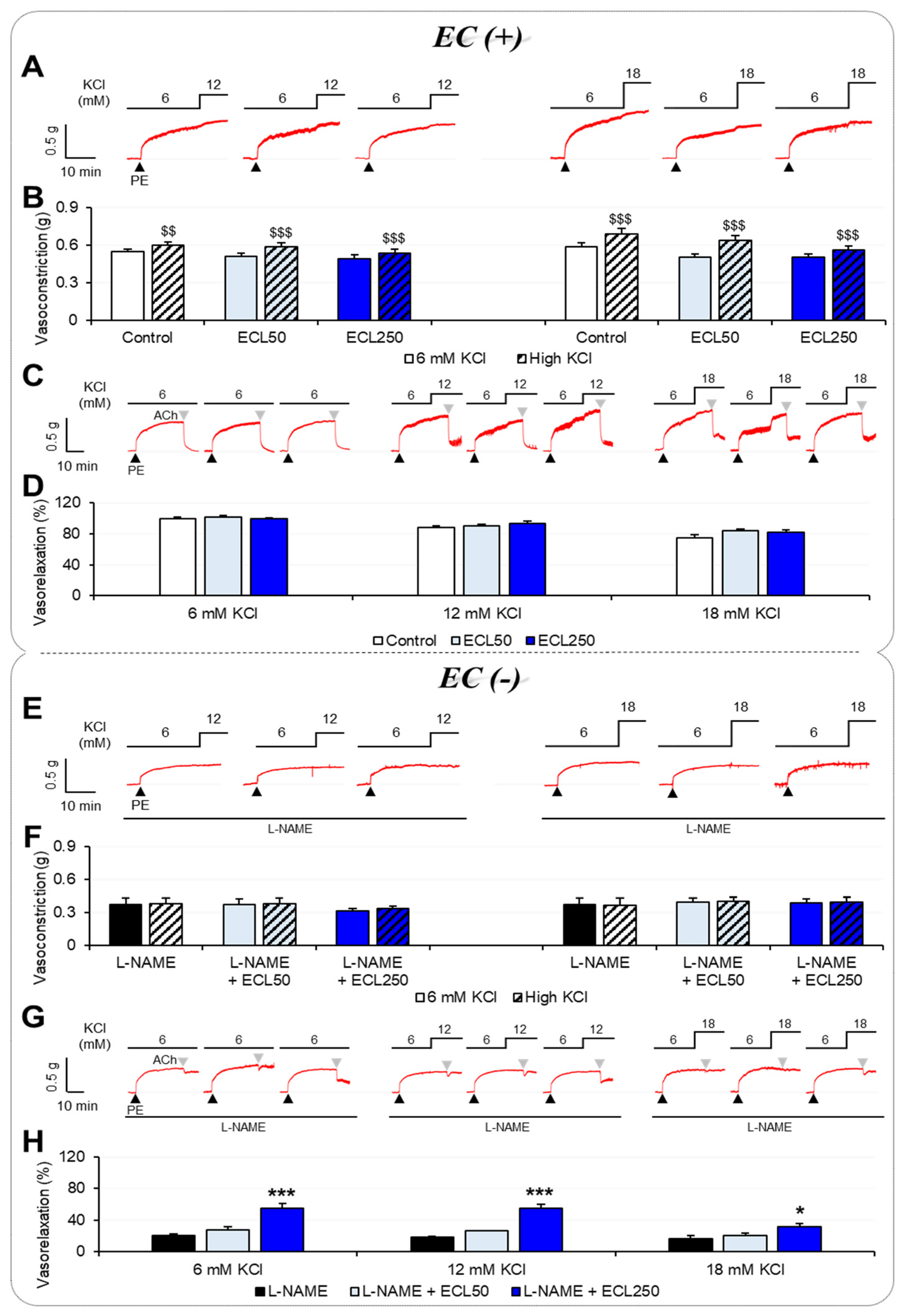
Figure 1. Effects of an ECL on the vascular tension of rat carotid arteries when exposed to normal or high [K+]o. Experimental traces with difference of PE-induced vasoconstriction (A,B) and ACh-induced vasorelaxation (C,D) when exposed to normal or high [K+]o (12 or 18 mM) in carotid arteries with intact endothelia. Experimental traces with differences in PE-induced vasoconstriction (E,F) and ACh-induced vasorelaxation (G,H) when exposed to normal or high [K+]o in endothelium-impaired carotid arteries. Results are displayed as means ± standard error of the mean and were analyzed via a paired t-test or one-way ANOVA followed by Tukey’s post hoc test (n = 5 to 22). $$ p < 0.01, $$$ p < 0.001 respective to 6 mM of KCl; * p < 0.05, *** p < 0.001 versus L-NAME. ACh, acetylcholine; EC, endothelial cell; ECL, extract of Codonopsis lanceolata; L-NAME, Nω-nitro-L-arginine methyl ester; PE, phenylephrine.
Nutrients 15 03791 g001
Figure 2. Underlying mechanism of action of ECL in vascular smooth muscle. Extracellular Ca2+ influx via voltage-dependent Ca2+ channel (A,B). Extracellular Ca2+ influx via receptor-operated Ca2+ channel (C,D). Intracellular Ca2+ release from sarcoplasmic reticulum (E,F). Activation of non-selective K+ channel or ATP-sensitive K+ channel (G,H). Results are expressed as means ± standard error of the mean. Analysis was performed via one-way ANOVA followed by Tukey’s post hoc or paired t-test (n = 5 to 13). ### p < 0.001 vs. Control; * p < 0.05, ** p < 0.01 vs. L-NAME. ECL, extract of Codonopsis lanceolata; GLB, glibenclamide; TEA, tetraethylammonium; L-NAME, Nω-nitro-L-arginine methyl ester; PE, phenylephrine.
Nutrients 15 03791 g002
Figure 3. Mechanism underlying the vasorelaxant effects of ECL on the vascular smooth muscle of rat carotid arteries exposed to high [K+]o. ECL, extract of Codonopsis lanceolata; IP3R, inositol 1,4,5-trisphosphate receptor; ROCC, receptor-operated Ca2+ channel; RyR, ryanodine receptor; VDCC, voltage-dependent Ca2+ channel; [Ca2+]i, intracellular Ca2+ concentration; [K+]o, extracellular K+ concentration.
Nutrients 15 03791 g003
Disclaimer/Publisher’s Note: The statements, opinions and data contained in all publications are solely those of the individual author(s) and contributor(s) and not of MDPI and/or the editor(s). MDPI and/or the editor(s) disclaim responsibility for any injury to people or property resulting from any ideas, methods, instructions or products referred to in the content.