Article

Analysis of Network Pharmacological Efficacy and Therapeutic Effectiveness in Animal Models for Functional Dyspepsia of Foeniculi fructus

1
Department of Longevity and Biofunctional Medicine, Pusan National University School of Korean Medicine, Yangsan 50612, Republic of Korea
2
Department of Pharmaceutical Engineering, Daegu Hanny University, Gyeongsan 38610, Republic of Korea
3
College of Oriental Medicine, Daegu Hanny University, Gyeongsan 38610, Republic of Korea
4
Department of Gastroenterology, College of Korean Medicine, Kyung Hee University, Seoul 02447, Republic of Korea
5
Department of Clinical Korean Medicine, Graduate School of Kyung Hee University, Seoul 02447, Republic of Korea
*
Authors to whom correspondence should be addressed.
Nutrients 2023, 15(12), 2644; https://doi.org/10.3390/nu15122644
Submission received: 15 May 2023 / Revised: 3 June 2023 / Accepted: 5 June 2023 / Published: 6 June 2023
(This article belongs to the Section Nutrition and Metabolism)

Abstract

:
For centuries, Foeniculi fructus (F. fructus) has been used as a traditional herbal medicine in China and Europe and is widely used as a natural therapy for digestive disorders, including indigestion, flatulence, and bloating. The mechanism of F. fructus that alleviates functional dyspepsia was analyzed through network pharmacology, and its therapeutic effect on an animal model of functional dyspepsia were investigated. The traditional Chinese medicine systems pharmacology (TCMSP) database was used to investigate the compounds, targets, and associated diseases of F. fructus. Information on the target genes was classified using the UniProtdatabase. Using the Cytoscape 3.9.1 software, a network was constructed, and the Cytoscape string application was employed to examine genes associated with functional dyspepsia. The efficacy of F. fructus on functional dyspepsia was confirmed by treatment with its extract in a mouse model of loperamide-induced functional dyspepsia. Seven compounds targeted twelve functional dyspepsia-associated genes. When compared to the control group, F. fructus exhibited significant suppression of symptoms in a mouse model of functional dyspepsia. The results of our animal studies indicated a close association between the mechanism of action of F. fructus and gastrointestinal motility. Based on animal experimental results, the results showed that F. fructus provided a potential means to treat functional dyspepsia, suggesting that its medical mechanism for functional dyspepsia could be described by the relationship between seven key compounds of F. fructus, including oleic acid, β-sitosterol, and 12 functional dyspepsia-related genes.

1. Introduction

Functional dyspepsia is characterized as a clinical syndrome where individuals experience recurring or persistent discomfort or pain in the upper abdomen, without any identifiable organic disorders underlying the symptoms [1]. In patients with functional dyspepsia, antagonists of histamine H2 receptors [2], inhibitors of proton pumps [3], or eradication with Helicobacter pylori have shown limited benefits [4], and the outcomes of controlled trials were generally unsatisfactory. Additionally, pharmacological agents (such as cisapride), despite their limited effectiveness, pose the risk of potential side effects such as arrhythmia, cardiovascular disease, headaches, and abdominal pain.
One attractive alternative through a natural approach is the use of herbal remedies, which are recognized to have a low risk of side effects. However, few rigorous clinical studies are available because of the insufficient standardization of herbal ingredients.
Foeniculi fructus (F. fructus; Foeniculum vulgare or fennel) is an umbelliferous plant that is indigenous to southern Europe and the Mediterranean region. It has a long history of traditional herbal medicine use in both China and Europe, dating back to ancient times [5,6,7,8,9,10,11,12,13,14]. This herb has been employed as a natural remedy for various digestive ailments, such as flatulence, bloating, and indigestion. Additionally, it possesses antipyretic, analgesic, and antioxidant properties [5,6,7]. F. fructus provides relief from symptoms associated with female menopausal syndrome, helps regulate menstruation, and enhances libido [8]. It also has galactagogue and emmenagogue properties [9]. F. fructus has hepatoprotective effects and may be used in pediatric colic [10,11]. In addition, it also functions as a 5-lipoxygenase inhibitor and is known to be effective in suppressing vomiting, gastrointestinal diseases, and anti-allergies [12]. Additionally, in traditional Turkish medicine, F. fructus is used as a diuretic, laxative, antispasmodic, lactating stimulant, and a wound dressing [13]. Although few clinical studies have been conducted on fennel, a clinical study in China reported that drinking F. fructus tea after open surgery on gynecological malignancies increased intestinal motility, reducing hospitalization periods and complications [14].
Conventional biological experimental methodologies frequently face limitations when studying the comprehensive mechanisms of action of traditional herbal medicines, owing to their intricate pharmacological properties [15,16,17,18,19,20,21,22]. To solve these difficulties, network pharmacology, an integrated research field using physics, mathematics, medicine, pharmacology, network science, and computational systems biology, is a new and effective approach [15,16,17,18,19,20,21,22]. The objective of this integrative scientific approach is to elucidate the interactions among biological components such as organs, tissues, cells, proteins, and genes, with the aim of identifying the mechanisms of drug activity and disease pathogenesis [15,16,17,18,19,20,21,22]. To date, network pharmacology studies have identified distinct system-level pharmacological effects, active compounds, and key therapeutic targets, as well as mechanisms (e.g., apoptosis, proliferation, oxidation, and by further confirming the therapeutic regulation of biological processes such as reduction, cell cycle regulation, insulin metabolism, and inflammation) and the multipharmacological properties of traditional herbal drugs exerted by synergistic interactions between multiple compounds and targets [15,16,17,18,19,20,21,22,23,24,25,26,27,28,29,30,31,32,33]. Figure 1 depicts a schematic representation of the study protocol. The objective of our network pharmacology study was to comprehensively understand the impact of F. fructus on the molecular mechanisms related to its digestive properties, taking a systems perspective.

2. Materials and Methods

2.1. Analysis of Network Pharmacology

2.1.1. Identifying Compounds of F. fructus

The traditional Chinese medicine systems pharmacology (TCMSP) database was utilized to identify the potentially active compounds in F. fructus. We entered ‘Foeniculi fructus’ as a search term for herbs.

2.1.2. Target Network

The target information was acquired through the utilization of TCMSP [34]. To associate target proteins with official gene names, the UniProtKB database (https://www.uniprot.org/uniprot, accessed on 7 February 2023) was employed [35].

2.1.3. Analysis of Network

To construct the compound-target network, we utilized Cytoscape 3.9.1 (https://cytoscape.org, accessed on 23 February 2023) [36]. Functional-dyspepsia-associated genes were collected using Cytoscape App., which organized and updated the data weekly [37].

2.1.4. Screening of Active Compound

Physiologically active compounds in F. fructus were subjected to screening based on specific criteria related to ADME (absorption, distribution, metabolism, and excretion) parameters. These criteria included MW (molecular weight), OB (oral bioavailability), Caco-2 permeability, and DL (drug similarity). The screening criteria used were as follows: OB ≥ 30%, DL ≥ 0.10, and Caco-2 ≥ −0.4. The compounds that fulfilled the specified criteria were chosen as the active compounds.

2.2. Analysis of F. fructus

2.2.1. Instrument and Reagent

A Waters ACQUITY ultra-performance LC system (USA) was utilized to conduct the ultra-performance liquid chromatography (UPLC). A Waters ACQUITYTM photodiode array detector (PDA) and HPLC column (Waters ACQUITYTM BEH C18 columns, 1.7 µm, 2.1 × 100), along with the software Empower, were employed for the analysis. The experiment involved the use of methanol (HPLC grade, Junsei, Tokyo, Japan), acetonitrile (HPLC grade, JT-BAKER, Radnor, PA, USA), and tertiary distilled water as reagents. The standard preparations of this experiment were obtained from Anethole (Sigma-Aldrich, St. Louis, MO, USA), R-(a)-phellandrene (Sigma-Aldrich, St. Louis, MO, USA), and 4-Methoxybenzoic acid (ChemFaces, Wuhan, China).

2.2.2. Preparation of the Standard Solution

An accurate measurement of Anethole, R-(a)-phellandrene, and 4-Methoxybenzoic acid was conducted, followed by their dissolution in dimethyl sulfoxide (DMSO) and methanol. Subsequently, a standard undiluted solution was prepared, containing 1 mg per ml of the compounds. In succession, the standard undiluted solution was diluted with methanol to 12.5, 25, 50, and 100 μg per mL, and they were used as standard solutions. All standard materials exhibited determination coefficient (R2) values exceeding 0.999 when establishing a standard curve.

2.2.3. Preparation of the Test Liquid for Quantitative Analysis

To perform quantitative analysis, the sample was thoroughly mixed with the test liquid, and precisely 0.2 g of the resulting mixture was added to 10 mL of ethyl alcohol. Subsequently, the mixture was subjected to microwave extraction for a duration of one hour. The resulting test liquid was then filtered using a 0.22 μm membrane filter.

2.2.4. Quantitation of the F. fructus Extract

Ultra-performance liquid chromatography (UPLC) was conducted using a Waters ACQUITYTM ultra-performance LC system (USA) and a Waters ACQUITYTM BEH C18 column (1.7 μm, 2.1 × 100). The temperature of the column was kept at room temperature. In PDA analysis, 4-Methoxybenzoic acid and R-(a)-phellandrene were examined at 330 nm, whereas anethole was analyzed at 306 nm (Table 1). The mobile phase used in the analysis consisted of a blend of acetonitrile and water with 0.1% formic acid. The analysis parameters were set as follows: a 2 μL sample injection and a flow rate of 0.4 mL/min. The qualitative analysis was conducted by verifying the retention time, followed by quantitation using the peak area method. The F. fructus samples were deposited at the College of Korean Medicine, Daegu Hanny University (Table 2; Figure 2).

2.3. Animal Testing

2.3.1. Design of Animal Experiment

A commercial animal breeder (Samtako, Gyeonggi, Republic of Korea) provided a total of 108 specific pathogen-free (SPF) ICR mice. These mice were all male, weighed between 19–21 g, and were five weeks old at the time of purchase. The mice were housed in a temperature-controlled room within a specific pathogen-free (SPF) facility, where the temperature was maintained at 22 ± 2 °C and the relative humidity at 60 ± 5%. The mice followed a 12/12 h light/dark cycle in their housing environment. The mice were provided with unlimited access to commercial standard chow (Samtako, Gyeonggi, Republic of Korea) and tap water. Following a one-week acclimatization period, the mice were randomly divided into three experimental groups: the first group for small intestine motility (6 mice/group, n = 36), the second group for gastric emptying test (6 mice/group, n = 36), and the third group for Western blot, qPCR, and histopathology (3 mice/group, n = 18). The sets were divided into six groups: control, loperamide (negative control, 10 mg/kg), three different doses of F. fructus (25, 50, and 100 mg/kg), and mosapride (positive control, 3 mg/kg). In general, the treatment dose of mosapride was 3.1 mg/kg in mice [38]. Distilled water was used to prepare Foeniculi fructus and mosapride. Each group received oral administration of either distilled water (control and loperamide groups), F. fructus, or mosapride for a duration of three consecutive days [39,40]. The experiments and animal care procedures followed the guidelines provided by the Animal Care and Use Committee of the Pusan National University Animal Research Institute (PNU-2022-0160) and the regulations outlined in the guidelines for the management and utilization of laboratory animals at the US National Institutes of Health.

2.3.2. Assessment of Gastric Weight and Gastric Emptying

The mice underwent a 19 h fasting period with unrestricted access to tap water. The volume of phenol red solution (500 µL) and the 50% delayed gastric emptying time point was based on previously established study protocols [41,42]. After a 30 min period following the administration of 0.05% phenol red (dyeing substance that checks the level of gastric emptying), the mice were humanely euthanized. The stomachs were promptly excised and weighed. Subsequently, the stomachs were treated with 5 mL of 0.1 N sodium hydroxide solution to measure the optical density of residual phenol red. Additionally, 0.5 mL of trichloroacetic acid (20% w/v) was added to the stomachs. The produced homogenate was centrifuged at 3000 rpm for 20 min and then mixed with 0.5 N sodium hydroxide solution with supernatant 1 milliliter. In addition, the optical density was measured at a wavelength of 560 nm with a spectrophotometer.
The emission values mentioned above were derived using the following formula:
gastric emptying (%) = (1 − X/Y) × 100
X: Optical density of the phenol red remaining on it. Y: Optical density of the phenol red mixture with sodium hydroxide under test tube conditions.

2.3.3. Assessment of Intestinal Transit Rate by Evans Blue

To measure the intestinal transit rate, the Evans blue diet method was used, in which 5% Evans blue (dyeing substance that checks the level of intestinal transit rate) was prepared in distilled water, as previously described [43]. Evans blue diet was orally administered (250 μL/20 g mouse) 30 min after IP injection of loperamide. After a 30 min period following the administration of the Evans blue dye, the mice were humanely euthanized. The distance covered by the Evans blue dye within the small intestine, specifically from the pylorus to the cecum, was measured to determine the intestinal transit distance. The above time points were selected as per the methods of an earlier study [44].

2.3.4. Western Blot Analysis for Check of Protein Level

To measure the gastric protein levels of neuronal nitric oxide synthase (nNOS), TEME16A, and TRPM7, gastric tissues were homogenized in RIPA lysis buffer. Following a 5 min denaturation period by boiling, the proteins were subjected to electrophoresis on a 10% polyacrylamide gel and subsequently transferred to a nitrocellulose (NC) membrane. After blocking in 5% skim milk for 30 min, membranes were tested overnight at 4 °C with nNOS (1:1000, ab76067), TEME16A (1:1000, a72984), TRPM7 (1:200, ab135817), or β-actin (1:5000, sc-47778). After washing the membranes, they were incubated with a horseradish peroxidase (HRP)-conjugated rabbit antibody (diluted 1:5000 against nNOS, TEME16A, and TRPM7) or an HRP-conjugated mouse antibody (diluted 1:5000 against β-actin) for a duration of 1 h at room temperature. Protein visualization was performed using Western Bright Sirius (Advansta, San Jose, CA, USA), and protein expression was examined using an ImageQuant LAS 4000 system (GE Healthcare, Chicago, IL, USA). Protein expression quantification was conducted using ImageJ software (NIH).

2.3.5. Quantitative Real-Time PCR to Evaluate Gene Expression

In order to determine the expression of genes related to muscle contraction, including anoctamin-1 (ANO1), ryanodine receptor 3 (RYR3), smooth muscle cell myosin light chain kinase (smMLCK), and 5HT4 receptor (5HT4R), total mRNA was extracted from the stomach tissues using Trizol reagents (Invitrogen, Waltham, MA, USA). cDNA reverse transcription kit (M-MLV Reverse Transcriptase, Promega, Madison, WI, USA) was utilized to synthesize cDNA from the entire RNA sample (1 μg). For qPCR analysis, iTaq Universal SYBR Green Supermix (Bio-Rad, Hercules, CA, USA) was employed along with the primers provided in Table 3. The StepOnePlus Real-Time PCR System (Applied Biosystems, Foster City, CA, USA) was utilized for the analysis of gene expression data.

3. Results

3.1. Target Information Derived by Examining Correlations between Compounds and Targets

A total of 45 potentially active compounds in F. fructus were identified with the TCMSP database (Supplementary Materials Table S1). Out of the identified compounds, 41 exhibited information regarding their targets (Supplementary Materials Table S2). These 41 compounds interacted with a total of 260 targets, involving a combination of 611 components. As shown in Figure 3, acetaldehyde was linked to the greatest number of targets (142 genes), followed by oleic acid (48 genes), β-sitosterol (38 genes), APIOL (31 genes), stigmasterol (31 genes), anisketone (24 genes), (−)-nopinene (21 genes), and terragon (20 genes).

3.2. A Total of Nine Compounds Met ADME Requirements for Active Compounds

A total of nine compounds satisfied the screening criteria for active compounds (Table 4): ammidin, β-sitosterol, EIC, oleic acid, majudin, oleic acid, petroselic acid, stigmasterol, and uvadex.

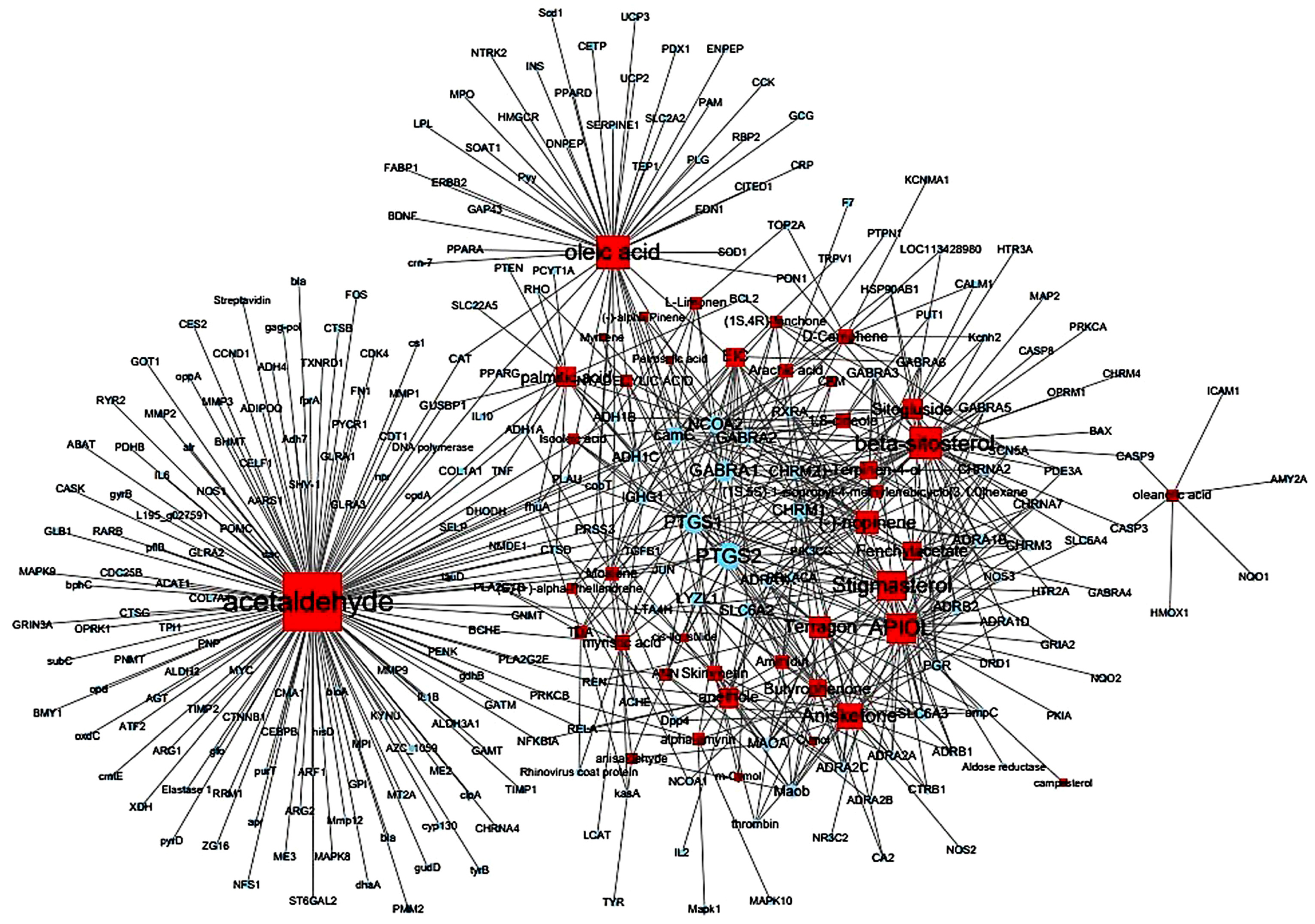
3.3. Thirty-Two Compounds Associated with Gastrointestinal (GI) Diseases Were Identified in F. fructus

Furthermore, we utilized the TCMSP database to establish relationships between compounds, targets, and diseases. It was observed that 32 compounds were linked to gastrointestinal (GI) diseases (Table 5). In particular, ammidin, β-sitosterol, EIC, isooleic acid, oleic acid, petroselic acid, and stigmasterol were related with gastrointestinal diseases. Other compounds associated with gastrointestinal diseases, including (−)-nopinene, (1S,5S)-1-isopropyl-4-methylenebicyclo [3.1.0]hexane, (S)-(+)-alpha-phellandrene, 1,8-cineole, acetaldehyde, alpha-amyrin, anethole, anisketone, ANN, APIOL, arachic acid, butyrophenone, cis-ligustilide, d-camphene, fenchylacetate, l-limonen, moslene, myristic acid, oleanolic acid, palmitic acid, pentadecylic acid, sitogluside, skimmetin, TDA, and terragon were confirmed as non-active compounds (Figure 4).

3.4. All 31 GI Disease-Related Compounds in F. fructus except Oleanolic Acid Were Associated with Functional Dyspepsia

To investigate the relationship between F. fructus and functional dyspepsia, we used the Cytoscape App to determine genetic information related to functional dyspepsia. With a score cutoff 0.40 and a maximum of 100 proteins, we initially identified 100 genes associated with functional dyspepsia (Supplementary Materials Table S3). Based on the obtained results, we constructed a network comprising functional dyspepsia-related genes and the target genes of activated compounds in F. fructus (Figure 5). Fourteen genes corresponding to two gene sets were identified, and the functional dyspepsia-related genes targeted by the activated F. fructus compound were ADRA2A, BDNF, CCK, CRP, GCG, JUN, Kcnh2, PTGS1, PTGS2, Pyy, SLC6A4, and TRPV.

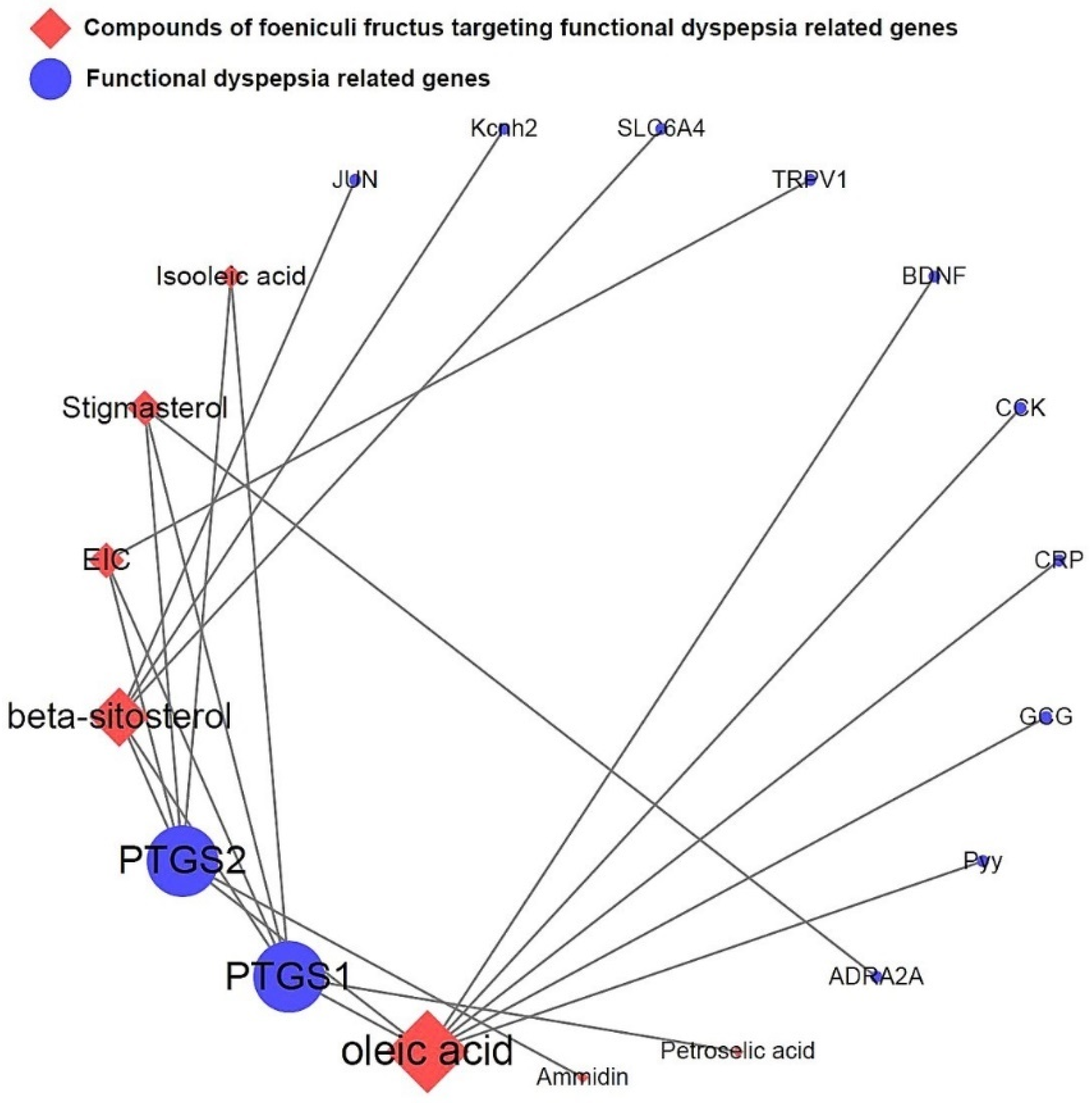
3.5. Network of Functional Dyspepsia-Associated Genes and Compounds

The network depicted in Figure 6 illustrates the relationship between activated compounds in F. fructus and target genes associated with functional dyspepsia. Notably, PTGS1 and PTGS2 exhibited the strongest associations with functional dyspepsia. In summary, ammidin, EIC, oleic acid, petroselic acid, stigmasterol, β-sitosterol, and oleic acid were active compounds that targeted functional dyspepsia-associated genes, suggesting that they could be potential drug candidates.

3.6. Mouse Experiment on Delayed Gastric Emptying

Loperamide injection induced gastric food retention, whereas pretreatment with F. fructus decreased this effect, as seen by macroscopic observation (Figure 7B). This finding was validated using quantitative analysis. The group treated with F. fructus exhibited a significantly lower gastric weight compared to the loperamide group (p < 0.05, as shown in Figure 7C). The pretreatment with F. fructus resulted in a significant reduction in the amount of phenol red retention in the stomach compared to the loperamide group (p < 0.05, as depicted in Figure 7D). Pretreatment with mosapride also had similar effects as the F. fructus treatment.

3.7. Mouse Experiment on Molecules Involved in Gastrointestinal Motility

Loperamide injection sharply attenuated nNOS, TEME16A, and TRPM7 protein expression in gastric tissue, whereas pretreatment with F. fructus sharply increased nNOS, TEME16A, and TRPM7 protein expression (p < 0.01, Figure 8A,B). Loperamide injection also decreased smooth-muscle-contraction-related gene expression, including 5HT4R, RYR3, ANO1, and smMLCK. These changes were inhibited by pretreatment with F. fructus (p < 0.05, p < 0.01, Figure 8C). Mosapride had a positive effect on nNOS, TEME16A, and TRPM7 proteins and 5HT4R, RYR3, ANO1, and smMLCK gene expression.

3.8. Mouse Experiment on Intestinal Motility

The administration of loperamide significantly decreased small intestine motility when compared to the control group. This suppression of small intestine motility was significantly restored by pretreatment with F. fructus (p < 0.01; Figure 9). Pretreatment with mosapride also sharply restored the motility of the small intestine, similar to that of F. fructus.

4. Discussion

For centuries, F. fructus has served as a renowned traditional herbal medicine in China and Europe, with extensive cultivation in southern Europe and the Mediterranean region. Multiple studies have demonstrated the antitumor, antioxidant, cytoprotective, hypoglycemic, hepatoprotective, and estrogenic properties of F. fructus [11,45,46,47,48]. Furthermore, it has proven effective in managing various infectious disorders caused by bacteria, fungi, mycobacteria, protozoa, and viruses [49,50,51]. The seeds of F. fructus are known to be associated with menstrual control and alleviation of symptoms of female menopausal syndrome [8], and the aqueous extract of F. fructus has a significant antiulcer effect against ethanol-induced gastric lesions [52]. In addition, the essential oil of F. fructus regulates intestinal smooth muscle motility and reduces intestinal gas. It is also used in the treatment of spasmodic gastrointestinal disorders and indigestion caused by gastrointestinal disorders along with other plant medicines [53]. However, this has not yet been studied.
To uncover the bioactive components and therapeutic mechanisms of F. fructus, a comprehensive approach combining network-based pharmacological analysis and experimental validation was employed in this study. The investigation yielded the identification of 45 compounds, among which 9 were found to be active compounds (Supplementary Materials Table S1). Additionally, target information was available for 41 out of the 45 compounds (Supplementary Materials Table S2), resulting in the identification of 260 target genes (Supplementary Materials Table S3). FD- and F. fructus-related genes included alpha-2A adrenergic receptor (ADRA2A), brain-derived neurotrophic factor (BDNF), cholecystokinin (CCK), C-reactive protein (CRP), glucagon (GCG), transcription factor Jun (JUN), hERG (Kcnh2), cyclooxygenase 1 (PTGS1), cyclooxygenase 2 (PTGS2), peptide YY (Pyy), transient receptor potential cation channel subfamily V member 1 (TRPV1), and serotonin transporter (SLC6A4) (Figure 5). The findings were in accordance with prior research studies. Particularly, as depicted in Figure 6, PTGS1 and PTGS2 were the targets of most of the activated FD-related compounds in F. fructus, suggesting that the compounds in F. fructus could synergistically modulate the levels of PTGS1 and PTGS2. PTGS1 was associated with dyspepsia and chronic cystitis [54] and contributed to the maintenance of the mucus barrier and mucosal blood flow in the stomach [55]. The involvement of PTGS2 was crucial in several key aspects of mucosal defense, making a substantial contribution to the resolution of gastroenteritis and playing a significant role in the regulation of ulcer healing. PTGS2 also contributed to long-term changes in gastrointestinal function following inflammation [56]. These results indicated that the effects of F. fructus PTGS1 and PTGS2 on the treatment mechanism of functional dyspepsia were related.
Functional dyspepsia-related active compounds including ammidin, EIC, oleic acid, petroselic acid, stigmasterol, β-sitosterol, and oleic acid were identified (Figure 6). Six compounds were found to target PTGS1 and PTGS2, and oleic acid targeted BDNF, CRP, CCK, GCG, PTGS1, PTGS2, and Pyy. β-sitosterol targeted JUN, Kcnh2, PTGS1, PTGS2, and SLC6A4. The association between major compounds and functional dyspepsia has been verified by multiple studies. The presence of oleic acid in emulsions triggers a nutrient-induced negative feedback mechanism within the small intestine. This mechanism effectively decelerates gastrointestinal transit and alleviates symptoms of diarrhea [57]. In mice, β-Sitosterol enhances antibacterial activity and effectively mitigates DSS-induced colitis [58].
Figure 7 demonstrated the multi-component multi-targeting attributes of herbal medicines, revealing their interaction with an average of around 15 target genes. The synergistic effects of various compounds found in F. fructus predicted its potential as a therapeutic agent for functional dyspepsia. In this study, we examined the therapeutic effects of F. fructus using a mouse model of functional dyspepsia. Our results showed that F. fructus has therapeutic potential for functional dyspepsia. In addition, it was found that there was a therapeutic effect on functional dyspepsia through a mechanism related to the interaction between seven major active ingredients of F. fructus, such as oleic acid and β-sitosterol, and 12 functional dyspepsia-related genes, including PTGS1 and PTGS2.
There are about 100 trillion microorganisms in the human gut [59]. The largest number of bacteria in the population of more than 100 species are Gram-positive Firmicutes that produce short-chain fatty acids and Gram-negative Bacteroidetes that produce hydrogen, Proteobacteria, and Actionobacteria [60]. This rich and diverse microbial ecosystem acts as an effective barrier to pathogens, interacts with the immune system, and represents a key factor in maintaining host homeostasis [61]. Many studies have shown that the occurrence of gastrointestinal diseases is caused by qualitative or quantitative changes in the composition of gut microbiota [62,63]. Functional dyspepsia mentioned in this paper is also known to be caused by gut microbiota [64,65] and thus is becoming our new treatment approach to functional dyspepsia. F. fructus is not well known for its regulation of gut microbiota, and there are few studies. In the future, it is thought that research on the control of gut microbiota by F. fructus may be needed.
We selected a functional dyspepsia animal model using loperamide to test the pharmacological effects of F. fructus and to identify the mechanism of action. Loperamide, an agonist of the μ-opioid receptor, is used to trigger dyspepsia [66].
The administration of loperamide via injection resulted in a delay in gastric emptying, as evidenced by the presence of postprandial satiety, an increase in gastric weight, and the retention of phenol red in the stomach. Pretreatment with F. fructus significantly prevented the delay in gastric emptying. (Figure 7). In the majority of clinical studies, delayed gastric emptying has been consistently observed as a characteristic feature of functional dyspepsia [67,68]. In our model, treatment with loperamide resulted in a significant reduction in GI motility (Figure 8). Overlap between functional dyspepsia and irritable bowel syndrome (IBS) has been reported in previous studies, with a substantial degree of concurrence estimated at approximately 19% [69,70]. Postprandial satiety is a major complaint in patients with IBS and FD, in which constipation predominates [71]. The delay in GI immobility induced by loperamide was significantly alleviated by pretreatment with F. fructus extract (Figure 9).
To elucidate the mechanism for the therapeutic effect of F. fructus, the protein level of nNOS and the expressions of four genes (5-HT4R, RYR3, ANO1, and smMLCK) were confirmed in the gastric tissue. NO produced by nNOS, a well-known neurotransmitter in the gastrointestinal tract, plays an important role in smooth muscle cell relaxation [72]. Furthermore, the presence of nNOS gene polymorphisms has been linked to an increased susceptibility to functional dyspepsia (FD) and the development of postprandial discomfort and epigastric pain [73]. Notably, pretreatment with F. fructus effectively mitigated the reduction in nNOS protein levels induced by loperamide while also leading to an elevation in the expression of smooth muscle contraction-related genes such as 5-HT4R, RYR3, ANO1, and smMLCK (Figure 8). The activation of intracellular calcium efflux into interstitial cells of Cajal (ICC) and the generation of slow waves both rely on the functioning of ANO1 [74,75]. The Ca2+ spark creates a slow wave and is regulated by ANO1 in the membrane of ICC and by RYR3 molecules in the endoplasmic reticulum [76,77]. A decrease in smMLCK activity in the smooth muscle in intestinal motility disorders is characterized by diminished peristalsis [78]. ANO1 and smMLCK were downregulated in an animal model of diabetic gastroparesis [79,80]. The findings implied that F. fructus exhibited pharmacological activity in the modulation of interstitial cells of Cajal (ICC) within the gastrointestinal tract. By modulating nNOS, F. fructus could restore normal peristalsis and activate contraction-related molecules.

5. Conclusions

Through the utilization of network-based pharmacological analysis, it was determined that seven compounds and 12 genes found in F. fructus were linked to functional dyspepsia. Our animal studies have shown that F. fructus suppressed functional dyspepsia-like symptoms in a mouse model of functional dyspepsia. The findings of this study indicated that F. fructus possesses therapeutic potential for treating functional dyspepsia.

Supplementary Materials

The following supporting information can be downloaded at: https://www.mdpi.com/article/10.3390/nu15122644/s1, Table S1: Potential active compounds of Foeniculi fructus. Table S2: Target genes of Foeniculi fructus. Table S3: One hundred functional dyspepsia-related genes.

Author Contributions

Conceptualization, N.-R.C., W.-G.C. and B.-J.K.; methodology, N.-R.C., W.-G.C. and B.-J.K.; software, N.-R.C., D.J., S.-C.K. and J.-W.P.; validation, N.-R.C., W.-G.C. and B.-J.K.; formal analysis, N.-R.C. and W.-G.C.; investigation, N.-R.C., D.J., S.-C.K. and J.-W.P.; resources, D.J., S.-C.K. and J.-W.P.; data curation, N.-R.C., W.-G.C. and B.-J.K.; writing—original draft preparation, W.-G.C. and B.-J.K.; writing—review and editing, W.-G.C. and B.-J.K.; visualization, N.-R.C. and W.-G.C.; supervision, W.-G.C. and B.-J.K.; project administration, B.-J.K.; funding acquisition, B.-J.K. All authors have read and agreed to the published version of the manuscript.

Funding

This study was supported by Basic Science Research Program through the National Research Foundation of Korea (NRF), funded by the Ministry of Education (2021R1I1A3042479).

Institutional Review Board Statement

The animal study protocol was approved by the Institutional Animal Care and Use Committee (IACUC) at Pusan National University (Busan, Korea; approval no. PNU-2022-0237).

Informed Consent Statement

Not applicable.

Data Availability Statement

The original data are available upon reasonable request to the corresponding author.

Conflicts of Interest

The authors declare no conflict of interest.

References

  1. Lacy, B.E.; Chase, R.C.; Cangemi, D.J. The treatment of functional dyspepsia: Present and future. Expert Rev. Gastroenterol. Hepatol. 2023, 17, 9–20. [Google Scholar] [CrossRef]
  2. Li, J.; Wang, F.; Lv, L.; Xu, L.; Zeng, E.; Tang, X. Histamine H2 antagonists for functional dyspepsia: A protocol for a systematic review and meta-analysis. Medicine 2019, 98, e18128. [Google Scholar] [CrossRef]
  3. Potter, M.D.E.; Wood, N.K.; Walker, M.M.; Jones, M.P.; Talley, N.J. Proton pump inhibitors and suppression of duodenal eosinophilia in functional dyspepsia. Gut 2019, 68, 1339–1340. [Google Scholar] [CrossRef]
  4. Ebik, B.; Aslan, N.; Ekin, N.; Bacaksiz, F.; Arpa, M.; Neselioglu, S.; Erel, O.; Ucmak, F. Oxidative stress and the importance of H. pylori eradication in patients with functional dyspepsia. Saudi J. Gastroenterol. 2022, 28, 434–440. [Google Scholar] [CrossRef] [PubMed]
  5. Das, B.; Rabalais, J.; Kozan, P.; Lu, T.; Durali, N.; Okamoto, K.; McGeough, M.D.; Lee, B.J.; Barrett, K.E.; Marchelletta, R.; et al. The effect of a fennel seed extract on the STAT signaling and intestinal barrier function. PLoS ONE 2022, 17, e0271045. [Google Scholar] [CrossRef] [PubMed]
  6. Choi, E.M.; Hwang, J.K. Antiinflammatory, analgesic and antioxidant activities of the fruit of Foeniculum vulgare. Fitoterapia 2004, 75, 557–565. [Google Scholar] [CrossRef] [PubMed]
  7. Guimarães, R.; Barros, L.; Carvalho, A.M.; Ferreira, I.C.F.R. Infusions and Decoctions of Mixed Herbs used in Folk Medicine: Synergism in Antioxidant Potential: Synergism in antioxidant potential of mixed herbs from folk medicine. Phytother. Res. 2011, 25, 1209–1214. [Google Scholar] [CrossRef]
  8. Ghaffari, P.; Hosseininik, M.; Afrasiabifar, A.; Sadeghi, H.; Hosseininik, A.; Tabatabaei, S.M.; Hosseini, N. The effect of Fennel seed powder on estradiol levels, menopausal symptoms, and sexual desire in postmenopausal women. Menopause 2020, 27, 1281–1286. [Google Scholar] [CrossRef]
  9. Mokaberinejad, R.; Rampisheh, Z.; Aliasl, J.; Akhtari, E. The comparison of fennel infusion plus dry cupping versus metformin in management of oligomenorrhoea in patients with polycystic ovary syndrome: A randomised clinical trial. J. Obstet. Gynaecol. 2019, 39, 652–658. [Google Scholar] [CrossRef]
  10. Savino, F.; Cresi, F.; Castagno, E.; Silvestro, L.; Oggero, R. A randomized double-blind placebo-controlled trial of a standardized extract of Matricariaerecutita, Foeniculum vulgare and Melissa officinalis (ColiMil®) in the treatment of breastfed colicky infants. Phytother. Res. 2005, 19, 335–340. [Google Scholar] [CrossRef]
  11. Özbek, H.; Uğraş, S.; Dülger, H.; Bayram, İ.; Tuncer, İ.; Öztürk, G.; Öztürk, A. Hepatoprotective effect of Foeniculum vulgare essential oil. Fitoterapia 2003, 74, 317–319. [Google Scholar] [CrossRef] [PubMed]
  12. Lee, J.H.; Lee, D.U.; Kim, Y.S.; Kim, H.P. 5-Lipoxygenase Inhibition of the Fructus of Foeniculum vulgare and Its Constituents. Biomol. Ther. 2012, 20, 113–117. [Google Scholar] [CrossRef] [Green Version]
  13. Badgujar, S.B.; Patel, V.V.; Bandivdekar, A.H. Foeniculum vulgare Mill: A Review of Its Botany, Phytochemistry, Pharmacology, Contemporary Application, and Toxicology. BioMed Res. Int. 2014, 2014, 842674. [Google Scholar] [CrossRef] [Green Version]
  14. Ma, H.; Zhao, J.; Zhao, X. The Effect of Fennel Tea Drinking on Postoperative Gut Recovery after Gynecological Malignancies Operation. Sichuan Da Xue Xue Bao Yi Xue Ban 2015, 46, 940–943. [Google Scholar] [PubMed]
  15. Poornima, P.; Kumar, J.D.; Zhao, Q.; Blunder, M.; Efferth, T. Network pharmacology of cancer: From understanding of complex interactomes to the design of multi-target specific therapeutics from nature. Pharmacol. Res. 2016, 111, 290–302. [Google Scholar] [CrossRef]
  16. Lee, W.Y.; Lee, C.Y.; Kim, Y.S.; Kim, C.E. The Methodological Trends of Traditional Herbal Medicine Employing Network Pharmacology. Biomolecules 2019, 9, 362. [Google Scholar] [CrossRef] [Green Version]
  17. He, R.; Ou, S.; Chen, S.; Ding, S. Network Pharmacology-Based Study on the Molecular Biological Mechanism of Action for Compound Kushen Injection in Anti-Cancer Effect. Med. Sci. Monit. 2020, 26, e918520. [Google Scholar] [CrossRef] [PubMed]
  18. Mi, J.L.; Liu, C.; Xu, M.; Wang, R.S. Network Pharmacology to Uncover the Molecular Mechanisms of Action of LeiGongTeng for the Treatment of Nasopharyngeal Carcinoma. Med. Sci. Monit. 2020, 26, e923431. [Google Scholar] [CrossRef]
  19. Wang, Y.; Dong, B.; Xue, W.; Feng, Y.; Yang, C.; Liu, P.; Cao, J.; Zhu, C. Anticancer Effect of Radix Astragali on Cholangiocarcinoma In Vitro and Its Mechanism via Network Pharmacology. Med. Sci. Monit. 2020, 26, e922837. [Google Scholar] [CrossRef] [PubMed]
  20. Xu, T.; Wang, Q.; Liu, M. A Network Pharmacology Approach to Explore the Potential Mechanisms of Huangqin-Baishao Herb Pair in Treatment of Cancer. Med. Sci. Monit. 2020, 26, e923199. [Google Scholar] [CrossRef] [PubMed]
  21. Zhang, S.Q.; Xu, H.B.; Zhang, S.J.; Li, X.Y. Identification of the Active Compounds and Significant Pathways of Artemisia Annua in the Treatment of Non-Small Cell Lung Carcinoma based on Network Pharmacology. Med. Sci. Monit. 2020, 26, e924. [Google Scholar] [CrossRef] [PubMed]
  22. Lee, H.S.; Lee, I.H.; Park, S.I.; Lee, D.Y. Network Pharmacology-Based Investigation of the System-Level Molecular Mechanisms of the Hematopoietic Activity of Samul-Tang, a Traditional Korean Herbal Formula. Evid. Based Complement. Altern. Med. 2020, 2020, 9048089. [Google Scholar] [CrossRef] [PubMed]
  23. Hu, Z.; Yang, M.; Yang, L.; Xie, C.; Gao, H.; Fu, X.; Xie, H.; Liu, Y. Network Pharmacology-Based Identification of the Mechanisms of Shen-Qi Compound Formula in Treating Diabetes Mellitus. Evid. Based Complement. Altern. Med. 2020, 2020, 5798764. [Google Scholar] [CrossRef]
  24. Jiang, Y.; Zhong, M.; Long, F.; Yang, R. Deciphering the Active Ingredients and Molecular Mechanisms of Tripterygium hypoglaucum (Levl.) Hutch against Rheumatoid Arthritis Based on Network Pharmacology. Evid. Based Complement. Altern. Med. 2020, 2020, 2361865. [Google Scholar] [CrossRef] [PubMed] [Green Version]
  25. Li, D.H.; Su, Y.F.; Sun, C.X.; Fan, H.F.; Gao, W.J. A Network Pharmacology-Based Identification Study on the Mechanism of Xiao-Xu-Ming Decoction for Cerebral Ischemic Stroke. Evid. Based Complement. Altern. Med. 2020, 2020, 2507074. [Google Scholar] [CrossRef] [PubMed]
  26. Liu, W.; Fan, Y.; Tian, C.; Jin, Y.; Du, S.; Zeng, P.; Wang, A. Deciphering the Molecular Targets and Mechanisms of HGWD in the Treatment of Rheumatoid Arthritis via Network Pharmacology and Molecular Docking. Evid. Based Complement. Altern. Med. 2020, 2020, 7151634. [Google Scholar] [CrossRef]
  27. Qian, H.; Jin, Q.; Liu, Y.; Wang, N.; Chu, Y.; Liu, B.; Liu, Y.; Jiang, W.; Song, Y. Study on the Multitarget Mechanism of Sanmiao Pill on Gouty Arthritis Based on Network Pharmacology. Evid. Based Complement. Altern. Med. 2020, 2020, 9873739. [Google Scholar] [CrossRef]
  28. Ren, B.; Tan, L.; Xiong, Y.; Ji, W.; Mu, J.; Pei, Y.; Cheng, F.; Wang, X.; Wang, Q. Integrated Analysis of the Mechanisms of Da-Chai-Hu Decoction in Type 2 Diabetes Mellitus by a Network Pharmacology Approach. Evid. Based Complement. Altern. Med. 2020, 2020, 9768414. [Google Scholar] [CrossRef]
  29. Wang, W.; Zhang, Y.; Luo, J.; Wang, R.; Tang, C.; Zhang, Y.; Borgatti, M. Virtual Screening Technique Used to Estimate the Mechanism of AdhatodavasicaNees for the Treatment of Rheumatoid Arthritis Based on Network Pharmacology and Molecular Docking. Evid. Based Complement. Altern. Med. 2020, 2020, 5872980. [Google Scholar]
  30. Xiao, K.; Li, K.; Long, S.; Kong, C.; Zhu, S. Potential Molecular Mechanisms of Chaihu-Shugan-San in Treatment of Breast Cancer Based on Network Pharmacology. Evid. Based Complement. Altern. Med. 2020, 2020, 3670309. [Google Scholar] [CrossRef]
  31. Yang, K.; Zeng, L.; Ge, J. Exploring the Pharmacological Mechanism of DanzhiXiaoyao Powder on ER-Positive Breast Cancer by a Network Pharmacology Approach. Evid. Based Complement. Altern. Med. 2018, 2018, 5059743. [Google Scholar] [CrossRef] [PubMed]
  32. Zhang, C.; Liao, Y.; Liu, L.; Sun, Y.; Lin, S.; Lan, J.; Mao, H.; Chen, H.; Zhao, Y. A Network Pharmacology Approach to Investigate the Active Compounds and Mechanisms of Musk for Ischemic Stroke. Evid. Based Complement. Altern. Med. 2020, 2020, 4063180. [Google Scholar] [CrossRef] [PubMed]
  33. Zhou, J.; Wang, Q.; Xiang, Z.; Tong, Q.; Pan, J.; Wan, L.; Chen, J. Network Pharmacology Analysis of Traditional Chinese Medicine Formula Xiao Ke Yin Shui Treating Type 2 Diabetes Mellitus. Evid. Based Complement. Altern. Med. 2019, 2019, 4202563. [Google Scholar] [CrossRef] [Green Version]
  34. Ru, J.; Li, P.; Wang, J.; Zhou, W.; Li, B.; Huang, C.; Li, P.; Guo, Z.; Tao, W.; Yang, Y.; et al. TCMSP: A database of systems pharmacology for drug discovery from herbal medicines. J. Cheminform. 2014, 6, 13. [Google Scholar] [CrossRef] [PubMed] [Green Version]
  35. The UniProt Consortium. UniProt: A worldwide hub of protein knowledge. Nucleic Acids Res. 2019, 47, D506–D515. [Google Scholar] [CrossRef] [Green Version]
  36. Choi, N.R.; Lee, K.; Seo, M.; Ko, S.J.; Choi, W.G.; Kim, S.C.; Kim, J.; Park, J.W.; Kim, B.J. Network Pharmacological Analysis and Experimental Validation of the Effect of Smilacis Glabrae Rhixoma on Gastrointestinal Motility Disorder. Plants 2023, 12, 1509. [Google Scholar] [CrossRef]
  37. Doncheva, N.T.; Morris, J.H.; Gorodkin, J.; Jensen, L.J. CytoscapeStringApp: Network Analysis and Visualization of Proteomics Data. J. Proteome Res. 2019, 18, 623–632. [Google Scholar] [CrossRef]
  38. Reagan-Shaw, S.; Nihal, M.; Ahmad, N. Dose translation from animal to human studies revisited. FASEB J. 2008, 22, 659–661. [Google Scholar] [CrossRef] [Green Version]
  39. Ekor, M. The growing use of herbal medicines: Issues relating to adverse reactions and challenges in monitoring safety. Front. Pharmacol. 2014, 4, 177. [Google Scholar] [CrossRef] [Green Version]
  40. Zheng, Y.; Zheng, M.; Shao, J.; Jiang, C.; Shen, J.; Tao, R.; Deng, Y.; Xu, Y.; Lu, Y. Upregulation of claudin-4 by Chinese traditional medicine Shenfu attenuates lung tissue damage by acute lung injury aggravated by acute gastrointestinal injury. Pharm. Biol. 2022, 60, 1981–1993. [Google Scholar] [CrossRef]
  41. Asano, T.; Aida, S.; Suemasu, S.; Mizushima, T. Anethole restores delayed gastric emptying and impaired gastric accommodation in rodents. Biochem. Biophys. Res. Commun. 2016, 472, 125–130. [Google Scholar] [CrossRef] [PubMed] [Green Version]
  42. Sprouse, J.; Sampath, C.; Gangula, P. 17β-Estradiol Suppresses Gastric Inflammatory and Apoptotic Stress Responses and Restores nNOS-Mediated Gastric Emptying in Streptozotocin (STZ)-Induced Diabetic Female Mice. Antioxidants 2023, 12, 758. [Google Scholar] [CrossRef] [PubMed]
  43. Nunes Marona, H.R.; Bastos Lucchesi, M.B. Protocol to refine intestinal motility test in mice. Lab. Anim. 2004, 38, 257–260. [Google Scholar] [CrossRef]
  44. Mittelstadt, S.W.; Hemenway, C.L.; Spruell, R.D. Effects of fasting on evaluation of gastrointestinal transit with charcoal meal. J. Pharmacol. Toxicol. Methods 2005, 52, 154–158. [Google Scholar] [CrossRef] [PubMed]
  45. El-Soud, N.A.; El-Laithy, N.; El-Saeed, G.; Wahby, M.S.; Khalil, M.; Morsy, F.; Shaffie, N. Antidiabetic Activities of Foeniculum vulgare Mill. Essential Oil in Streptozotocin-Induced Diabetic Rats 8. Maced. J. Med. Sci. 2011, 4, 139–146. [Google Scholar]
  46. Pourjafari, F.; Haghpanah, T.; Nematollahi-Mahani, S.N.; Pourjafari, F.; Ezzatabadipour, M. Hydroalcoholic extract and seed of Foeniculum vulgare improve folliculogenesis and total antioxidant capacity level in F1 female mice offspring. BMC Complement. Med. Ther. 2020, 20, 294. [Google Scholar] [CrossRef]
  47. Oktay, M.; Gülçin, İ.; Küfrevioğlu, Ö.İ. Determination of in vitro antioxidant activity of fennel (Foeniculum vulgare) seed extracts. LWT Food Sci. Technol. 2003, 36, 263–271. [Google Scholar] [CrossRef]
  48. Pradhan, M.; Sribhuwaneswari, S.; Karthikeyan, D.; Minz, S.; Sure, P.; Chandu, A.N.; Mishra, U.; Kamalakannan, K.; Saravanankumar, A.; Sivakumar, T. In-vitro Cytoprotection Activity of Foeniculum vulgare and Helicteresisora in Cultured Human Blood Lymphocytes and Antitumour Activity against B16F10 Melanoma Cell Line. Res. J. Pharm. Tech. 2008, 1, 450–452. [Google Scholar]
  49. Orhan, İ.E.; ÖzçelİK, B.; Kartal, M.; Kan, Y. Antimicrobial and antiviral effects of essential oils from selected Umbelliferae and Labiatae plants and individual essential oil components. Turk. J. Biol. 2012, 36, 239–246. [Google Scholar]
  50. Thaler, K.; Kaminski, A.; Chapman, A.; Langley, T.; Gartlehner, G. Bach Flower Remedies for psychological problems and pain: A systematic review. BMC Complement. Altern. Med. 2009, 9, 16. [Google Scholar] [CrossRef] [Green Version]
  51. Morales, P.; Carvalho, A.M.; Sánchez-Mata, M.C.; Cámara, M.; Molina, M.; Ferreira, I.C.F.R. Tocopherol composition and antioxidant activity of Spanish wild vegetables. Genet. Resour. Crop. Evol. 2012, 59, 851–863. [Google Scholar] [CrossRef]
  52. Birdane, F.M.; Cemek, M.; Birdane, Y.O.; Gülçin, I.; Büyükokuroğlu, M.E. Beneficial effects of Foeniculum vulgare on ethanol-induced acute gastric mucosal injury in rats. World J. Gastroenterol. 2007, 13, 607–611. [Google Scholar] [CrossRef] [PubMed] [Green Version]
  53. Noreen, S.; Tufail, T.; Badar Ul Ain, H.; Awuchi, C.G. Pharmacological, nutraceutical, functional and therapeutic properties of fennel (foeniculum vulgare). Int. J. Food Prop. 2023, 26, 915–927. [Google Scholar] [CrossRef]
  54. Chan, F.K.; To, K.F.; Ng, Y.P.; Lee, T.L.; Cheng, A.S.; Leung, W.K.; Sung, J.J. Expression and cellular localization of COX-1 and -2 in Helicobacter pylori gastritis. Aliment. Pharmacol. Ther. 2001, 15, 187–193. [Google Scholar] [CrossRef] [PubMed]
  55. Vane, J.R.; Bakhle, Y.S.; Botting, R.M. Cyclooxygenases 1 and 2. Annu. Rev. Pharmacol. Toxicol. 1998, 38, 97–120. [Google Scholar] [CrossRef]
  56. Wallace, J.L.; Devchand, P.R. Emerging roles for cyclooxygenase-2 in gastrointestinal mucosal defense. Br. J. Pharmacol. 2005, 145, 275–282. [Google Scholar] [CrossRef] [Green Version]
  57. Lin, H.C.; van Citters, G.W.; Heimer, F.; Bonorris, G. Slowing of gastrointestinal transit by oleic acid: A preliminary report of a novel, nutrient-based treatment in humans. Dig. Dis. Sci. 2001, 46, 223–229. [Google Scholar] [CrossRef]
  58. Ding, K.; Tan, Y.; Ding, Y.; Fang, Y.; Yang, X.; Fang, J.; Xu, D.; Zhang, H.; Lu, W.; Li, M.; et al. β-Sitosterol improves experimental colitis in mice with a target against pathogenic bacteria. J. Cell. Biochem. 2019, 120, 5687–5694. [Google Scholar] [CrossRef]
  59. Eckburg, P.B.; Bik, E.M.; Bernstein, C.N.; Purdom, E.; Dethlefsen, L.; Sargent, M.; Gill, S.R.; Nelson, K.E.; Relman, D.A. Diversity of the human intestinal microbial flora. Science 2005, 308, 1635–1638. [Google Scholar] [CrossRef] [Green Version]
  60. Backhed, F.; Ley, R.E.; Sonnenburg, J.L.; Peterson, D.A.; Gordon, J.I. Host-bacterial mutualism in the human intestine. Science 2005, 307, 1915–1920. [Google Scholar] [CrossRef] [Green Version]
  61. Lynch, S.V.; Pedersen, O. The Human Intestinal Microbiome in Health and Disease. N. Engl. J. Med. 2016, 375, 2369–2379. [Google Scholar] [CrossRef] [Green Version]
  62. Barbara, G.; Feinle-Bisset, C.; Ghoshal, U.C.; Quigley, E.M.; Santos, J.; Vanner, S.; Vergnolle, N.; Zoetendal, E.G. The Intestinal Microenvironment and Functional Gastrointestinal Disorders. Gastroenterology 2016, 150, 1305–1318. [Google Scholar] [CrossRef] [PubMed] [Green Version]
  63. Gkolfakis, P.; Dimitriadis, G.; Triantafyllou, K. Gut microbiota and non-alcoholic fatty liver disease. Hepatobiliary Pancreat. Dis. Int. 2015, 14, 572–581. [Google Scholar] [CrossRef] [PubMed]
  64. Talley, N.J. What Causes Functional Gastrointestinal Disorders? A Proposed Disease Model. Am. J. Gastroenterol. 2020, 115, 41–48. [Google Scholar] [CrossRef] [PubMed]
  65. Tziatzios, G.; Giamarellos-Bourboulis, E.J.; Papanikolaou, I.S.; Pimentel, M.; Dimitriadis, G.D.; Triantafyllou, K. Is small intestinal bacterial overgrowth involved in the pathogenesis of functional dyspepsia? Med. Hypotheses 2017, 106, 26–32. [Google Scholar] [CrossRef]
  66. Lee, M.C.; Ha, W.; Park, J.; Kim, J.; Jung, Y.; Kim, B.J. Effects of Lizhong Tang on gastrointestinal motility in mice. World J. Gastroenterol. 2016, 22, 7778. [Google Scholar] [CrossRef] [PubMed] [Green Version]
  67. Hafeez, M.; Hussain, F.; Salamat, A.; Khan, M.B. Gastric emptying scintigraphy in postprandial distress syndrome. Pak. J. Med. Sci. 2018, 34, 27–31. [Google Scholar] [CrossRef] [PubMed]
  68. Shanahan, E.R.; Kang, S.; Staudacher, H.; Shah, A.; Do, A.; Burns, G.; Chachay, V.S.; Koloski, N.A.; Keely, S.; Walker, M.M.; et al. Alterations to the duodenal microbiota are linked to gastric emptying and symptoms in functional dyspepsia. Gut 2023, 72, 929–938. [Google Scholar] [CrossRef]
  69. Futagami, S.; Yamawaki, H.; Shimpuku, M.; Izumi, N.; Wakabayashi, T.; Kodaka, Y.; Nagoya, H.; Shindo, T.; Kawagoe, T.; Sakamoto, C. Impact of Coexisting Irritable Bowel Syndrome and Non-erosive Reflux Disease on Postprandial Abdominal Fullness and Sleep Disorders in Functional Dyspepsia. J. Nippon. Med. Sch. 2013, 80, 362–370. [Google Scholar] [CrossRef] [Green Version]
  70. Kaji, M.; Fujiwara, Y.; Shiba, M.; Kohata, Y.; Yamagami, H.; Tanigawa, T.; Watanabe, K.; Watanabe, T.; Tominaga, K.; Arakawa, T. Prevalence of overlaps between GERD, FD and IBS and impact on health-related quality of life: Overlap of FGIDs and HR-QOL. J. Gastroenterol. Hepatol. 2010, 25, 1151–1156. [Google Scholar] [CrossRef]
  71. Choi, Y.J.; Kim, N.; Yoon, H.; Shin, C.M.; Park, Y.S.; Kim, J.W.; Kim, Y.S.; Lee, D.H.; Jung, H.C. Overlap between irritable bowel syndrome and functional dyspepsia including subtype analyses: Overlap of IBS and dyspepsia. J. Gastroenterol. Hepatol. 2017, 32, 1553–1561. [Google Scholar] [CrossRef]
  72. Terauchi, A.; Kobayashi, D.; Mashimo, H. Distinct roles of nitric oxide synthases and interstitial cells of Cajal in rectoanal relaxation. Am. J. Physiol. Gastrointest. Liver Physiol. 2005, 289, G291–G299. [Google Scholar] [CrossRef] [Green Version]
  73. Park, J.M.; Baeg, M.K.; Lim, C.H.; Cho, Y.K.; Choi, M.G. Nitric Oxide Synthase Gene Polymorphisms in Functional Dyspepsia. Dig. Dis. Sci. 2014, 59, 72–77. [Google Scholar] [CrossRef]
  74. Gomez-Pinilla, P.J.; Gibbons, S.J.; Bardsley, M.R.; Lorincz, A.; Pozo, M.J.; Pasricha, P.J.; de Rijn, M.V.; West, R.B.; Sarr, M.G.; Kendrick, M.L.; et al. Ano1 is a selective marker of interstitial cells of Cajal in the human and mouse gastrointestinal tract. Am. J. Physiol. Gastrointest. Liver Physiol. 2009, 296, G1370–G1381. [Google Scholar] [CrossRef] [Green Version]
  75. Sanders, K.M.; Hwang, S.J.; Ward, S.M. Neuroeffector apparatus in gastrointestinal smooth muscle organs: Neural regulation of GI smooth muscle. J. Physiol. 2010, 588, 4621–4639. [Google Scholar] [CrossRef] [PubMed]
  76. Saeki, T.; Kimura, T.; Hashidume, K.; Murayama, T.; Yamamura, H.; Ohya, S.; Suzuki, Y.; Nakayama, S.; Imaizumi, Y. Conversion of Ca2+ oscillation into propagative electrical signals by Ca2+-activated ion channels and connexin as a reconstituted Ca2+ clock model for the pacemaker activity. Biochem. Biophys. Res. Commun. 2019, 510, 242–247. [Google Scholar] [CrossRef] [PubMed]
  77. Liu, H.N.; Ohya, S.; Wang, J.; Imaizumi, Y.; Nakayama, S. Involvement of ryanodine receptors in pacemaker Ca2+ oscillation in murine gastric ICC. Biochem. Biophys. Res. Commun. 2005, 328, 640–646. [Google Scholar] [CrossRef]
  78. He, W.; Peng, Y.; Zhang, W.; Lv, N.; Tang, J.; Chen, C.; Zhang, C.; Gao, S.; Chen, H.; Zhi, G.; et al. Myosin Light Chain Kinase Is Central to Smooth Muscle Contraction and Required for Gastrointestinal Motility in Mice. Gastroenterology 2008, 135, 610–620.e2. [Google Scholar] [CrossRef] [PubMed] [Green Version]
  79. Hu, W.; Feng, P. Myosin Light Chain Kinase Is Involved in the Mechanism of Gastrointestinal Dysfunction in Diabetic Rats. Dig. Dis. Sci. 2012, 57, 1197–1202. [Google Scholar] [CrossRef]
  80. Mazzone, A.; Bernard, C.E.; Strege, P.R.; Beyder, A.; Galietta, L.J.V.; Pasricha, P.J.; Rae, J.L.; Parkman, H.P.; Linden, D.R.; Szurszewski, J.H.; et al. Altered Expression of Ano1 Variants in Human Diabetic Gastroparesis. J. Biol. Chem. 2011, 286, 13393–13403. [Google Scholar] [CrossRef] [Green Version]
Figure 1. The study protocol schematic.
Nutrients 15 02644 g001
Figure 2. UPLC profiles of three major compounds identified in F. fructus. (A) UPLC profile of the commercial standard compounds. (B) UPLC profile of three major compounds in F. fructus. 4-Methoxybenzoic acid and R-(a)-phellandrene were analyzed at 330 nm, and the anethole was analyzed at 306 nm. Black line: 4-Methoxybenzoic acid. Green line: R-(a)-phellandrene. Red line: Anethole.
Nutrients 15 02644 g002
Figure 3. Compound-target network of F. fructus. The node size depends on the number of connected edges. The compound is represented as a red square-shaped node, and the targets are represented as a blue round-shaped node.
Nutrients 15 02644 g003
Figure 4. The Venn diagram of bioactive compounds of F. fructus related to GI disease.
Nutrients 15 02644 g004
Figure 5. Network of functional dyspepsia related genes and F. fructus target genes. There were 12 genes in common in two places.
Nutrients 15 02644 g005
Figure 6. Network of compounds of F. fructus and functional dyspepsia-related genes.
Nutrients 15 02644 g006
Figure 7. Results of F. fructus (FF) on gastric emptying. The experimental schedule is summarized in (A). For 3 days, mice (n = 6/group) were treated by po with 25, 50, and 100 mg/kg of FF or 3 mg/kg of mosapride and then treated by IP injection with 10 mg/kg of loperamide. After the treatment of phenol red, results of visualization (B), weight of stomach (C), and results of gastric emptying (D) are presented. The data are organized as the mean ± SEM. * p < 0.05, ** p < 0.01 for the Control group; # p < 0.05 for the loperamide group.
Nutrients 15 02644 g007
Figure 8. Results of F. fructus (FF) on GI motility-associated molecules in stomach tissue. The analyses of Western blot for nNOS, TMEM16A, and TRPM7 (A) and semi-quantifications (B) were conducted (n = 3). The analyses of mRNA expression of GI motility-associated genes were performed (C) (n = 3) in the stomach tissue. The data are organized as the mean ± SEM. * p < 0.05, *** p < 0.001 for the Control group; # p < 0.05, ## p < 0.01, ### p < 0.001 for the loperamide group.
Nutrients 15 02644 g008
Figure 9. Results of F. fructus (FF) on small intestinal motility. For 3 days, mice (n = 6/group) were treated by po with 25, 50, and 100 mg/kg of FF or 3 mg/kg of mosapride and then treated by IP injection with 10 mg/kg of loperamide. After 30 min of treatment with Evans blue, the distances stained were checked and quantified. The data are organized as the mean ± SEM. *** p < 0.001 for the Control group; ### p < 0.001 for the loperamide group.
Nutrients 15 02644 g009
Table 1. The analysis condition of 4-Methoxybenzoic acid, Anethole, and R-(α)-Phellandrene.
Time (min)0.1% FA/Water (%)0.1% FA/Acetonitrile (%)Flow Rate (mL/min)
09820.40
1.09820.40
3.085150.40
5.075250.40
6.055450.40
8.050500.40
9.030700.40
10.010900.40
12.02980.40
14.09820.40
16.09820.40
Table 2. Contents of the F. fructus marker compounds by UPLC.
F. fructus (Unit: mg/kg)
4-Methoxybenzoic acid0.219 ± 0.042
Anethole63.029 ± 2.076
R-(a)-Phellandrene0.792 ± 0.059
Table 3. Summary for gene sequence.
GenePrimerSequence (5′ to 3′)Product Length (bp)
5HT4RForwardAGTTCCAACGAGGGTTTCAGG92
ReverseCAGCAGGTTGCCCAAGATG
ANO1ForwardGGCATTTGTCATTGTCTTCCAG140
ReverseTCCTCACGCATAAACAGCTC
RYR3ForwardGGCCAAGAACATCAGAGTGACTAA79
ReverseTCACTTCTGCCCTGTCAGTTTC
smMLCKForwardAGAAGTCAAGGAGGTAAAGAATGATGT76
ReverseCGGGTCGCTTTTCATTGC
GAPDHForwardCATGGCCTTCCGTGTTCCT103
ReverseCCTGCTTCACCACCTTCTTGA
Table 4. Active compounds of F. fructus.
Molecule NameStructureMWOB(%)Caco-2DL
AmmidinNutrients 15 02644 i001270.334.551.130.22
beta-sitosterolNutrients 15 02644 i002414.7936.911.320.75
EICNutrients 15 02644 i003280.541.91.160.14
Isooleic acidNutrients 15 02644 i004282.5233.131.150.14
MajudinNutrients 15 02644 i005216.242.210.940.13
oleic acidNutrients 15 02644 i006282.5233.131.170.14
Petroselic acidNutrients 15 02644 i007282.5233.131.170.14
StigmasterolNutrients 15 02644 i008412.7743.831.440.76
UvadexNutrients 15 02644 i009216.235.31.050.13
Table 5. Compounds and targets related to GI diseases.
Molecule NameGene NameDisease Name
(−)-nopinenePTGS1* Functional dyspepsia
PTGS2* Functional dyspepsia
Colorectal cancer
Peutz–Jeghers syndrome
Oropharyngeal squamous cell carcinoma
(1S,5S)-1-isopropyl-4-methylenebicyclo [3.1.0]hexanePTGS2* Functional dyspepsia
Colorectal cancer
Peutz–Jeghers syndrome
Oropharyngeal squamous cell carcinoma
(S)-(+)-alpha-PhellandreneACHE* Functional dyspepsia
1,8-cineoleNOS3Colorectal cancer
PTGS2* Functional dyspepsia
Colorectal cancer
Peutz–Jeghers syndrome
Oropharyngeal squamous cell carcinoma
acetaldehydeBCHE* Functional dyspepsia
CTNNB1Colorectal cancer
FOS* Functional dyspepsia
IL1B* Functional dyspepsia
IL6* Functional dyspepsia
JUN* Functional dyspepsia
LTA4HEsophageal cancer
MAPK8Crohn’s Disease, unspecified
MAPK9Crohn’s Disease, unspecified
MMP1Kaposi’s Sarcoma
Pancreatic Cancer
Mmp12Crohn’s Disease, unspecified
Gastro-intestinal ulcers
Ulcerative colitis
MMP2Kaposi’s Sarcoma
Pancreatic Cancer
MMP3Pancreatic Cancer
NOS1* Functional dyspepsia
OPRK1Diarrhea
POMC* Functional dyspepsia
PTGS1* Functional dyspepsia
PTGS2* Functional dyspepsia
Colorectal cancer
Peutz–Jeghers syndrome
Oropharyngeal squamous cell carcinoma
RARBPancreatic Cancer
RRM1Pancreatic Neoplasms
TNF* Functional dyspepsia
Crohn’s Disease, unspecified
alpha-amyrinPTGS2* Functional dyspepsia
Colorectal cancer
Peutz–Jeghers syndrome
Oropharyngeal squamous cell carcinoma
AmmidinPTGS2* Functional dyspepsia
Colorectal cancer
Peutz–Jeghers syndrome
Oropharyngeal squamous cell carcinoma
anetholeJUN* Functional dyspepsia
AnisketoneACHE* Functional dyspepsia
ADRA2A* Functional dyspepsia
CA2Pancreatic Cancer
NOS3Colon cancer
PTGS1* Functional dyspepsia
PTGS2* Functional dyspepsia
Colorectal cancer
Peutz–Jeghers syndrome
Oropharyngeal squamous cell carcinoma
ANNPTGS1* Functional dyspepsia
PTGS2* Functional dyspepsia
Colorectal cancer
Peutz–Jeghers syndrome
Oropharyngeal squamous cell carcinoma
APIOLADRA2A* Functional dyspepsia
LTA4HEsophageal cancer
NOS3Colon cancer
PTGS1* Functional dyspepsia
PTGS2* Functional dyspepsia
Colorectal cancer
Peutz–Jeghers syndrome
Oropharyngeal squamous cell carcinoma
SLC6A4* Functional dyspepsia
Arachic acidHSP90AB1Gastrointestinal Stromal Tumors (GIST)
PTGS1* Functional dyspepsia
PTGS2* Functional dyspepsia
Colorectal cancer
Peutz–Jeghers syndrome
Oropharyngeal squamous cell carcinoma
beta-sitosterolHSP90AB1Gastrointestinal Stromal Tumors (GIST)
JUN* Functional dyspepsia
Kcnh2* Functional dyspepsia
OPRM1Diarrhea
Opioid-induced bowel dysfunction
PTGS1* Functional dyspepsia
PTGS2* Functional dyspepsia
Colorectal cancer
Peutz–Jeghers syndrome
Oropharyngeal squamous cell carcinoma
SLC6A4* Functional dyspepsia
ButyrophenoneCA2Pancreatic Cancer
PTGS1* Functional dyspepsia
PTGS2* Functional dyspepsia
Colorectal cancer
Peutz–Jeghers syndrome
Oropharyngeal squamous cell carcinoma
cis-ligustilidePTGS2* Functional dyspepsia
Colorectal cancer
Peutz–Jeghers syndrome
Oropharyngeal squamous cell carcinoma
D-CampheneKcnh2* Functional dyspepsia
PTGS2* Functional dyspepsia
Colorectal cancer
Peutz–Jeghers syndrome
Oropharyngeal squamous cell carcinoma
EICPTGS1* Functional dyspepsia
PTGS2* Functional dyspepsia
Colorectal cancer
Peutz–Jeghers syndrome
Oropharyngeal squamous cell carcinoma
TRPV1* Functional dyspepsia
FenchylacetatePTGS2* Functional dyspepsia
Colorectal cancer
Peutz–Jeghers syndrome
Oropharyngeal squamous cell carcinoma
Isooleic acidPTGS1* Functional dyspepsia
PTGS2* Functional dyspepsia
Colorectal cancer
Peutz–Jeghers syndrome
Oropharyngeal squamous cell carcinoma
L-LimonenPTGS2* Functional dyspepsia
Colorectal cancer
Peutz–Jeghers syndrome
Oropharyngeal squamous cell carcinoma
MosleneACHE* Functional dyspepsia
PTGS2* Functional dyspepsia
Colorectal cancer
Peutz–Jeghers syndrome
Oropharyngeal squamous cell carcinoma
myristic acidBCHE* Functional dyspepsia
PTGS1* Functional dyspepsia
PTGS2* Functional dyspepsia
Colorectal cancer
Peutz–Jeghers syndrome
Oropharyngeal squamous cell carcinoma
oleanolic acidAMY2APancreatic disease
oleic acidBDNF* Functional dyspepsia
CCk* Functional dyspepsia
CRP* Functional dyspepsia
GCG* Functional dyspepsia
PTGS1* Functional dyspepsia
PTGS2* Functional dyspepsia
Colorectal cancer
Peutz–Jeghers syndrome
Oropharyngeal squamous cell carcinoma
Pyy* Functional dyspepsia
palmitic acidIL10* Functional dyspepsia
PTGS1* Functional dyspepsia
PTGS2* Functional dyspepsia
Colorectal cancer
Peutz–Jeghers syndrome
Oropharyngeal squamous cell carcinoma
TNF* Functional dyspepsia
Crohns’s Disease, unspecified
PENTADECYLIC ACIDPTGS1* Functional dyspepsia
PTGS2* Functional dyspepsia
Colorectal cancer
Peutz–Jeghers syndrome
Oropharyngeal squamous cell carcinoma
Petroselic acidPTGS1* Functional dyspepsia
SitoglusideHSP90AB1Gastrointestinal Stromal Tumors (GIST)
HTR3A* Functional dyspepsia
Diarrhea
Postoperative nausea and vomiting
Irritable bowel syndrome
Chemotherapy-induced nausea and vomiting
Kcnh2* Functional dyspepsia
PTGS1* Functional dyspepsia
PTGS2* Functional dyspepsia
Colorectal cancer
Peutz–Jeghers syndrome
Oropharyngeal squamous cell carcinoma
SkimmetinADRA2A* Functional dyspepsia
LTA4HEsophageal cancer
PTGS1* Functional dyspepsia
PTGS2* Functional dyspepsia
Colorectal cancer
Peutz–Jeghers syndrome
Oropharyngeal squamous cell carcinoma
StigmasterolADRA2A* Functional dyspepsia
LTA4HEsophageal cancer
PTGS1* Functional dyspepsia
PTGS2* Functional dyspepsia
Colorectal cancer
Peutz–Jeghers syndrome
Oropharyngeal squamous cell carcinoma
TDAPTGS1* Functional dyspepsia
TerragonADRA2A* Functional dyspepsia
PTGS1* Functional dyspepsia
PTGS2* Functional dyspepsia
Colorectal cancer
Peutz–Jeghers syndrome
Oropharyngeal squamous cell carcinoma
* After utilizing the Cytoscape StringApp to explore the association between F. fructus and functional dyspepsia, genes relevant to functional dyspepsia were included in this table.
Disclaimer/Publisher’s Note: The statements, opinions and data contained in all publications are solely those of the individual author(s) and contributor(s) and not of MDPI and/or the editor(s). MDPI and/or the editor(s) disclaim responsibility for any injury to people or property resulting from any ideas, methods, instructions or products referred to in the content.

