Article

Wogonin, a Compound in Scutellaria baicalensis, Activates ATF4–FGF21 Signaling in Mouse Hepatocyte AML12 Cells

1
Division of Complex Biosystem Research, Department of Research and Development, Institute of Natural Medicine, University of Toyama, Toyama 930-0194, Japan
2
Department of Endocrinology and Metabolism, Faculty of Medicine, University of Tsukuba, Tsukuba 305-8575, Japan
3
Section of Pharmacognosy, Institute of Natural Medicine, University of Toyama, Toyama 930-0194, Japan
4
Life Science Center for Survival Dynamics, Tsukuba Advanced Research Alliance (TARA), University of Tsukuba, Tsukuba 305-8577, Japan
5
Transborder Medical Research Center (TMRC), University of Tsukuba, Tsukuba 305-8575, Japan
6
Department of Hematology, Endocrinology and Metabolism, Niigata University Faculty of Medicine, Niigata 951-8510, Japan
7
International Institute for Integrative Sleep Medicine (WPI-IIIS), University of Tsukuba, Tsukuba 305-8575, Japan
8
Japan Agency for Medical Research and Development-Core Research for Evolutional Science and Technology (AMED-CREST), Tokyo 100-0004, Japan
*
Author to whom correspondence should be addressed.
Nutrients 2022, 14(19), 3920; https://doi.org/10.3390/nu14193920
Submission received: 30 August 2022 / Revised: 13 September 2022 / Accepted: 17 September 2022 / Published: 21 September 2022
(This article belongs to the Section Nutrition and Metabolism)

Abstract

:
Fibroblast growth factor 21 (FGF21), which is mainly synthesized and secreted by the liver, plays a crucial role in systemic glucose and lipid metabolism, ameliorating metabolic diseases. In this study, we screened the WAKANYAKU library derived from medicinal herbs to identify compounds that can activate Fgf21 expression in mouse hepatocyte AML12 cells. We identified Scutellaria baicalensis root extract and one of its components, wogonin, as an activator of Fgf21 expression. Wogonin also enhanced the expression of activating transcription factor 4 (ATF4) by a mechanism other than ER stress. Knockdown of ATF4 by siRNA suppressed wogonin-induced Fgf21 expression, highlighting its essential role in wogonin’s mode of action. Thus, our results indicate that wogonin would be a strong candidate for a therapeutic to improve metabolic diseases by enhancing hepatic FGF21 production.

1. Introduction

Fibroblast growth factor 21 (FGF21) is expressed in various tissues including the liver, pancreas, brown adipose tissue (BAT), and white adipose tissue (WAT) [1]. The receptor for FGF21 forms complexes. The FGF21 receptor complex comprises FGF receptor 1c (FGFR1c) and the co-factor β-Klotho [2,3]. β-Klotho is highly expressed in the liver, gall bladder, colon, pancreas, BAT, and WAT [1]. FGFR1c is widely expressed, but little or not expressed in the liver [1]. Thus, the effects of FGF21 on the liver are thought to be indirect. Under normal conditions, plasma FGF21 is secreted from the liver [4]. FGF21 is mainly secreted into the bloodstream by the liver and affects various peripheral tissues to normalize systemic glucose and lipid metabolism. FGF21 reduces plasma glucose levels by increasing glucose uptake by adipose tissues [5]. It also induces thermogenic gene expression and browning in the white adipose tissue by increasing the levels of peroxisome proliferator-activated receptor (PPAR)-γ coactivator-1α (PGC-1α). Hence, FGF21 knockout mice showed impaired adaptation to cold exposure [6]. FGF21 has been shown to normalize plasma glucose, insulin, and triglyceride levels in some type 2 diabetes mouse models [5,7]. Thus, it is a therapeutic target for metabolic diseases.
Hepatic gene expression of FGF21 was up-regulated in response to fasting by PPARα [8] and cyclic adenosine monophosphate-responsive element-binding protein H (CREBH) [9]. Other transcription factors that activate Fgf21 expression include activating transcription factor 4 (ATF4) [10], ATF6 [11], carbohydrate-responsive element-binding protein (ChREBP) [12], nuclear factor-like 2 (NRF2) [13], and X-box binding protein 1s (XBP1s) [14].
ATF4 is a basic leucine zipper domain-containing transcription factor that regulates a gene expression program in the integrated stress response (ISR), including the activation of autophagy during amino acid deprivation [15,16], the stimulation of anti-oxidant defenses during oxidative stress [17], and the inhibition of mRNA translation and elevation of protein folding capacity during endoplasmic reticulum (ER) stress [18]. The accumulation of unfolded proteins in the ER causes ER stress, which activates the ISR [19]. ATF4 activates gene expression, including that of Fgf21, by directly binding to amino acid response element sequences in the promoter region of its target genes [20]. The expression of ATF4 is up-regulated by NRF2 [21], transcription factor E3 (TFE3), and TFEB [22], while it is down-regulated by CCAAT/enhancer-binding protein β (C/EBPβ) [23]. The translation of ATF4 is activated by double-stranded RNA-dependent protein kinase (PKR)-like endoplasmic reticulum kinase (PERK)-phosphorylated eukaryotic initiation factor 2α (eIF2α) [20]. The role of ISR in metabolic diseases is dichotomous. The ISR induces hepatic steatosis. On the other hand, ISR induces Fgf21 expression [10,24], which improves hepatic steatosis and glucose intolerance [25,26].
The crude drug Scutellaria baicalensis root (Scutellaria root) is widely used as a traditional oriental medicine. S. baicalensis root extract (SBE) has antioxidant [27], antitumor [28], anti-inflammatory [29], antiviral [30], and neuroprotective effects [31]. SBE has been shown to ameliorate non-alcoholic fatty liver disease [32] and diabetes [33,34]. All these effects are due to the flavonoids in SBE, chiefly, wogonin, baicalin, and baicalein [35]. Wogonin counters hyperglycemia and hyperlipidemia in db/db mice by stimulating PPARα and adiponectin expression by activating adenosine monophosphate (AMP)-activated protein kinase (AMPK) in adipose tissue [36]. Wogonin activates PPARα and adiponectin receptor 2 in the liver of diet-induced obesity mice, ameliorating the metabolic disorder [37].
In this study, we screened natural compounds from the Natural Medicine (WAKANYAKU) library that can activate Fgf21 expression in mouse hepatocyte AML12 cells. We identified that SBE and one of its components, wogonin, activates Fgf21 expression. We found that wogonin is a potential compound to activate Fgf21 expression, mediated by ATF4 in AML12 cells.

2. Materials and Methods

2.1. Chemicals

The WAKANYAKU library consisted of 122 extracts of crude drugs (Table S1), which were provided by the Institute of Natural Medicine, University of Toyama. All crude drugs were purchased from Tochimoto Tenkaido Co., Ltd (Osaka, Japan). The voucher specimens of these crude drugs were deposited in the Museum of Materia Medica, Institute of Natural Medicine (TMPW), University of Toyama. The 30.0 g of each crude drug was extracted with purified water (300 mL) by boiling for 60 min. The filtrated decoction was lyophilized to obtain dry extract powder. Each extract was dissolved in water (10 mg/mL). Wogonin (TOKYO CHEMICAL INDUSTRY CO., LTD, Tokyo, Japan, W0010), baicalin (Combi-Blocks, San Diego, CA, USA, QB-9653), and baicalein (BLDpharm, Shanghai, China, BD6296) were purchased.

2.2. Cell Culture

AML12 cells were cultured at 37 °C in a 5% CO2 environment in D-MEM/Ham’s F-12 medium (WAKO, Osaka, Japan, 048-29785) supplemented with 10% fetal bovine serum (CORNING, NY, USA, 35-079-CV), 100 U/mL penicillin, 100 μg/mL streptomycin (Nacalai Tesque, Kyoto, Japan), and 1% ITS-G Supplement (WAKO, 090-06741). Cells were treated with 10, 50, or 100 µg/mL of SBE and 10 or 20 µM of wogonin, baicalin, and baicalein.

2.3. Plasmids and Small Interfering RNA (siRNA)

pGL3-FGF21 contained –2 kbp to –40 bp of the mouse Fgf21 promoter [9]. pGL3-ATF4 contained –0.5 kbp to –100 bp of the mouse Atf4 promoter. pRK-ATF4, the human ATF4 expression vector, was a gift from Yihong Ye (Addgene plasmid #26114) [38]. siRNAs against luciferase (siLuc) (Invitrogen, Waltham, MA, USA, 12935-146) and ATF4 (Santa Cruz, Dallas, TX, USA, sc-35113) were purchased. These plasmids and siRNAs were transfected into AML12 cells with Lipofectamine 3000 (Thermo Fisher Scientific, Waltham, MA, USA).

2.4. Screening Analysis to Increase Fgf21 Expression Using the WAKANYAKU Library

AML12 cells were transfected with pGL3-FGF21 and pRL-CMV (Promega, Madison, WI, USA), as a reference, with Lipofectamine 3000 (Thermo Fisher Scientific). After 24 h of transfection, each crude drug from the WAKANYAKU library was added to the medium at 10 µg/mL. After an additional 24 h incubation, cells were collected. Firefly and renilla luciferase activity were measured using the Dual-Luciferase® Reporter Assay System (Promega). Firefly luciferase activities were normalized to renilla luciferase activities.

2.5. Luciferase Analysis

AML12 cells were transfected with the indicated luciferase vector and pRL-CMV (Promega), as a reference, using Lipofectamine 3000 (Thermo Fisher Scientific). After a 24 h incubation, cells were treated with the indicated concentrations of flavonoids for 24 h. Firefly and renilla luciferase activity was measured using the Dual-Luciferase® Reporter Assay System (Promega). Firefly luciferase activities were normalized to renilla luciferase activities.

2.6. Quantitative Polymerase Chain Reaction (qPCR)

Total RNA was isolated from collected cells using Sepasol®-RNA I Super G (Nacalai Tesque, 09379-55) according to the manufacturer’s protocol. All samples passed the RNA quality control as assessed on the NanoDrop 1000 Spectrophotometer. cDNA was generated using the PrimeScript™ RT Master Mix (Perfect Real Time) (Takara Bio, Kusatsu, Japan, RR036). qPCR was performed on a CFX Connect Real-Time PCR Detection System (Bio-Rad, Hercules, CA, USA) using TB Green® Premix Ex Taq™ II (Tli RNaseH Plus) (Takara Bio. RR820) or THUNDERBIRD® Next SYBR® qPCR Mix (Toyobo, Osaka, Japan, QPX-201). Samples were quantified by the ΔΔCt method and normalized to Cyclophilin levels to quantify the relative mRNA expression. qPCR primer sequences are listed in Table 1.

2.7. Western Blotting

Cells were lysed in the lysis buffer containing 50 mM HEPES, 200 mM NaCl, 1% NP-40, 100 mM NaF, 0.5% sodium pyrophosphate, 10% glycerol, and cOmplete protease inhibitor (Roche, Basel, Switzerland). The total cell lysates were subjected to sodium dodecyl sulfate-polyacrylamide gel electrophoresis (SDS-PAGE) and transferred to Immobilon-P PVDF membranes (Millipore, Burlington, MA, USA). The membranes were incubated with anti-ATF4 (Santa Cruz, sc-390063, 1:000), anti-phosho-eIF2α (Cell Signaling Technologies, Danvers, MA, USA, 3298, 1:1000), anti-eIF2α (Cell Signaling Technologies, 5324, 1:1000), and anti-GAPDH (WAKO, 016-25523, 1:5000) antibodies. GAPDH was used as an internal control. After washing, the membranes were incubated with horseradish peroxidase-conjugated mouse IgG (Cell Signaling Technologies, 7076, 1:5000) and rabbit IgG (Cell Signaling Technologies, 7074, 1:5000). The immunoreactive bands were detected by ChemiDoc XRS+ (BioRad) using ImmunoStar LD (WAKO, 290-69904). The intensity of immunoreactive bands was quantified with the Image Lab software (BioRad).

2.8. Statistical Analyses

All data were expressed as the mean ± standard deviation (SD). Statistical significance between the two groups was calculated with the unpaired Student’s t-test. Statistical significance among multiple groups was calculated with one-way ANOVA, followed by Tukey’s post hoc test using GraphPad Prism 7 (GraphPad Prism software, San Diego, CA, USA). p-values < 0.05 were considered statistically significant.

3. Results

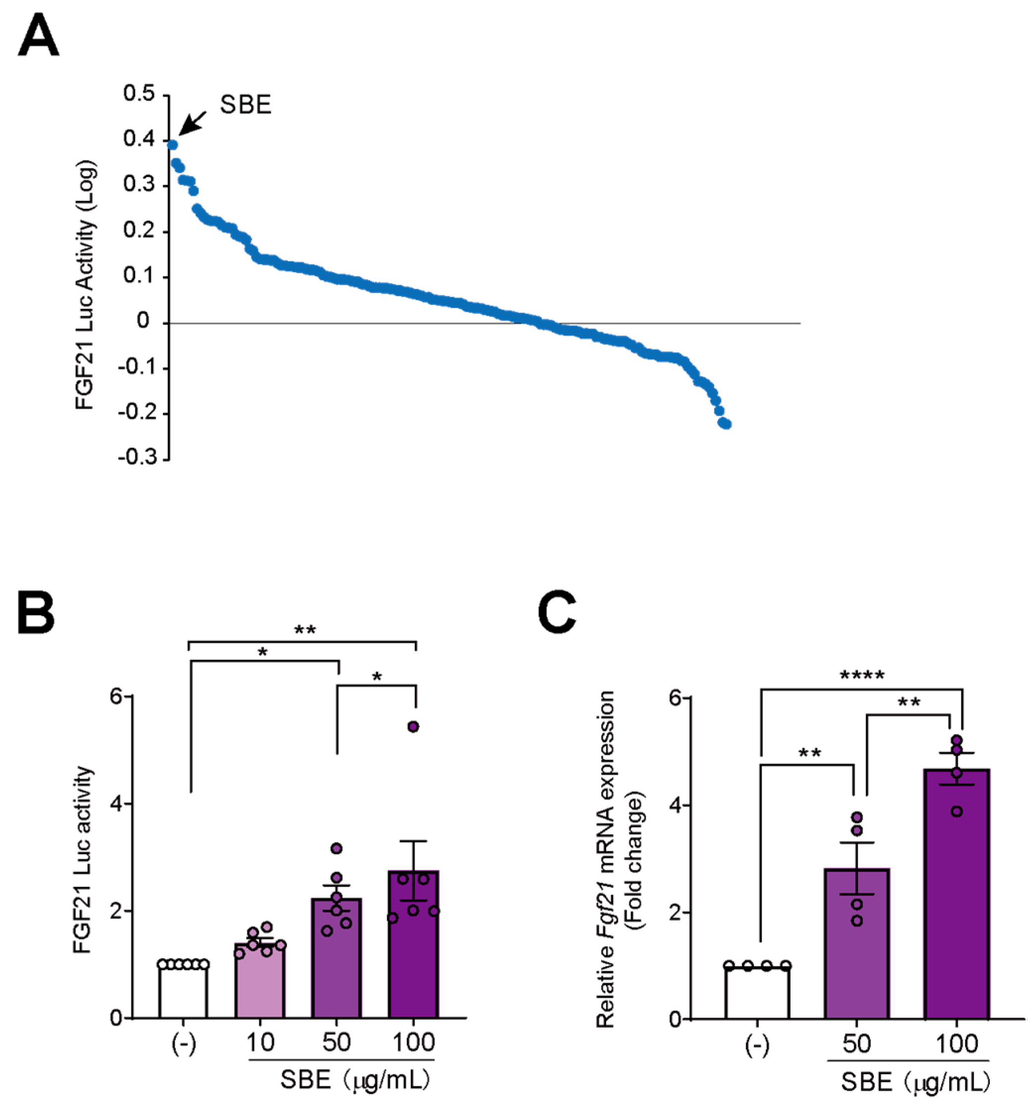
3.1. Cell-Based Screening Using a Natural Medicine Library Identified That SBE Induced Fgf21 Expression

To identify novel activators of Fgf21 expression, we screened 122 crude drugs contained in the WAKANYAKU library by performing the luciferase assay on pGL3-FGF21-transfected AML12 cells after treating them with 10 µg/mL of each drug for 24 h. SBE increased FGF21-luciferase activity to the greatest extent (Figure 1A). To confirm the stimulatory effects of SBE, we performed a dose-dependent experiment and found that FGF21-luciferase activity increased with the dose of SBE (Figure 1B). To further confirm the effects of SBE on Fgf21 expression, we performed qPCR analysis. SBE also induced Fgf21 mRNA expression in AML12 cells in a dose-dependent manner (Figure 1C). Taken together, we identified SBE to be a novel activator of Fgf21 expression.

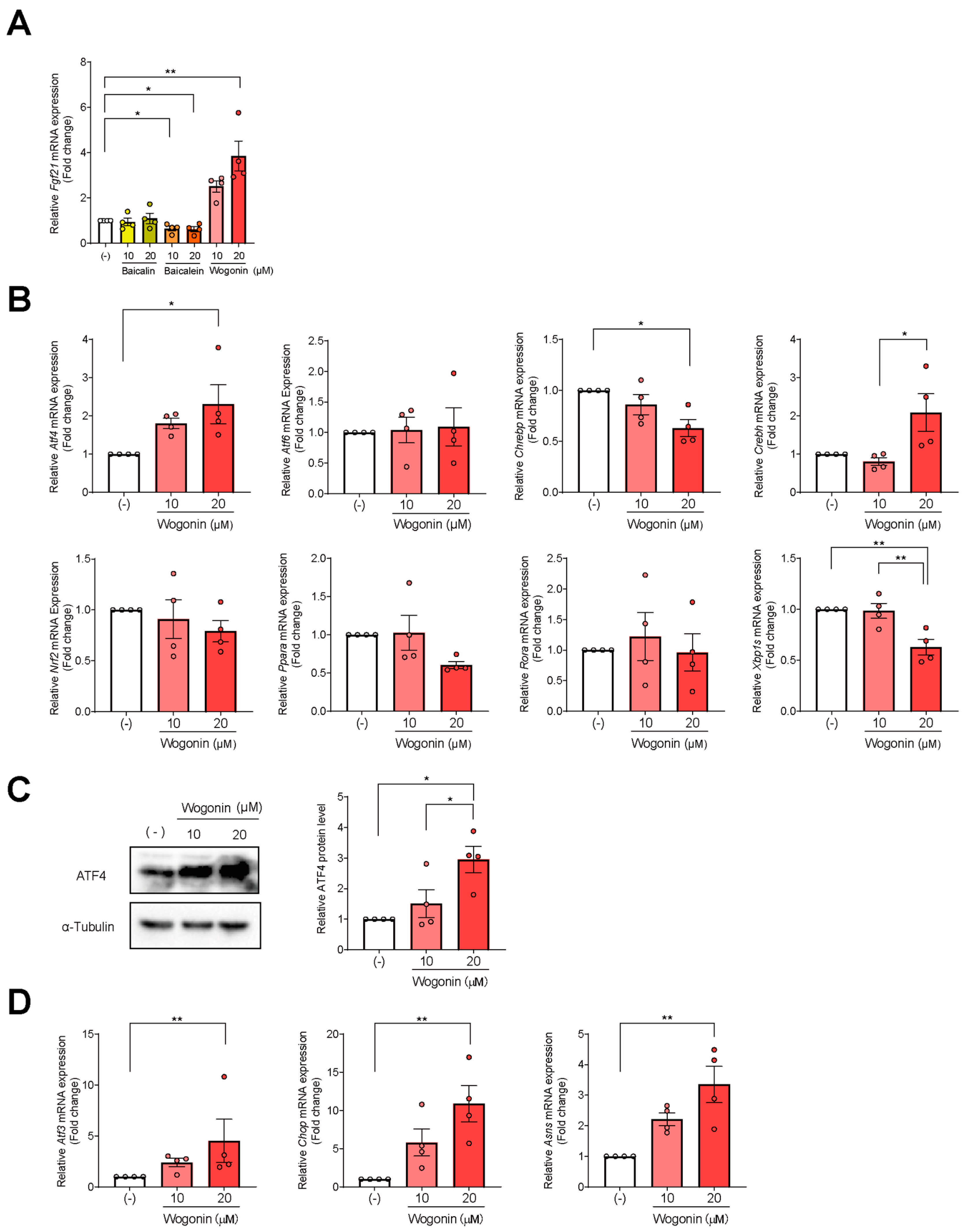
3.2. Wogonin, a Flavonoid in SBE, Induces Fgf21 Expression in AML12 Cells

SBE mainly comprises the flavonoids baicalin, baicalein, and wogonin [35,39]. To identify the major compound(s) from SBE that contribute(s) to its Fgf21-enhancing activity, AML12 cells were treated with the predominant SBE flavonoids—baicalein, baicalin, and wogonin—at 10 and 20 µM for 48 h, followed by measuring Fgf21 expression by qPCR. Wogonin increased Fgf21 expression in a dose-dependent manner, while baicalein and baicalin showed no effect (Figure 2A). These findings support that wogonin could be a candidate to increase Fgf21 expression.

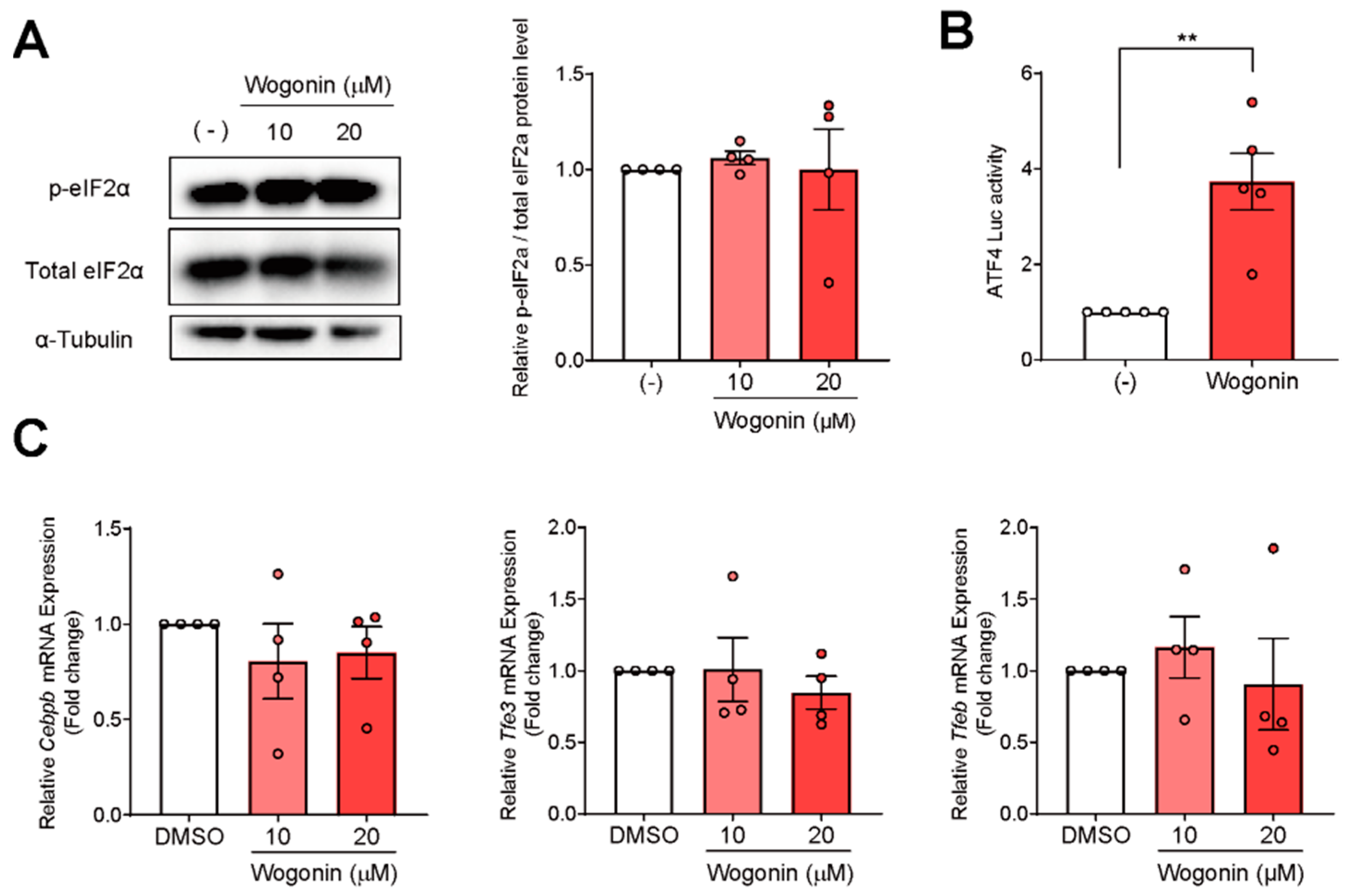
3.3. ATF4 Increased Fgf21 Expression in Response to Wogonin

Fgf21 expression is up-regulated by transcription factors such as ATF4, ATF6, ChREBP, CREBH, NRF2, PPARα, retinoic acid-related orphan receptor α (RORα), and XBP1s. To identify the master transcription factor controlling wogonin-mediated Fgf21 expression, we examined the gene expression of these factors. Wogonin increased Atf4 expression alone in a dose-dependent manner; CrebH expression was enhanced only at high doses of wogonin (Figure 2B). The expression of the other transcription factors was either unchanged or decreased in response to wogonin (Figure 2B). These results support that wogonin would regulate Fgf21 expression via ATF4. Consistent with Atf4 mRNA levels, wogonin increased ATF4 protein levels (Figure 2C) as well as the expression of some typical target genes of ATF4, such as Atf3, asparagine synthetase (Asns), and CCAAT-enhancer-binding protein homologous protein (Chop), in a dose-dependent manner (Figure 2D). These results suggest that ATF4 regulates wogonin-mediated Fgf21 expression in AML12 cells and confirm that wogonin increases ATF4 transcriptional activity.

3.4. Wogonin Controls ATF4 at the Transcription Level

Atf4 expression is controlled by the PERK-eIF2α signaling pathway. ER stress induces PERK, which activates eIF2α by phosphorylating it, eventually inducing the translation of ATF4. Therefore, we determined the protein levels of phospho-eIF2α (p-eIF2α) and total eIF2α by Western blotting. We found that wogonin did not elevate the levels of either p-eIF2α or total eIF2α (Figure 3A), suggesting that its stimulatory effect on ATF4 expression was not mediated via eIF2α signaling. To determine whether wogonin activates Atf4 promoter activity, we performed a luciferase assay with pGL3-ATF4 vector containing –0.5 kbp to –100 bp of the mouse Atf4 promoter. Wogonin significantly increased the luciferase activity inside pGL3-ATF4-transfected AML12 cells (Figure 3B). Taken together, these results indicate that wogonin enhanced Atf4 expression by acting on its promoter. Concerning the transcriptional regulation of ATF4, Atf4 expression has been shown to be up-regulated by NRF2 [21], TFE3, and TFEB [22], and down-regulated by C/EBPβ [23]. However, the expressions of these genes were unchanged after wogonin treatment (Figure 3C), indicating that the known Atf4-regulating transcription factors are not involved in wogonin’s mode of action.

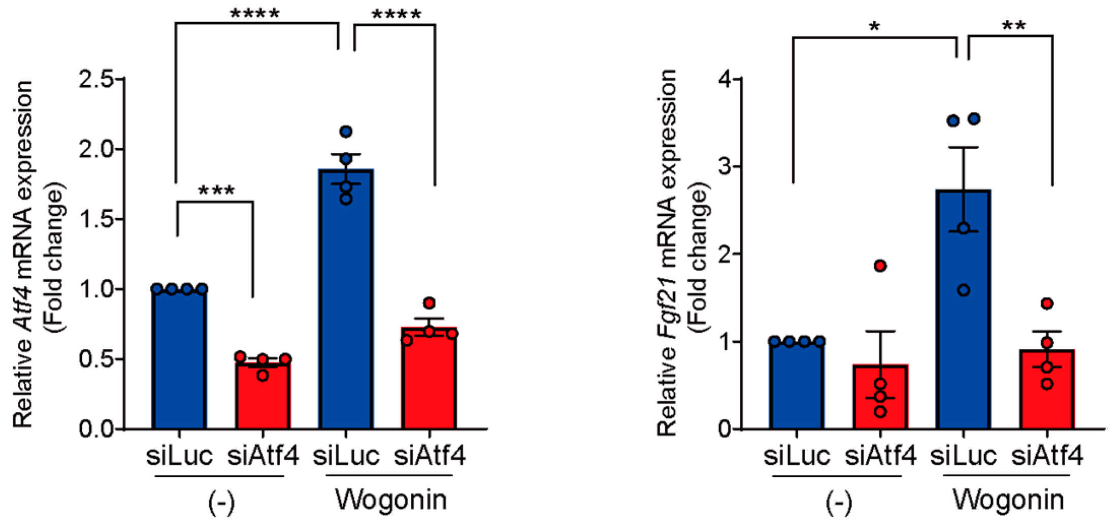
3.5. Knockdown of Atf4 Suppresses Wogonin-Induced Fgf21 Expression

To confirm the necessity of ATF4 for wogonin-induced Fgf21 expression, we performed a loss-of-function analysis by knocking down ATF4 using siRNA (siAtf4) in AML12 cells. AML12 cells were transfected with siRNAs and then treated with wogonin. We confirmed that siAtf4 efficiently reduced Atf4 expression with/without wogonin (Figure 4). ATF4 knockdown eliminated wogonin-induced Fgf21 expression (Figure 4), demonstrating that ATF4 plays a crucial role in wogonin’s effects on Fgf21.

4. Discussion

FGF21 is mainly secreted by the liver, and it systemically improves nutrient metabolism with paracrine action; thus, finding a drug that induces FGF21 in the liver is important. Our study identified that wogonin, a component of SBE, activates Fgf21 expression in AML12 cells—a process mediated by the transcription factor ATF4—by using the WAKANYAKU library.
S. baicalensis has been traditionally used as a medicinal herb in Asia, including Japan, China, and Korea. SBE is broadly used for the clinical treatment of hyperlipidemia, atherosclerosis, hypertension, and inflammatory diseases [29]. SBE suppresses sterol regulatory element binding protein-1c (SREBP-1c) activity by down-regulating Srebf1c expression and activates AMPK in the liver, improving non-alcoholic fatty liver disease [32]. In type 2 diabetic db/db mice, SBE also activates AMPK activity and improves metabolic disorders in the liver, ameliorating this disease [33]. SBE enhances the activity of metformin, a drug for treatment of type 2 diabetes, activates AMPK in type 1 diabetes mice and streptozotocin (STZ)-induced diabetic rats [34]. SBE contains flavonoids, such as baicalin, baicalein, and wogonin, which exert anti-obesity and antihyperlipidemic effects [40]. It has been reported that baicalin and baicalein activates AMPK [32,41]. The effect of wogonin on AMPK activation remains unknown. Wogonin activates PPARα, ameliorating the diabetic phenotypes in db/db mice [36]. Study of the molecular mechanism underlying the improvement effects of SBE on metabolic disorders is still insufficient. FGF21, a master regulator for metabolic homeostasis, is focused on the treatment of metabolic disorders. Baicalein has been reported to increase Fgf21 expression in C2C12 myotubes by activating the transcription factor RORα [42]. However, whether SBE itself can up-regulate Fgf21 expression has not been reported yet. Here, we found that SBE and one of its main components, wogonin, increased Fgf21 expression in AML12 cells, while baicalein and baicalin did not. As the liver is the main organ to secrete FGF21, wogonin might be a therapeutic that can ameliorate metabolic disorders. This could explain the multiple effects of SBE on metabolic disorders.
Fgf21 expression, critically regulated by PPARα and CREBH, is induced in the liver during fasting [43]. Wogonin activates PPARα in the liver and adipose tissues [36]. However, the gene expression of these transcription factors was unchanged in wogonin-treated cells. Additionally, the expression of other known Fgf21 regulators, such as ATF6, ChREBP, NRF2, RORα, and XBP1s, was unchanged. The only factor whose gene expression was enhanced by wogonin was ATF4. Knockdown of ATF4 blunted wogonin-induced Fgf21 expression, confirming its essential role in wogonin’s mechanism of action.
ER stress has three branches that increase Fgf21 expression: (1) ATF6, (2) Inositol-requiring enzyme 1–XBP1, and (3) PERK–ATF4 [13]. During ER stress, the translation of ATF4 is activated by PERK-mediated eIF2α phosphorylation [44]. eIF2α is also phosphorylated by the kinases other than PERK including PKR, hemeregulated inhibitor (HRI), and general control nonderepressible 2 (GCN2). These kinases are activated in response to different stresses: PKR by the infection with certain viruses, HRI by the limitation of heme, GCN2 by the deprivation of essential amino acids [45]. These stimuli finally converge on eIF2α phosphorylation and induce Atf4 expression. However, wogonin-induced ATF4 activation was not mediated by this pathway, as seen by the unchanged levels of p-eIF2α after wogonin treatment. Wogonin activates ATF4 without activating ER stress pathways. Additionally, wogonin did not up-regulate either Atf6 or Xbp1s. Taken together, wogonin cannot activate ER stress pathways. C/EBPβ has been identified as a suppressor of Atf4 expression [23], while TFE3, TFEB, and NRF2 have been identified as activators [21]. However, the expression of these molecules was unchanged as well upon wogonin treatment. Thus, the mechanism of wogonin-induced Atf4 expression remains a mystery that needs to be investigated in future studies.
Chronic inflammation underlies metabolic syndromes, including obesity, diabetes, hyperlipidemia, and high blood pressure [46]. Wogonin has displayed anti-inflammatory activity in several animal models, including lipopolysaccharide-induced acute liver injury, acute lung injury, and kidney injury [47]. Wogonin activates the expression of PPARγ and subsequently suppresses the nuclear factor-κB pathway [47]. Besides these effects, wogonin has been shown to ameliorate metabolic diseases [48]. In this study, we revealed that wogonin can induce Fgf21 expression in AML12 cells, which are derived from the liver, the main FGF21-secreting organ. FGF21 can improve the status of various metabolic diseases; thus, our findings present wogonin as an attractive therapeutic for metabolic syndromes.

Supplementary Materials

The following supporting information can be downloaded at: https://www.mdpi.com/article/10.3390/nu14193920/s1, Table S1: A library consisting of 122 herbal extracts.

Author Contributions

Conceptualization, Y.N.; methodology, Y.Y. and Y.N.; investigation, Y.Y., H.S. (Hodaka Saito), M.A., Y.T. and Y.N.; resources, K.T.; writing—original draft preparation, Y.N.; writing—review and editing, Y.Y., H.S. (Hodaka Saito), M.A., K.T. and Y.N.; visualization, Y.Y.; supervision, S.-i.M., K.S., J.-D.K., T.M., H.S. (Hirohito Sone) and H.S. (Hitoshi Shimano); project administration, Y.N.; funding acquisition, Y.N. All authors have read and agreed to the published version of the manuscript.

Funding

This work was supported by JSPS KAKENHI Grant Numbers JP21H04861 and JP22K19705 (to Y.N.), AMED-CREST Grant Number 22gm1110008s0305 (to Y.N.) and the Cooperative Research Project Program of Life Science Center for Survival Dynamics, Tsukuba Advanced Research Alliance (TARA Center), University of Tsukuba (to Y.N.).

Institutional Review Board Statement

Not applicable.

Informed Consent Statement

Not applicable.

Data Availability Statement

Not applicable.

Acknowledgments

This research was performed by utilizing the INM-deposited WAKANYAKU library, Institute of Natural Medicine, University of Toyama in 2020.

Conflicts of Interest

The authors declare no conflict of interest.

References

  1. Fon Tacer, K.; Bookout, A.L.; Ding, X.; Kurosu, H.; John, G.B.; Wang, L.; Goetz, R.; Mohammadi, M.; Kuro-o, M.; Mangelsdorf, D.J.; et al. Research resource: Comprehensive expression atlas of the fibroblast growth factor system in adult mouse. Mol. Endocrinol. 2010, 24, 2050–2064. [Google Scholar] [CrossRef] [PubMed]
  2. Foltz, I.N.; Hu, S.; King, C.; Wu, X.; Yang, C.; Wang, W.; Weiszmann, J.; Stevens, J.; Chen, J.S.; Nuanmanee, N.; et al. Treating diabetes and obesity with an FGF21-mimetic antibody activating the betaKlotho/FGFR1c receptor complex. Sci. Transl. Med. 2012, 4, 162ra153. [Google Scholar] [CrossRef] [PubMed]
  3. Adams, A.C.; Yang, C.; Coskun, T.; Cheng, C.C.; Gimeno, R.E.; Luo, Y.; Kharitonenkov, A. The breadth of FGF21’s metabolic actions are governed by FGFR1 in adipose tissue. Mol. Metab. 2012, 2, 31–37. [Google Scholar] [CrossRef] [PubMed]
  4. Markan, K.R.; Naber, M.C.; Ameka, M.K.; Anderegg, M.D.; Mangelsdorf, D.J.; Kliewer, S.A.; Mohammadi, M.; Potthoff, M.J. Circulating FGF21 is liver derived and enhances glucose uptake during refeeding and overfeeding. Diabetes 2014, 63, 4057–4063. [Google Scholar] [CrossRef] [PubMed]
  5. Kharitonenkov, A.; Shiyanova, T.L.; Koester, A.; Ford, A.M.; Micanovic, R.; Galbreath, E.J.; Sandusky, G.E.; Hammond, L.J.; Moyers, J.S.; Owens, R.A.; et al. FGF-21 as a novel metabolic regulator. J. Clin. Investig. 2005, 115, 1627–1635. [Google Scholar] [CrossRef]
  6. Fisher, F.M.; Kleiner, S.; Douris, N.; Fox, E.C.; Mepani, R.J.; Verdeguer, F.; Wu, J.; Kharitonenkov, A.; Flier, J.S.; Maratos-Flier, E.; et al. FGF21 regulates PGC-1alpha and browning of white adipose tissues in adaptive thermogenesis. Genes Dev. 2012, 26, 271–281. [Google Scholar] [CrossRef]
  7. Xu, J.; Lloyd, D.J.; Hale, C.; Stanislaus, S.; Chen, M.; Sivits, G.; Vonderfecht, S.; Hecht, R.; Li, Y.S.; Lindberg, R.A.; et al. Fibroblast growth factor 21 reverses hepatic steatosis, increases energy expenditure, and improves insulin sensitivity in diet-induced obese mice. Diabetes 2009, 58, 250–259. [Google Scholar] [CrossRef]
  8. Inagaki, T.; Dutchak, P.; Zhao, G.; Ding, X.; Gautron, L.; Parameswara, V.; Li, Y.; Goetz, R.; Mohammadi, M.; Esser, V.; et al. Endocrine regulation of the fasting response by PPARalpha-mediated induction of fibroblast growth factor 21. Cell Metab. 2007, 5, 415–425. [Google Scholar] [CrossRef]
  9. Nakagawa, Y.; Satoh, A.; Yabe, S.; Furusawa, M.; Tokushige, N.; Tezuka, H.; Mikami, M.; Iwata, W.; Shingyouchi, A.; Matsuzaka, T.; et al. Hepatic CREB3L3 controls whole-body energy homeostasis and improves obesity and diabetes. Endocrinology 2014, 155, 4706–4719. [Google Scholar] [CrossRef]
  10. De Sousa-Coelho, A.L.; Marrero, P.F.; Haro, D. Activating transcription factor 4-dependent induction of FGF21 during amino acid deprivation. Biochem. J. 2012, 443, 165–171. [Google Scholar] [CrossRef] [Green Version]
  11. Chen, X.; Zhang, F.; Gong, Q.; Cui, A.; Zhuo, S.; Hu, Z.; Han, Y.; Gao, J.; Sun, Y.; Liu, Z.; et al. Hepatic ATF6 Increases Fatty Acid Oxidation to Attenuate Hepatic Steatosis in Mice Through Peroxisome Proliferator-Activated Receptor alpha. Diabetes 2016, 65, 1904–1915. [Google Scholar] [CrossRef] [PubMed]
  12. Iizuka, K.; Takeda, J.; Horikawa, Y. Glucose induces FGF21 mRNA expression through ChREBP activation in rat hepatocytes. FEBS Lett. 2009, 583, 2882–2886. [Google Scholar] [CrossRef] [PubMed]
  13. Yu, Y.; He, J.; Li, S.; Song, L.; Guo, X.; Yao, W.; Zou, D.; Gao, X.; Liu, Y.; Bai, F.; et al. Fibroblast growth factor 21 (FGF21) inhibits macrophage-mediated inflammation by activating Nrf2 and suppressing the NF-kappaB signaling pathway. Int. Immunopharmacol. 2016, 38, 144–152. [Google Scholar] [CrossRef] [PubMed]
  14. Jiang, S.; Yan, C.; Fang, Q.C.; Shao, M.L.; Zhang, Y.L.; Liu, Y.; Deng, Y.P.; Shan, B.; Liu, J.Q.; Li, H.T.; et al. Fibroblast growth factor 21 is regulated by the IRE1alpha-XBP1 branch of the unfolded protein response and counteracts endoplasmic reticulum stress-induced hepatic steatosis. J. Biol. Chem. 2014, 289, 29751–29765. [Google Scholar] [CrossRef] [PubMed]
  15. B’Chir, W.; Maurin, A.C.; Carraro, V.; Averous, J.; Jousse, C.; Muranishi, Y.; Parry, L.; Stepien, G.; Fafournoux, P.; Bruhat, A. The eIF2alpha/ATF4 pathway is essential for stress-induced autophagy gene expression. Nucleic Acids Res. 2013, 41, 7683–7699. [Google Scholar] [CrossRef]
  16. Kilberg, M.S.; Shan, J.; Su, N. ATF4-dependent transcription mediates signaling of amino acid limitation. Trends Endocrinol. Metab. 2009, 20, 436–443. [Google Scholar] [CrossRef]
  17. Harding, H.P.; Zhang, Y.; Zeng, H.; Novoa, I.; Lu, P.D.; Calfon, M.; Sadri, N.; Yun, C.; Popko, B.; Paules, R.; et al. An integrated stress response regulates amino acid metabolism and resistance to oxidative stress. Mol. Cell 2003, 11, 619–633. [Google Scholar] [CrossRef]
  18. Ron, D.; Harding, H.P. Protein-folding homeostasis in the endoplasmic reticulum and nutritional regulation. Cold Spring Harb. Perspect. Biol. 2012, 4, a013177. [Google Scholar] [CrossRef]
  19. Harding, H.P.; Zhang, Y.; Ron, D. Protein translation and folding are coupled by an endoplasmic-reticulum-resident kinase. Nature 1999, 397, 271–274. [Google Scholar] [CrossRef]
  20. Pakos-Zebrucka, K.; Koryga, I.; Mnich, K.; Ljujic, M.; Samali, A.; Gorman, A.M. The integrated stress response. EMBO Rep. 2016, 17, 1374–1395. [Google Scholar] [CrossRef] [Green Version]
  21. Afonyushkin, T.; Oskolkova, O.V.; Philippova, M.; Resink, T.J.; Erne, P.; Binder, B.R.; Bochkov, V.N. Oxidized phospholipids regulate expression of ATF4 and VEGF in endothelial cells via NRF2-dependent mechanism: Novel point of convergence between electrophilic and unfolded protein stress pathways. Arterioscler. Thromb. Vasc. Biol. 2010, 30, 1007–1013. [Google Scholar] [CrossRef] [PubMed]
  22. Martina, J.A.; Diab, H.I.; Brady, O.A.; Puertollano, R. TFEB and TFE3 are novel components of the integrated stress response. EMBO J. 2016, 35, 479–495. [Google Scholar] [CrossRef] [PubMed]
  23. Dey, S.; Savant, S.; Teske, B.F.; Hatzoglou, M.; Calkhoven, C.F.; Wek, R.C. Transcriptional repression of ATF4 gene by CCAAT/enhancer-binding protein beta (C/EBPbeta) differentially regulates integrated stress response. J. Biol. Chem. 2012, 287, 21936–21949. [Google Scholar] [CrossRef]
  24. Kim, K.H.; Jeong, Y.T.; Oh, H.; Kim, S.H.; Cho, J.M.; Kim, Y.N.; Kim, S.S.; Kim, D.H.; Hur, K.Y.; Kim, H.K.; et al. Autophagy deficiency leads to protection from obesity and insulin resistance by inducing Fgf21 as a mitokine. Nat. Med. 2013, 19, 83–92. [Google Scholar] [CrossRef]
  25. Laeger, T.; Albarado, D.C.; Burke, S.J.; Trosclair, L.; Hedgepeth, J.W.; Berthoud, H.R.; Gettys, T.W.; Collier, J.J.; Munzberg, H.; Morrison, C.D. Metabolic Responses to Dietary Protein Restriction Require an Increase in FGF21 that Is Delayed by the Absence of GCN2. Cell Rep. 2016, 16, 707–716. [Google Scholar] [CrossRef] [PubMed]
  26. Zarei, M.; Barroso, E.; Leiva, R.; Barniol-Xicota, M.; Pujol, E.; Escolano, C.; Vazquez, S.; Palomer, X.; Pardo, V.; Gonzalez-Rodriguez, A.; et al. Heme-Regulated eIF2alpha Kinase Modulates Hepatic FGF21 and Is Activated by PPARbeta/delta Deficiency. Diabetes 2016, 65, 3185–3199. [Google Scholar] [CrossRef] [PubMed]
  27. Huang, W.H.; Lee, A.R.; Yang, C.H. Antioxidative and anti-inflammatory activities of polyhydroxyflavonoids of Scutellaria baicalensis GEORGI. Biosci. Biotechnol. Biochem. 2006, 70, 2371–2380. [Google Scholar] [CrossRef]
  28. Ye, F.; Xui, L.; Yi, J.; Zhang, W.; Zhang, D.Y. Anticancer activity of Scutellaria baicalensis and its potential mechanism. J. Altern. Complement. Med. 2002, 8, 567–572. [Google Scholar] [CrossRef]
  29. Kim, E.H.; Shim, B.; Kang, S.; Jeong, G.; Lee, J.S.; Yu, Y.B.; Chun, M. Anti-inflammatory effects of Scutellaria baicalensis extract via suppression of immune modulators and MAP kinase signaling molecules. J. Ethnopharmacol. 2009, 126, 320–331. [Google Scholar] [CrossRef]
  30. Nagai, T.; Moriguchi, R.; Suzuki, Y.; Tomimori, T.; Yamada, H. Mode of action of the anti-influenza virus activity of plant flavonoid, 5,7,4′-trihydroxy-8-methoxyflavone, from the roots of Scutellaria baicalensis. Antivir. Res. 1995, 26, 11–25. [Google Scholar] [CrossRef]
  31. Heo, H.J.; Kim, D.O.; Choi, S.J.; Shin, D.H.; Lee, C.Y. Potent Inhibitory effect of flavonoids in Scutellaria baicalensis on amyloid beta protein-induced neurotoxicity. J. Agric. Food Chem. 2004, 52, 4128–4132. [Google Scholar] [CrossRef] [PubMed]
  32. Chen, Q.; Liu, M.; Yu, H.; Li, J.; Wang, S.; Zhang, Y.; Qiu, F.; Wang, T. Scutellaria baicalensis regulates FFA metabolism to ameliorate NAFLD through the AMPK-mediated SREBP signaling pathway. J. Nat. Med. 2018, 72, 655–666. [Google Scholar] [CrossRef] [PubMed]
  33. Song, K.H.; Lee, S.H.; Kim, B.Y.; Park, A.Y.; Kim, J.Y. Extracts of Scutellaria baicalensis reduced body weight and blood triglyceride in db/db Mice. Phytother. Res. 2013, 27, 244–250. [Google Scholar] [CrossRef] [PubMed]
  34. Waisundara, V.Y.; Hsu, A.; Huang, D.; Tan, B.K. Scutellaria baicalensis enhances the anti-diabetic activity of metformin in streptozotocin-induced diabetic Wistar rats. Am. J. Chin. Med. 2008, 36, 517–540. [Google Scholar] [CrossRef] [PubMed]
  35. Huang, S.T.; Wang, C.Y.; Yang, R.C.; Chu, C.J.; Wu, H.T.; Pang, J.H. Wogonin, an active compound in Scutellaria baicalensis, induces apoptosis and reduces telomerase activity in the HL-60 leukemia cells. Phytomedicine 2010, 17, 47–54. [Google Scholar] [CrossRef]
  36. Bak, E.J.; Kim, J.; Choi, Y.H.; Kim, J.H.; Lee, D.E.; Woo, G.H.; Cha, J.H.; Yoo, Y.J. Wogonin ameliorates hyperglycemia and dyslipidemia via PPARalpha activation in db/db mice. Clin. Nutr. 2014, 33, 156–163. [Google Scholar] [CrossRef]
  37. Chen, J.; Liu, J.; Wang, Y.; Hu, X.; Zhou, F.; Hu, Y.; Yuan, Y.; Xu, Y. Wogonin mitigates nonalcoholic fatty liver disease via enhancing PPARalpha/AdipoR2, in vivo and in vitro. Biomed. Pharmacother. 2017, 91, 621–631. [Google Scholar] [CrossRef]
  38. Wang, Q.; Mora-Jensen, H.; Weniger, M.A.; Perez-Galan, P.; Wolford, C.; Hai, T.; Ron, D.; Chen, W.; Trenkle, W.; Wiestner, A.; et al. ERAD inhibitors integrate ER stress with an epigenetic mechanism to activate BH3-only protein NOXA in cancer cells. Proc. Natl. Acad. Sci. USA 2009, 106, 2200–2205. [Google Scholar] [CrossRef]
  39. Makino, T.; Hishida, A.; Goda, Y.; Mizukami, H. Comparison of the major flavonoid content of S. baicalensis, S. lateriflora, and their commercial products. J. Nat. Med. 2008, 62, 294–299. [Google Scholar] [CrossRef]
  40. Baradaran Rahimi, V.; Askari, V.R.; Hosseinzadeh, H. Promising influences of Scutellaria baicalensis and its two active constituents, baicalin, and baicalein, against metabolic syndrome: A review. Phytother. Res. 2021, 35, 3558–3574. [Google Scholar] [CrossRef]
  41. Sun, W.; Liu, P.; Wang, T.; Wang, X.; Zheng, W.; Li, J. Baicalein reduces hepatic fat accumulation by activating AMPK in oleic acid-induced HepG2 cells and high-fat diet-induced non-insulin-resistant mice. Food Funct. 2020, 11, 711–721. [Google Scholar] [CrossRef] [PubMed]
  42. Hirai, T.; Nomura, K.; Ikai, R.; Nakashima, K.I.; Inoue, M. Baicalein stimulates fibroblast growth factor 21 expression by up-regulating retinoic acid receptor-related orphan receptor alpha in C2C12 myotubes. Biomed. Pharmacother. 2019, 109, 503–510. [Google Scholar] [CrossRef] [PubMed]
  43. Nakagawa, Y.; Shimano, H. CREBH Regulates Systemic Glucose and Lipid Metabolism. Int. J. Mol. Sci. 2018, 19, 1396. [Google Scholar] [CrossRef] [PubMed]
  44. Jackson, R.J.; Hellen, C.U.; Pestova, T.V. The mechanism of eukaryotic translation initiation and principles of its regulation. Nat. Rev. Mol. Cell Biol. 2010, 11, 113–127. [Google Scholar] [CrossRef]
  45. Dang Do, A.N.; Kimball, S.R.; Cavener, D.R.; Jefferson, L.S. eIF2alpha kinases GCN2 and PERK modulate transcription and translation of distinct sets of mRNAs in mouse liver. Physiol. Genom. 2009, 38, 328–341. [Google Scholar] [CrossRef]
  46. Matsuzawa, Y.; Funahashi, T.; Nakamura, T. Molecular mechanism of metabolic syndrome X: Contribution of adipocytokines adipocyte-derived bioactive substances. Ann. N. Y. Acad. Sci. 1999, 892, 146–154. [Google Scholar] [CrossRef]
  47. Yao, J.; Pan, D.; Zhao, Y.; Zhao, L.; Sun, J.; Wang, Y.; You, Q.D.; Xi, T.; Guo, Q.L.; Lu, N. Wogonin prevents lipopolysaccharide-induced acute lung injury and inflammation in mice via peroxisome proliferator-activated receptor gamma-mediated attenuation of the nuclear factor-kappaB pathway. Immunology 2014, 143, 241–257. [Google Scholar] [CrossRef]
  48. Dai, J.M.; Guo, W.N.; Tan, Y.Z.; Niu, K.W.; Zhang, J.J.; Liu, C.L.; Yang, X.M.; Tao, K.S.; Chen, Z.N.; Dai, J.Y. Wogonin alleviates liver injury in sepsis through Nrf2-mediated NF-kappaB signalling suppression. J. Cell. Mol. Med. 2021, 25, 5782–5798. [Google Scholar] [CrossRef]
Figure 1. SBE is identified as an activator of Fgf21 expression after screening the WAKANYAKU library. (A) The screening of natural medicines from the WAKANYAKU library that can induce Fgf21 expression using an FGF21–luciferase assay. AML12 cells were co-transfected with the reporter vector pGL3-FGF21 and pRL-SV40 as a reference. After 24 h of transfection, cells were treated with 10 µg/mL of natural medicines for 24 h. The luciferase activity was measured and normalized to the renilla luciferase activity. (B) SBE activated FGF21-luciferase activity in AML12 cells. Cells were co-transfected with pGL3-FGF21 and pRL-SV40 vectors. After 24 h of transfection, cells were treated with 10, 50, 100 µg/mL of SBE for 24 h. n = 4 per group. (C) SBE increased Fgf21 expression in AML12 cells. Cells were treated with 50 and 100 µg/mL of SBE for 48 h. n = 4 per group. Data are represented as mean ± SD. * p < 0.05; ** p < 0.01; **** p < 0.0001. Comparisons among multiple groups were assessed using one-way ANOVA, followed by Tukey’s post hoc test.
Nutrients 14 03920 g001
Figure 2. Wogonin induces Atf4 and Fgf21 expression. (A) Wogonin increased Fgf21 expression in AML12 cells. Cells were treated with 10 and 20 µM of baicalin, baicalein, and wogonin for 48 h. n = 4 per group. (B) Gene expression of FGF21-regulating transcription factors in AML12 cells. Cells were treated with 10 and 20 µM of wogonin for 48 h. n = 4 per group. (C) Wogonin increased the protein levels of ATF4 in AML12 cells. Cells were treated with 10 and 20 µM of wogonin for 48 h. The protein bands were quantified. n = 4 per group. (D) Wogonin increased the expression of genes regulated by ATF4 in AML12 cells. Cells were treated with 10 and 20 µM of wogonin for 48 h. n = 4 per group. Data are represented as mean ± SD. * p < 0.05; ** p < 0.01. Comparisons among multiple groups were assessed using one-way ANOVA, followed by Tukey’s post hoc test.
Nutrients 14 03920 g002
Figure 3. Wogonin affects the promoter activity of Atf4. (A) The protein levels of phospho- and total-eIF2α were not changed in AML12 cells. Cells were treated with 10 and 20 µM of wogonin for 48 h. The protein bands were quantified. n = 4 per group. (B) Wogonin increased ATF4-luciferase activity. AML12 cells were co-transfected with the pGL3-ATF4 and pRL-SV40 vectors. After 24 h of transfection, cells were treated with 20 μM of wogonin for 24 h. n = 5 per group. Data are represented as mean ± SD. (C) The gene expression of ATF4-regulating transcription factors in AML12 cells. Cells were treated with 10 and 20 µM of wogonin for 48 h. n = 4 per group. Data are represented as mean ± SD. ** p < 0.01. Comparisons between two groups were assessed using unpaired two-tailed t tests and those among multiple groups were assessed using one-way ANOVA, followed by Tukey’s post hoc test.
Nutrients 14 03920 g003
Figure 4. Deficiency of ATF4 suppresses wogonin-induced Fgf21 expression. Fgf21 expression was suppressed by transfecting AML12 cells with siRNA against ATF4. After 24 h of transfection, cells were treated with 20 µM of wogonin for 48 h. n = 4 per group. Data are represented as mean ± SD. * p < 0.05; ** p < 0.01; *** p < 0.001; **** p < 0.0001. Comparisons among multiple groups were assessed using one-way ANOVA, followed by Tukey’s post hoc test.
Nutrients 14 03920 g004
Table 1. Primers used for real-time PCR analysis.
GeneForward Primer (5′ to 3′)Reverse Primer (5′ to 3′)
AsnsTTACCTGTCTCTGCCGCCAGATCACTGAAGGCTTCTTTGGGTCG
Atf3TTTGCTAACCTGACACCCTTTGAGAGGACATCCGATGGCAGA
Atf4CCTGAACAGCGAAGTGTTGGTGGAGAACCCATGAGGTTTCAA
Atf6GGACGAGGTGGTGTCAGAGGACAGCTCTTCGCTTTGGAC
CebpbTACGAGCCCGACTGCCTGTCGGAGAGGAAGTCGTGGTG
ChopGGAGGTCCTGTCCTCAGATGAAGCTCCTCTGTCAGCCAAGCTAG
ChrebpAATGGGATGGTGTCTACCGCGGCGAAGGGAATTCAGGACA
Fgf21GGCAAGATATACGGGCTGATTCCATTTCCTCCCTGAAGGT
CrebHAGATCAGGGAGGATGGAACATCAAAGTGAGGCGATCCATA
CyclophilinTGGCTCACAGTTCTTCATAACCAATGACATCCTTCAGTGGCTTGTC
Nrf2CAAGACTTGGGCCACTTAAAAGACAGTAAGGCTTTCCATCCTCATCAC
PparaACGCGAGTTCCTTAAGAACCTGGTGTCATCTGGATGGTTGCTCT
RoraGATGACCTCAGCACCTATATGGACGGGTTTGATCCCATTGATGTC
Tfe3AGGATCAAAGAGCTGGGCACCCGGCTCTCCAGGTCTTTG
TfebCAGAAGCGAGAGCTAACAGATTGTGATTGTCTTTCTTCTGCCG
Xbp1sCTGAGTCCGAATCAGGTGCAGGTCCATGGGAAGATGTTCTGG
Publisher’s Note: MDPI stays neutral with regard to jurisdictional claims in published maps and institutional affiliations.

